�
�
Università degli Studi di Padova
Dipartimento Territorio e Sistemi Agro Forestali
SCUOLA DI DOTTORATO DI RICERCA IN
TERRITORIO AMBIENTE RISORSE E SALUTE
CICLO XXVI
DNA Repair is Modulated by Cellular and
Circadian Cycles
Direttore della scuola: Ch.mo Prof. Mario Aristide Lenzi
Supervisore: Ch.mo Prof. Lucia Celotti
Co-supervisore: Ch.mo Prof. Urs Albrecht
Dottorando: Leonardo Bee

Contents
1
1. Summary 3
2. Riassunto 7
3. Introduction 13
4. Aim of the study 33
5. Papers
5.1. The efficiency of Homologous Recombination and Non Homologous End Joining systems in repairing double-strand breaks during cell cycle progression
5.2. Mammalian ribonucleotide reductase subunit p53R2 is required for mitochondrial DNA replication and DNA repair in quiescent cells
35
35
75
6. Unpublished studies: “The circadian control of UV-induced
DNA damage sensitivity and repair”
6.1. Introduction
6.2. Materials and Methods
6.3. Results
6.4. Discussion
6.5. Supporting Information
113
117
119
127
139
143
7. Conclusions and future perspectives 145
8. References 149
9. Acknowledgements 157


Summary
3
Summary
Background DNA repair is an essential cellular function aiming to maintain genomic
stability during dangerous environmental conditions, such as exposure to UV light
and ionizing radiation. At the molecular level, cell life is marked by rhythmic
events resulting from the two major oscillatory systems: the circadian clock and
the cell cycle, which share some striking similarities. Both consist of interlocked
auto-regulatory feedback loops and both rely on chromatin remodeling events that
produce sequential transcription, translation, post-translational modification, and
degradation phases. In mammals, DNA double-strand breaks (DSBs) are rejoined
by two highly regulated pathways: non-homologous end joining (NHEJ), which is
always operative, and homologous recombination (HR), which is active only in
late S- and G2-phases when both sister chromatids are present. DNA adducts
induced by exposure to UV light, such as cyclobutane pyrimidine dimers (CPDs)
and pyrimidine pirimidone photoproducts ([6-4]PPs), can be repaired only by the
nucleotide excision repair (NER) system. DNA damage response (DDR), which
includes DNA repair, checkpoint activation, chromatin remodeling, and apoptosis,
seems to be strictly modulated by the rhythmicity induced by the cell cycle and
the circadian clock, and this could affect its efficiency, making the cells more
sensitive or resistant to genotoxic stress at different times of the day.
Methodology and Principal Findings
DNA double-strand break repair is modulated by cell cycle progression
Human lung fibroblasts were tested for DNA repair efficiency during some
phases of the cell cycle by monitoring formation and disappearance of γ-H2AX
foci at the sites of DSBs induced by γ-ray irradiation. The decrease in γ-H2AX
signals at DSB sites was more pronounced in G2- with respect to G1-phase cells.
Moreover, G2 cells treated with the HR inhibitor (RI-1) exhibited a higher level of
unrepaired DSBs starting six hours after irradiation, showing that HR is active
early after DNA damage. Our data demonstrated that in G2-phase cells, NHEJ and

Summary
4
HR repair systems cooperate in DSB rejoining not only long times after
irradiation but also during the first hours of post-irradiation incubation. The
relevance of HR during DSB repair and, in particular, the possibility that this
pathway could compensate for NHEJ deficiency, was assayed in DNA-PKcs
deficient cells as well as in those treated with DNA-PKcs inhibitor. In both
conditions, the kinetics of γ-H2AX demonstrated that DSB repair was strongly
affected during all phases of the cell cycle, even in the G2-one when HR should
have been active, confirming previous observations according to which DSB
rejoining is strictly dependent on the integrity of the NHEJ repair system (Bee e
al. 2013).
DNA repair efficiency is modulated during the circadian cycle
We evaluated whether the circadian clock affects the efficiency of DNA repair in
non-proliferating human primary skin fibroblasts irradiated with UV light.
Quiescent fibroblasts (in the G0 phase) were synchronized by dexamethasone
incubation to induce rhythmic expression of the circadian clock genes. We found,
by using qRT-PCR, a strong circadian rhythmicity in PER2 mRNA levels, which
showed two negative peaks at 16 h and 40 h after dexamethasone incubation and a
positive one at 28 h, with an overall period of about 24 h. BMAL1 mRNA
expression also showed robust oscillations in the anti-phase of PER2 transcript (as
expected). The rhythmicity of the transcripts was confirmed by Western blot
analyses showing clear oscillations of BMAL1 and PER2 protein levels. We
identified, thus, two time-points separated by a 12 h interval during which
administration of UV light occurred at the two opposite levels of clock gene
expression.
Following the PER2 protein expression level, the cells were irradiated at 16 h
after dexamethasone, which corresponded to the PER2 nadir, and at 28 h after
dexamethasone, corresponding to the PER2 zenith. By measuring H2AX
phosphorylation, a marker of damaged DNA, we found that zenith-irradiated cells
exhibited a significantly higher extension of γ-H2AX, especially 2 and 4 h after
irradiation. Analyzing the renjoining of the gaps in DNA molecules produced
during the NER process, using fluorometric analysis of DNA unwinding (FADU),

Summary
5
we uncovered, moreover, that the cells irradiated at the PER2 zenith contained
less dsDNA than those irradiated at the nadir, suggesting that a greater residual
damage results from slower repair kinetics. By measuring the removal of [6-
4]PPs, we were able to determine that both treatment groups efficiently removed
photoproducts within the first 3 h after irradiation, but the fibroblasts irradiated at
the zenith exhibited a significantly slower kinetic of [6-4]PPs removal with
respect to those irradiated at the nadir. By assessing XPA protein expression, we
did not, as opposed to other authors, find any evidence of an oscillating pattern.
Analysis of the XPA promoter using luciferase assay or by blocking the core
clock machinery by RNA interference of BMAL1 expression and in BMAL1
mutant cells revealed no connection between this protein and circadian clock
transcription factors. Interestingly, our results showed that in addition to the faster
kinetics of [6-4]PPs removal, fibroblasts irradiated at the nadir of PER2 induced a
significantly higher formation of both [6-4]PPs and CPDs with respect to zenith-
irradiated cells. These data were confirmed by Unscheduled DNA Synthesis assay
(UDS), which showed an higher incorporation of [3H]-TdR at the nadir- with
respect to the zenith-irradiated cells. Transfection of fibroblasts with siRNAs
targeting BMAL1 gene expression abrogated the difference observed in the
amount of [6-4]PPs formed and reduced the photoproduct removal efficiency of
nadir-irradiated cells. In this condition we observed no differences between nadir
and zenith-irradiated cells, confirming BMAL1’s role in UV light sensitivity and
DNA damage repair.
Conclusions Taken as a whole, even if preliminary, our results indicate that DNA
damage response is an event that is strictly modulated by the rhythmicity of the
cell cycle and the circadian clock and that its efficiency varies over a 24 h
interval. This indicates that the time of exposure to genotoxic stress, such as
ionizing radiation and/or UV light, could increase the risk of genomic instability.
We observed that both the sensitivity of DNA to UV and repair were higher when
PER2 reached its nadir (and therefore BMAL1 its zenith), perhaps as a
Summary
6
consequence of a particular chromatin structure which leads to higher DNA
accessibility. We observed, nevertheless, that DSB rejoining was more efficient in
the S-G2 phases, when both HR and NHEJ cooperate in DNA repair. In mice
PER2 nadir was found to occur during the day while the PER2 zenith and S-G2
phases occurred during the night far from the UV component of sunlight. We
speculate that the temporal separation of these events is a form of adaptation to
environmental conditions (such as sunlight) and that the disruption of this
oscillatory equilibrium affects DNA repair process.

Riassunto
7
Riassunto
Contesto La riparazione dei danni al DNA è una funzione essenziale per il
mantenimento della stabilità genomica in seguito all’esposizione a particolari
condizioni ambientali, tra le quali le radiazioni UV e le radiazioni ionizzanti. A
livello molecolare la vita della cellula è scandita da eventi ritmici risultanti dai due
principali sistemi oscillatori: l’orologio circadiano e il ciclo cellulare, i quali
condividono notevoli somiglianze. Entrambi, infatti, consistono in cicli di auto-
regolazione ed entrambi si basano su eventi di rimodellamento della cromatina per
produrre fasi sequenziali di trascrizione, traduzione, modificazione post-
traduzionale, e fasi di degradazione. Nei mammiferi, le rotture del doppio
filamento del DNA sono riparate mediante due vie altamente regolate: la saldatura
delle estremità non omologhe (NHEJ), attiva in tutte le fasi del ciclo cellulare, e la
ricombinazione omologa (HR), attiva solamente nelle fasi S e G2, quando sono
presenti entrambi i cromatidi fratelli. Gli addotti di DNA indotti dall’esposizione
ai raggi UV, come i dimeri ciclo butano-pirimidina (CPD) ed i fotoprodotti
pirimidina-pirimidone ([6-4]PP), sono, invece, riparati unicamente dal sistema di
rimozione dei nucleotidi (NER). La risposta ai danni al DNA (DDR) che include
la riparazione del DNA, l’attivazione dei checkpoint del ciclo cellulare, il
rimodellamento della cromatina e l’apoptosi, sembrano essere strettamente
modulati dalla ritmicità indotta sia dal ciclo cellulare sia da quello circadiano.
Questa interazione può quindi influenzare la risposta ai danni al DNA, rendendo
la cellula più sensibile o più resistente agli stress genotossici nei diversi momenti
del giorno.
Scopo dello studio
1. Analizzare in fibroblasti umani irradiati con raggi γ (a) il coinvolgimento e
l’efficienza dei due sistemi di riparazione principali durante le fasi del
ciclo cellulare e (b) la tempistica del reclutamento di HR nella riparazione
delle doppie rotture al DNA.

Riassunto
8
2. Investigare se difetti nella NHEJ coinvolgono anche l’efficienza di HR
nella riparazione delle doppie rotture in fase G2.
3. Sviluppare un sistema per il mantenimento delle colture cellulari in
quiescenza a lungo termine e validare un insieme di tecniche che
consentano di saggiare con precisione la riparazione dei danni al DNA
indotti dalla luce UV.
4. Sincronizzare colture cellulari umani e indurre l’espressione circadiana dei
vari geni orologio che oscillano in animali vivi.
5. Investigare se l’efficienza della risposta ai danni al DNA è influenzata
dalla fase circadiana nella quale le cellule vengono trattate con agenti
genotossici.
Metodologie e Risultati Principali
La riparazione delle doppie rotture al DNA è regolata dalla progressione del
ciclo cellulare
In questo lavoro, è stata valutata l’efficienza della riparazione del DNA
nelle varie fasi del ciclo cellulare in colture di fibroblasti umani di polmone
seguendo la formazione e la scomparsa dei foci di γ-H2AX nei siti delle doppie
rotture al DNA indotte da raggi γ. I risultati ottenuti hanno evidenziato una
diminuzione del segnale di γ-H2AX più elevata nelle cellule in fase G2 rispetto a
quelle in G1. Dopo sei ore dall’irradiazione, inoltre, le cellule in fase G2 trattate
con l’inibitore di HR (RI-1), esibiscono un livello più elevato di DSBs non
riparate, indicando che HR opera attivamente dopo tempi brevi dall’irradiazione. I
dati dimostrano, quindi, che nelle cellule in fase G2, NHEJ e HR cooperano nella
risoluzione delle DSB non solo a tempi lunghi dall’irradiazione, ma anche durante
le prime ore di incubazione post-irradiazione. Data la rilevanza di HR nelle
riparazione delle DSB, è stata valutata la capacità di HR di sopperire alla
mancanza di NHEJ, utilizzando cellule difettive per DNA-PKcs o trattate con il
suo specifico inibitore. In entrambe le condizioni, la cinetica dei foci di γ-H2AX
ha dimostrato che la riparazione delle doppie rotture del DNA è fortemente

Riassunto
9
compromessa in tutte le fasi del ciclo cellulare, perfino in G2 dove HR dovrebbe
essere attiva, confermando che la riparazione delle DSB è strettamente dipendente
dall’integrità del sistema NHEJ.
L’efficienza della riparazione del DNA è controllata dall’orologio circadiano
In questo lavoro è stata valutata la possibilità che l’orologio circadiano
possa influenzare l’efficienza della riparazione del DNA in fibroblasti primari di
pelle umana quiescenti irradiati con luce UV. Attraverso un trattamento con
dexametasone, i fibroblasti quiescenti (fase G0) sincronizzati al fine di indurre
l’espressione ritmica dei geni orologio. Mediante qRT-PCT, è stato possibile
apprezzare una forte induzione nella ritmicità del mRNA di PER2, che mostra due
picchi negativi a 16 h e 40 h dopo l’incubazione con dexametasone, e un picco
positivo a 28 h, con un periodo complessivo di circa 24 h. L’espressione di
BMAL1 mostra anch’essa robuste oscillazioni e, come atteso, in anti-fase con il
trascritto di PER2. La ritmicità dei trascritti è stata confermata mediante Western
blot, la quale ha evidenziato chiare oscillazioni dei livelli delle proteine PER2 e
BMAL1. È stato possibile identificare, quindi, due momenti temporali, separati da
12 h , nei quali la somministrazione di luce UV avviene in due livelli opposti di
espressione dei geni orologio. Seguendo l’espressione della proteina PER2, le
cellule sono state irradiate a 16 e 28 h in seguito al trattamento con dexametasone,
corrispondenti rispettivamente al nadir e allo zenit di PER2. Quantificando la
fosforilazione dell’istone H2AX, come marcatore dei danni al DNA, abbiamo
trovato che le cellule irradiate allo zenit esibiscono un’estensione
significativamente più elevata di γ -H2AX, in modo particolare a 2 e 4 h post-
irradiazione. È stata quindi esaminata la risaldatura delle regioni di DNA a
singolo filamento, prodotte durante il processo NER, mediante l’analisi
fluorimetrica dello svolgimento del DNA (FADU). I risultati hanno evidenziato
che le cellule irradiate allo zenit di PER2 contengono meno DNA a doppia elica
rispetto a quelle irradiate al Nadir, suggerendo un maggiore danno residuo, come
riflesso di una cinetica di riparazione più lenta. In accordo con ciò, quantificando
la rimozione dei [6-4]PP è stato osservato che entrambi i gruppi rimuovono i foto

Riassunto
10
prodotti in modo efficiente entro le prime 3 h dall’irradiazione, ma i fibroblasti
irradiati allo zenit di PER2 mostrano una cinetica di rimozione significativamente
più lenta rispetto a quella osservata per il gruppo irradiato al nadir di PER2.
Osservando l’espressione della proteina XPA, contrariamente dai risultati riportati
da altri autori, non abbiamo trovato nessuna evidenza che segua un andamento
oscillatorio. Una serie di analisi sul promotore di XPA mediante saggio della
luciferasi, mediante il silenziamento di BMAL1 e in cellule mutanti in BMAL1
hanno dimostrato che non c’è una diretta connessione tra questa proteina e i fattori
di trascrizione dell’orologio circadiano. Interessante, analizzando la formazione
dei CPD e dei [6-4]PP immediatamente dopo l’irradiazione mediante saggio
ELISA, abbiamo trovato che i fibroblasti irradiati al nadir di PER2 sono soggetti a
una formazione significativamente più elevata di entrambi i CPD e [6-4]PP
rispetto alle cellule irradiate allo zenit di PER2. Questi dati sono stati confermati
dal saggio della Sintesi del DNA Non Programmata (UDS), che rivela una
un’incorporazione più elevata di [3H]-TdR nelle cellule irradiate al nadir di PER2
rispetto a quelle irradiate al suo zenit. La trasfezione dei fibroblasti con siRNA
aventi come target l’espressione del gene BMAL1, elimina la differenza osservata
nell’ammontare dei [6-4]PP formati e riduce l’efficienza di rimozione dei dimeri
delle cellule irradiate al nadir di PER2. In queste condizioni non è stato più
possibile apprezzare differenze tra le cellule irradiate al nadir o allo zenit di PER2,
suggerendo che BMAL1 giochi un ruolo importante nella sensibilità e nella
riparazione dei danni al DNA indotti dalla luce UV.
Conclusioni Nel loro insieme i dati, anche se preliminari, indicano che la risposta ai
danni al DNA è un evento che è strettamente modulato dalla ritmicità del ciclo
cellulare e dall’orologio circadiano, e la sua efficienza varia nel corso delle 24 ore.
Questo significa che il tempo in cui avviene l’esposizione a stress genotossici,
quali le radiazioni ionizzanti e UV, rappresenta un ulteriore fattore di pericolo per
la stabilità genomica. È stato osservato, infatti, che sia la sensibilità che la

Riassunto
11
riparazione del DNA alla luce UV è elevata quando PER2 raggiunge il suo nadir
(e quindi BMAL1 il suo zenit), forse come conseguenza di particolari
conformazioni della cromatina che incrementano l’accessibilità del DNA. D’altra
parte, è stato osservato che la riparazione delle doppie rotture al DNA è più
efficiente nelle fasi S-G2 del ciclo cellulare, quando HR e NHEJ possono
cooperare. Nel topo è stato osservato che il nadir di PER2 avviene durante il
giorno, mentre lo zenit di PER2 e le fasi S-G2 avvengono durante la notte, lontano
dal componente UV della luce del sole. Possiamo quindi ipotizzare che la
separazione temporale di questi eventi sia una forma di adattamento alle
condizioni ambientali (come la luce del sole) e che la rottura di questo equilibrio
oscillatorio possa influenzare i processi di riparazione del DNA.


Introduction
13
Introduction
DNA repair is an essential cellular function aiming to maintain genomic
stability during dangerous environmental conditions, such as exposure to UV light
and ionizing radiation. At the molecular level, cell life is marked by rhythmic
events resulting from the two major oscillatory systems: the circadian clock and
the cell cycle, which share some striking similarities. Both consist of interlocked
auto-regulatory feedback loops and both rely on chromatin remodeling events that
produce sequential transcription, translation, post-translational modification, and
degradation phases. After damage has been induced, the cells activate a complex
DNA damage response (DDR) process, which includes checkpoint activation,
chromatin remodeling, DNA repair, and/or apoptosis, all seemingly strictly
modulated by the rhythmicity linked to the cell cycle and the circadian clock.
Both systems are able to control the DDR by affecting its efficiency and
consequently making the cells more sensitive and/or resistant to genotoxic stress
at different times in their cycle.
The environment surrounding us is saturated with high levels of radiation that can
generate severe damage to DNA molecules such as single and double strand
breaks, cross-links between DNA and proteins, and modifications in the double
helix structure due to incorrect bond formation between adjacent nucleobases.
All organisms are, nevertheless, equipped with sophisticated, efficient systems to
detect and repair radiation-damaged DNA. In many organisms, including humans,
Nucleotide Excision Repair (NER) corrects any type of DNA lesions, such as
cyclobutane pyrimidine dimers (CPDs) and pyrimidine pirimidone photoproducts
([6-4]PPs) induced by exposure to UV light. In mammals, DNA double strand
breaks are rejoined by two highly regulated pathways: Non-Homologous End
Joining (NHEJ), which is always operative, and Homologous Recombination
(HR), which is active only in late S- and G2-phases when both sister chromatids
are present.
This work focuses on research aiming to elucidate two important questions linked
to this subject:

Introduction
14
1. The involvement and the efficiency of the two main repair systems during
cell cycle phases.
2. How the effectiveness of DDR is affected by the specific circadian time
when the cells are stressed.
Radiation qualities Radiation may be defined as energy in transit in the form of
electromagnetic waves or particles. The environment that surrounds us is
saturated with radiation, some forms perceptible to the senses (light, heat) and
others requiring special instruments to be detected (radio waves, cosmic radiation,
decay of radioactive isotopes). Depending on the amount of energy it possesses,
radiation can be divided into two large groups: ionizing (e.g. γ-rays) and non-
ionizing (e.g. ultraviolet light). The difference between the two groups lies in the
higher energy possessed by the former, allowing it to induce the ionization of
atoms and molecules with respect to the latter that does not possess enough
energy to trigger the loss of electrons. Radiation’s ability to cause biological
effects does not, however, depend solely on the amount of energy it possesses but
also on its capacity to penetrate substrates, which corresponds to the linear energy
transfer (LET), representing the quantity of energy released per unit of distance
traveled. In view of the complex nature of high LET radiation-induced damage, it
is generally acknowledged that high-LET radiation forms (protons, alpha and beta
particles) are biologically more effective than low-LET ones (γ- and X-rays) (Xue
et al., 2009).
As far as non-ionizing radiations are concerned, due to solar irradiance,
ultraviolet light (UV) is considered the kind of radiation to which organisms are
most frequently exposed. UV light is an electromagnetic radiation with a
wavelength between 100 and 400 nm that can be broken down into three bands:
UV-A (400-320 nm), UV-B (320-280 nm), and UV-C (280-100 nm). Since
frequency is inversely proportional to wavelength, despite its lower energy, UV-A
penetrates the skin reaching the dermal layer damaging fibroblasts, the cells
responsible for collagen secretion. UV-A is, as a result, the major contributor to

Introduction
15
skin aging. While UV-C possesses low penetration proprieties, due to its higher
energy (4.43-12.4 eV of UV-C vs. 3.10-3.94 eV of UV-A), direct exposure can
cause more severe damage over time with respect to UV-A and UV-B. Despite the
solar emission that covers all three bands, following absorption by the
atmosphere, 95% of the rays that reach the earth's surface are represented by UV-
A and the remaining 5% by UV-B. The ozone layer, in fact, absorbs 100% of UV-
C and most of the UV-B rays.
The Biological Effect of Radiations DNA damage represent the more serious kind of biological effect induced
to organisms by radiations because they can potentially alter several fundamental
cell life processes, such as cell cycle control and apoptosis, leading to cancer
development. Consisting in chemical and structural modifications, DNA damage
can be produced directly by energy absorption or indirectly through the
production of reactive oxidative species (ROS), such as hydrogen peroxide
(H2O2), superoxide anion (O2°-) and/or the hydroxyl radical (OH°). Following
exposure to radiation, single (SSBs) and double-stranded DNA (DSBs) breaks,
cross-links between DNA and proteins, and/or modifications in the double helix
structure consequent to incorrect bond formation between adjacent nucleobases
can be generated.
UV light can be directly absorbed by DNA, which contains many ring
structures with conjugated double bonds, leading to damage caused by
photochemical reactions. Adjacent pyrimidine bases react to form cyclobutane
pyrimidine dimers (CPDs), accounting for the most common damage induced by
UV (75%), with pyrimidine-pirimidone photoproduct [6-4]PPs, representing the
remaining 25% (Figure 1). Dimers, in turn, cause a distortion in the DNA helix,
which appears to be more pronounced in the case of photoproducts, which
interfere with the integrity and stability of the genome as well as with
fundamental processes such as DNA replication and gene transcription.

Introduction
16
Figure 1 | Cyclobutane pyrimidine dimers (CPDs) and pyrimidine pirimidone
photoproduct [6-4]PPs formation following UV irradiation.
Exposure to ionizing radiation (IR) is known to cause many types of DNA
damage; among these double-strand breaks (DSBs) are considered the most
dangerous threat to genomic integrity (Ohnishi et al., 2009; Vilenchik et al.,
2003). If unrepaired, DSBs can lead to permanent cell cycle arrest, induction of
apoptosis, or mitotic cell death caused by loss of genomic material (Rothkamm et
al., 2003); if repaired improperly, they can induce carcinogenesis through
translocations, inversions, or deletions (Hoeijmakers, 2001; van Gent et al., 2001).
High doses of ionizing radiation can also lead to complex DNA damage (Ward,
2010) consisting of DSBs associated with base damages as well as non-DSB
clusters composed of base lesions, apyrimidinic or apurinic sites and single-strand
breaks that can produce additional DSBs due to damage processing (Eccles et al.,
2011).
Mammalian DNA Damage Response (DDR) DNA damage response (DDR) is a complex process involving highly
specialized systems which need to be efficiently and rapidly activated after DNA
damage has been induced. Each of these signaling cascades and subsequent repair

Introduction
17
mechanisms involve several unique, overlapping mechanisms generally classified
as sensors, mediators, transducers, and effectors whose activation ultimately leads
to the spatio-temporal assembly of multi-protein complexes in the region of the
damaged DNA (Collis et al., 2007) (Figure 2). DNA damage activates a
checkpoint signalling system triggering the phosphoinositol-3-kinase-related
kinases, ATM and ATR. These are activated in response to different types of
DNA lesions: for the former, double-strand breaks and regions of ssDNA
normally associated with stalled and/or collapsed replication forks, and for the
latter, by lesion processing. It is now known, however, that there is some
overlapping of functions and cross-talk between these pathways (Cuadrado et al.,
2006; Collis et al., 2007). Activation of ATM and ATR is dependent upon the
respective DNA-binding properties of the MRE11-RAD50-NBS1 (MRN; recruits
ATM) complex and replication protein A (RPA; recruits ATR via its binding
partner ATRIP), which are responsible for the recruitment and subsequent
activation of ATM and ATR to the sites of DNA damage (Bartek et al. 2004;
Harrison and Haber 2006; Collis et al., 2007). Activation of ATM and ATR
triggers a signalling cascade through the phosphorylation of many downstream
proteins, which ultimately leads to rapid alterations in the expression profiles of
proteins involved in the transition of cell cycle phases (such as Chk1 and Chk2),
recruitment of effectors of DNA repair, or in the regulation of cell survival (p53).
The functional importance of DDR integrity is highlighted by its conservation
throughout eukaryotes. DDR defects in human cells can, in fact, lead to genomic
instability, mutagenesis, chromosomal abnormalities and an increased risk of
cancer induction and progression (Chen et al., 2004; Zhou et al., 2000).

Introduction
18
Figure 2 | The DNA damage response (DDR). DNA damage induced by different
genotoxic stresses triggers a cascade of signals and subsequent repair mechanisms
involving several unique and overlapping factors (Zhou et al., 2004).
DNA Double-Strand Break Repair Following DNA double-strand break formation (DSBs), protein kinase
ATM is activated and relocated through interaction with the Rad50/Mre11/NBS1
(MRN) complex (Lavin et al., 2008). ATM is a 370 kDa protein that, in the form
of inactive dimer, is localized mainly in the nucleus. Following exposure to even
low doses of ionizing radiation, a rapid autophosphorylation occurs at serine 1981
of ATM, leading to the dissociation of the dimer and the formation of active
monomers (Kurz et al., 2004). Activated ATM has been shown to phosphorylate
hundreds of proteins (Matsuoka et al., 2007), including those involved in
checkpoint activation (e.g., p53 and Chk2) and in DNA-repair, such as Brca1 and
53BP1. A critical target of ATM is phosphorylation of the Ser-139 in the C
terminus of the histone variant H2AX (γ-H2AX). Phosphorylation of H2AX by
Introduction
19
ATM spreads away from the DSB, creating a γ-H2AX domain that extends for
about two megabases along the chromatin from the DSB (Rogakou et al., 1999).
Eukaryotic cells rely on two highly regulated DSB repair pathways: the
non-homologous end joining (NHEJ) and homologous recombination (HR)
(Figure 3|A and 3|B, respectively). The former, which rejoins DNA ends without
requiring sequence homologies, is carried out by the DNA-dependent protein
kinase (DNA-PK) holoenzyme, consisting of the heterodimer KU70/KU80 and
the DNA-PK catalytic subunit (DNA-PKcs) and by the DNA LIG4-XLF
(Cernunnos)- XRCC4 complex. The process starts with binding of the Ku70-
Ku80 complex on both ends of the damaged DNA, creating the scaffold for the
assembly of other NHEJ enzymes. In the early repair stages, the DNA-Ku70/80
scaffold attracts the DNA-dependent protein kinase catalytic subunit (DNA-
PKcs). DNA-PK holoenzyme plays multiple roles in DSBs repair, including the
formation of a synaptic complex that holds the two DNA ends together. Damaged
extremities, in fact, must often be processed by multiple enzymes, primarily
nucleases and polymerases, which add or remove sequences to make them
compatible for joining. This process can result in the occasional loss of
nucleotides, which makes NHEJ an error-prone repairing system. Finally, the
XRCC4 ligase IV complex catalyzes the ligation of the filaments carrying out the
DNA repair.
Homologous recombination, instead, uses long homologous sequences
from the undamaged sister chromatid or the homologous chromosomes as the
template to faithfully restore the DNA strands at the broken site. HR’s central
activity is coordinated by RAD51 protein, associated with RAD52 and RPA,
which catalyze the strand capture and invasion of broken ends of DSBs into intact
homologous DNA sequences. Finally, the homologous sequence guides the
synthesis and resolution of the two strands, which complete the repair process
without introducing any errors (error-free system) (Lisby et al., 2009; Shrivastav
et al., 2005; Shrivastav et al., 2009; Tracker et al., 2005). As the two chromatids
are identical, genomic stability should be ensured. A potential risk is represented,
however, by recombination between different chromatids or between homologous

Introduction
20
sequences dispersed in the other chromosomes. The close proximity and structural
cohesion promote, however, the use of sister chromatids as templates for HR
repair of DSBs (Fabre, et al., 1984; Johnson, et al., 2000).
Figure 3 | DNA double-strand break repair systems. (A) A simplified overview of
homologous recombination (HR) and (B) non-homologous end-joining (NHEJ).
UV-Induced DNA Repair: The Nucleotide Excision Repair system Nucleotide excision repair (NER) is a universal, versatile repair system
that corrects all types of lesions to DNA bases. In many organisms including
humans it is, moreover, the only system that corrects DNA adducts such as
cyclobutane pyrimidine dimers (CPDs) and pyrimidine-pirimidone photoproducts
([6-4]PPs) (Sancar et al., 2010). Defects in NER, in fact, underlie genetic
disorders featuring genomic instability and segmental progeria. Mutations in NER
proteins are responsible for severe genetic disorders, such as Xeroderma
pigmentosum (XP), Cockayne syndrome (CS) and Trichothiodystrophy (TTD).
Patients suffering from these disorders are predisposed to sun-induced skin cancer

Introduction
21
incidence by more than a thousand-fold with respect to wild type controls, and
show skin hypersensitivity to sunlight, a high frequency of internal tumors,
accelerated neurodegeneration and developmental abnormalities.
The system can be broadly divided into two major pathways: the first sub-
pathway, the global genome NER (GG-NER), is able to repair lesions throughout
the entire genome through recognition of the reduced DNA rigidity resulting from
the helix distortion. The second sub-pathway, transcription-coupled NER (TC-
NER), specifically repairs DNA lesions in genes that block the actively
transcribing RNA polymerases II (RNAPII) (Figure 4|A). The two mechanisms
differ only in the initial recognition of DNA damage. The GG-NER initially
requires the XPC-HR23B and DDB1-DDB2 (DNA damage binding protein 1 and
2) complexes to recognize the lesion. In TC-NER, instead, the RNA polymerase is
itself involved in damage recognition and in the subsequent recruitment of CSA
and CSB proteins which replace it. After this phase, the two mechanisms proceed
along the same steps (Figure 4|B): the DNA double helix is unwound by the
TFIIH complex in a reaction that requires ATP-dependent helicase activity of the
subunits XPB (3'-5’ strand) and XPD (5'- 3' strand). The resulting single-stranded
DNA is stabilized by XPA and RPA proteins (replication protein A). Then the
XPG and ERCC1-XPF nucleases proceed by cutting a segment of 27-30
nucleotides around the damaged site. Finally, the gap created is filled by a process
that requires DNA polymerases δ or ε, as well as the accessory replication
proteins and a correctly balanced pool of dNTPs (Palomera-Sanchez et al., 2010;
Pontarin et al., 2012).
It has recently been demonstrated that repair is dependent on the type of
lesion: apart from the different amount, [6-4]PPs are repaired five-fold faster than
CPDs, probably due to the affinity of the damage sensor XPC for this type of
lesion or its higher accessibility to DNA, facilitating in both cases the recruitment
of other proteins involved in the repair process. The repair of transcriptionally
active genes takes place, moreover, more rapidly with respect to transcriptionally
silent DNA regions (Rastogi et al., 2010).

Introduction
22
Figure 4 | The nucleotide excision repair system. (A) Thymidine dimer recognition by global
genome repair (GGR), responsible for repairing lesions in the wide genome, and transcription-
coupled repair (TCR), responsible for repairing damage in transcriptionally active genes (B) The
repair is carried out by cutting a 27-30 nucleotide segment around the damaged site followed by
repair synthesis and ligation (Friedberg et al., 2001).
A
B

Introduction
23
This difference could depend on stalling of RNA polII at the DNA lesion that acts
as a damage recognition signal in TC-NER, without requiring recognition of helix
distortion by the XPC-RAD23B and DDB complexes (Mellon, 2005).
The Circadian Clock The circadian clock is the molecular system that confers rhythmicity to
daily physiological functions. From an evolutionary point of view, it is a form of
adaptation on the part of organisms to the regular cycles of the Earth's rotation on
its axis in a 24-hour solar day. The system’s major function is to synchronize
internal metabolic processes with daily and seasonal environmental
variations/signals in order to ensure optimal performance of the different organs.
In mammals, the control of circadian rhythmicity is localized in the central
nervous system, in a region of the anterior hypothalamus called the
suprachiasmatic nucleus (SCN), whose primary function is to synchronize the
activity of the peripheral clocks which operate in almost every cell and tissue. The
basic molecular architecture of the SCN clock is the same as those of the
peripheral clocks; the SCN, however, has the capability of signaling to, and thus
synchronizing, the peripheral clocks with itself and with one another (Sancar et
al., 2010). The master clock is influenced by several stimuli, among which the
light-dark cycle that plays a predominant role. The retina of the eye contains
photoreceptors that absorb sunlight and send nervous signals to the brain which
modulates the circadian clock’s molecular machinery allowing it to control the
organisms’ physiological activities through the synchronization of the peripheral
clocks. The regulation of the ‘peripheral’ organs’ circadian rhythms occurs
through the release of hormones, such as glucocorticoids, which are secreted,
under the control of the central nervous system, in a rhythmic pattern by the
adrenal glands. Glucocorticoids bind to intracellular receptors, causing a
conformational change that leads to the translocation of the receptors into the
nucleus where, by binding to glucocorticoid response elements (gRES), they

Introduction
24
modulate the expression of circadian clock genes, such as Per1 (Yamamoto et al.,
2005) and Rev-erbα (Torra et al., 2000).
Figure 5 | The mammalian circadian clock. The transcription factors CLOCK and BMAL1 bind
to the E-box motifs of Per and Cry gene promoters activating their transcription. The CRY and
PER proteins make heterodimers which, after a delay, enter the nucleus and inhibit CLOCK-
BMAL1-activated transcription (Antoch et al., 2010).
The circadian clock’s molecular mechanisms consist of a small group of
genes/proteins, which operate in such a way as to generate transcription-
translation feedback loops (TTFLs) (Figure 5). The positive components of the
loop are the basic helix-loop-helix-PAS domain (bHLH-PAS domain)
transcription factors CLOCK and BMAL1. In the form of a heterodimer in the
nucleus, they drive the rhythmic expression of numerous genes through the E-box
elements (CACGTG) in their promoter regions. Among the transcriptional targets
of the CLOCK/BMAL1 complex are the Period (Per1 and Per2) and
Cryptochrome (Cry1 and Cry2) genes. PER and CRY proteins function as
negative components of the circadian loop by inhibiting CLOCK/BMAL1-
mediated trans-activation. The Bmal1 gene is also regulated by two of its
transcriptional targets, the nuclear receptors REV-ERBα and RORα (retinoic acid
receptor–related orphan receptor α), which function, respectively, as the repressor
and activator of Bmal1 transcription by competing for the RORE (RORα response

Introduction
25
element, (A/T rich)6GGTCA) in its promoter (Antoch et al., 2010). Clock-BMal1
also controls the transcription of about 10% of the genes in a given cell, causing
rhythmic expression of these so-called clock-controlled genes (CCG) (Hughes et
al., 2007).
Cell Cycle Control of DNA Repair Cell cycle regulation involves processes crucial to cell survival, including
the detection and repair of genetic damages as well as the prevention of
uncontrolled cell division. Progression through cell cycle phases requires the
successive activation of different cyclin-dependent protein kinases (CDKs). These
enzymes are controlled by transient associations with cyclin regulatory subunits,
binding of inhibitory polypeptides, and reversible phosphorylation reactions. To
promote progression towards DNA replication (S-phase), CDK/cyclin complex
phosphorylate proteins required for the activation of genes involved in DNA
synthesis, as well as components of the DNA replication machinery (Nigg, 1995).
Following detection of DNA damage, a rapid phosphorylation-driven signaling
cascade results in immediate inhibition of Cdk/cyclin complexes and in a delayed
transcriptional response that promotes a prolonged cell cycle arrest through the
induction of Cdk inhibitors, such as p21 (Boucas et al., 2012). Besides controlling
cell cycle progression, Cdk/cyclin complexes represent the core of the
Homologous Recombination system in DNA double-strand break repair.
Although both NHEJ and HR contribute to DSB rejoining, their involvement
varies during the different cell cycle phases as NHEJ is active throughout all cell
cycles while HR is active only during the S- and G2- phases when the sister
chromatids are available (Figure 6). The key regulator of HR is represented by the
phosphorylation on S3291 of the Breast Cancer Type 2 susceptibility protein
(BRCA2), which is crucial for modulating the activity of the essential
recombination protein RAD51. Phosphorylation of S3291 in the C-terminal region
of BRCA2 increased throughout G2/M during the cell cycle and was reduced in
G1, respectively allowing or inhibiting RAD51 activity (Esashi et al., 2005).

Introduction
26
As the two repair systems (NHEJ and HR) can be operating at the same time in
the G2-cell cycle phase with respect to G1-phase during which only NHEJ can
operate, it has been hypothesized that the efficiency of DSBs repair could be
different. Some authors have, however, identified HR as part of a “slow
component” in DSBs repair as it requires the recruitment of ATM and the
endonuclease Artemis in order to process the damaged DNA extremities (Beucher
et al., 2009). Consistent with this hypothesis, the majority of IR-induced DSBs
(approx 80%) are repaired by NHEJ with fast kinetics in the G1- and in G2-
phases. Other researchers have reported that HR is involved even during the first
hours following IR, demonstrating that within 5 min of irradiation, homologous
chromosomes make contact at the sites of DSBs induced by ionizing radiation
(Gandhi et al., 2013). The question if NHEJ and HR are two competitive repair
systems or cooperate to enhance the efficiency of DSBs repair in the S- and G2-
phases is as yet unanswered.
Figure 6 | The different contributions of Non-homologous End Joining and Homologous
Recombination systems during cell cycle progression.
Recent works have, moreover, reported that there are differences, during
cell cycle progression, even in Nucleotide Excision Repair efficiency (Li et al.,
2011; Li et al., 2013). They demonstrated that NER efficiency is regulated by the
ATR/p53 checkpoint via modulation of importin-α4-mediated XPA nuclear
import in cell cycle synchronized cells. As this regulation occurs in a cell cycle-

Introduction
27
dependent manner, the removal of cyclobutane pyrimidine dimers (CPDs) has
been found to be more efficient in the S- phase with respect to G1- phase cells.
The Circadian Clock, DNA Damage Response, and Cancer The role of the circadian clock in cancer development and progression is
intimately linked to the role of the circadian system in the genotoxic stress
response of an organism (Kang et al., 2009). Results of several epidemiological
studies involving employees working the night shift have shown that abnormal
work schedules are correlated with a higher risk of developing different types of
carcinomas, including breast and prostate cancer. These findings are consistent
with an interconnection between functional defects in the circadian system and
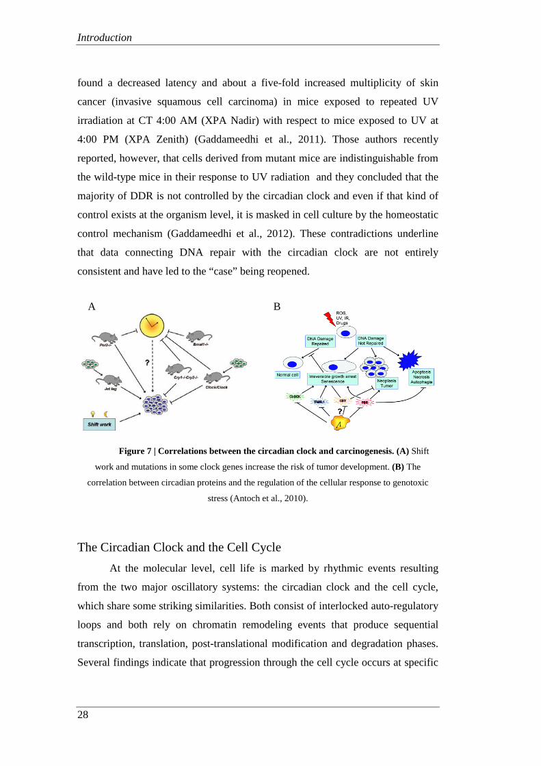
carcinogenesis. Experiments carried out on mice have revealed, in fact, that
interruption of normal rhythmicity due to surgical removal of the suprachiasmatic
nucleus or due to chronic exposure to frequent changes in the light-dark cycle
resulted in an accelerated growth of implanted tumors (Figure 7) (Antoch et al.,
2010). In agreement with an interconnection between functional defects in the
circadian system and carcinogenesis, cancer prognosis has, moreover, been found
to be less favorable in patients with an altered with respect to a normal circadian
rhythm (Sahar et al., 2009).
Several key cancer-related genes and signalling pathways have been found
to be potential targets for the circadian clock. It has been hypothesized that
potential molecular mechanisms underlying the link between the circadian clock
and cancer development include several DNA damage response components, such
as cell cycle progression control, DNA repair and apoptosis (Kondratov, 2012).
Recent studies have suggested that the principal mechanism involved in UV-
induced DNA repair, the Nucleotide Excision Repair (NER), is strictly controlled
by the circadian clock (Kang et al., 2009). Those authors analyzed the expression
of the main proteins involved in DNA repair of UV-induced damage over a 24 h
period. Their results showed that, unlike all other proteins of the Nucleotide
Excision Repair, XPA, the protein involved of the recognition of DNA dimers and
signal transduction, exhibits a circadian expression pattern. Other investigators
Introduction
28
found a decreased latency and about a five-fold increased multiplicity of skin
cancer (invasive squamous cell carcinoma) in mice exposed to repeated UV
irradiation at CT 4:00 AM (XPA Nadir) with respect to mice exposed to UV at
4:00 PM (XPA Zenith) (Gaddameedhi et al., 2011). Those authors recently
reported, however, that cells derived from mutant mice are indistinguishable from
the wild-type mice in their response to UV radiation and they concluded that the
majority of DDR is not controlled by the circadian clock and even if that kind of
control exists at the organism level, it is masked in cell culture by the homeostatic
control mechanism (Gaddameedhi et al., 2012). These contradictions underline
that data connecting DNA repair with the circadian clock are not entirely
consistent and have led to the “case” being reopened.
Figure 7 | Correlations between the circadian clock and carcinogenesis. (A) Shift
work and mutations in some clock genes increase the risk of tumor development. (B) The
correlation between circadian proteins and the regulation of the cellular response to genotoxic
stress (Antoch et al., 2010).
The Circadian Clock and the Cell Cycle At the molecular level, cell life is marked by rhythmic events resulting
from the two major oscillatory systems: the circadian clock and the cell cycle,
which share some striking similarities. Both consist of interlocked auto-regulatory
loops and both rely on chromatin remodeling events that produce sequential
transcription, translation, post-translational modification and degradation phases.
Several findings indicate that progression through the cell cycle occurs at specific
A B

Introduction
29
times of the day/night cycle and suggest that this process is controlled by the
circadian clock. Analysis of microarrays have revealed that many of the key
components of the cell cycle (such as cyclin D1, cyclin B1, cyclin E, cyclin A,
Wee1 and c -myc) have circadian expression. The three main components of cell
cycle network, p21, c-Myc and Wee1, were found to be regulated by the clock
proteins (Figure 8). The p21 gene, responsible for the G1-S-phase transition, is
under the control of RORα and REV-ERBα, which, in turn, respectively induce
and repress its transcription. The oncogene c-Myc, which plays a key role in cell
proliferation, is inhibited by the Per1 and Per2 genes, while Wee1, which
regulates the transition G2-M, is activated by the CLOCK-BMAL1 heterodimer
and repressed by CRYs proteins (Borgs et al., 2009). It has recently been
hypothesized that the RNA-binding protein NONO (p54nrb) is a synchronizing
factor between the circadian clock and the cell cycle (Kowalska et al., 2012). This
hypothesis is based on observations that NONO disruption completely abrogates
circadian gating of the cell cycle in fibroblasts without severely disrupting either
the circadian clock or the cell cycle. NONO, moreover, was found to be necessary
for correct dermal wound healing, and when this control was lacking there was a
loss of circadian cell cycle gating in vitro and tissue over-proliferation in vivo.
Figure 8 | Regulation of the cell cycle by the circadian clock. (Sahar et al., 2009)

Introduction
30
The Circadian Clock and Chromatin remodeling It has become increasingly clear that chromatin remodeling is one of the
processes through which the circadian clock regulates the transcription of genes
controlled by it (Sahar et al., 2013). The transcription process requires a rhythmic
assembly and recruitment of specific multiprotein complexes in the appropriate
sites of chromatin. This event is accompanied by modifications of clock proteins
and chromatin components, such as acetylation, deacetylation and methylation of
histones. Histone acetylation is a marker for transcription activation which is
achieved by remodeling the chromatin to make it more accessible to the
transcription machinery (Jenuwein and Allis, 2001). Histone methylation, on the
other hand, acts as a signal for recruitment of chromatin remodeling factors which
can either activate or repress transcription (Sahar et al., 2013). The key of this
epigenetic control of gene expression is CLOCK, a central component of the
circadian pacemaker, recently found to have histone acetyltransferase (HAT)
activity, essential for circadian clock-controlled gene expression. CLOCK is able
to acetylate the lysines 9 and 14 of histone H3 (H3K9 and H3K14), stimulating
the opening of the chromatin and promoting gene transcription. BMAL1, the
heterodimerization partner of CLOCK, seems to be involved in enhancing the
HAT function (Doi et al., 2006). The histone deacetylase sirtuin 1 (SIRT1) was,
moreover, found to regulate circadian rhythms by counteracting the HAT activity
of CLOCK (Nakahata et al., 2008). SIRT1 operates, thus, as a rheostat of the
circadian machinery, modulating the amplitude and “tightness” of CLOCK-
mediated acetylation and the consequent transcription cycles in metabolic tissues.
The prominent role of SIRT1 in longevity provides another interesting link
between the circadian clock and ageing. Perturbations in the expression or
activities of these regulators (CLOCK and SIRT1) could contribute to cancer
development by causing higher proliferation and defects in the metabolic
pathways (Sahar et al., 2013). Some investigators have recently reported that the
most pervasive circadian regulation observed on a genome scale are rhythms in
H3K4me3, H3K9ac (responsible for chromatin relaxation), H3K27ac, and
RNAPII recruitment and transcription initiation, which occur at thousands of

Introduction
31
expressed genes regardless of whether RNA cycling was detectable (Koike et al.,
2012). This demonstrates that gene expression per se exhibits circadian rhythms
with temporally separated activation and repression phases (Figure 9).
Figure 9 | Phase distributions of circadian transcriptional regulators, intron cycling RNA
transcripts, and histone modifications (Koike et al., 2012).
Access to genomic DNA for events such as DNA repair must be facilitated
by processes that directly alter chromatin structure leading to increased DNA
accessibility. Since different conformational states of chromatin could affect both
DNA damage incidence and repair processes, it is possible that the circadian clock
regulates DNA damage response by transcription-coupled chromatin remodeling.


Aim of the Study
33
Aim of the Study
The overall aim of this study was to investigate whether the rhythmic
progression of events resulting from the two major oscillatory systems, the cell
cycle and the circadian clock, affect the efficiency of DNA repair following
genotoxic stress induction. Specifically we planned the following objectives:
1. To analyze in human fibroblasts irradiated with γ-rays (a) the involvement
and the efficiency of the two main repair systems during the phases of cell
cycle and (b) the timing of HR recruitment during the repair incubation.
2. To investigate whether the impairment of NHEJ affects the efficiency of
HR to rejoin DSBs in G2 phase cells.
3. To develop a system to maintain cell culture in a long-term quiescence
status and to validate a set of techniques which allow to accurately
quantify the kinetics of UV-induced DNA damage repair.
4. To synchronize human cell cultures and induce the circadian expression of
various genes which oscillate in living animals.
5. To investigate whether the effectiveness of DDR is affected by the
circadian time at which the cells are irradiated.


35
Leonardo Bee1*, Sonia Fabris1*, Roberto Cherubini2, Maddalena Mognato1, and
Lucia Celotti1,2
1 Dipartimento di Biologia, Università di Padova, Padova, Italy.
2 Laboratori Nazionali di Legnaro, Istituto Nazionale di Fisica Nucleare, Padova, Italy.
*Equally contributing authors.
PAPER I
PLoS One 2013 Jul 11 8(7):e69061
[ THE EFFICIENCY OF
HOMOLOGOUS RECOMBINATION
AND NON-HOMOLOGOUS END
JOINING SYSTEMS IN REPAIRING
DOUBLE-STRAND BREAKS DURING
CELL CYCLE PROGRESSION ]


Paper I - Abstract
37
Abstract This study investigated the efficiency of Non-Homologous End Joining
(NHEJ) and Homologous Recombination (HR) repair systems in rejoining DNA
double-strand breaks (DSB) induced in CCD-34Lu cells by different γ-ray doses.
The kinetics of DNA repair was assessed by analyzing the fluorescence decrease
of γ-H2AX foci measured by SOID (Sum Of Integrated Density) parameter and
counting foci number in the time-interval 0.5-24 hours after irradiation.
Comparison of the two methods showed that the SOID parameter was useful in
determining the amount and the persistence of DNA damage signal after exposure
to high or low doses of ionizing radiation. The efficiency of DSB rejoining during
the cell cycle was assessed by distinguishing G1, S, and G2 phase cells on the
basis of nuclear fluorescence of the CENP-F protein. Six hours after irradiation, γ-
H2AX foci resolution was higher in G2 compared to G1 cells in which both NHEJ
and HR can cooperate. The rejoining of γ-H2AX foci in G2 phase cells was,
moreover, decreased by RI-1, the chemical inhibitor of HR, demonstrating that
homologous recombination is at work early after irradiation. The relevance of HR
in DSB repair was assessed in DNA-PK-deficient M059J cells and in CCD-34Lu
treated with the DNA-PKcs inhibitor, NU7026. In both conditions, the kinetics of
γ-H2AX demonstrated that DSBs repair was markedly affected when NHEJ was
absent or impaired, even in G2 phase cells in which HR should be at work. The
recruitment of RAD51 at DSB sites was, moreover, delayed in M059J and in
NU7026 treated-CCD-34Lu, with respect to DNA-PKcs proficient cells and
continued for 24 hours despite the decrease in DNA repair. The impairment of
NHEJ affected the efficiency of the HR system and significantly decreased cell
survival after ionizing radiation, confirming that DSB rejoining is strictly
dependent on the integrity of the NHEJ repair system.


Paper I - Introduction
39
Introduction It is known that exposure to ionizing radiation (IR) causes many types of
DNA damage, and, among these, double-strand breaks (DSBs) are considered the
most dangerous threat to genomic integrity [1, 2]. Radio-induced DSBs can have a
different complexity with respect to the ionization density of radiation. It has been
demonstrated that high-LET radiation induces clusters of DNA lesions along the
particle track while low-LET radiation causes sparse ionizations. When
administered at high doses, low-LET radiation can also, nevertheless, lead to
complex DNA damage [3] consisting of DSBs associated with base damages as
well as non-DSB damage clusters comprised of base lesions, apyrimidinic or
apurinic sites and single-strand breaks that can produce additional DSBs due to
damage processing [4].
The efficiency of DNA repair after exposure to IR depends on the
complexity of the radio-induced damage [5]. The presence of DSBs, whatever
their origin may be, elicits a complex DNA-Damage Response (DDR) consisting
of a cascade of events, involving damage sensing, signal transduction to the
effectors of DNA repair, cell cycle arrest, and induction of apoptosis [6]. After
exposure to IR, the extensive phosphorylation of histone H2AX at Ser139 results
in the formation of discrete γ-H2AX foci which can be easily identified by
immunostaining, a valuable tool highlighting the presence of DSBs [7, 8]. Since
phosphorylation of H2AX at Ser 139 is abundant, fast, and correlates well with
each DSB, it is the most sensitive marker that can be used to examine DNA
damage and subsequent lesion repair [9]. Apart from γ-H2AX, numerous
additional proteins that participate in DDR form Ionizing Radiation Induced Foci
(IRIF) through their recruitment and accumulation at DNA damaged sites and
often closely overlap with the relatively large γ-H2AX foci. One of these, the
tumor suppressor p53-binding protein 1 (53BP1) rapidly localizes at DSB sites
and activates p53 along with specific kinases. The number of 53BP1 foci has a
linear relationship with the irradiation dose, and the time course of 53BP1 foci
formation and disappearance is similar to that of γ-H2AX foci [10-14]. Another,
smaller type of foci restricted to stretches of single-stranded (ss) DNA produced
Paper I - Introduction
40
from DSB end resection is formed by the components of the homologous
recombination (HR) repair pathway, including Rad51 and RPA proteins. RPA
binds to ssDNA during the initial phase of homologous recombination. Just as in
DNA replication, this keeps ssDNA from binding to itself, in such a way that the
resulting nucleoprotein filament can then be bound by Rad51 and its cofactors
[15]. Broadly similar to the γ-H2AX foci detection, these additional foci provide
convenient surrogate markers useful for monitoring the presence of DNA DSBs or
the recruitment of HR repair proteins.
Eukaryotic cells rely on two highly regulated DSB repair pathways: the
non-homologous end joining (NHEJ) and homologous recombination (HR). The
former, which rejoins the DNA ends without requiring sequence homologies, is
carried out by the DNA-dependent protein kinase (DNA-PK) holoenzyme,
consisting of the heterodimer KU70/KU80 and the DNA-PK catalytic subunit
(DNA-PKcs) and by the DNA LIG4-XLF (Cernunnos)-XRCC4 complex. HR’s
central activity is coordinated by RAD51 protein, which catalyzes the strand
capture and invasion of broken ends of DSBs into intact homologous DNA
sequences, which are the sister chromatid or the homologous chromosome, to
ensure the fidelity of the repair process [16-19]. Although both NHEJ and HR
contribute to DSB rejoining, their involvement varies during the different cell
cycle phases as NHEJ is active throughout all cell cycle while HR is active during
the S and G2 phases when sister chromatids are available. Some authors observed
that the cell cycle control of DSB pathway choice can be bypassed in IR-exposed
cells, thus promoting a preferential repair by HR [20-22].
The involvement and efficiency of NHEJ and HR repair systems during
cell cycle phases in normal human CCD-34Lu fibroblasts exposed to different γ-
ray doses were analyzed here. The study aimed, moreover, to determine if the
impairment of DNA-PKcs protein by NU7026, a chemical inhibitor, or due to a
frameshift mutation in M059J cells, alters RAD51 protein activity during the
repair of γ-ray-induced DSBs. In order to analyze DSB repair at different stages of
the cell cycle, G1and G2 phases were distinguished on the basis of the nuclear
fluorescence intensity of CENP-F protein, whose expression and localization are
cell cycle-dependent. CENP-F is a protein of the nuclear matrix that gradually
Paper I - Introduction
41
accumulates during the cell cycle until it reaches peak levels in G2 and M phase
cells and is rapidly degraded after mitosis is complete [23]. It is thus detectable by
in situ immunofluorescence throughout the late S, G2, and M phases of the cell
cycle, but it is absent in the G1 one [24].
We used different methods based on quantifying foci fluorescence as an indicator
of DNA damage and repair to study the kinetics of DNA DSB rejoining during the
cell cycle. From our experiments, the Sum Of Integrated Density (SOID)
parameter [25] results a valuable tool which takes into account the number and the
size of IR-induced foci, allowing to accurately quantify DNA damage signal after
exposure to high or low doses of ionizing radiation [26].
Our data indicated that the NHEJ and HR repair systems cooperate in G2
phase cells in DSB rejoining not only long after irradiation, but also during the
first hours of post-irradiation incubation. We also noted that besides decreasing
the general efficiency of DNA repair, the impairment of NHEJ in CCD-34Lu
treated with the DNA-PKcs inhibitor, NU7026, as well as in DNA-PKcs deficient
M059J cells likewise affected RAD51 recruitment to DSB sites.


Paper I - Materials and Methods
43
Materials and Methods Cell lines
Normal human neonatal lung fibroblasts CCD-34Lu (ATCC N. CRL-
1491TM) were grown in high glucose (4.5 g/l) Dulbecco's Modified Eagle Medium
(DMEM) containing GlutaMAX (Gibco, Life Technologies), supplemented with
10% heat-inactivated fetal calf serum (FCS, Biochrom KG, Seromed), HEPES 20
mM (Sigma-Aldrich), 1% MEM non-essential amino acids (Gibco, Life
Technologies). At the time the experiments were carried out the cells were at 27 to
40 population doublings and actively proliferating, as confirmed by flow
cytometry analysis. Human malignant M059J glioblastoma cells were purchased
from ATCC (CRL-2366™), while M059K cells were kindly provided by
Professor S.C. West (Cancer Research UK London Research Institute, Clare Hall
Laboratories, South Mimms, UK). Both cell lines were grown in a 1:1 mixture of
DMEM and Ham’s F-12 medium (DMEM/F-12, Gibco, Life Technologies),
HEPES 20mM, 1% of MEM non-essential amino acids and 10% of heat-
inactivated FCS.
Cell irradiation
Gamma irradiation was performed at the Department of Oncological and
Surgical Sciences of the University Padova Medical Center with a 137Cs source
(dose rate of 2.8Gy/min). Cells (0.4x 10⁶) were seeded 48h before the experiment
and irradiated on Petri dishes (60x15mm), with or without coverslips, kept on ice
before and after irradiation, and cultured at 37°C in fresh medium for different
repair times. Except for irradiation, the control cells were subjected to the same
experimental conditions.
Immunofluorescence staining
The cells were fixed at 0.5, 2, 6 and 24h after irradiation for Fluorescence
Intensity (FI) analyses. Non-irradiated and irradiated cells were rinsed once with
cold Phosphate Buffered Saline (PBS) and fixed with a 4% solution of
formaldehyde (Sigma-Aldrich) at 37°C for 15 min and washed three times with
PBS. The cells were permeabilized with 0.5% Triton X-100 in PBS at 37°C for 10

Paper I - Materials and Methods
44
min and non-specific binding sites were masked with goat serum (10% in PBS) at
room temperature for 1h. The cells were incubated for 2h at room temperature
with anti-53BP1 (Bethyl Laboratories, 1:100), anti- γ-H2AX (Ser139) (Abcam or
Millipore Chemicon Upstate Clone JBW301, 1:100), anti-RAD51 (Santa Cruz
Biotechnology, H-92: sc-8349, lot. G0811, 1:100), anti–CENP-F (BD
Bioscience, 610768, Clone 11, 1:100 or Abcam, ab5, lot. GR73067-3, 1:300)
primary antibodies followed by three washings in PBS and once in PBS + 0.1%
Triton X-100. The cells were subsequently incubated at room temperature for 1h
with Alexa Fluor 488 goat anti-mouse secondary antibodies and Alexa Fluor 594
donkey anti-rabbit antibodies (Life Technologies, 1:250 and 1:350, respectively)
and washed, as described above. Immunofluorescent staining for RPA and R2
were performed using the MAX-Stain™ reagents (Active Motif) according to the
manufacturer’s instructions. Briefly, fixed cells were incubated for 1h at 37°C
with MAX-Block™ Blocking medium, washed 10 min with PBS, and incubated
for 1h at 37°C with primary anti-R2 (Santa Cruz, N-18 sc:10844, lot. K061,
1:200) and anti-RPA32/RPA2 antibodies (Abcam, 9H8 lot. GR92538-1, ab2175,
1:200) diluted in MAX-Bind™ staining medium. The cells were then washed
three times in PBS + 0.05% Tween-20, incubated for 1h at 37°C with secondary
antibodies diluted in MAX-Bind™ staining medium, and washed, as described
above. Cover slips were then mounted on glass slides with Vectashield mounting
medium (Vector Laboratories) containing DAPI 0.2 µg/ml.
Images of 53BP1, γ-H2AX and RAD51 and RPA foci were taken using a
Leica TCS SP5 confocal microscope (Leica Microsystems) with 40X or 63X oil
immersion objectives. All images were acquired under the same laser intensity,
PMC voltage, pinhole aperture, and 8-bit intensity value conditions. Z-plane stack
scanning (500 nm thickness) was performed using sequential scanning to prevent
crosstalk due to overlap of the emission spectra from the various fluorophores.
Manual counts of γ-H2AX and 53BP1 foci were performed using the maximum
intensity projection (MIP) images. The red and green images were superimposed
by ImageJ software (NIH) to obtain merged images. The number of γ-H2AX and
53BP1 foci was determined for each time point on an average of 100 nuclei in
Paper I - Materials and Methods
45
three independent experiments and listed in the figures after the number of foci in
the non-irradiated cells was subtracted.
Nocodazole treatment
We added the spindle poison nocodazole (Sigma-Aldrich) to CCD-34Lu to arrest
cell cycle progression during the M phase 1h before irradiation at 5 and 10 Gy at
the final concentration of 50ng/mL. After irradiation the cells were maintained for
2h in the drug’s presence and then analyzed by immunofluorescence for the
presence of CENP-F nuclear protein and RAD51 foci.
Quantification of nuclear fluorescence
The images acquired with the confocal microscope were processed and analyzed
with ImageJ software, using a specifically designed Macro to enable automated
analysis of a larger number of nuclei (average of 200 nuclei) for each time point.
All the images were processed to remove the background. The nuclear area for
each image was determined by 4’, 6-Diamidino-2-phyenylindole (DAPI)
fluorescent signal and saved as a list of coordinates for subsequent analyses.
Nuclear fluorescence was calculated as the mean intensity of all the pixels
included in the nuclear area. In accordance with Mistrik et al. [25] and Ishikawa et
al. [26], the SOID parameter was calculated for each nucleus as the product of the
sum of the area of the foci and the mean fluorescence intensity. An intensity
threshold was set to calculate the SOID so that only foci were included in the
analysis.
The nuclear fluorescence intensity (FI) of CENP-F protein was used to
discriminate the γ-H2AX and RAD51 SOID signal in the G2 and G1 cells. We
also assigned the specific range of CENP-F FI values to G1, S, and G2 cells
correlating CENP-F nuclear FI with the DNA replication phase using the EdU (5-
ethynyl-2-deoxyuridine, Life Technologies) staining method described by Salic et
al. and Buck et al. [27, 28], with minor modifications. Non-irradiated cells were
seeded on Petri dishes with glass coverslips 48h before labeling for EdU assay.
The cells were then incubated with EdU (30 µM) for 1h, rinsed three times with
PBS and fixed with 4% of formaldehyde for 15 min at 37°C. The cells were

Paper I - Materials and Methods
46
washed again before the “Click” stain reaction was performed and permeabilized
with Triton X-100 0.5% in TBS for 5 min at RT. The “Click” reaction was
performed incubating the cells for 30 min with a freshly prepared mix of 50 mM
Tris-HCl pH 7.3, 2 mM CuSO4, 5 µl/ml fluorescent 647-azide, 10 mM ascorbic
acid and used immediately after ascorbate was added. EdU-stained coverslips
were immunostained with CENP-F antibody, as described. Double stained slides
were acquired using a Leica TCS SP5 confocal microscope and nuclear
fluorescence was quantified. The range of CENP-F FI associated with the 95% of
EdU positive cells identifying S-phase cells was calculated while the CENP-F FI
values associated with EdU negative nuclei were assigned to the G1 cells. Finally,
the values of CENP-F FI associated with EdU negative nuclei but higher than the
maximum value of S-phase cells were assigned to the G2 cells. Throughout the
different cell cycle phases during analyses of DNA repair we excluded nuclei
with CENP-F intensity values within the confidence interval of S-phase, estimated
as the mean CENP-F values ± S.D.
Due to the incompatibility between CENP-F and RPA antibodies, we used
the presence of cytosolic RPA fluorescence of the ribonucleotide reductase R2
subunit as a marker of G1 and G2 phases [29]. R2 positive cells (S-G2) were
discriminated by the presence of cytosolic fluorescence.
FACS analyses
The cell cycle distribution of irradiated and non-irradiated control cells was
assessed by flow cytometry analysis of DNA content following staining with 50
µg/µl of propidium iodide (PI, Sigma-Aldrich), as previously described [30].
To analyze CENP-F content throughout the cell cycle, the cells were fixed
in 70% cold ethanol, rinsed twice in PBS, centrifuged at 200 g for 10 min at 4 °C,
and permeabilized in PBS with 0.1% TritonX-100 and 4% goat serum for 10 min
on ice. After centrifugation, the cells were incubated over night with primary
antibody diluted in permeabilization solution (mouse anti-CENP-F, 1:100). Then
the cells were rinsed three times in PBS with 2% of goat serum and incubated at
room temperature for 1h with agitation with secondary antibody (Alexa Fluor 488
goat anti-mouse) diluted in permeabilization solution. After three washings in

Paper I - Materials and Methods
47
PBS with 2% goat serum, the cells were stained with at 37 °C for 1h. FACS
analysis of total γ-H2AX content was carried out in a similar way, using a rabbit
anti-γ-H2AX (1:500) as the primary antibody and Alexa Fluor 488 goat anti-
mouse as the secondary one.
Data concerning FI were collected from 10x103-25x103 cells/sample using a
BD FACSCantoTM II flow cytometer (Becton Dickinson, BD Biosciences) and
analyzed using the ModFit LT software (Verity Software House).
NU7026 and RI-I treatments
To specifically inhibit NHEJ or HR, 24h before irradiation CCD-34Lu
cells were incubated with 10 µM NU7026 (DNA-PKcs inhibitor, Sigma-Aldrich)
or 10 µM of RI-1 (RAD-51 inhibitor, CALBIOCHEM), both diluted in DMSO.
After irradiation, the medium was replaced with a fresh one containing the
inhibitor, and the cells were incubated for the fixed repair times. Non-irradiated
cells were treated with DMSO only, NU7026 only, or RI-1 only to exclude any
potential toxicity from contributing to the effects of radiation; no differences were
detected in the various treatment conditions.
Cell viability
Cell viability was determined by a clonogenic assay in non-irradiated and
irradiated CCD-34Lu, incubated with or without NU7026, the DNA-PKcs
inhibitor, in M059K and M059J cells. After irradiation, 200 viable CCD34-Lu
cells were seeded together with feeder layer cells (IMR90, 15x105 cells/plate),
previously irradiated with 40 Gy of γ-rays in complete medium supplemented
with 15% serum in 10 cm diameter Petri dishes. When CCD-34Lu cells were
treated with NU7026 they were maintained for 24h with the inhibitor and then the
medium was replaced with a fresh one without the inhibitor. During clonogenic
assay, 500 viable M059K and M059J cells were seeded in complete medium,
without a feeder layer. Culture plates were scored for colony formation 14 days
later by staining cells with crystal violet 0.4%. Only colonies containing at least
50 cells were considered positive. Cell survival was calculated as the percentage
of cloning efficiency of treated cells over that of control cells.

Paper I - Materials and Methods
48
Statistical Analysis
Data from at least three separate experiments are presented as means ±
standard deviation (S.D.). All comparisons, with the exception of the cell survival
experiments, were calculated using Student’s t-test, in which case the P values are
based on a two-way ANOVA analysis. Differences with a <0.05 P-value are
considered significant.

Paper I - Results
49
Results
Kinetics of the formation and repair of DNA double-strand breaks
The formation and rejoining of DNA DSBs were analyzed by determining
the number of ionizing radiation induced foci (IRIF) of γ-H2AX and 53BP1
proteins in CCD-34Lu cells irradiated with 0.5 Gy of γ-rays. Our data indicated
that the kinetics of DSB rejoining is characterized by a complete resolution of
IRIF within 24h of irradiation and the almost complete co-localization of γ-H2AX
and 53BP1 foci (Figure 1A, B). Although the number of foci is correlated with the
number of DNA DBSs [9], this parameter alone cannot precisely quantify the
amount of DNA damage signal, which is linked to the size and persistence of foci
during DNA-repair kinetics. The SOID parameter, which accounts for the
number, the size, and the fluorescence density of ionizing radiation-induced foci,
was thus utilized to accurately quantify the DNA damage. The results obtained,
indicated in the text and in the figures as foci fluorescence intensity (FI), showed
that the resolution of foci in cells irradiated with 0.5 and 5 Gy occurred with
similar kinetics for both doses, even if with different values, according to the
dose-related intensity of DNA damage signal (Figure 1C). By comparing the
results of γ-H2AX kinetics obtained on the same samples of irradiated cells by
manual foci counting and SOID parameter, we observed that in 0.5 Gy irradiated
cells the kinetics of DNA damage signal (SOID parameter) and the DSB
resolution (foci number) were rather similar, as both methods showed a complete
DBS resolution in the 0.5-24h time-interval (Figure 1D). Following irradiation
with 5 Gy, it was impossible to count the foci manually as shortly after irradiation
the foci number was too high for reliable eye resolution. As a result we could not
compare the kinetics obtained using the two methods. We were, however, able to
observe that the decrease in the fluorescence of the foci detected by SOID
proceeded in a slower manner with respect to the decrease in the number of foci,
as demonstrated by the higher SOID values 2 and 6 hours after irradiation, mainly
due to the increase over time of the size of the foci (Figure S1). We also measured
the kinetics of γ-H2AX fluorescence using flow cytometry, which is a convenient

Paper I - Results
50
Figure 1. Kinetics of DSB rejoining in cells irradiated with γ-rays plotted against 24h of the
repair time. (A) DBS rejoining was measured by manual counting of γ-H2AX and 53BP1 foci
induced by irradiation with 0.5 Gy. (B) Representative pictures of γ-H2AX (green) and 53BP1 foci
(red) in nuclei (blue) at 0.5 and 2 h from γ-irradiation, showing the almost complete co-
localization of γ-H2AX and 53BP1 foci. (C) Fluorescence intensity (FI) of γ-H2AX foci has been
determined by SOID parameter in cells irradiated with 0.5 Gy and 5 Gy of γ-rays. (D) Comparison

Paper I - Results
51
of FI of γ-H2AX foci, induced by irradiation with 0.5 Gy and 5 Gy, analyzed by manual counting
and SOID parameter, expressed as increase over control. Error bars represent standard deviation of
the mean calculated from 3-4 experiments (*P<0.05, **P<0.01, ***P<0.001, SOID vs. foci
number, Student’s t-test).
method to analyze a high number of cells. In our experiments the method did not,
however, prove to be sensitive enough to detect differences in γ-H2AX FI at the
different time-points after irradiation with the lower IR dose (Figure S2). On the
basis of our experiments, we concluded that the SOID parameter was the most
useful method to evaluate both high and low values of γ-H2AX FI, reflecting the
amount and the persistence of DNA damage signal.
To evaluate the efficiency of DSB rejoining in the different cell cycle
phases, we used nuclear fluorescence intensity of CENP-F protein, whose
expression is cell cycle-dependent, to discriminate G1, S and G2 cells. By staining
the cells with CENP-F antibody, positive G2 cells can be easily distinguished
from negative G1 cells, but weakly stained cells, probably in the late S-phase,
cannot be reliably identified by this method (Figure 2B). To overcome this
difficulty, we determined the range of CENP-F fluorescence intensity of G1, S,
and G2 cells by labeling the S-phase of non-irradiated cells with EdU staining
(Figure 3). Cell distribution through cell cycle phases determined with CENP-F
fluorescence by confocal microscopy has been confirmed by propidium iodide
FACS analysis (Figure S3). By using SOID parameter, we were thus able to
monitor the kinetics of DSB resolution, based on the disappearance of damage
signaling, in G1, S, and G2 cells irradiated with 0.5 and 5 Gy of γ-rays. Our
results showed that the majority of IR-induced DSBs were repaired within the first
6 hours of irradiation in all cell

Paper I - Results
52
Figure 2. Cell cycle distribution of CCD-34Lu cells at 6 and 24h from irradiation with 0.5
and 5 Gy of γ-rays. (A) X axis, DNA content; Y axis, number of cells.*P<0.05; **P<0.01;
***P<0.001 (Student’s t-test). (B) Pictures of γ-H2AX foci (red) in nuclei counterstained with
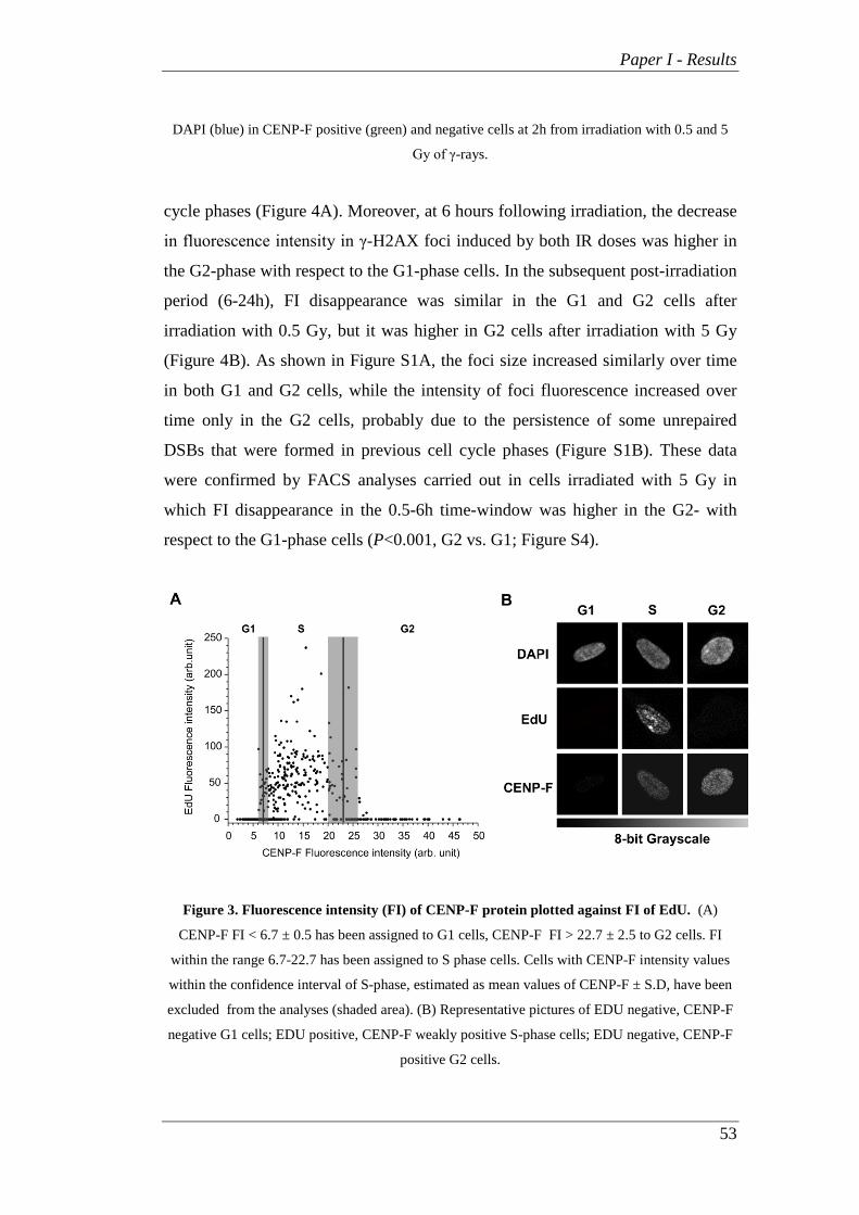
Paper I - Results
53
DAPI (blue) in CENP-F positive (green) and negative cells at 2h from irradiation with 0.5 and 5
Gy of γ-rays.
cycle phases (Figure 4A). Moreover, at 6 hours following irradiation, the decrease
in fluorescence intensity in γ-H2AX foci induced by both IR doses was higher in
the G2-phase with respect to the G1-phase cells. In the subsequent post-irradiation
period (6-24h), FI disappearance was similar in the G1 and G2 cells after
irradiation with 0.5 Gy, but it was higher in G2 cells after irradiation with 5 Gy
(Figure 4B). As shown in Figure S1A, the foci size increased similarly over time
in both G1 and G2 cells, while the intensity of foci fluorescence increased over
time only in the G2 cells, probably due to the persistence of some unrepaired
DSBs that were formed in previous cell cycle phases (Figure S1B). These data
were confirmed by FACS analyses carried out in cells irradiated with 5 Gy in
which FI disappearance in the 0.5-6h time-window was higher in the G2- with
respect to the G1-phase cells (P<0.001, G2 vs. G1; Figure S4).
Figure 3. Fluorescence intensity (FI) of CENP-F protein plotted against FI of EdU. (A)
CENP-F FI < 6.7 ± 0.5 has been assigned to G1 cells, CENP-F FI > 22.7 ± 2.5 to G2 cells. FI
within the range 6.7-22.7 has been assigned to S phase cells. Cells with CENP-F intensity values
within the confidence interval of S-phase, estimated as mean values of CENP-F ± S.D, have been
excluded from the analyses (shaded area). (B) Representative pictures of EDU negative, CENP-F
negative G1 cells; EDU positive, CENP-F weakly positive S-phase cells; EDU negative, CENP-F
positive G2 cells.

Paper I - Results
54
Figure 4. Kinetics of γ-H2AX foci in G1, S and G2 cells irradiated with γ-rays. (A) Values of
γ-H2AX foci FI are mean ± S.D. from SOID determinations carried out in at least four
independent experiments in cells irradiated with 0.5 and 5Gy. (B) Disappearance of γ-H2AX foci
FI in G1- and G2-phases cells calculated in the 0.5-6h and 6-24h time-windows after irradiation
(*P<0.05, **P<0.01, G2 cells vs. G1 cells, t-test).
Since our results suggest that DSBs are repaired faster in the G2-phase
cells during the first time-window after irradiation (0.5-6h), we analyzed the
kinetics of RAD51 and RPA foci formation, representative of HR repair system
involvement, in CCD-34Lu cells irradiated with 5 Gy (Figure 5). RAD51 foci
were clearly visible beginning 2 hours after irradiation, they peaked at 6h, and at
24h they were mostly disassembled. Foci of RPA displayed similar kinetics, with
fluorescence intensity values lower than those of RAD51. We can thus conclude
that some HR proteins are recruited at DSB sites during the first hours after
irradiation.

Paper I - Results
55
Figure 5. Analysis of RAD51 and RPA recruitment in CCD-34Lu cells irradiated with γ-
rays. Fluorescence intensity (FI) of foci was determined at 2, 6 and 24h after irradiation with 5Gy.
Values of FI are mean ± S.D. from SOID determinations carried out in at least three independent
experiments and plotted as increase over controls.
We then evaluated the contribution of HR and NHEJ in rejoining IR-
induced DSBs in G1- and G2-phase cells by quantifying γ-H2AX foci FI in CCD-
34Lu treated with NHEJ and HR inhibitors, NU7026 and RI-1, before irradiation
with 5Gy. As previously shown, the majority of DSBs (~60%) in untreated CCD-
34Lu cells were repaired during the first 6 hours after irradiation both in the G1-
and G2- phases (Figure 4), while HR- and NHEJ-inhibited cells showed a delay in
DSB resolution in both phases (Figure 6A). In particular, six hours after
irradiation the remaining FI in G1-phase cells was significantly higher in the

Paper I - Results
56
Figure 6. Contribution of HR and NHEJ pathways to DSB repair in G1- and G2-phases. (A)
The quantification of γ-H2AX FI induced by 5 Gy was performed by SOID parameter in untreated
CCD-34Lu and in NHEJ- and HR-inhibited cells, incubated respectively with the inhibitor of
DNA-PKcs, NU7026, and the inhibitor of RAD51, RI-1. (B) In G1 cells the remaining FI at 6h
after irradiation with 5 Gy increased in NHEJ-inhibited cells with respect to untreated cells (58%
vs. 35%, ***P<0.001, t-test). In G2 cells both inhibitors significantly increased the remaining FI at
6h after irradiation with respect to untreated cells (in NU7026-treèated cells FI was 63% vs. 39%
in untreated cells and 55% in RI-1 treated cells, **P<0.01, t-test). (C) Disappearance of γ-H2AX
foci FI in G1- and G2-phase of cells with similar initial SOID values obtained by irradiating the
cells respectively with 5 and 2.5 Gy. The values of FI at 0.5h after irradiation were 1132 ± 166 in
G2 cells irradiated with 2.5 Gy and 1450 ± 45 in G1 cells irradiated with 5Gy (see the box on the
right). At 6h after irradiation the remaining γ-H2AX foci FI was significantly higher in G2 cells
(**P<0.01, t-test).

Paper I - Results
57
NU7026-treated cells compared to the untreated ones (P<0.001, Figure
6B). After treatment with RI-1 inhibitor, instead, the percentage of remaining γ-
H2AX FI was similar to that in the untreated cells. Both inhibitors significantly
affected the repair of DSBs in G2-phase cells by increasing the fraction of
remaining γ-H2AX FI (P<0.01, Figure 6B). Since DSB repair kinetics have been
analyzed in cells that probably have different amounts of initial DNA damage, we
analyzed DSB rejoining of γ-H2AX foci in G1 cells irradiated with 5 Gy and in
G2 cells irradiated with 2.5 Gy. The initial values of SOID parameter for γ-H2AX
foci in G1 and G2-phase cells were very similar in these experimental conditions:
1450 ± 45 for G1 cells and 1132 ± 166 for G2 cells. The results outlined in Figure
6C confirmed that the decrease of γ-H2AX foci FI was slightly but significantly
higher in the G2 cells compared to the G1 cells (P<0.01).
DSB rejoining in DNA-PKcs-deficient cells
After we verified that the chemical inhibition of DNA-PKcs in CCD-34Lu
cells affected the rejoining of DSBs in the G1 and G2 cell cycle phases, we
analyzed the DNA repair kinetics in the DNA-PKcs-deficient M059J cells, and,
for comparison, in the isogenic DNA-PKcs proficient M059K cell line. As
reported in the literature [31] and detected in our experiments (not shown), both
cell lines are G1/ S checkpoint deficient. By contrast, and differently from M059K
cells, M059J retained the G2/M checkpoint, as shown by the significant increase
in G2-phase cells 24 hours after irradiation with 5 Gy (Figure S5). Figure 7 shows
that M059K cells rejoined almost completely the DSBs induced by 0.5 and 5 Gy
during the 24h of post-irradiation incubation, as was similarly observed in CCD-
34Lu (Figure 6A). In M059J cells, both in G1- and G2-phases, almost 60% of γ-
H2AX foci fluorescence persisted 24 hours after irradiation with 0.5 Gy, but there
was no fluorescence decrease after irradiation with 5Gy.
We analyzed the kinetics of RAD51 foci in CCD-34Lu cells incubated
with the DNA-PKcs inhibitor and in M059J cells to verify if the impairment of
NHEJ can alter the recruitment of HR proteins at DSB sites (Figure 8). In cells
proficient for DNA-PKcs (i.e. untreated CCD-34Lu cells and M059K cells) the
fluorescence intensity of RAD51 foci after irradiation with 5 Gy peaked at 6h and

Paper I - Results
58
then fell at 24h in both the G1 and G2 cells. On the contrary, in NU7026-treated
CCD-34Lu and in M059J cells the formation of RAD51 foci in the G2-phase was
strongly affected by the absence of DNA-PKcs activity, with an initial delay in the
RAD51 recruitment, followed by a progressive increase in foci FI up to 24h. The
kinetics of RAD51 foci was similar in NU7026-treated and untreated CCD-34Lu
cells in G1-phase, while it was very different in M059J and M059K cells, which
was true also in the G2-phase (Figure 8A).
Figure 7. DSB repair in cells with DNA-PKcs inhibited or absent. (A) Fluorescence intensity
(FI) of the remaining γ-H2AX foci was measured in G1- and G2-phase of M059K and M059J cells
irradiated with 0.5 and 5 Gy of γ-rays. (B) Representative pictures of γ-H2AX foci in CCD-34Lu
cells, with and without inhibitor, and in M059K and M059J cells at 0.5h and 24h after irradiation
with 0.5Gy.
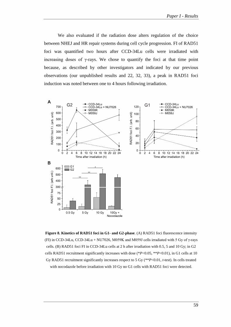
Paper I - Results
59
We also evaluated if the radiation dose alters regulation of the choice
between NHEJ and HR repair systems during cell cycle progression. FI of RAD51
foci was quantified two hours after CCD-34Lu cells were irradiated with
increasing doses of γ-rays. We chose to quantify the foci at that time point
because, as described by other investigators and indicated by our previous
observations (our unpublished results and 22, 32, 33), a peak in RAD51 foci
induction was noted between one to 4 hours following irradiation.
Figure 8. Kinetics of RAD51 foci in G1- and G2-phase. (A) RAD51 foci fluorescence intensity
(FI) in CCD-34Lu, CCD-34Lu + NU7026, M059K and M059J cells irradiated with 5 Gy of γ-rays
cells. (B) RAD51 foci FI in CCD-34Lu cells at 2 h after irradiation with 0.5, 5 and 10 Gy; in G2
cells RAD51 recruitment significantly increases with dose (*P<0.05, **P<0.01), in G1 cells at 10
Gy RAD51 recruitment significantly increases respect to 5 Gy (**P<0.01, t-test). In cells treated
with nocodazole before irradiation with 10 Gy no G1 cells with RAD51 foci were detected.

Paper I - Results
60
As expected, the recruitment of RAD51 at the sites of DSBs significantly
increased with the γ-ray dose in the G2 cells as well as after irradiation with 10
Gy, also in G1 cells (Figure 8B). The quantification of RPA foci FI was in
agreement with that of RAD51 in G2 cells, while there was a negative or very
scanty recruitment of RPA in G1 cells irradiated with 5-10 Gy (data not shown).
To check the origin of RAD51 loaded at the DBS sites of G1 cells irradiated with
10 Gy, we blocked cell-cycle progression from G2/M to G1-phase using
nocodazole, the inhibitor of the mitotic spindle. Under these experimental
conditions, no G1 cells with RAD51 foci were detected, indicating that the foci
previously observed were probably due to the persistence of unrepaired DSBs
formed in the G2-phase of cells that 2h later passed to G1-phase.
Figure 9. Cell survival after irradiation with increasing doses of γ-rays. Cells impaired for
NHEJ repair pathway showed a significant lower survival at 0.5, 1 and 2 Gy respect to their proper
control cells (CCD-34Lu+NU7026 vs. CCD-34Lu cells and M059J vs. M059K cells, ****P<
0.0001, two-way ANOVA). Data are means ± S.D. of 3-5 independent experiments.
The decrease in DNA repair efficiency in DNA-PKcs-inhibited CCD-43Lu and in
DNA-PKcs-deficient M059J cells markedly affected cell viability after irradiation
with increasing IR doses (Figure 9). Cell survival of NU7026-treated CCD-34Lu

Paper I - Results
61
was significantly lower than that of the untreated cells for all the doses
(P<0.0001). Similar results were observed in the M059J with respect to the DNA-
PK proficient M059K cells (P<0.0001). At the lowest dose of γ-rays (0.5 Gy), the
viability of untreated CCD-34Lu was slightly affected, while it was significantly
lower than that in non-irradiated cells when incubated with the NU7026 inhibitor
(62% vs. 92% ). The same γ-ray dose reduced the survival of M059K to 65% and
that of M059J to 15% with respect to the non-irradiated cells.


Paper I - Discussion
63
Discussion Increasing doses of γ-rays were utilized to investigate if DNA damage of
different structural complexities affects DSB signaling and repair. The questions
of whether and to what extent HR or NHEJ are involved in repairing radio-
induced DSBs were also addressed. The shift from NHEJ toward HR as the cell
cycle progresses from G1 to S/G2 is regulated by CDK activity. It has been seen
that during homologous recombination repair process CDK-mediated
phosphorylation at ser3291 of BRCA2 blocks its interaction with the RAD51
protein required for homology searching and strand invasion. HR is, thus, blocked
when ssDNA resection is prevented by CDK1 inhibition and early during G1-
phase when CDK1 is inactive. Some investigators have reported that in cells
exposed to IR cell cycle checkpoint control can be bypassed, leading to a rapid
decrease in ser3291 phosphorylation and an increased association of BRCA2 with
RAD51 to promote DSB repair by HR [18, 34, 35].
In agreement with previous data demonstrating that the time course of
53BP1 foci formation and disappearance is similar to that of γ-H2AX foci, our
results in human fibroblasts irradiated with 0.5 Gy of γ-rays indicate that the
kinetics of γ-H2AX and 53BP1 foci is very similar (Figure 1) [10-14]. To verify
whether DSB repair efficiency was dose-related, the kinetics of γ-H2AX cellular
content after irradiation with increasing doses of γ-rays was assessed using
different methods : manual counting of nuclear foci, fluorescence intensity of foci
measured by the SOID parameter and flow cytometry. In agreement with
Ishikawa et al.’s findings, our results indicated that the SOID parameter is an
accurate method to quantify even slight variations of γ-H2AX fluorescence [26].
By contrast, γ-H2AX kinetics after 5 Gy irradiation could not be evaluated by
manual foci counting since the number of foci induced by that dose was too high
for a reliable determination; nor does the method take into account the increase in
foci size detected at later times after irradiation. When the kinetics of DSB
rejoining was analyzed by means of the SOID parameter, it was possible to
quantify not only the number of the remaining foci but also the total fluorescence
of DDR proteins still associated to DSBs. In particular, the size increase of γ-

Paper I - Discussion
64
H2AX foci over time markedly influences the SOID parameter value, highlighting
the persistence of some DSBs difficult to repair during the post-irradiation
incubation.
By comparing the efficiency in rejoining radiation-induced DSBs during
cell cycle progression, we found that in G2-phase cells, whose frequency
significantly increased after irradiation (Figure 2A), DNA repair proceeded very
efficiently, both during the first period of repair-incubation (0.5-6h) and at later
times (6-24h, Figure 4). Previous studies reported that the kinetics of IR-induced
DSB repair in the G1-phase exhibit fast as well as slow components [36, 37]. The
former removes the majority of DSBs within the first 2 h, while the latter
represents a sub-pathway of the NHEJ pathway [38, 39]. Just as in G1, DSB-
repair kinetics are biphasic in G2-phase with the fast component representing
NHEJ and accounting for the majority of DSB-repair events, while the slow
component represents HR and accounts for 15-20% of DSB-repair events [22].
We analyzed the kinetics of γ-H2AX foci in the G1- and G2-phases of HR-
inhibited cells to evaluate the contribution of HR in rejoining DNA DSBs during
cell cycle progression. According to our results, G2-phase cells treated with the
HR inhibitor (RI-1) exhibited a higher level of unrepaired DSBs 6 hours after
irradiation (Figure 6). By comparing the repair rate in G1 and G2 phase cells
having a similar amount of initial DNA damage, we were able to confirm that the
HR repair system contributed to rejoining DSBs also during the first hours
following irradiation (Figure 6C), differently from what reported by Beucher et al.
[22]. The differences between our and Beucher’s findings could be explained by
the two different methods adopted to measure the decreased level of γ-H2AX:
counting the number of foci used by Beucher [22] and the SOID parameter used
during our experiments. The hypothesis that HR is involved in the first hours after
IR is, moreover, supported by recent observations by Gandhi et al., [40] who
demonstrated that within 5 min of irradiation, homologous chromosomes make
contact at the sites of DSBs induced by ionizing radiation in human G0-G1 cells.
Proteins that mediate the recruitment of HR and NHEJ are generally
distinct, but in some cases they are implicated in both pathways. Among those
proteins, DNA-PK primarily regulates DSB repair by NHEJ, but it can also

Paper I - Discussion
65
influence HR. Several studies have reported that when DNA-PKcs is absent and
NHEJ is compromised, HR repair is enhanced [41]. Other authors have, instead,
reported that HR involvement is reduced during DNA repair of cells in which
DNA-PKcs is physically present but functionally compromised [42]. It would
seem then that functionally compromised DNA-PKcs has more severe
consequences than the enzyme’s complete absence, probably blocking its auto-
phosphorylation that allows the NHEJ pathway to proceed [37]. Experiments on
CCD34-Lu cell line in which DNA-PKcs was impaired through chemical
inhibition (NU7026) and DNA-PK deficient M059J cells as a consequence of a
nonsense frame-shift mutation [43] were carried out to evaluate contribution of
HR in DSB repair. The presence of DNA-PKcs inhibitor significantly decreased
the efficiency of DSB rejoining in irradiated CCD-34Lu cells, both in G1- and
G2-phase, as well as in M059J cells (Figures 6 and 7). Moreover, when NHEJ
was compromised, the recruitment of RAD51 at DSB sites in both NU7026-
treated CCD-34Lu and M059J cells was low early after irradiation and continued
up to 24h in both G1 and G2 cells (Figure 8). In M059J cells, DSBs present in the
G2-phase were partially caused by lesions induced in this phase of the cell cycle
as well as by unrepaired lesions that occurred in the G1-phase and accumulated
24h later in the G2-phase due to the lack of a G1/S checkpoint. However, despite
the extensive recruitment of RAD51 at DSB sites 24h after irradiation, the HR
pathway, which should be fully functional in those cells, is unable to compensate
for the impairment of NHEJ, as evidenced by the persistence of γ-H2AX
phosphorylation and the low level of cell survival, even after irradiation with low
doses of ionizing radiation.
As we analyzed if radiation dosage alters the choice between the NHEJ
and the HR repair systems during cell cycle progression, we observed that RAD51
was recruited at DSB sites even in G1 cells irradiated with the highest dose.
Although in accordance with data outlined by Kim et al. [33] and by Rapp and
Greulich [44] showing the presence of RAD51 foci in the G1-phase of irradiated
cells, these observations can be more accurately interpreted by blocking the
transition of cells from the G2/M- to the G1-phase. Indeed, cells irradiated with
high doses of IR in the G2-phase could be able to progress to the G1-phase with

Paper I - Discussion
66
unrepaired DSBs. By blocking the progression of G2 cells with unrepaired DSBs,
we demonstrated that the presence of G1 cells positive for RAD51 foci can be
explained by the advancement of cells with RAD51 foci formed in the S-G2
phases.
Our data generally agree with the model that NHEJ is the major pathway
for IR-induced DSBs repair. They also demonstrate that RAD51, the main protein
in the HR pathway, participates, together with NHEJ, in DSB rejoining even
during the first hours after irradiation. RAD51 recruitment and activity at DSB
sites appears, however, to be strictly dependent on the integrity of NHEJ
components, highlighting the dominant role that DNA-PKcs play in regulating the
cell response to DNA damage throughout cell cycle progression.

Paper I - References
67
Acknowledgements We gratefully acknowledge Vito Barbieri
for cell irradiation with γ-rays, and Prof.
S.C. West (Cancer Research, UK London
Research Institute, Clare Hall Laboratories,
South Mimms) for providing M059K cells.
We would also like to thank Chiara
Romualdi for statistical analysis and
helpful discussion, Giovanna Pontarin for
assistance with R2 immunostaining,
Antonella Russo for providing
experimental protocols for nocodazole
treatment, Cristina Girardi for foci
counting, and Renzo Mazzaro for help
with graphics.
Author Contributions
Conceived and designed the experiments:
LC LB MM SF. Performed the
experiments: LB SF MM. Analyzed the
data: LC LB MM RC. Wrote the paper: LC
MM LB SF.
References 1. Ohnishi T, Mori E, Takahashi A (2009)
DNA double-strand breaks: Their
production, recognition, and repair in
eukaryotes. Mutat Res 66: 8-12.
2. Vilenchik MM, Knudson AG (2003)
Endogenous DNA double-strand breaks:
production, fidelity of repair, and induction
of cancer. Proc Natl Acad Sci USA 100:
12871-876.
3. Ward JF (2000) Complexity of damage
produced by ionizing radiation. Cold
Spring Harb Symp Quant Biol 65: 377-
382.
4. Eccles LJ, O'Neill P, Lomax ME (2011)
Delayed repair of radiation induced
clustered DNA damage: friend or foe?
Mutat Res 711: 134-141.
5. Nikjoo H, O'Neill P, Wilson WE,
Goodhead DT (2001) Computational
approach for determining the spectrum of
DNA damage induced by ionizing
radiation. Radiat Res 156: 577-583.
6. Bassing CH, Alt WF (2004) The cellular
response to general and programmed DNA
double strand breaks. DNA Repair (Amst)
3: 781-796.
7. Fernandez-Capetillo O, Lee A,
Nussenzweig M, Nussenzweig A (2004)
H2AX: the histone guardian of the
genome. DNA Repair (Amst) 3: 959-967.
8. Svetlova MP, Solovjeva LV, Tomilin
NV (2010) Mechanism of elimination of
phosphorylated histone H2AX from
chromatin after repair of DNA double-
strand breaks. Mutat Res 685: 54-60.
9. Sharma A, Singh K, Almasan A (2012)
Histone H2AX Phosphorylation: A Marker
for DNA Damage. Methods in Molecular
Biology 920:613-626.
10. Ward IM, Minn K, Jorda KG, Chen J
(2003) Accumulation of checkpoint protein
53BP1 at DNA breaks involves its binding
to phosphorylated histone H2AX. J Biol

Paper I - References
68
Chem 278: 19579-582.
11. Schultz LB, Chehab NH, Malikzay A,
Halazonetis TD (2000) p53 binding protein
1 (53BP1) is an early participant in the
cellular response to DNA double-strand
breaks. J Cell Biol 151: 1381-390.
12. Rappold I, Iwabuchi K, Date T, Chen J
(2001) Tumor suppressor p53 binding
protein 1 (53BP1) is involved in DNA
damage-signaling pathways. J Cell Biol
153: 613-620.
13. Anderson L, Henderson C, Adachi Y
(2001) Phosphorylation and rapid
relocalization of 53BP1 to nuclear foci
upon DNA damage. Mol Cell Biol 21:
1719-729.
14. Asaithamby A, Chen DJ (2009)
Cellular responses to DNA double-strand
breaks after low-dose gamma-irradiation.
Nucleic Acids Res 37: 3912-923.
15. Wold MS (1997). "Replication protein
A: heterotrimeric, single-stranded DNA-
binding protein required for eukaryotic
DNA metabolism". Annual Review of
Biochemistry 66 (1): 61–92.
16. Lisby M, Rothstein R (2009)
Choreography of recombination proteins
during the DNA damage response. DNA
Repair 8: 1068-076.
17. Shrivastav M, De Haro LP, Nickoloff
JA (2005) Regulation of DNA double-
strand break repair pathway choice. Cell
Res 18: 134-147.
18. Shrivastav M, Miller CA, De Haro LP,
Durant ST, Chen BPC, et al. (2009) DNA-
PKcs and ATM co-regulate DNA double-
strand break repair. DNA Repair 8: 920-
929.
19. Tracker J (2005) The RAD51 gene
family, genetic instability and cancer.
Cancer Lett 219: 125-135.
20. Sedelnikova OA, Rogakou EP,
Panyutin IG, Bonner WM (2002)
Quantitative detection of (125)IdU-
induced DNA double-strand breaks with
gamma-H2AX antibody. Radiat Res 4:
486-492.
21. Asaithamby A, Uematsu N, Chatterjee
A, Story MD, Burma S, et al. (2008)
Repair of HZE-particle-induced DNA
double-strand breaks in normal human
fibroblasts. Radiat Res 169: 437-446.
22. Beucher A, Birraux J, Tchouandong L,
Barton O, Shibata A, et al. (2009) ATM
and Artemis promote homologous
recombination of radiation-induced DNA
double-strand breaks in G2. EMBO J 28:
3413-427.
23. Liao H, Winkfein RJ, Mack G, Rattner
JB, Yen TJ (1995) CENP-F is a protein of
the nuclear matrix that assembles onto
kinetochores at late G2 and is rapidly
degraded after mitosis. J Cell Biol 130:
507-18.
24. Varis A, Salmela AL, Kallio MJ (2006)
Cenp-F (mitosin) is more than a mitotic
marker. Chromosoma 115: 288-295.

Paper I - References
69
25. Mistrik M, Oplustilova L, Lukas J,
Bartek J (2009) Low-dose DNA damage
and replication stress responses quantified
by optimized automated single-cell image
analysis. Cell Cycle 8: 2592-599.
26. Ishikawa A, Yamauchi M, Suzuki K,
Yamashita S (2010) Image-based
quantitative determination of DNA damage
signal reveals a threshold for G2
checkpoint activation in response to
ionizing radiation. Genome Integr 1:10.
27. Salic A, Mitchison TJ (2008) A
chemical method for fast and sensitive
detection of DNA synthesis in vivo. Proc
Natl Acad Sci USA 105: 2415-420.
28. Buck SB, Bradford J, Gee KR, Agnew
BJ, Salic A (2008) Detection of S-phase
cell cycle progression using 5-ethynyl-2'-
deoxyuridine incorporation with click
chemistry, an alternative to using 5-bromo-
2'-deoxyuridine antibodies. Biotechniques
44: 927-929.
29. Pontarin G, Fijolek A, Pizzo P, Ferraro
P, Rampazzo C, et al. (2008)
Ribonucleotide reduction is a cytosolic
process in mammalian cells independently
of DNA damage. Proc Natl Acad Sci USA.
105: 17801–17806.
30. Canova S, Fiorasi F, Mognato M,
Grifalconi M, Reddi E, et al. (2005)
“Modeled microgravity" affects cell
response to ionizing radiation and
increases genomic damage. Radiat Res
163: 191-199.
31. Holgersson A, Heiden T, Castro J,
Edgren MR, Lewensohn R, et al. (2005)
Different G2/M accumulation in M059J
and M059K cells after exposure to DNA
double-strand break-inducing agents. Int J
Radiat Oncol Biol Phys. 61(3):915-21.
32. Short SC, Giampieri S, Worku M,
Alcaide-German M, Sioftanos G, et al.
(2011) Rad51 inhibition is an effective
means of targeting DNA repair in glioma
models and CD133+ tumor-derived cells.
Neuro Oncol 13: 487-99.
33. Kim JS, Krasieva TB, Kurumizaka H,
Chen DJ, Taylor AM, et al. (2005)
Independent and sequential recruitment of
NHEJ and HR factors to DNA damage
sites in mammalian cells. J Cell Biol
170(3):341-7.
34. Mao Z, Bozzella M, Seluanov A,
Gorbunova V (2008) Comparison of non-
homologous end joining and homologous
recombination in human cells. DNA
Repair 7: 1765-771.
35. Mukherjee B, Camacho CV,
Tomimatsu N, Miller J, Burma S (2008)
Modulation of the DNA-damage response
to HZE particles by shielding. DNA Repair
7: 1717-730.
36. Löbrich M, Jeggo PA (2005)
Harmonising the response to DSBs: a new
string in the ATM
bow. DNA Rep (Amst) 4: 749–759.
37. Wu W, Wang M, Wu W, Singh SK,
Mussfeldt T, et al. (2008) Repair of

Paper I - References
70
radiation induced DNA double strand
breaks by backup NHEJ is enhanced in G2.
DNA Repair (Amst) 7: 329-338.
38. Rothkamm K, Krüger I, Thompson
LH, Löbrich M (2003) Pathways of DNA
double-strand
break repair during the mammalian cell
cycle. Mol Cell Biol 23: 5706–5715.
39. Kühne M, Riballo E, Rief N,
Rothkamm K, Jeggo PA, et al. (2004) A
double-strand
break repair defect in ATM-deficient cells
contributes to radiosensitivity. Cancer Res
64:
500–508.
40. Gandhi M, Evdokimova VN, Cuenco
KT, Bakkenist CJ, Nikiforov YE (2013)
Homologous chromosomes move and
rapidly initiate contact at the sites of
double-strand breaks in genes in G₀-phase
human cells. Cell Cycle 12(4):547-52.
41. Allen C, Halbrook J, Nickoloff JA
(2003) Interactive competition between
homologous recombination and non-
homologous end joining. Mol Cancer Res
1: 913-920.
42. Convery E, Shin EK, Ding Q, Wang
W, Douglas P, et al. (2005) Inhibition of
homologous recombination by variants of
the catalytic subunit of the DNA-
dependent protein kinase (DNA-PKcs).
Proc Natl Acad Sci USA 102: 1345-350.
43. Anderson CW, Allalunis-Turner MJ
(2000) Human TP53 from the malignant
glioma-derived cell lines M059J and
M059K has a cancer-associated mutation
in exon 8. Radiat Res 154: 473-476.
44. Rapp A, Greulich KO (2004) After
double-strand break induction by UV-A,
homologous recombination and non-
homologous end joining cooperate at the
same DSB if both systems are available. J
Cell Sci. 117: 4935-45.

Paper I - Supporting Information
71
Supporting information
Figure S1. Analysis of γ-H2AX foci physical parameters in G1 and G2 cells after irradiation with
5 Gy of γ-rays. (A) γ-H2AX foci size increased significantly with time after irradiation in both G1
and G2 phases (*P<0.05, **P<0.01, t-test) without differences between the two phases. (B) Signal
intensities of γ-H2AX foci were determined by SOID parameter in the same cells. The intensity of
foci fluorescence increased with time only in G2 cells (***P<0.001, t-test). The values are mean
fluorescence intensity ± S.D. of single γ-H2AX foci determined from at least 30-50 nuclei for each
time-point.
Figure S2. Kinetics of γ-H2AX total fluorescence determined by FACS analysis in cells irradiated
with 0.5 and 5 Gy of γ-rays.

Paper I - Supporting Information
72
Figure S3. Comparison of data of cell distribution in cell cycle phases obtained from FACS
analysis and from confocal microscopy of CENP-F F.I. in the same cell population.
Figure S4. γ-H2AX total fluorescence determined by FACS analysis. Kinetics of γ-H2AX in G1,
S and G2 cells irradiated with 5 Gy of γ-rays. (B) In the time-interval 0.5-6h, the γ-H2AX FI
disappearance was significantly higher in G2 vs. G1 cells ( P<0.001, G2 vs. G1).

Paper I - Supporting Information
73
Figure S5. Cell cycle distribution in M059J and M059K cells after irradiation with 5Gy of γ-rays.
In both cell lines γ-irradiation did not induce the G1/S checkpoint, whereas only in M059J cells the
G2/M checkpoint was activated. Data are mean ± S.D. from three independent experiments.


75
Giovanna Pontarin*, Paola Ferraro*, Leonardo Bee, Peter Reichard, and Vera
Bianchi
Dipartimento di Biologia, Università di Padova, Padova, Italy.
*Equally contributing authors.
Author contributions: G.P., P.F., L.B., P.R., and V.B. designed research; G.P., P.F., and L.B. (Figure 7 and Figure S3) performed research; G.P., P.F., L.B. (Figure 7 and Figure S3), P.R., and V.B. analyzed data; and G.P., P.F., P.R., and V.B. wrote the paper.
PAPER II
Proc Natl Acad Sci USA 2012 Aug 14 109(33):13302-7
[ MAMMALIAN RIBONUCLEOTIDE
REDUCTASE SUBUNIT P53R2 IS
REQUIRED FOR MITOCHONDRIAL
DNA REPLICATION AND DNA
REPAIR IN QUIESCENT CELLS ]


Paper II - Abstract
77
Abstract In postmitotic mammalian cells, protein p53R2 substitutes for protein R2 as a
subunit of ribonucleotide reductase. In human patients with mutations in RRM2B,
the gene for p53R2, mitochondrial (mt) DNA synthesis is defective, and skeletal
muscle presents severe mtDNA depletion. Skin fibroblasts isolated from a patient
with a lethal homozygous missense mutation of p53R2 grow normally in culture
with an unchanged complement of mtDNA. During active growth, the four dNTP
pools do not differ in size from normal controls, whereas during quiescence, the
dCTP and dGTP pools decrease to 50% of the control. We investigate the ability
of these mutated fibroblasts to synthesize mtDNA and repair DNA after exposure
to UV irradiation. Ethidium bromide depleted both mutant and normal cells of
mtDNA. On withdrawal of the drug, mtDNA recovered equally well in cycling
mutant and control cells, whereas during quiescence, the mutant fibroblasts
remained deficient. Addition of deoxynucleosides to the medium increased
intracellular dNTP pools and normalized mtDNA synthesis. Quiescent mutant
fibroblasts were also deficient in the repair of UV-induced DNA damage, as
indicated by delayed recovery of dsDNA analyzed by fluorometric analysis of
DNA unwinding and the more extensive and prolonged phosphorylation of
histone H2AX after irradiation. Supplementation by deoxynucleosides improved
DNA repair. Our results show that in nontransformed cells only during
quiescence, protein p53R2 is required for maintenance of mtDNA and for optimal
DNA repair after UV damage.


Paper II - Introduction
79
Introduction DNA replication and repair require the continued synthesis of the four
dNTPs. They are synthesized by evolutionary-related ribonucleotide reductases
operating with slightly different mechanisms in aerobic and anaerobic organisms
(1). Each ribonucleotide reductase provides the required amounts of all four
dNTPs. A similar allosteric mechanism, maintained throughout evolution,
regulates both the enzyme’s activity and its substrate specificity. Cells contain
small dNTP pools of similar sizes, approximately 10-fold larger during DNA
replication than during quiescence. Regulation of pool sizes by ribonucleotide
reductases is of great importance for correct DNA replication, and changes in the
actual sizes or in their balance lead to increased mutation rates (2). For
mammalian cells, the induction of mutations by pool imbalances has been
described in detail, along with possible mechanisms (3). In yeast, a recent elegant
study (4) linked specific amino acid substitutions in the catalytic subunit of
ribonucleotide reductase to defined pool imbalances, which result in increased
mutation rates.
In mammalian cells, the canonical ribonucleotide reductase is a complex
between two proteins: the large catalytic protein R1 that contains the allosteric
sites and the smaller protein R2 that contributes a stable tyrosyl free radical during
the reaction (1). Both proteins are transcriptionally activated during early S-phase
(5) and are present in roughly equal amounts (6, 7) to deliver the dNTPs required
for DNA replication. R2 is degraded during late mitosis (8); thus, postmitotic
quiescent cells are essentially devoid of R2 but retain some R1. In the year 2000,
a second radical-providing small subunit, termed p53R2, was discovered in
mammalian cells (9). p53R2 has the same function as the homologous R2, but is
not degraded in mitosis. Quiescent cells contain an undiminished amount of
p53R2 (6, 7) but little or no R2. After DNA damage, p53R2 is transcriptionally
activated by p53 and was reported to translocate into the nucleus (9). It was
therefore thought to be primarily involved in DNA repair. However, with affinity-
purified antibodies, we found p53R2, as well as R1 and R2, in the cytosol also
after DNA damage (10). Moreover the concentrations of the three proteins at

Paper II - Introduction
80
different stages of the cell cycle speak against a specific requirement of p53R2 for
DNA repair. During S-phase, murine or human fibroblasts contain roughly
equimolar amounts of R1 and R2, whereas p53R2 amounts to only 3% of R2 and
increases to 13% after DNA damage (6), indicating that the main catalytically
active enzyme is an R1/R2 and not an R1/p53R2 complex. In a study of quiescent
human fibroblasts, ribonucleotide reduction was catalyzed largely by an
R1/p53R2 complex at a rate amounting to only 2–3% of that of cycling cells (7).
In 2007, Bourdon et al. (11) opened a new chapter in the history of p53R2
by reporting that in humans, genetic inactivation of p53R2 causes a severe
mitochondrial disease characterized by profound depletion of mtDNA in
differentiated cells and lethality shortly after birth. They found that skeletal
muscle of patients with mutations in the RRM2B gene coding for p53R2
completely lacked mtDNA (11). Similar patients were described in later studies
from several other laboratories (12–16). It is now clear that p53R2 activity is
required for the stability of the mt genome in differentiated tissues, shifting the
attention from DNA repair to mtDNA maintenance.
Less clear are the extent to which p53R2 is required for DNA repair and
the extent to which it is required only in quiescent cells. Most experiments
concerning p53R2’s function have been carried out with transformed cell lines (9,
17–19), which are not suitable for addressing these questions. To investigate
p53R2 in nontransformed cells, we recently examined in vitro the consequences
of p53R2 inactivation with fibroblasts from a patient with a lethal homozygous
missense mutation in the iron-binding center of p53R2 who had died at aged 3 mo
with severe muscular mtDNA depletion (16, 20). Compared with age-matched
controls, the mutant fibroblasts grew normally in culture and contained a normal
complement of mtDNA (20); however, once they became quiescent, their ability
to reduce ribonucleotides was strongly curtailed, resulting in smaller dCTP and
dGTP pools. The profound changes in deoxyribonucleotide metabolism did not
result in a depletion of mtDNA in vitro, unlike in the patient. We hypothesized
that this ostensible paradox might be explained by the low copy number of

Paper II - Introduction
81
mtDNA in fibroblasts, only <5% of that in skeletal muscle cells, requiring much
less dNTPs for its maintenance.
In the present work, we tested this hypothesis with cycling and quiescent
fibroblasts. We induced mtDNA depletion by treating the cells with ethidium
bromide (EtBr) (21), and followed the recovery of mtDNA after removal of the
drug. In addition, we investigated the involvement of p53R2 in DNA repair by
analyzing the ability of the mutated fibroblasts to repair DNA after UV damage.
Our data demonstrate the importance of p53R2 for both mtDNA replication and
DNA repair in quiescent cells that contain insufficient R2 for dNTP synthesis.
The data indicate that correct pool balances are required not only for the fidelity
of nuclear DNA replication, but also for optimal mtDNA synthesis and DNA
repair after UV damage.


Paper II - Materials and Methods
83
Materials and Methods
Cell culture
The fibroblasts from a patient with an inactivating mutation in the gene for
p53R2 (16) were identical to those used in a previous study (20). G. Kollberg and
E. Holme (Sahlgrenska University Hospital, Goteborg, Sweden) provided the
original frozen cells (second passage) and suitable age-matched normal controls.
Before use, both cell lines were immortalized with plasmid CMV-hTERT/PGK-
Pura, as described previously (36). The cells for the experiments were seeded at
0.35 × 10⁶ cells/10-cm dish and grown in MEM/10% FCS. Quiescent cultures
were obtained by growing cells to confluence (usually for 7 d), changing the
medium to MEM/0.1% dialyzed FCS, and keeping the cells in this medium until
use in the experiments at different times, usually after 7 d. Cultures received fresh
medium every third day. Where indicated, the medium was supplemented with 5
μM deoxynucleosides along with a medium change. To prevent degradation of
individual deoxynucleosides, 0.5 μM immucillin (37) was added together with
GdR and 10 μM erythro-9-2-hydroxy-3-nonyladenine (38) with AdR.
Materials
MEM and New Zealand FCS were obtained from Invitrogen. Erythro-9-2-
hydroxy 3-nonyladenine was a gift from Staffan Eriksson (Swedish University of
Agricultural Sciences, Uppsala, Sweden), and Immucillin H was a gift from Vern
Schramm(Yeshiva University,NewYork, NY). Ethidium bromide (EtBr),
deoxynucleosides, and deoxynucleotides were purchased from Sigma-Aldrich.
The following mouse monoclonal antibodies were used: anti-cyclobutane
pyrimidine dimers (CPDs) (clone TDM-2; CosmoBio), anti-(6,4) photoproducts
(6-4PPs) (clone 64M-2; CosmoBio), and anti–phospho-histone H2AX (Ser139)
(clone JBW301; Millipore).

Paper II - Materials and Methods
84
Analytical Procedures
Intracellular concentrations of the four dNTPs were determined by an
enzymatic assay (1) modified as described recently (2). The number of mtDNA
copies was determined by real-time PCR (3) as described previously (4).
Depletion of mtDNA.
To deplete cells of mtDNA, EtBr (5) was added at a final concentration of
either 20 or 50 ng/mL to medium supplemented with 1 mM sodium pyruvate and
50 μg/mL
of uridine. For cycling cells, cells were transplanted every third day in the EtBr-
containing medium containing 10% FCS. To obtain quiescent mtDNA-depleted
cultures, the cells were incubated in EtBr-containing medium with 10% FCS
before they reached confluence, followed by a second 7-d incubation in EtBr
medium with 0.1% dialyzed FCS. With both cycling and quiescent cells,
treatment was stopped by removing the EtBr medium, washing the monolayer
with warm PBS, and adding fresh medium without EtBr.
UV Irradiation of Cells
Cell monolayers were irradiated with a 254-nm UVS-11 mineral light
lamp at a fluency rate of 2 J/m2/sec. Then fresh medium was added, and the cells
were returned to culture conditions.
Analysis of Photoproducts After UV Irradiation
Dot blot immunoassays (6) were performed to determine the relative
amounts of CPDs and 6-4PPs in total genomic DNA from cells collected at
different times after UV irradiation. Genomic DNA was isolated using the
Puregene Core Kit B (Qiagen). After equal amounts of DNA were denatured from
each sample by boiling for 5 min, the samples were placed on ice, and equal
volumes of Tris-EDTA buffer and 20× sodium chloride-sodium phosphate-EDTA
buffer were added. Using a dot-blot apparatus (BioRad), each sample was blotted
in triplicate (10 ng DNA/dot for CPDs and 100 ng DNA/dot for 6-4PPs) onto

Paper II - Materials and Methods
85
nitrocellulose membranes previously soaked in 6× SSC. DNA was fixed to the
membranes by heating for 2 h at 80 °C, after which the membranes were blocked
in PBS/ 0.2% Tween 20 containing 5% (wt/vol) blocking agent (GE Healthcare)
for 1 h at room temperature. The membranes were then incubated with either the
CPD-specific monoclonal antibody TDM-2 (dilution 1:6,000) or the 6-4PP–
specific monoclonal antibody 64M-2 (dilution 1:6,000) overnight at 4 °C. After
washing with PBS-Tween, the membranes were incubated with anti-mouse HRP-
conjugated Ig (dilution 1:20,000) for 1 h at room temperature. After further
washing, the signals were developed with the ECL Advanced Chemiluminescence
Kit (GE Healthcare) in accordance with the manufacturer’s instructions. The
relative intensity of each signal was determined using a Kodak 440CF one
dimensional imaging station.
Fluorometric Analysis of DNA Unwinding
Fluorometric analysis of DNA unwinding (FADU) (7) was performed
using five different buffer solutions: solution B, composed of 0.25 M myoinositol,
1 mMMgCl2, and 10mMNa-phosphate buffer (pH 7.2); solution C, 9 M urea, 10
mM NaOH, 5 mM cyclohexanediaminetetraacetate, 0.1% SDS; solution D, 0.45
vol/vol solution C in 0.2 M NaOH; solution E, 0.40 vol/vol solution C in
0.2MNaOH; and solution F, 1 M glucose and 14 mM dithiotreitol. For FADU
analyses, the cells were trypsinized at fixed time points after UV irradiation,
counted, and washed twice with cold PBS. At each time point, the cells were
suspended in solution B and divided (triplicate samples of 3.5 × 10⁵ cells/0.1 mL
of solution B) into each of three sets of tubes: B, blank samples, completely
unwound DNA; T, total fluorescence of native DNA, and P, samples for
determination of DNA unwinding rate. After addition of 0.1 mL of solution C, all
samples were incubated on ice for 10 min. Samples T were neutralized with 0.2
mL of solution F with mixing. Then 0.05 mL of solution D and 0.05 mL of
solution E were added very gently and without mixing to each triplicate sample B,
P, and T. After a 30- min incubation on ice, samples B were sonicated for 15 min
at 50 W in a Fisher 300 sonicator and then incubated for 1 h at room temperature.

Paper II - Materials and Methods
86
Samples P were incubated in parallel for 1 h at 16 °C, and samples T were kept on
ice for 1 h. Denaturation was stopped by adding 0.2 mL of solution F and chilling
on ice. The percentage of dsDNA was estimated by staining with EtBr (0.5 μg/mL
in 13.3 mM NaOH), which selectively binds to dsDNA. Fluorescence was read in
a Jasko 821-FP spectrofluorimeter
(excitation, 520 nm; analyzer, 590 nm). The % dsDNA values (D) were calculated
from the fluorescence of B, T, and P samples using the equation D = (P − B)/(T −
B).
γH2AX Determination by Flow Cytometry.
For flow cytometry analysis (8), cells that had been fixed in 70% ethanol
and stored at 4 °C until analysis were washed in PBS, centrifuged, resuspended in
1 mL of cold PBS/4% FCS/0.1% Triton X-100 (T-PBS) and placed on ice for 10
min to rehydrate. They were then centrifuged, resuspended in 0.2 mL of mouse
monoclonal anti-γH2AX antibody (1:500 in T-PBS), and incubated for 2 h at
room temperature under shaking. After rinsing in T-PBS and centrifugation, cell
pellets were resuspended in 0.2 mL of secondary antibody (donkey anti-mouse
Alexa Fluor 488, 1:200 in T-PBS) and shaken for 1 h at room temperature. The
cells were rinsed in T-PBS and counterstained with 50 μg/mL of propidium iodide
in 1 mL of PBS containing RNase A (0.1 mg/mL) for 1 h at 37 °C. Samples of
∼25,000 cells were analyzed with a BD dual-laser FACSCantoII flow cytometer.
Data were analyzed using BD FACSDiva software.
γH2AX Determination by Immunofluorescence Microscopy.
Cells were grown in 35-mm thin-bottomed Petri dishes for high-end
microscopy (Ibidi), fixed with 2% paraformaldehyde/0.3 M sucrose/ 0.5% Triton
X for 20 min on ice, and blocked with MAXblock blocking medium (Active-
Motif) for 1 h at 37 °C. The fixed cells were incubated with mouse monoclonal
anti-γH2AX antibody (1:500) for 1 h at 37 °C. After three 10-min washes with
PBS + 0.05% Tween 20, the cells were incubated with donkey antimouse Alexa
Fluor 488 (1:500) for 1 h at 37 °C. The cells were counterstained with 20 ng/mL
of DAPI for 20 min at room temperature, and after washing with PBS-Tween, the

Paper II - Materials and Methods
87
coverslips were mounted for fluorescence microscopy. The immunostained cells
were visualized using a Leica TCS SP5 confocal microscope equipped with a 63×
oil immersion objective.


Paper II - Results
89
Results
Recovery of mtDNA After Depletion with EtBr.
Cells growing in the presence of EtBr lose their mtDNA (21). In
preliminary experiments, p53R2 mutant and control fibroblasts cultured for 7 d in
medium containing 10% FCS with either 20 or 50 ng EtBr/mL were rapidly
depleted of mtDNA. When growth continued in the absence of EtBr, all cultures
rapidly recovered mtDNA (Fig. S1A), with no clear difference between mutant
and control cells. We repeated the experiment with quiescent cells. As before, we
grew the fibroblasts with EtBr in 10% FCS for 7 d, but when the cultures became
confluent we shifted them to 0.1% FCS + EtBr for 7 d longer. The cells reached
quiescence, lost protein R2, and became dependent on p53R2. We then removed
EtBr and followed the reappearance of mtDNA in low serum. Now only the
control cells fully recovered their mtDNA (Fig S1B), suggesting that the cells
with mutated p53R2 did not produce sufficient dNTPs for sustained mtDNA
synthesis.
Previous studies have shown that quiescent mutant fibroblasts contain
smaller dCTP and dGTP pools than controls, whereas dATP is unaffected and
dTTP is slightly increased (20). If the inability of the mutant fibroblasts to
reconstitute their mtDNA indeed depended on insufficient production of dNTPs,
then it might be possible to compensate the deficiency by exploiting the salvage
pathway of dNTP synthesis (22). Low micromolar amounts of deoxycytidine
(CdR) and deoxyguanosine (GdR) added to the media of quiescent cultures
increased the corresponding dNTP pools in both normal and mutant fibroblasts
(Table 1). GdR alone diminished the dCTP pool of mutant cells in a
concentration-dependent manner with a decrease to 50% after an 18-h incubation
with 5 μM GdR (Table S1). The combination of the two deoxynucleosides
increased both the dCTP and dGTP pools of mutant cells slightly above the pool
sizes of the controls.

Paper II - Results
90
Table 1. Effect of deoxynucleosides in the medium on dNTP pool sizes in quiescent control and
p53R2 mutant fibroblasts. Summary of dNTP pools from between five and seven separate
experiments with quiescent mutant and control cells. Pools were measured after an 18-h incubation
with deoxynucleosides. Values are mean ± SD.
We next tested whether changes in intracellular dNTPs arising from the
addition of deoxynucleosides to the medium affected the recovery of mtDNA in
the quiescent mutant cells after depletion by EtBr. We found no clear increase in
mtDNA after the addition of CdR and GdR either alone or in combination.
However, when we also included deoxysadenosine (AdR), the restoration of EtBr-
depleted mtDNA was almost complete at 7 d after removal of the drug (data not
shown). Thus, in two independent time curves in quiescent fibroblasts, we
compared the effects of AdR + CdR + GdR with those of CdR + GdR on the
recovery of mtDNA depleted by 20 ng/mL EtBr (Fig. 1A) and 50 ng/mL EtBr
(Fig. 1B). At both concentrations, control cells recovered a full complement of
mtDNA independent of the presence of deoxynucleosides. In the mutant cells, in
agreement with the preliminary results, mtDNA fully recovered only in the
presence of all three deoxynucleosides, confirming the importance of AdR. Fig.
1C also shows the changes in dNTP concentrations induced by the two
combinations of deoxynucleosides after removal of EtBr. We found no systematic
differences in the concentrations at the various times of recovery, and thus report
their average values here.

Paper II - Results
91
Fig. 1. Stimulation of mtDNA recovery in mutant cells by deoxynucleosides present in the
medium. We depleted the mtDNA of quiescent mutant or control fibroblasts in 0.1% FCS with
EtBr 20 ng/mL (A) or 50 ng/mL (B). After removal of the drug, the cultures were divided into
three groups during a 2-wk recovery period. Group 1 (blue) was contained in a medium composed
of 5 μM AdR, CdR, and GdR; group 2 (red) was in a medium composed of only CdR and GdR;
and group 3 (black) served as control without deoxynucleosides. At the indicated time intervals, in
cells from each group we determined the copy number of mtDNA by real-time PCR (A and B) and
measured the sizes of the four dNTP pools (C). Pool sizes are mean ± SEM of the values measured
at the three time points after removal of EtBr. nt (white), pool sizes in parallel cultures not treated
with EtBr.
DNA Repair After UV Damage: dNTP Pools.
We first investigated whether and how UV irradiation affects the size of
the four dNTP pools in quiescent mutant and control cells during a 3-h period
after irradiation. In the control fibroblasts, the pools did not change, but in the
mutant cells, the dCTP pool (originally 50% of the control pool) decreased further
to 25% after irradiation. UV irradiation had no clear effect on the very small
dGTP pool (Fig. 2A). Neither dATP nor dTTP was affected (data not shown).

Paper II - Results
92
Fig. 2. Effects of UV irradiation and hydroxyurea on dCTP and GTP pools. (A) Pool changes after
irradiation. We irradiated quiescent mutant or control fibroblasts with UV (24 J/m2) and measured
the size of the dCTP and dGTP pools during a 3-h repair period. (B) Effect of hydroxyurea. The
same experiment as in A was run but with 2 mM hydroxyurea (HU) in the medium starting at 30
min before irradiation and during repair. Bars indicate SEM. In most cases, the values were too
low to be visible in the figure.
Does a remaining activity of ribonucleotide reductase support the dNTP
pools in the mutant cells? The addition of 2 mM hydroxyurea to the incubation
medium at 30 min before irradiation and during the subsequent 3 h decreased the
size of the dCTP pool in both mutant and control cells (Fig. 2B). Hydroxyurea
inactivates ribonucleotide reductase by sequestering the free tyrosyl radical of R2
(23). Its inhibitory effect on the dCTP pool of both types of cells indicates that in
the mutated fibroblasts, ribonucleotide reduction was responsible for maintenance
of the dCTP pool, implying either that p53R2 was not completely inactivated by
the mutation or that the quiescent cells retained some R2 activity.
DNA Repair After UV Damage: Disappearance of Primary Damage.
The primary UV-induced damage involves mainly two types of cross-links
in the DNA structure (24): cyclobutane pyrimidine dimers (CPDs) and a smaller

Paper II - Results
93
number of 6-4 pyrimidone photoproducts (6-4PPs). Early during nucleotide
excision repair (NER), specific proteins recognize the DNA damages and remove
the cross-links, with 6-4PPs disappearing rapidly during the first 6 h after
irradiation and CPDs possibly persisting longer (24).
Using specific antibodies, we determined the time-dependent
disappearance of the two types of DNA damage in quiescent mutant and control
cells after exposure to UV irradiation (Fig. 3). The 6-4PPs were gone after 6 h,
whereas CPD removal took considerably longer. In both instances, we found no
clear difference between mutant and control cells, indicating that the mutant cells
were not deficient in the early recognition and removal of the damaged sites.
Fig. 3. Disappearance of primary DNA damage in mutant and control fibroblasts. We treated
quiescent mutant or control cells with UV (12 or 24 J/m2) and used specific monoclonal
antibodies to identify the disappearance of the two main types of DNA damage products (CPDs
and 6-4PPs) over the subsequent 24 h.
dNTPs are required during NER to fill the gaps arising from excision of
the UV-induced photoproducts. At the end of the process, the nicks are sealed by
ligases, restoring the original dsDNA. Before the ligation step, alkali treatment of

Paper II - Results
94
the nicked DNA produces single-stranded regions at the sites of the initial
damage. Thus, the extent of DNA double-strandedness provides a measure of the
ongoing but still incomplete repair (25, 26). This can be quantified by
fluorometric analysis of DNA unwinding (FADU) (27) from the fluorescence of
DNA-bound EtBr. Immediately after the damage occurs, fluorescence of the
DNA-bound EtBr shows a rapid drop in alkali. When the nicks are sealed after
resynthesis with fresh dNTPs, the ensuing recovery of fluorescence reflects
completion of the gap-filling process.
In a series of experiments, we used FADU to evaluate UV-induced DNA
repair in cultures of the two cell lines maintained in low serum for various time
periods. We irradiated cells with 24 J/m2 UV after 0 d (confluent cultures), 3 d, 7
d, or 14 d in 0.1% FCS, and determined the percentage of dsDNA at 3 h after
irradiation (Fig. 4A). At all time points, the mutant contained less dsDNA than the
control, suggesting a greater residual damage caused by slower repair. We next
investigated the time course of DNA repair after UV irradiation with 12 or 24
J/m2 immediately after serum change (Fig. 4B1) or after 7 d in low serum (Fig.
4B2). In both instances, the fluorescence dropped to low values immediately after
irradiation, indicating the rapid loss of dsDNA, followed over the next 6 h by a
slow recovery that was almost complete after 24 h. There were some
quantitatively distinct features, however; the drop in fluorescence was larger in
cells irradiated with the higher UV dose, after incubation in low serum, and in
mutant cells. These data suggest that the timing of fluorescence recovery is related
to the efficiency of DNA repair. Such a relationship is also supported by the
effects of hydroxyurea (23) and aphidicolin (28), two inhibitors of DNA synthesis
(Fig. 4C). Neither drug affected the immediate loss of fluorescence after UV
irradiation, but both drugs completely abolished the recovery phase in both cell
lines. In fact, at the first time point after irradiation (i.e., 30 min), fluorescence
was already higher in the cultures without inhibitors than in those with the
inhibitors. The difference was particularly marked in the controls, reflecting the
efficiency of repair synthesis.

Paper II - Results
95
Fig. 4. Repair of UV-damaged DNA analyzed by FADU, which measures the fraction of dsDNA
from the fluorescence of EtBr bound to alkali-treated DNA. (A) Mutant and control fibroblasts
were maintained for up to 14 d in low-serum medium, as indicated on the abscissa, before
irradiation with UV (24 J/m2). The percentage dsDNA was determined after 3 h of repair. (B)
Time course of DNA repair in mutant and control cells after irradiation with UV (12 or 24 J/m2)
after 0 (B1) or 7 (B2) days in low serum. (C) Inhibition of DNA repair by 2 mM hydroxyurea
(HU) or 2 μM aphidicolin (APC) in quiescent mutant or control cells maintained for 7 d in low
serum before UV irradiation (24 J/m2). The drugs were present 30 min before and for 4 h after
irradiation. nt, not drug-treated.
CdR and GdR were converted to their triphosphates, and in the mutant
fibroblasts, their combination “cured” the dCTP and dGTP pool deficiencies
(Table 1). In a FADU experiment with mutant cells, the two deoxynucleosides did
not affect the initial decay of DNA fluorescence (Fig. 5A), suggesting that they do
not affect the detection and removal of DNA cross-links, that is, the early steps of
NER that do not require dNTPs. However, after 3 h, DNA fluorescence was
already higher in the presence of deoxynucleosides than in the absence of
deoxynucleosides and continued to increase until the end of the experiment,
indicating faster repair. GdR alone had the opposite effect (Fig. 5A), reducing the
recovery of fluorescence. This suggests that a deficiency of dCTP delays repair
(Fig. 5B and Table S1).

Paper II - Results
96
Fig. 5. Effects of CdR and/or GdR on UV-induced DNA repair in quiescent mutant fibroblasts.
After 7 d in low serum, we irradiated mutant cells with UV (12 J/m2). The indicated
deoxynucleosides were added at final concentrations of 5 μM 18 h before irradiation and during
DNA repair. (A) Fraction of dsDNA determined by FADU at various time points after irradiation.
(B) dCTP and dGTP pool sizes at the time of irradiation. nt, cells not treated with
deoxynucleosides.
We conducted three additional FADU experiments, comparing results
from mutant and control cells kept in low serum for 4, 7, or 11 d before
irradiation, to substantiate a connection between dNTP pools and DNA repair
(Fig. S2). In all cases, mutant cells showed delayed DNA repair, with
progressively more marked effects with increasing quiescence time. The addition
of CdR + GdR counteracted the delay. The combined FADU data strongly suggest
that the p53R2 mutation reduces the cells’ ability to repair UV-induced DNA
damage because of limitations in the supply of dNTPs.
DNA Repair in Mutant Fibroblasts: Histone H2AX Phosphorylation.
The phosphorylation of histone H2AX on Ser-139 (γH2AX) is considered
a marker of repair of double-strand breaks induced by ionizing radiation (29, 30).
DNA damage by UV also induces γH2AX in both proliferating and quiescent

Paper II - Results
97
cells (31, 32), but its relevance to this connection is unclear. Thus, it was proposed
that γH2AX might act as a biomarker of UV damage rather than as a participant in
repair (33). During NER, the presence of γH2AX signals a persistence of DNA
gaps created by excision of the UV-induced damage, and its disappearance signals
filling of the gaps by dNTP polymerization. A comparison of γH2AX kinetics in
mutant and control fibroblasts after UV irradiation further illuminates the
importance of dNTPs and the p53R2 mutation for DNA repair.
We analyzed the γH2AX content in quiescent mutant and control cells at
different times after UV irradiation by flow cytometry and confocal fluorescence
microscopy. In cells maintained in low-serum medium, flow cytometry showed
that G1/G0 cells accounted for 95% or more of the total cell population; thus, we
limited our analysis to these phases of the cell cycle. In cycling cultures, the
intensity of the γH2AX signal remained almost unchanged after irradiation, with
no differences between mutant and control fibroblasts (Fig. 6). In contrast, in
quiescent cultures, the γH2AX signal increased in the irradiated cells and reached
higher values in the mutant fibroblasts compared with the control fibroblasts. This
difference increased with the time of quiescence (Fig. 6).
Fig. 6. Histone H2AX phosphorylation after UV-induced DNA damage in cycling and quiescent
mutant and control fibroblasts. Control (A) and mutant (B) cells were irradiated with UV (12

Paper II - Results
98
J/m2) during proliferation in 10% serum (cycling) or after 3, 7, or 18 d in 0.1% FCS medium.
γH2AX was measured during a 24-h repair period by flow cytometry after staining with a specific
monoclonal antibody. Note the difference in ordinates in the two panels.
We then examined the effects of deoxynucleosides on H2AX
phosphorylation. Mutant and control fibroblasts were incubated with or without
CdR + GdR for 7 d, from the shift to low serum to the time of UV irradiation, and
during the 24 h after the irradiation. Parallel sets of cultures were maintained
without added deoxynucleosides. Direct microscopic analysis of the accumulation
and decay of γH2AX in irradiated mutant and control fibroblasts showed that
addition of deoxynucleosides reduced the intensity and the persistence of the
signal in the mutant cells, whereas it had no effect in the control cells (Fig S3).
In agreement with earlier observations (31, 33) immunofluorescence
revealed that the cell distribution of γH2AX at each time point was not
homogeneous (Fig S3). In a parallel experiment, flow cytometry demonstrated
greater heterogeneity of the fluorescent signal in mutant fibroblasts than in control
fibroblasts during the 24-h repair period (Fig. 7). In the control fibroblasts, the
distribution of the γH2AX fluorescence was unimodal at all times after UV
irradiation exception the 8-h time point. Peak fluorescence was already reached by
3 h and then shifted back to the original preirradiation value by 24 h. In the
mutant fibroblasts, a bimodal pattern already began to appear by 1 h, with a high
fluorescence peak that increased in intensity for up to 8 h postirradiation. The
subpopulation with lower fluorescence expanded progressively, shifting toward
lower signal intensities. The addition of deoxynucleosides enhanced expansion of
the lower fluorescence subpopulation, suggesting that the distinctive
heterogeneity of the γH2AX signal in the mutant fibroblasts is related to a
limitation of DNA precursors during repair synthesis. Here again,
deoxynucleosides had no effect on the behavior of the p53R2-proficient cells.

Paper II - Results
99
Fig. 7. Heterogeneity of the γH2AX signal after UV irradiaton of quiescent cells; effect of
deoxynucleosides. Control and mutant cells were irradiated with UV (12 J/m2), and the
distribution of γH2AX in the nuclei of G1/G0 cells during the subsequent 24 h was detected by
flow cytometry after immunostaining. The gray histograms refer to cultures incubated without
deoxynucleoside addition; the white histograms, to cultures incubated with 5 μM CdR + GdR for 7
d before and 24 h after irradiation. The vertical line indicates the median value of γH2AX
fluorescence in nonirradiated cells, denoted by an arrow.


Paper II - Discussion
101
Discussion Several levels of control regulate the intracellular concentrations of the
four dNTPs required for DNA synthesis. Pool imbalances lead to mutations (2–4),
and thus the supply of dNTPs is tightly controlled. The de novo synthesis of
dNTPs is regulated by the sophisticated allosteric regulation of ribonucleotide
reduction (1). In mammalian cells, their degradation to and resynthesis from
deoxynucleosides in substrate cycles (34) provides further fine-tuning of pool
sizes. Attesting to the importance of appropriate pool sizes are human genetic
diseases caused by both deficiencies in and overproduction of dNTPs (35).
The p53R2 protein is a recent addition to this picture (9). This protein is a
subunit of mammalian ribonucleotide reductase with the same radical-providing
function as the canonical R2 protein. In postmitotic resting cells in which R2 has
disappeared, only p53R2 can provide the free radical required for ribonucleotide
reduction, acting as the functioning small subunit of mammalian ribonucleotide
reductase (6, 7).
The results of our experiments with the mutant human fibroblasts reported
here strongly support this concept. These cells carry a homozygous RR2MB
mutation, resulting in the exchange of a highly conserved glycine residue located
in the iron center of p53R2 with a valine (16). We found that the loss of p53R2
activity has no consequences for cycling cells containing a large excess of R2
over p53R2; however, in quiescent cells, when R2 function should be taken over
by p53R2, mtDNA synthesis and DNA repair are disturbed. In our experiments,
the final degradation of R2 occurred during maintenance of the cells in serum-
depleted medium after they had reached confluency. At confluency, the cultures
still contained a minor fraction of S-phase cells (20), yet the mutant already
exhibited delayed repair (Fig. 4B1). After 1 wk in 0.1% FCS, the cells still
contained a small amount of R2, along with small dNTP pools (20). Hydroxyurea,
a known inhibitor of ribonucleotide reductase, blocked dNTP synthesis (Fig. 2B),
demonstrating that the dNTPs are synthesized de novo. Additional evidence for
some residual ribonucleotide reductase activity was the decrease in dCTP and
dTTP pools after addition of GdR to the medium (Table S1). GdR increased the

Paper II - Discussion
102
intracellular dGTP pool, resulting in diminished size of the two pyrimidine dNTPs
by allosteric regulation of the substrate specificity of a functioning ribonucleotide
reductase, probably containing R2 (1). Despite the small remaining R2 activity in
the mutant cells, the absence of p53R2 resulted in deficiencies of dCTP and
dGTP, which were further accentuated by UV irradiation (Fig. 2A).
The mutant fibroblasts had been obtained from an individual completely
lacking mtDNA in muscle cells. Nevertheless, they grew normally in culture and
contained a normal amount of mtDNA even during prolonged quiescence (20).
We hypothesized that the low mtDNA content in fibroblasts compared with
muscle cells made the fibroblasts less sensitive to a deficiency of dNTPs. Thus,
we set up conditions that forced the cells to renew all of their mtDNA during a
relatively short period after depletion with EtBr. The R2-containing cycling
fibroblasts efficiently resynthesized the mtDNA independent of their p53R2
status, whereas quiescent mutant fibroblasts were deficient (Fig. 1). In response to
the increased demand for mtDNA precursors, the supply of dNTPs in the mutant
cells was insufficient. This deficiency was cured by adding deoxynucleosides,
which expanded the dNTP pools. The combination of CdR, GdR, and AdR was
the most effective addition.
The mutant cells also exhibited defective repair of DNA damage after UV
irradiation. Deoxynucleotides participate in NER only toward the end of the
process, after excision of the damaged sites. The early steps involving recognition
and removal of the cross-links in the damaged DNA functioned normally in the
quiescent mutant cells (Fig. 3); however, the later steps requiring the participation
of dNTPs were delayed, and the damage was more extensive and persistent. This
delay was evident in the FADU experiments when the irradiated mutant cells
contain more single-stranded DNA after alkaline treatment and required more
time to reseal the nicked DNA. The addition of deoxynucleosides had a beneficial
effect and favored repair. Particularly remarkable are the divergent results seen
after the addition of GdR + CdR or GdR alone. The combination of the
deoxynucleosides normalized the dCTP and dGTP pools (Table 1) and improved
DNA repair. GdR alone increased the dGTP pool but decreased the dCTP pool,

Paper II - Discussion
103
resulting in delayed repair. Such a close correlation between the size of the dCTP
pool and the cellular ability to repair DNA is strong evidence for a causal relation
between the two parameters.
Surprisingly, the mutant cells required a different constellation of
deoxynucleosides for mtDNA maintenance (AdR + CdR + GdR) and DNA repair
(CdR + GdR). Given that the pool differences between mutant and control
fibroblasts concern mainly dCTP and dGTP, we did not expect to see a
requirement for AdR. However, considering that CdR is a precursor of both
pyrimidine dNTPs via the conversion of dCMP to dTMP, whereas GdR feeds
only the dGTP pool, we reasoned that adding AdR to the deoxynucleoside mix
would create a condition favoring the synthesis of all four DNA precursors. The
main effect of AdR was a twofold to threefold increase in dATP and concomitant
similar decreases in the dCTP and dGTP pool expansions caused by the presence
of CdR and GdR (Fig. 1C). The dTTP pool was also decreased by the addition of
AdR. Thus, AdR provokes a large disturbance of the balance between dNTP
pools. An important consideration in relation to the different requirements for
deoxynucleosides to support mtDNA reexpansion and nuclear DNA repair is that
different DNA polymerases are involved in the two processes.
The mutant cells also differed from the control cells in the level of
phosphorylated histone H2AX. Kinetic flow cytometry analyses of UV-irradiated
quiescent cells demonstrated larger amounts and a more extended persistence of
γH2AX in the mutant fibroblasts than in the control fibroblasts. The distribution
of γH2AX was not homogeneous and differed between the two cell populations.
Confocal fluorescence microscopy yielded essentially similar results. The most
important finding with both types of analysis was that the presence of CdR + GdR
in the medium had little effect on the control cells, but shifted the behavior of the
mutants toward that of the controls, again indicating that the defective DNA repair
exhibited by the mutant fibroblasts in the absence of added deoxynucleosides
depends on insufficient de novo synthesis of dNTPs.


Paper II - References
105
Acknowledgments We thank Dr. Arianna Calistri for advice and
help with the immortalization of the
fibroblasts. This work was supported by Italian
Telethon Grant GGP09019 (to V.B.), Italian
Association for Cancer Research (AIRC) Grant
1091 (to V.B.), the University of Padova,
Strategic Project 2008 “Models of
Mitochondrial Diseases,” and the Cariparo
Foundation.
References 1. Nordlund P, Reichard P. Ribonucleotide
reductases. Annu Rev Biochem. 2006;75:681–
706.
2. Niida H, Shimada M, Murakami H,
Nakanishi M. Mechanisms of dNTP supply
that play an essential role in maintaining
genome integrity in eukaryotic cells. Cancer
Sci. 2010;101:2505–2509.
3. Meuth M. The molecular basis of mutations
induced by deoxyribonucleoside triphosphate
pool imbalances in mammalian cells. Exp Cell
Res. 1989;181:305–316.
4. Kumar D, et al. Mechanisms of mutagenesis
in vivo due to imbalanced dNTP pools. Nucleic
Acids Res. 2011;39:1360–1371.
5. Chabes AL, Björklund S, Thelander L. S
phase-specific transcription of the mouse
ribonucleotide reductase R2 gene requires both
a proximal repressive E2F-binding site and an
upstream promoter activating region. J Biol
Chem. 2004;279:10796–10807.
6. Håkansson P, Hofer A, Thelander L.
Regulation of mammalian ribonucleotide
reduction and dNTP pools after DNA damage
and in resting cells. J Biol Chem.
2006;281:7834–7841.
7. Pontarin G, et al. p53R2-dependent
ribonucleotide reduction provides
deoxyribonucleotides in quiescent human
fibroblasts in the absence of induced DNA
damage. J Biol Chem. 2007;282:16820–16828.
8. Chabes AL, Pfleger CM, Kirschner MW,
Thelander L. Mouse ribonucleotide reductase
R2 protein: A new target for anaphase-
promoting complex Cdh1-mediated
proteolysis. Proc Natl Acad Sci USA.
2003;100:3925–3929.
9. Tanaka H, et al. A ribonucleotide reductase
gene involved in a p53-dependent cell-cycle
checkpoint for DNA damage. Nature.
2000;404:42–49.
10. Pontarin G, et al. Ribonucleotide reduction
is a cytosolic process in mammalian cells
independently of DNA damage. Proc Natl
Acad Sci USA. 2008;105:17801–17806.
11. Bourdon A, et al. Mutation of RRM2B,
encoding p53-controlled ribonucleotide
reductase (p53R2), causes severe
mitochondrial DNA depletion. Nat Genet.
2007;39:776–780.
12. Bornstein B, et al. Mitochondrial DNA
depletion syndrome due to mutations in the
RRM2B gene. Neuromuscul Disord.
2008;18:453–459.
13. Acham-Roschitz B, et al. A novel mutation
of the RRM2B gene in an infant with early
fatal encephalomyopathy, central

Paper II - References
106
hypomyelination, and tubulopathy. Mol Genet
Metab. 2009;98:300–304.
14. Tyynismaa H, et al. A heterozygous
truncating mutation in RRM2B causes
autosomal-dominant progressive external
ophthalmoplegia with multiple mtDNA
deletions. Am J Hum Genet. 2009;85:290–295.
15. Shaibani A, et al. Mitochondrial
neurogastrointestinal encephalopathy due to
mutations in RRM2B. Arch Neurol.
2009;66:1028–1032.
16. Kollberg G, et al. A novel homozygous
RRM2B missense mutation in association with
severe mtDNA depletion. Neuromuscul Disord.
2009;19:147–150.
17. Devlin HL, et al. Impairment of the DNA
repair and growth arrest pathways by p53R2
silencing enhances DNA damage-induced
apoptosis in a p53-dependent manner in
prostate cancer cells. Mol Cancer Res.
2008;6:808–818.
18. Kunos CA, et al. Ribonucleotide reductase
inhibition enhances chemoradiosensitivity of
human cervical cancers. Radiat Res.
2010;174:574–581.
19. Nakano K, Bálint E, Ashcroft M, Vousden
KH. A ribonucleotide reductase gene is a
transcriptional target of p53 and p73.
Oncogene. 2000;19:4283–4289.
20. Pontarin G, et al. Deoxyribonucleotide
metabolism in cycling and resting human
fibroblasts with a missense mutation in p53R2,
a subunit of ribonucleotide reductase. J Biol
Chem. 2011;286:11132–11140.
21. King MP, Attardi G. Isolation of human
cell lines lacking mitochondrial DNA. Methods
Enzymol. 1996;264:304–313.
22. Arnér ES, Eriksson S. Mammalian
deoxyribonucleoside kinases. Pharmacol Ther.
1995;67:155–186.
23. Krakoff IH, Brown NC, Reichard P.
Inhibition of ribonucleoside diphosphate
reductase by hydroxyurea. Cancer Res.
1968;28:1559–1565.
24. Friedberg E, et al. DNA Repair and
Mutagenesis. Washington, DC: ASM Press;
2006.
25. Erixon K, Ahnström G. Single-strand
breaks in DNA during repair of UV-induced
damage in normal human and xeroderma
pigmentosum cells as determined by alkaline
DNA unwinding and hydroxylapatite
chromatography: Effects of hydroxyurea, 5-
fluorodeoxyuridine and 1-beta-D-
arabinofuranosylcytosine on the kinetics of
repair. Mutat Res. 1979;59:257–271.
26. Baumstark-Khan C, Hentschel U,
Nikandrova Y, Krug J, Horneck G.
Fluorometric analysis of DNA unwinding
(FADU) as a method for detecting repair-
induced DNA strand breaks in UV-irradiated
mammalian cells. Photochem Photobiol.
2000;72:477–484.
27. Birnboim HC, Jevcak JJ. Fluorometric
method for rapid detection of DNA strand
breaks in human white blood cells produced by
low doses of radiation. Cancer Res.
1981;41:1889–1892.

Paper II - References
107
28. Huberman JA. New views of the
biochemistry of eucaryotic DNA replication
revealed by aphidicolin, an unusual inhibitor of
DNA polymerase alpha. Cell. 1981;23:647–
648.
29. Rogakou EP, Pilch DR, Orr AH, Ivanova
VS, Bonner WM. DNA double-stranded breaks
induce histone H2AX phosphorylation on
serine 139. J Biol Chem. 1998;273:5858–5868.
30. Paull TT, et al. A critical role for histone
H2AX in recruitment of repair factors to
nuclear foci after DNA damage. Curr Biol.
2000;10:886–895.
31. Marti TM, Hefner E, Feeney L, Natale V,
Cleaver JE. H2AX phosphorylation within the
G1 phase after UV irradiation depends on
nucleotide excision repair and not DNA
double-strand breaks. Proc Natl Acad Sci USA.
2006;103:9891–9896.
32. Matsumoto M, et al. Perturbed gap-filling
synthesis in nucleotide excision repair causes
histone H2AX phosphorylation in human
quiescent cells. J Cell Sci. 2007;120:1104–
1112.
33. Cleaver JE. γH2Ax: Biomarker of damage
or functional participant in DNA repair “all
that glitters is not gold!” Photochem Photobiol.
2011;87:1230–1239.
34. Rampazzo C, et al. Regulation by
degradation, a cellular defense against
deoxyribonucleotide pool imbalances. Mutat
Res. 2010;703:2–10.
35. Spinazzola A, et al. Clinical and molecular
features of mitochondrial DNA depletion
syndromes. J Inherit Metab Dis. 2009;32:143–
158.
36. Chaouch S, et al. Immortalized skin
fibroblasts expressing conditional MyoD as a
renewable and reliable source of converted
human muscle cells to assess therapeutic
strategies for muscular dystrophies: Validation
of an exon-skipping approach to restore
dystrophin in Duchenne muscular dystrophy
cells. Hum Gene Ther. 2009;20:784–790.
37. Schramm VL. Development of transition
state analogues of purine nucleoside
phosphorylase as anti–T-cell agents. Biochim
Biophys Acta. 2002;1587:107–117.
38. Carson DA, Seegmiller JE. Effect of adenosine deaminase inhibition upon human lymphocyte blastogenesis. J Clin Invest. 1976;57:274–282.


Paper II - Supporting Information
109
Supporting information
Fig. S1. Depletion and recovery of mtDNA in cycling and quiescent fibroblasts. Mutant and
control fibroblasts were treated with EtBr 20 or 50 ng/mL for 7 d (cycling cells) or 14 d (quiescent
cells), followed by a recovery period in the absence of the drug. The copy number of mtDNA
(mtDNA molecules/cell) was determined at time intervals by real-time PCR. (A) Cycling cells,
recovery period in 10% FCS. (B) Quiescent cells, recovery period in 0.1% FCS.
Fig. S2. FADU analysis of UV-induced DNA repair in quiescent mutant and control cells
maintained for various periods in low serum; effects of deoxynucleosides. The cells were kept for
4 d (A), 7 d (B), or 11 d (C) in low serum before exposure to UV irradiation (12 J/m2). The time
course of DNA repair was analyzed by FADU as recovery of dsDNA. In each experiment, half of
the cultures received 5 μM CdR + GdR 18 h before UV irradiation and over the next 24 h. The
tables show dCTP and dGTP pool sizes (pmol/106cells) measured at the time of irradiation. nt,
cells not treated with deoxynucleosides.

Paper II - Supporting Information
110
Fig. S3. Deoxynucleosides affect the heterogeneity of the UV-induced H2AX phosphorylation in
quiescent mutant cells. We maintained control and mutant fibroblasts in the absence or presence of

Paper II - Supporting Information
111
CdR + GdR for 7 d in low serum and for 24 h after exposure to UV irradiation (12 J/m2). (A)
γH2AX was detected by immunofluorescence analysis at the indicated times. (B) Nuclear DNA
was counterstained with DAPI. In both cell lines, the γH2AX signal produced dim nuclear foci at 1
h after irradiation. After 3 h, most cells had brighter foci, and a few cells showed a far stronger
pan-nuclear staining. After 6 h, the signal had declined in the control cells, whereas the mutant
cells maintained the bright nuclear foci and the pan-nuclear staining. In both cell lines, the γH2AX
signal virtually disappeared by 24 h. In the presence of deoxynucleosides, the γH2AX fluorescence
pattern of mutant cells was closer to that of the control cells.
Table S1. Deoxynucleotide pools in quiescent mutant and control fibroblasts incubated with
deoxynucleosides. Cells were maintained for 7 days in low serum and incubated for 18 h with
deoxynucleosides before pool extraction.


113
Leonardo Bee1, Selena Marini1, Giovanna Pontarin1, Paola Ferraro1, Rodolfo
Costa1, Urs Albrecht2, and Lucia Celotti1
1Department of Biology, University of Padova, Padova, Italy.
1Department of Biology, Unit for Biochemistry, University of Fribourg, Fribourg, Switzerland.
UNPUBLISHED STUDIES
[ THE CIRCADIAN CLOCK
CONTROLS THE DNA SENSITIVITY
TO UV-INDUCED DAMAGE AND
REPAIR KINETICS ]


Unpublished Studies - Abstract
115
Abstract Several studies have recently indicated that the circadian clock, the
molecular oscillatory system which confers rhythmicity to many biochemical
processes, could have a role in regulating the cell response to DNA damages. In
cultures of human quiescent fibroblasts we investigated whether the efficiency
and kinetics of UV-induced DNA repair are affected by the circadian phase at
which the cells are stressed. The cells were incubated with dexamethasone to
induce rhythmic expression of the circadian clock genes and irradiated at two
different circadian times corresponding at two opposite levels of clock gene
expression, CT16 (PER2 nadir, BMAL1 zenith) and CT28 (PER2 zenith, BMAL1
nadir). Our results showed that the cells irradiated at CT 28 exhibited a slower
repair kinetics with respect to CT16-irradiated cells, as suggested by the more
extensive phosphorylation of histone H2AX and delayed recovery of double
stranded DNA in this set of samples. By measuring the removal of pyrimidine
pirimidone ([6-4]PP) photoproducts we found that the fibroblasts irradiated at
PER2 zenith (CT28) exhibited a significantly slower kinetics of photoproduct
removal with respect to those irradiated at CT16. In addition to the faster kinetics
of photoproduct removal, fibroblasts irradiated at CT16 showed a significantly
higher formation of both [6-4]PPs and cyclobutane pyrimidine dimers (CPDs).
We evaluated whether in our cell system the XPA protein, the main limiting factor
of nucleotide excision repair process, showed an oscillatory pattern as suggested
by some authors. In contrast, we did not find any evidence of such oscillating
pattern or direct connection between this protein and the circadian clock.
Transfection of fibroblasts with siRNAs targeting BMAL1 gene expression
confirmed that DNA damage formation and repair efficiency were strictly
dependent to the circadian time of irradiation, although no direct involvement of
some clock components has been detected.

Unpublished Studies - Introduction
117
Introduction
The efficient repair of DNA is crucial for the maintenance of genomic
stability during dangerous environmental conditions such as exposure to the UV
component of sunlight. Its exposure is responsible for the formation of two major
classes of UV-induced DNA lesions: the cyclobutane pyrimidine dimer (CPD)
and the pyrimidine-pirimidone photoproduct [6-4]PP, both of which play an
important role in skin aging and wrinkling (photoaging) and skin cancer (Meeran
et al., 2008; Gaddameedhi et al., 2011). Dimer formation triggers a complex
process, the DNA damage response (DDR), which includes checkpoint activation,
chromatin remodeling, DNA repair and/or apoptosis. In many organisms
including humans, the Nucleotide excision repair (NER) system represents the
only one that is able to repair a wide range of DNA adducts (Sancar et al., 2010).
The system can be broadly divided into two major pathways: the global genome
NER (GG-NER), which is responsible for repairing lesions throughout the entire
genome, and the transcription-coupled NER (TC-NER), which specifically repairs
DNA lesions in genes that block the actively transcribing RNA polymerases II
(RNAPII) (Lagerwerf et al., 2011; Ray et al., 2013). Despite differences in
recognizing photoproducts, removal of dimers proceeds in the same way through
a dual incision of DNA around the damaged site and the consequent excision of a
27-30 nucleotide oligomer in a process carried out by six excision repair factors:
RPA, xeroderma pigmentosum group A (XPA), XPC, TFIIH, XPG, and XPF-
ERCC1 (Mu et al., 1995). In the end, the gap created is filled by a process that
requires DNA polymerases δ or ε as well as the accessory replication proteins and
a correctly balanced pool of dNTPs (Palomera-Sanchez et al., 2010; Pontarin et
al., 2012).
A growing number of studies have recently indicated that the circadian
clock, the molecular oscillatory system which confers rhythmicity to many
biochemical processes, is also involved in the control of the DNA damage
response. It has been reported, in fact, that circadian clock components such as
BMAL1-CLOCK, PER1, PER2, PER3 and RORα are involved in controlling the

Unpublished Studies - Introduction
118
cellular response to genotoxic stresses (Kang et al., 2009; Im et al., 2010; Kim et
al., 2011; Geyfman et al., 2012). Beyond the regulatory connection between the
clock and UV-induced DNA damage repair, it has been reported that the NER
system displays a circadian oscillation in mice, possibly through oscillations in the
expression of Xeroderma pigmentosum group A protein (XPA), the DNA damage
recognition protein for this pathway (Gaddameedhi et al., 2011). Since XPA is
involved in the recognition of DNA dimers and represents the rate-limiting factor
in excision repair, a time-dependent variation in its relative abundance results in
an impaired DNA repair capability when UV-irradiation is performed in anti-
phase with its expression (Kang et al., 2009; Gaddameedhi et al., 2011).
Subsequent analysis revealed, however, that the response to UV radiation in cells
derived from mice mutant in circadian clock proteins is indistinguishable from
that in their wild-type counterparts, leading to the conclusion that the majority of
DDR is not controlled by the circadian clock or that this control exists only at the
organism level (Gaddameedhi et al., 2012), which of course complicates the
picture.
We used cultures of human quiescent fibroblasts synchronized with
dexamethasone to investigate if the effectiveness of UV-induced DNA repair is
affected by the circadian phase at which cells are stressed. Our data demonstrated
that despite indications that the NER system is not regulated by the circadian
clock, both DNA sensitivity and repair efficiency are strictly correlated to the
circadian time when cells are exposed to UV radiation.

Unpublished Studies - Materials and Methods
119
Materials and methods
Cell cultures and Circadian clock synchronization
Primary human skin fibroblasts C63 and NIH-3T3 were grown in DMEM
high glucose (Life Technologies) supplemented with 10% of heat-inactivated
bovine serum (FBS, Biochrom), MEM Non-Essential Aminoacids and 100 U/ml
of penicillin-streptomycin (25 mg/ml - 30 mg/ml), at 37°C and 5% CO2 in a
humidified incubator. Quiescent cultures were obtained by growing cells to
confluence (usually for 7 d), changing the medium to DMEM-0.1% FBS, and
keeping the cells in this medium for further 7-10 days. Cultures received fresh
medium every 3-4 day. Quiescence was analyzed by flow cytometry after
propidium iodide staining as previously described (Bee et al., 2013).
Circadian clock gene expression was synchronized by addition of DMEM
containing 100 nM dexamethasone (Balsalobre et al. 2000). After 2 h the medium
was removed and cells were washed two times with fresh medium w/o serum.
After the dexamethasone shock quiescent C63 cells were incubated again with
DMEM-0.1% FBS while NIH-3T3 were incubated in DMEM-5% FBS.
Transfection with siRNAs
C63 fibroblasts were seed and grown up to confluence in DMEM-10%
FBS (usually 7 days). We then transfected the confluent cells with siRNA (30 nM
final concentration, Life Technologies) in DMEM-0.1% FBS without antibiotics
using RNAiMAX (Life Technologies), as reported by Franzolin et al. (2013).
After three days medium was diluted 1:1 with fresh DMEM-0.1% FBS and cells
were incubated for additional 3 days in presence of 15 nM siRNA. Fibroblasts
were thus transfected a second time with 30 nM of siRNA in DMEM-0.1% FBS
without antibiotics. After 24 h cells medium was exchanged with DMEM-0.1%
FBS + 100 nm dexamethasone for 2 h, washed twice and incubated in the
previous medium containing siRNA until UV irradiation.

Unpublished Studies - Materials and Methods
120
RNA extraction, Reverse Transcription and Real-Time PCR
At specific circadian time (CT) after synchronization with dexamethasone,
cells were harvested (0.5x10⁶) and total RNAs extracted by using TRIzol reagent
(Life Technologies). The quality and quantity of RNA were assessed by using
NanoDrop 1000 spectrophotometer (Thermo Scientific) and by denaturing
agarose gel electrophoresis with ethidium bromide staining. 1 µg of total RNA
was transcribed to cDNAs using the ImProm-II™ Reverse Transcription System
(Promega) and random hexamers (Life Technologies), according to the
manufacturer’s instructions. For quantitative real-time PCR 25 ng of cDNA was
used and transcript levels of genes were detected by Sybr Green GoTaq® qPCR
Master Mix (Promega). Reactions were performed in quadruplicate in 96-well
optical plates with a 7500 real-time PCR system (Applied Biosystems). GAPDH
and RPL32 were used as endogenous control for normalizations.
Nuclear Extracts and Western Blot
Pellets of 1-2 million cells were collected and washed 2 times in cold PBS.
Nuclear protein extracts were obtained using the CelLytic™ NuCLEAR™
Extraction Kit (Sigma) according to manufacturer. Protein concentration were
determinate by Bradford assay (Bio-Rad), and appropriate amounts (20-30 µg)
were loaded and electrophoresed. Proteins were blotted on Hybond-C Extra
nitrocellulose membrane (GE Healthcare). Membranes were washed 4 times in
TBS-0.05% Tween-20 (TBS-T) and saturated with 2% ECL Prime Blocking
Agent (GE Healthcare) in TBS-T for 1 h at room temperature. Membranes were,
thus, incubated with the primary antibodies overnight at 4°C: anti-PER2 (1:200,
Santa Cruz), anti-BMAL1 (1:1000, Abcam), anti-XPA (1:1000, Abcam), anti-
GAPDH (1:10000, Millipore). After 4 washing with TBS-T for 10 min,
membranes were incubated with the appropriate HRP-secondary antibodies
(1:40000, GE Healthcare). After further 4 washing membranes were developed
using the ECL Select chemiluminescence kit (GE Healthcare). The signal were
detected on Kodak films and quantified with ImageJ software.

Unpublished Studies - Materials and Methods
121
UV irradiation
Clock synchronized cells were irradiated with a 254-nm UVS-11 mineral
light lamp at a fluency rate of 1.7 J/m2/sec. Before irradiation medium was
completely removed and subsequently fresh medium was added to cells.
Analysis of γ-H2AX
Flow cytometer analysis of γ-H2AX were performed as previously
described (Bee et al., 2013). Briefly at 0, 0.5, 1, 2, 4, 6 and 24 h after irradiation,
cells were fixed in 70% cold ethanol, rinsed twice in PBS and centrifuged at 200 g
for 10 min at 4 °C. Samples were, thus, permeabilized for 10 min on ice with
0.1% Triton X-100 in PBS, supplemented with 4% goat serum. After
centrifugation, the cells were incubated over night with primary antibody diluted
in permeabilization solution (mouse anti-γ-H2AX, 1:500, Millipore). Cells were
rinsed three times in PBS with 2% of goat serum and incubated at room
temperature for 1 h with agitation with secondary antibody (Alexa Fluor 488 goat
anti-mouse, 1:200, Life Technologies) diluted in permeabilization solution. After
three washings in PBS with 2% goat serum, cells were resuspended in PBS and
analyzed by FACS. Data concerning FI were collected from 20,000 cells/sample
using a BD FACSCantoTM II flow cytometer (Becton Dickinson, BD
Biosciences).
For immunofluorescence analysis of γ-H2AX, irradiated cells were rinsed
once with cold PBS, fixed with 4% of formaldehyde (Sigma-Aldrich) at 37°C for
15 min and washed three times with PBS. The cells were permeabilized with
0.5% Triton X-100 in PBS at 37°C for 10 min and non-specific binding sites were
masked with goat serum (10% in PBS) at room temperature for 1h. Samples were
incubated for 2 h at room temperature with anti- γ-H2AX (Ser139) (Abcam or
Millipore Chemicon Upstate Clone JBW301, 1:100), followed by three washings
in PBS and once in PBS + 0.1% Triton X-100. Cells were subsequently incubated
at room temperature for 1 h with Alexa Fluor 488 goat anti-mouse secondary
antibodies (Life Technologies, 1:250) and washed, as described above. Cover
slips were then mounted on glass slides with Vectashield mounting medium

Unpublished Studies - Materials and Methods
122
(Vector Laboratories) containing DAPI 0.2 µg/ml. Images were taken using a
Leica TCS SP5 confocal microscope (Leica Microsystems) with 40X oil
immersion objective.
Analysis of [6-4]-PPs and CPDs after UV irradiation
Dot blot and ELISA immunoassays were performed to determine the
relative amounts of CPDs and [6-4]PPs in total genomic DNA from cells collected
at different times after UV irradiation (Pontarin et al., 2012). Genomic DNA was
isolated using the Puregene Core Kit B (Qiagen). The quality and quantity of
DNA were assessed by using NanoDrop 1000 spectrophotometer (Thermo
Scientific) and by agarose gel electrophoresis with ethidium bromide staining.
For Dot-Blot analysis, DNA was denaturated by boiling it for 5 min
followed by rapid chill in ice. Equal volumes of Tris-EDTA buffer and 20X
sodium chloride-sodium phosphate-EDTA buffer were added to samples. Using a
dot-blot apparatus (BioRad), each sample was blotted in triplicate (10 ng
DNA/dot for CPDs and 100 ng DNA/dot for [6-4]PPs) onto nitrocellulose
membranes previously soaked in 6X SSC. DNA was fixed to the membranes by
heating for 2 h at 80°C, after which the membranes were blocked in PBS/0.2%
Tween 20 containing 5% (wt/vol) blocking agent (GE Healthcare) for 1 h at room
temperature. The membranes were then incubated with either the CPD-specific
monoclonal antibody TDM-2 (1:12,000) or the 6-4PP–specific monoclonal
antibody 64M-2 (1:12,000) overnight at 4°C. After washing with PBS-Tween, the
membranes were incubated with anti-mouse HRP-conjugated IgG (1:20,000) for 1
h at room temperature. After further washing, the signals were developed with the
ECL Select Chemiluminescence Kit (GE Healthcare) in accordance with the
manufacturer’s instructions. The relative intensity of each signal was determined
using Amersham Hyperfilm ECL film, acquired by scanner and quantified using
ImageJ software.
For ELISA assay DNA was denatured boiling samples at 100°C for 10 min
and rapidly chilled in a ice bath for 15 min. 50 μl of/well of denatured DNA (4
ng/µl for [6-4]PPs and 0.2 ng/µl for CPDs) were distributed to protamine sulfate-

Unpublished Studies - Materials and Methods
123
coated PVC microplates in quadruplicate and completely dried overnight at 37°C.
Plates were thus washed 5 times with 150 μl/well of PBS-T (0.05% Tween-20 in
PBS) and incubated 30 min at 37°C with 150 μl/well of 2% goat serum in PBS to
prevent non-specific antibody binding. Plates were washed 5 times with 150
μl/well of PBS-T and incubated 30 min at 37°C with 100 μl/well of mouse anti-
CPDs (TDM-2, 1:1000, CosmoBio) or mouse anti-6-6PPs (64M-2, 1:1500,
CosmoBio) antibodies in PBS. Plates were washed 5 times with 150 μl/well of
PBS-T and incubated 30 min at 37°C with 100 μl/well of Biotin-F(ab’)2 fragment
of goat anti-mouse IgG (H+L) secondary antibody (1:2000, Life Techonolgy) in
PBS. Plates were washed 5 times with 150 μl/well of PBS-T and incubated 30 min
at 37°C with 100 μl/well of Peroxidase-Streptavidin (1:10000, Life Technology)
in PBS. Plates were washed 5 times with 150 μl/well of PBS-T and once with 150
μl/well of Citrate-phosphate buffer (Citric acid monohydrate 5.10 g, Na2HPO4
7.30 g, ddH2O 1 l, pH 5.0). Buffer was thus thrown away and plates were
incubated 30 min at 37°C with 100 μl/well of substrate solution (o-Phenylene
diamine 8 mg, H2O2 30% 4.7 μl, citrate-phosphate buffer 20 ml). Enzyme reaction
was stopped distributing 50 μl/well of 2 M H2SO4 and absorbance was read at 492
nm with a SynergyTM HT multi-mode 96-well plate reader (BioTek).
Flourometric Analysis of DNA Unwinding (FADU)
Fluorometric analysis of DNA unwinding (FADU) was performed according
to Pontarin et al., 2012: Solution B (0.25 M myoinositol, 1 mM MgCl2, and 10
mM Na-phosphate buffer, pH 7.2), Solution C (9 M urea, 10 mM NaOH, 5 mM
cyclohexane-diaminetetraacetate, 0.1% SDS), Solution D (0.45 vol/vol solution C
in 0.2 M NaOH), Solution E (0.40 vol/vol solution C in 0.2 M NaOH) and
Solution F (1 M glucose and 14 mM dithiotreitol).
For FADU analyses, the cells were trypsinized at fixed time points after UV
irradiation, counted, and washed twice with cold PBS. At each time point, the
cells were suspended in solution B and divided (triplicate samples of 3.5x10⁵
cells/0.1 mL of solution B) into each of three sets of tubes: B, blank samples,
completely unwound DNA; T, total fluorescence of native DNA, and P, samples
for determination of DNA unwinding rate. After addition of 0.1 mL of solution C,

Unpublished Studies - Materials and Methods
124
all samples were incubated on ice for 10 min. Samples T were neutralized with
0.2 mL of solution F with mixing. Then 0.05 mL of solution D and 0.05 mL of
solution E were added very gently and without mixing to each triplicate sample B,
P, and T. After a 30-min incubation on ice, samples B were sonicated for 15 min
at 50 W in a Fisher 300 sonicator and then incubated for 1 h at room temperature.
Samples P were incubated in parallel for 1 h at 16 °C, and samples T were kept on
ice for 1 h. Denaturation was stopped by adding 0.2 mL of solution F and chilling
on ice. The percentage of dsDNA was estimated by staining with EtBr (0.5 μg/mL
in 13.3 mM NaOH), which selectively binds to dsDNA. Fluorescence was read in
a Jasko 821-FP spectrofluorimeter (excitation, 520 nm; analyzer, 590 nm). The %
dsDNA values (D) were calculated from the fluorescence of B, T, and P samples
using the equation D = (P − B)/(T − B). Trans-activation assay
For trans-activation assay, 0.3x10⁶ NIH-3T3 cells were seeded in 6-well
plate and incubated in normal conditions for 24 h. Cells were transfected with 100
ng of pSCT1-lacZ, 500 ng of luciferase reporter constructs and the indicated
amounts of expression vectors using JetPEI (Polyplus Transfection). Briefly, 10 μl
lysate were added to 100 μl 1 mM ATP (Sigma), 10 mM MgAc, 0.1 mg/ml BSA
in 250 mM Tris-HCl, pH 7.5. Samples were injected with 100 μl 200 μg
coenzyme A, 30 μg/ml luciferine (Sigma) in 12.5 mM PIPES, pH 6.5. Light
emission was measured after a delay of 0.3 seconds during a 10 second interval.
Transfection efficiency was normalized by β-galactosidase activity by
incubating 5 μl of lysate in 140 μl of MUG reaction buffer (10 mg/ml 4-
methylumbelliferone β-D-galactopyranoside in DMF, 100 mM Na3PO4, 2 mM
MgCl2, pH 8.0) for 30 min at 37°C protected from light. The reaction was stopped
by addition of 100 μl of MUG reaction-stop buffer (300 mM Glycin, 15 mM
EDTA, pH 11.5) and fluorescence was measured (360/460 nm,
excitation/emission) with a SynergyTM HT multi-mode 96-well plate reader
(BioTek).

Unpublished Studies - Materials and Methods
125
Real-time Bioluminescence Monitoring
0.3x10⁶ NIH-3T3 cells were seeded in 3.5 mm dishes and incubated in
normal conditions for 24 h. Cells were transfected with 100 ng of pSCT1-SEAP
and 1500 ng of luciferase reporter constructs using JetPEI (Polyplus
Transfection). 24 h after transfection cells were synchronized with 100 nM
dexamethasone, washed 2 times and incubated with red phenol-free medium
supplemented with 5% FCS and 0.1 mM luciferin. Plates were thus loaded into
the Lumicycle apparatus (Actimetrics) and bioluminescence was continuously
recorded for 4-5 days. Transfection efficiency was normalized by quantification of
secreted alkaline phosphatase (SEAP). From every sample, 20 μl of culture
medium were collected before dexamethasone shock and processed by the
chemiluminescence SEAP Reporter Gene Assay kit (Roche) according to the
manufacturer. Light emission was measured during an interval of 1 sec using a
MicroLumat Plus microplate luminometer (Berthold).
Unscheduled DNA Synthesis (UDS)
Immediately after irradiation, the medium of quiescent C63 cells was
substituted with pre-wormed DMEM-0.1% FBS supplemented with 0.3 µM of
[3H]-TdR (specific activity 20,000 CPM). At specific time-points cells were
harvested, centrifuged at 4°C and washed twice with cold PBS. Pellets (0.6x10⁶
cells) were thus dissolved in 150 ul of lyses buffer (10 mM Tris-HCl pH 7.4, 10
mM NaCl, 5% SDS and 10 mM EDTA). Lysates were spotted in glass microfiber
GF/C filters and dried 10 min under infrared lamps. Filters were thus washed 5
times in trichloroacetic acid (TCA), 2 times in 100% ethanol and dried again
under infrared lamps. Filters were placed inside scintillation tubes with 4 ml of
scintillation liquid. Radioactivity of samples was measured for 10 min/sample and
showed as count per minute/106 cells (CPM/106 cells).


Unpublished Studies - Results
127
Results
The Synchronization of Circadian Gene Expression in Quiescent
Human Fibroblasts Since our previous finding and literature data have shown that cell cycle
can modulate DNA repair activity, we decided to explore the contribution of
circadian clock alone in regulating DNA repair by carrying cells to a quiescent
status (G0). Human primary skin fibroblasts (C63) were thus allowed to grow
until confluence and then cultured in low-serum medium (0.1%) for 7 more days.
Flow cytometer analysis of cell cycle distribution (Figure 1|A) confirmed that
98.6% of the fibroblasts were blocked in the G1-phase while 1.3% were in the S
or G2/M phases (0.1%).
Quiescent C63 cells were then incubated for 2 h with dexamethasone, and
mRNA was collected every 4 hours from the 12th to 44th hour of circadian time
(CT). Relative amounts of BMAL1 and PER2 transcripts were analyzed by real-
time PCR (Figure 1|B). Synchronization with dexamethasone induced a rythmic
expression of BMAL1 and PER2 mRNAs in quiescent cells, as has been reported
in cycling cells (Nagoshi et al., 2004). The time course of PER2 expression
showed two negative peaks at CT 16 and CT 40 and a positive one at CT 28, with
an overall period of about 24 h. BMAL1 mRNA exhibited two antiphase PER2
transcriptional oscillations showing a negative peak at CT 24-28 and a positive
one at CT 36-40. The rhythmicity in qRT-PCR was confirmed by Western blot as
BMAL1 and PER2 protein levels were quantified in the CT 12-48 window
(Figure 1|C and |D). On the basis of these results, it was possible to identify two
time points separated by a 12 h interval so that genotoxic stress by UV irradiation
could be administered at two opposite ends of BMAL1 and PER2 expression. We
decided to follow the PER2 protein concentration, which reached a nadir region
corresponding to CT 16 and a zenith one corresponding to CT 28.

Unpublished Studies - Results
128
Figure 1 | The synchronization of quiescent skin fibroblasts. (A) The cell cycle distribution of
C63 fibroblasts after 7 days in DMEM + 0.1% FBS. The X axis shows DNA content; the Y axis
shows the number of cells. (B) The relative mRNA levels of PER2 and BMAL1 were analyzed
every 4 h after synchronization over a 32 h interval (C) A representative Western Blot. (D)
BMAL1 and PER2 relative protein levels measured every 4 h over a 32 h interval.
DNA Repair is Affected by Circadian Time While it is well known that the phosphorylation of histone H2AX on S139
(γ-H2AX) after ATM activation is an important step in double-strand break
(DSB) repair to assemble signaling and repair protein complexes, the functional
role of this event after UV irradiation is less apparent. Experiments carried out in
cells mutant on S139 showed that this site has no influence on UV damage
response, indicating that γ-H2AX is a biomarker rather than a participant in the
response (Cleaver 2011). The presence of γ-H2AX is correlated during the NER
process to persistent DNA gaps created by the excision of photoproducts, and its
disappearance signals that the gaps have been filled by dNTP polymerization
(Cleaver 2011; Pontarin et al., 2012). Our next step, then, was to analyze γ-H2AX

Unpublished Studies - Results
129
content in skin fibroblasts irradiated at the zenith and nadir of PER2 protein by
flow cytometry (Figure 2).
Figure 2: Histone H2AX phosphorylation after UV-induced DNA damage. (A) Flow
cytometer analysis of γ-H2AX in C63 irradiated at the nadir and zenith values of PER2. (B) A
representation of γ-H2AX (green) in quiescent C63 cells 4 h after irradiation. The nuclei were
counterstained with DAPI (blue). The experiments were carried out in triplicate and plotted as
mean ± SEM. The P-value refers to 2-way ANOVA. *P<0.05, **P<0.01, Bonferroni post-test.
The γ-H2AX signal started to increase 0.5 h after irradiation in the nadir-
irradiated cells, reached a peak at 4 h, and decreased until 24 h at which time the
Unpublished Studies - Results
130
fluorescence intensity was similar to that of the non-irradiated cells. The kinetics
of the γ-H2AX fluorescence signal in the zenith-irradiated fibroblasts followed the
same parabolic trend as did the nadir-irradiated ones, but with significantly higher
values, especially at 2 h (4.4 vs. 6, nadir vs. zenith, respectively) and 4 h (4.9 vs.
6.6, nadir vs. zenith, respectively) after irradiation.
The main classes of UV-induced photoproducts are cyclobutane
pyrimidine dimers (CPDs), which represent 75% of all the dimers formed, and 6-4
pyrimidone photoproducts ([6-4]PPs), which represent the remaining 25%. During
the early steps in the NER process, the pyrimidine dimers are recognized by
protein complexes (XPC, XPA) which bind DNA and recruit specific
endonucleases (XPG and XPF) that remove the dimers by cutting the DNA
strands at the damaged site. Strand integrity is thus restored by repair synthesis
activity after which the new polynucleotide segment is ligated to the DNA
molecule.
Using specific antibodies, it was possible to monitor the kinetics of [6-
4]PP removal and CPDs by Dot-blot assay (Figure 3|A and |B, respectively).
Quiescent skin fibroblasts were synchronized with dexamethasone incubation and
divided into two groups: one was irradiated at the CT corresponding to the nadir
(CT 16) and the other at the one corresponding to the zenith (CT 28) of PER2
protein expression. The cells were harvested at different times (0, 0.5, 1, 3, 6 and
24 h) after UV irradiation, and total DNA was extracted. The results of Dot-blot
showed that both groups efficiently removed the [6-4]PPs within the first 3 h after
irradiation, although the kinetics of fibroblasts irradiated at the zenith (CT 28)
were significantly slower with respect to the nadir (CT 16) time. The difference
was marked at 0.5 h after irradiation (20% vs. 44% of 6-4PPs removed, zenith vs.
nadir) and persisted up to 1 h after irradiation (32% vs. 69% of 6-4PPs removed,
zenith vs. nadir).

Unpublished Studies - Results
131
Figure 3: Analysis of [6-4]PP and CPD removal by Dot-blot. (A) The kinetics of [6-4]PP
removal in C63 irradiated with 12 J/m2 of UV light at the zenith (dashed line) and the nadir (solid
line) of PER2 protein and a representative Dot-blot (right). (B) The kinetics of CPD removal in
C63 irradiated with 12 J/m2 of UV light at the zenith (dashed line) and nadir (solid line) of PER2
protein expression and a representative Dot-blot (right). The experiments were carried out in
triplicate and plotted as mean ± SEM. The P-value refers to a 2-way ANOVA. ***P<0.001,
Bonferroni post-test.
CPD removal took a considerably longer time with respect to [6-4]PP and nearly
no repair took place within the first 6 h after irradiation (Figure 3|B). About 50%
of the CPDs was removed within 24 h after irradiation, but there were no
differences between the two groups, suggesting that the circadian clock affects the
recognition and removal of damaged sites only during the early stages of the

Unpublished Studies - Results
132
repair process and, over time, dimer removal occurs efficiently at the zenith as
well as at the nadir.
The second step in the NER process consists in restoring the integrity of
the DNA molecule by filling in the 27–30 nucleotide gap created by the dimer
removal. This process requires DNA polymerases δ or ε as well as the accessory
replication proteins and a correctly balanced pool of dNTPs. Prior to the ligation
step, alkali treatment of the nicked DNA produces single-stranded regions at the
sites of the initial damage. The amounts of double-stranded DNA (dsDNA)
provides, thus, a measure of the ongoing, although incomplete, repair process
(Baumstark-Khan et al., 2000; Erixon et al., 1979). This parameter can be
quantified by fluorometric analysis of DNA unwinding (FADU) (Pontarin at al.,
2012) from the fluorescence of DNA-bound EtBr. Immediately after damage, the
fluorescence of the DNA-bound EtBr shows a rapid drop that is proportional to
the number of remaining gaps. When the nicks are sealed by resynthesis, the
ensuing recovery of fluorescence indicates that the gap-filling process has been
completed.
The FADU assay was used to investigate the time course of DNA repair in
the clock-synchronized C63 cells irradiated with 12 J/m2 of UV light at the nadir
and the zenith of PER2 expression (Figure 4). In both groups the fluorescence
intensity dropped immediately after irradiation following a rapid loss of dsDNA
associated to the excision repair step. Proceeding slowly, the dsDNA recovery
was nearly complete within 24 h, and interesting differences were noted between
the zenith- and nadir-irradiated cells. First, the former contained less dsDNA at all
the time-points with respect to the latter, suggesting that slower DNA repair
kinetics reflected a higher residual damage. Second, after the initial drop in the
dsDNA percentage, the nadir group began a rapid recovery, as opposed to the
zenith one in which the quantity of dsDNA was unaltered for up to1 h after
irradiation due to the slower kinetics of [6-4]PP removal and/or defective DNA
synthesis. Finally, 6 h after irradiation, the zenith group contained only 68% of
dsDNA, as opposed to 88% in the nadir group.

Unpublished Studies - Results
133
Figure 4 | Fluorometric analysis of DNA unwinding (FADU). The experiments were carried out
in triplicate and plotted as mean ± SEM. The P-value refers to a 2-way ANOVA. **P<0.01,
***P<0.001, Bonferroni post-test.
XPA Is Not a Clock-Controlled Gene
We next assessed if differences in the removal of the photoproducts during
early NER steps could be attributed to a different expression of the XPA protein
during the initial phase of dimer recognition and signal transduction. Some
investigators have recently reported that repair of UVB-induced DNA damage in
mice is defective at night due to decreased levels of XPA-mediated excision repair
(Gaddameedhi et al., 2011). Those same authors, however, later reported that
mice mutant for different circadian clock genes are indistinguishable from the
wild-type in their response to UV irradiation (Gaddameedhi et al., 2012).
We set out to verify if the amount of XPA protein, which is supposedly
regulated by the circadian clock, shows oscillations during 12-44 hours of CT in
quiescent C63 skin fibroblasts. Our results showed that there were no appreciable
variations in XPA protein abundance during the 12-44 h time-interval in which
protein expression controlled by the circadian clock could oscillate (Figure 5|A).
We then analyzed XPA expression also in NIH-3T3 cells transfected with a
plasmid coding for the firefly luciferase driven by a mouse XPA promoter (Figure
5|B). Once again, the cells were shocked with dexamethasone, and the

Unpublished Studies - Results
134
bioluminescence was monitored in real-time for 4 days using the Lumicycle
apparatus (Actimetrics). Just as the C63 cells, XPA::luciferase did not show
circadian rhythmicity, in particular when compared to BMAL1::luciferase and
PER2::luciferase, which were considered the circadian positive controls,
suggesting that XPA expression does not follow circadian rhythms.
Figure 5 | The effect of the circadian clock on XPA expression. (A) The protein in XPA was
analyzed in human quiescent C63 fibroblasts every 4 h after synchronization during a 32 h time-
interval (B) Real-time bioluminescence of mouse XPA::luciferase, BMAL1::luciferase, and
PER2::luciferase in NIH-3T3 cells after synchronization with dexamethasone. (C) Luciferase
trans-activation assay. Mouse BMAL1-NPAS2 and RORα transcriptional activities were tested on
mouse XPA::luciferase in NIH-3T3 cells. Data from 3-4 independent experiments are expressed as
mean ± SEM. Transfection efficiency was normalized by βGal. (D) The relative level of XPA
mRNA in primary dermal fibroblasts deriving from wild type, BMAL1 mutant and REV-ERBα
mutant mice. ** p<0.01, *** p<0.001, ANOVA was followed by Bonferroni post-test.

Unpublished Studies - Results
135
We then attempted to verify if the main clock transcription factors interact
with the XPA promoter and if they could be involved in its transcription. Utilizing
a luciferase transactivation assay, we analyzed the dose response of
XPA::luciferase to transfection by testing different amounts of BMAL1, NPAS2,
and RORα coding plasmids (Figure 5|C). In contrast to the strong activation
observed for PER1::luciferase, which was highly significant for both plasmid
quantities assessed (7.6 folds and 10.4 folds, 200 ng and 800 ng, respectively.
p<0.001), no relevant changes were detected in the expression of XPA::luciferase
after co-transfection with BMAL1/NPAS2. As opposed to a strong activation of
BMAL1::luciferase, transfection with RORα did not produce significant
modifications in XPA::luciferase expression.
We investigated, finally, the effect of mutations in components of the
circadian clock on XPA transcripts. Mouse dermal fibroblast (MDF) cultures were
obtained from the dorsal flanks of wild type newborn mice and mice carrying
mutations in BMAL1 or in REV-ERBα genes, and the relative amount of XPA
transcript was analyzed by real-time PCR. mRNA expression was unmodified in
both the BMAL1m/m and REV-ERBαm/m mutants in the MDFs as compared to the
transcripts collected from the wild type cells, demonstrating that the circadian
clock does not control XPA transcription (Figure 5|D).
Clock Control of DNA Damage Sensitivity and Early Response to
UV-irradiation
We next investigated if differences in the kinetics of dimers removal are
correlated to different sensitivities to UV light when human fibroblasts are
irradiated at various circadian times. To investigate the photoproduct amount
formed immediately after UV irradiation more precisely, we analyzed CPD and
[6-4]PP induction using an ELISA assay to evaluate the amount of photoproducts
formed immediately after exposure to UV irradiation. Surprisingly, our results
showed that in addition to the faster kinetics of [6-4]PPs removal, fibroblasts

Unpublished Studies - Results
136
irradiated at the nadir of PER2 induce a significantly higher formation of both [6-
4]PPs and CPDs with respect to zenith-irradiated cells (Figure 6|A). This finding
was confirmed by the outcome of DNA resynthesis by the Unscheduled DNA
Synthesis assay (UDS). We found, in fact, a higher incorporation of [3H]-TdR
within the first 30 min in nadir- with respect to zenith-irradiated fibroblasts
(Figure 6|B). Over time, however, the incorporation of [3H]-TdR increased in
both the cell groups as the repair resynthesis step of NER progressed, with little or
no differences between them.
To evaluate if different sensitivities to UV-induced damage can be caused
by the circadian time at which the cells are irradiated, we transfected the cells with
siRNAs targeting BMAL1 gene expression. In those experimental conditions,
fibroblasts were found to become arrhythmic even after dexamethasone shock
(Figure S1); as a consequence, nadir and zenith cells had the same level of clock
gene expression at the time of irradiation. Fibroblasts transfected with siBMAL1
or siCTRL were thus irradiated at CT 16 and CT 28, and dimer formation was
analyzed immediately after UV exposure by ELISA assay. As expected, the
difference in the amount of [6-4]PPs formed at the CT of radiation persisted in the
fibroblasts transfected with siCTRL, which retained an intact circadian
rhythmicity, but it was totally abrogated in cells transfected with siBMAL1
(Figure 6|C). The amount of dimers formed in siBMAL1-transfected fibroblasts
was almost the same as that observed in siCTRL-transfected cells at the zenith of
PER2 (when BMAL1 reached the minimum expression level), suggesting that
BMAL1 could play an important role in DNA sensitivity to UV light.
Interestingly, when the removal of photoproducts early after irradiation (0.5 and 1
h) was analyzed, we found that despite the low level of photoproducts formed at
the time of irradiation, zenith-irradiated cells exhibited a delay in damage removal
(Figure 6|D). One half hour after irradiation, in fact, no removal activity was
observed in those cells as opposed to 44% of [6-4]PPs removed in nadir-irradiated
cells. By silencing BMAL1 expression, the efficiency of DNA damage removal
was markedly reduced in nadir-irradiated fibroblasts in which 23% of dimer had
been removed 0.5 h after irradiation with respect to 55% in cells transfected with

Unpublished Studies - Results
137
Figure 6 | Analysis of DNA sensitivity to UV and early DNA repair activity. (A) The quantity
of [6-4]PPs and CPDs immediately after UV irradiation was determined using an ELISA assay.
(B) Unscheduled DNA Synthesis (UDS) in nadir- and zenith-irradiated fibroblasts (C) [6-4]PPs in
nadir and zenith-irradiated cells after they were transfected with siBMAL1 and siCTRL (D) The
kinetics of [6-4]PP removal by ELISA assay in the nadir- and zenith-irradiated fibroblasts. (E)
Transfection with siBMAL1 reduced the efficiency of [6-4]PP removal in nadir-irradiated cells.
**P<0.01, ***P<0.001, Bonferroni post-test.

Unpublished Studies - Results
138
siCTRL and irradiated at the same circadian time (Figure 6|E). In that case as
well, the efficiency of DNA repair in siBMAL1-transfected fibroblasts was almost
the same as that observed in siCTRL-transfected cells at the zenith of PER2 when
BMAL1 reached the minimum expression level, confirming BMAL1’s role in the
UV-induced DNA repair process.

Unpublished Studies - Discussion
139
Discussion
The role of the circadian clock in DNA damage response (DDR) has
attracted increasing attention recently because of its possible implications to
cancer development (Gery et al, 2010; Gaddameedhi et al., 2011). While the
relationships between the circadian clock, cell cycle control, and apoptosis have
been clarified to some extent (Gery et al., 2006; Kowalska et al., 2012), the effect
of the circadian clock on DNA damage response pathways is still unclear
(Gaddameedhi et al., 2012; Kondratov, 2012). This work aimed to analyze cell
response to UV irradiation in clock-synchronized human skin fibroblasts in the
attempt to evaluate if and how the efficiency of DNA repair varies over a 24 h
time-interval. Quiescent fibroblasts were incubated with dexamethasone to induce
a robust rhythmic expression of circadian clock genes. After clock
synchronization, we identified two time-points separated by a 12 h interval at
which cells could be irradiated when clock gene expression was at two opposite
levels. Following PER2 protein expression, skin fibroblasts were irradiated at CT
16, which corresponds to the PER2 nadir, and at CT 28 h, which corresponds to
its zenith.
Although preliminary, our results showed significant differences in DNA
repair when UV irradiation was performed at the zenith and nadir of PER2 protein
expression. Analysis of the γ-H2AX quantity and the state of winding of the DNA
double helix revealed that greater residual damage persists in the zenith-irradiated
cells due to slower DNA repair activity. This finding was supported by the Dot-
blot kinetics of [6-4]PP removal, showing a significant delay within the first 3
hours after irradiation at the PER2 zenith with respect to its nadir. No difference
was, instead, noted in CPD removal during the first hours after irradiation at the
PER2 nadir and zenith circadian times. The kinetics of removal of this type of
photoproduct was relatively slow and 24 h after irradiation 40-50% of the initial
CPDs was still present; no significant differences were noted between the two
irradiation groups. As opposed to authors reporting on the circadian oscillation of
XPA protein expression (Kang et al., 2009; Gaddameedhi et al., 2011), we did not

Unpublished Studies - Discussion
140
observe any rhythmicity in its expression during a 32 h time-interval after
circadian clock synchronization. Moreover, when fibroblasts were assessed for
transactivation assay or derived from mice mutant in BMAL1 or REV-ERBα
genes, there was no correlation between XPA and the circadian clock. Our results,
in addition to reports that other proteins involved in the NER system (Kang et al.,
2009) are not linked to circadian oscillations, led us to conclude that NER is not
directly controlled by the circadian clock.
Interestingly, when we analyzed the induction of CPDs and [6-4]PPs
immediately after irradiation, we found that fibroblasts irradiated at the nadir of
PER2 formed a significantly higher amount of both photoproducts with respect to
zenith-irradiated ones. Unscheduled DNA Synthesis assay (UDS) confirmed this
result showing that, within the first 30 min of irradiation, the incorporation of
[3H]-TdR during the resynthesis step of NER was higher in cells irradiated at the
nadir of PER2 expression with respect to zenith-irradiated cells. By transfecting
the fibroblasts with siRNAs targeting BMAL1 gene expression, which was
reduced to 9%, the differences in the amounts of [6-4]PPs disappeared, but they
persisted when the fibroblasts were transfected with siCTRL. Moreover, by
silencing BMAL1 expression, the efficiency with which [6-4]PPs were removed
was reduced with respect to control cells, and differences between the nadir and
and the zenith-irradiated cells were abolished.
Taken together, these results led to two major conclusions. First of all,
because cells transfected with siBMAL1 were arrhythmic with respect to siCTRL
transfected cells, which retained an intact circadian clock, we demonstrated that
DNA damage formation and repair efficiency are strictly dependent on the
circadian time of UV exposure. Second, although BMAL1 is not essential to carry
out DNA repair and the molecular mechanisms underlying it are not yet fully
understood, we found that it enhances the efficiency of dimer removal.
Geyfman et al. (2012) recently reported that mouse epidermis had different
susceptibilities to UVB-induced DNA damage at different times during a 24 hour
period of the circadian clock. The authors correlated the higher amount of
Unpublished Studies - Discussion
141
photoproducts formed at Zeitgeber Time (ZT) 20, with respect to ZT 8, to the
DNA replication phase that culminates at ZT 20, making DNA more sensitive to
UV damage. Unlike the experiments carried out by those authors, ours were
conducted in quiescent cells blocked in the G1/G0 phase; we cannot, as a
consequence, explain the different susceptibilities to UV light by events
dependent on cell cycle phases. As demonstrated by the higher amount of
photoproducts formed in cells with relaxed as opposed to more condensed
chromatin, chromatin conformation could also play an important role in
susceptibility to UV-damage (Ogara et al., 2013). It is well known that the
chromatin remodeling process is strictly regulated by the circadian clock, as
demonstrated by the circadian modulation of RNAPII recruitment and the
rhythmicity of chromatin remodeling on a genome-wide scale (Koike et al., 2012).
Access to genomic DNA for activities such as DNA repair should be facilitated by
processes that directly alter the chromatin structure leading to increased DNA
accessibility. Since different conformational states of chromatin could affect both
the susceptibility to DNA damage and the repair processes, we speculate that the
circadian clock regulates DNA damage response by transcription-coupled
chromatin remodeling.


Unpublished Studies - Supporting Information
143
Supporting information
Figure S1 | Circadian clock disruption by transfection of C63 fibroblasts with siBMAL1.
Fibroblasts transfected with siBMAL1 were arrhythmic even after dexamethasone shock; as a
consequence, nadir and zenith cells had the same level of BMAL1 and PER2 expression at the
time of irradiation. As expected, clock disruption had not influence on XPA gene expression.


Conclusions and Future Perspectives
145
CONCLUSIONS AND FUTURE PERSPECTIVES
In the first part of this project our attention was focused on evaluating if
different levels of efficiency characterize DNA DSB repair during cell cycle
progression. Since NHEJ and HR can virtually operate at the same time in the G2-
with respect to the G1-phase (in the latter only NHEJ is active) we hypothesized
that the efficiency of DSB repair could vary. We thus set out to examine the
kinetics of γ-H2AX formation and disappearance after irradiation using increasing
doses of γ-rays and comparing the efficiency of DSB rejoining during cell cycle
progression. Using advanced fluorescence quantification techniques, we found
that in the G2-phase cells, in which the frequency of gaps was significantly higher
after irradiation, DNA repair proceeded faster with respect to that in G1 cells both
immediately and long after irradiation. In agreement with these results, G2-phase
cells treated with the HR inhibitor exhibited a higher level of unrepaired DSBs 6
hours after irradiation. When we compared the repair rate in G1- and G2-phase
cells after a similar amount of initial DNA damage, we were, moreover, able to
confirm that the HR repair system contributed to rejoining DSBs; repair efficiency
was, in fact, higher in the G2 phase during which the HR and NHEJ cooperate
with respect to that in the G1 cells in which only the NHEJ is active.
We then assessed if impairment in NHEJ affects the efficiency of HR in
rejoining DSBs in CCD-34Lu treated with the DNA-PKcs inhibitor, NU7026, and
in DNA-PKcs deficient M059J cells. In both conditions, the kinetics of γ-H2AX
demonstrated that DSB repair was strongly affected by DNA-PKcs absence in all
phases of the cell cycle, even in the G2-phase when HR should be active. This
finding confirmed previous observations demonstrating that DSB rejoining is
strictly dependent on the integrity of NHEJ repair system.
When we set out to grow human fibroblasts until they reached confluence
followed by serum deprivation (0.1% FBS), we were able to maintain cell cultures
in a long-term quiescence of up to 14 days with a good level of cell viability.
Methods aiming to accurately quantify the repair of UV-induced DNA damage
were utilized to assess if it was affected by the absence of the ribonucleotide

Conclusions and Future Prespectives
146
reductase subunit p53R2, which is responsible for the maintenance of the dNTP
pool in quiescent cells. Consistent with the fact that dNTPs are required in NER
only toward the end of the process after the damaged sites had been excised, our
results showed that the early steps involving recognition and removal of the cross-
links in the damaged DNA function normally in the quiescent mutant cells as well
as in the control ones. The effect of mutations in DNA repair was evident during
the FADU experiments when the irradiated mutant cells contained more single-
stranded DNA after alkaline treatment and required more time to reseal the nicked
DNA demonstrating that damage was more extensive and persistent in these cells
with respect to normal ones. The mutant cells also differed from the control ones
with regard to the level of phosphorylated histone H2AX. Kinetic flow cytometer
analyses of UV-irradiated quiescent cells demonstrated larger amounts and a
greater persistence of γH2AX in the mutant fibroblasts with respect to those in the
control ones. Overall the results showed that p53R2 protein in quiescent cells is
necessary for optimal DNA repair after UV damage.
We assessed, finally, if the circadian clock could modulate the response to
UV-induced DNA damages. After a short treatment with dexamethasone, we
successfully synchronized primary cultures of quiescent human skin fibroblasts.
The rhythmicity of PER2 and BMAL1 clock genes and protein expression was
confirmed by qRT-PCR and Western blot analysis. By irradiating the cells at the
nadir and zenith of PER2 protein expression, we noted that the sensitivity of DNA
to UV light and the repair of the UV-induced damages were both higher at the
PER2 nadir (when BMAL1 was at its zenith). In contrast to what was reported by
Gaddameedhi et al., (2011), in these conditions we did not observe any
rhythmicity in the expression of XPA protein, the main rate-limiting factor in
NER. Experiments in which fibroblasts were transfected with the specific siRNA
targeting BMAL1 gene confirmed that DNA damage formation and repair
efficiency were strictly correlated to the circadian time of irradiation, although no
direct involvement of clock components has been demonstrated.
On the whole, our results indicate that DNA damage response is an event
that is strictly modulated by the rhythmicity of the cell cycle and the circadian
Conclusions and Future Perspectives
147
clock, with its efficiency varying over the 24 h cycle. This means that the time of
exposure to genotoxic stress such as ionizing radiation and/or UV can be a factor
that contributes to increasing the risk of genomic instability. We observed that
both the sensitivity of DNA to UV and repair were higher when PER2 reached its
nadir (and therefore BMAL1 its zenith), perhaps as a consequence of a particular
chromatin structure which leads to increased DNA accessibility. We observed,
nevertheless, that DSB rejoining was more efficient in S-G2 phases when both
HR and NHEJ co-operate in DNA repair. In mice it was found that the PER2
nadir occurred during the day while the PER2 zenith and the S- G2-phases
occurred during the night, distant from the UV component of sunlight. We
speculate that the temporal separation of these events is a form of adaptation to
environmental conditions (such as sunlight) and the disruption of this oscillatory
equilibrium affects the DNA repair process.
Future research will aim to examine the molecular link between the circadian
clock and DNA damage and repair induced by UV light. The question if different
conformational states of chromatin can affect DNA damage incidence as well as
the efficiency of the repair processes will be investigated. It is well known that the
chromatin remodeling process is strictly regulated by the circadian clock, as
demonstrated by the circadian modulation of RNAPII recruitment and the
rhythmicity of chromatin remodeling on a genome-wide scale. It is possible then
that the circadian clock regulates DNA damage sensitivity and repair by a
transcription-coupled chromatin remodeling mechanism.


References
149
REFERENCES
- Antoch MP, Kondratov RV (2010) Circadian proteins and genotoxic stress
response. Circ Res. 2010 Jan 8;106(1):68-78.
- Balsalobre A, Brown SA, Marcacci L, Tronche F, Kellendonk C,
Reichardt HM, Schütz G, Schibler U (2000) Resetting of circadian time in
peripheral tissues by glucocorticoid signaling. Science 289(5488):2344-7.
- Bartek J, Lukas C, Lukas J (2004) Checking on DNA damage in S phase.
Nat Rev Mol Cell Biol 5:792-804.
- Baumstark-Khan C, Hentschel U, Nikandrova Y, Krug J, Horneck G
(2000) Fluorometric analysis of DNA unwinding (FADU) as a method for
detecting repair-induced DNA strand breaks in UV-irradiated mammalian
cells. Photochem Photobiol 72(4):477-84.
- Bee L, Fabris S, Cherubini R, Mognato M, Celotti L (2013) The efficiency
of homologous recombination and non-homologous end joining systems in
repairing double-strand breaks during cell cycle progression. PLoS One.
2013 Jul 11;8(7):e69061.
- Beucher A, Birraux J, Tchouandong L, Barton O, Shibata A, Conrad S,
Goodarzi AA, Krempler A, Jeggo PA, Löbrich M (2009) ATM and
Artemis promote homologous recombination of radiation-induced DNA
double-strand breaks in G2. EMBO J. 28(21):3413-27.
- Borgs L, Beukelaers P, Vandenbosch R, Belachew S, Nguyen L, and
Malgrange B (2009) Cell ‘‘circadian’’ cycle: new role for mammalian core
clock genes. Cell Cycle 8, 832–837.
- Boucas J, Riabinska A, Jokic M, Herter-Sprie GS, Chen S, Höpker K,
Reinhardt HC (2012) Posttranscriptional regulation of gene expression-
adding another layer of complexity to the DNA damage response. Front
Genet. 3:159.
- Chen Y, Sanchez Y (2004) Chk1 in the DNA damage response: conserved
roles from yeasts to mammals. DNA Repair (Amst) 3(8-9):1025-32.
- Ciccia A, Elledge SJ (2010) The DNA damage response: making it safe to
play with knives. Mol Cell. 40(2):179-204.

References
150
- Collis SJ, Boulton SJ (2007) Emerging links between the biological clock
and the DNA damage response. Chromosoma 116(4):331-9.
- Cuadrado M, Martinez-Pastor B, Murga M, Toledo LI, Gutierrez-
Martinez P, Lopez E, Fernandez-Capetillo O (2006) ATM regulates ATR
chromatin loading in response to DNA doublestrand breaks. J Exp Med
203:297–303.
- Doi M, Hirayama J, Sassone-Corsi P (2006) Circadian regulator CLOCK
is a histone acetyltransferase. Cell. 2006 May 5;125(3):497-508.
- Eccles LJ, O’Neill P, Lomax ME (2011) Delayed repair of radiation
induced clustered DNA damage: friend or foe? Mutat Res 711:134–141.
- Erixon K, Ahnström G (1979) Single-strand breaks in DNA during repair
of UV-induced damage in normal human and xeroderma pigmentosum
cells as determined by alkaline DNA unwinding and hydroxylapatite
chromatography: effects of hydroxyurea, 5-fluorodeoxyuridine and 1-beta-
D-arabinofuranosylcytosine on the kinetics of repair. Mutat Res.
59(2):257-71.
- Esashi F, Christ N, Gannon J, Liu Y, Hunt T, Jasin M, West SC (2005)
CDK-dependent phosphorylation of BRCA2 as a regulatory mechanism
for recombinational repair. Nature, 434:598-604.
- Fabre F, Boulet A, Roman H (1984) Gene conversion at different points in
the mitotic cycle of Saccharomyches cerevisiae. Mol Genet, 195:139-43.
- Franzolin E, Pontarin G, Rampazzo C, Miazzi C, Ferraro P, Palumbo E,
Reichard P, Bianchi V (2013) The deoxynucleotide triphosphohydrolase
SAMHD1 is a major regulator of DNA precursor pools in mammalian
cells. Proc Natl Acad Sci U S A 110(35):14272-7.
- Friedberg EC (2001) How nucleotide excision repair protects against
cancer. Nature Reviews Cancer 1:22-33.
- Gaddameedhi S, Selby CP, Kaufmann WK, Smart RC, Sancar A (2011)
Control of skin cancer by the circadian rhythm. Proc Natl Acad Sci U S A.
108(46):18790-5.

References
151
- Gaddameedhi S, Reardon JT, Ye R, Ozturk N, Sancar A (2012) Effect of
circadian clock mutations on DNA damage response in mammalian cells.
Cell Cycle. 11(18):3481-91.
- Gandhi M, Evdokimova VN, Cuenco KT, Bakkenist CJ, Nikiforov YE
(2013) Homologous chromosomes move and rapidly initiate contact at the
sites of double-strand breaks in genes in G0-phase human cells. Cell Cycle
12(4): 547–52.
- Gery S, Koeffler HP (2010) Circadian rhythms and cancer. Cell Cycle
9(6):1097-103.
- Geyfman M, Kumar V, Liu Q, Ruiz R, Gordon W, Espitia F, Cam E,
Millar SE, Smyth P, Ihler A, Takahashi JS, Andersen B (2012) Brain and
muscle Arnt-like protein-1 (BMAL1) controls circadian cell proliferation
and susceptibility to UVB-induced DNA damage in the epidermis. Proc
Natl Acad Sci U S A 109(29):11758-63.
- Harrison JC, Haber JE (2006) Surviving the breakup: the DNA damage
checkpoint. Annu Rev Genet 40:209-235.
- Hoeijmakers JH (2001) Genome maintenance mechanisms for preventing
cancer. Nature 411(6835):366-74.
- Hughes M, Deharo L, Pulivarthy SR, Gu J, Hayes K, Panda S, Hogenesch
JB (2007) High-resolution time course analysis of gene expression from
pituitary. Cold Spring Harb Symp Quant Biol. 72:381-6.
- Johnson CH (2010) Circadian clocks and cell division: what's the
pacemaker? Cell Cycle, 9(19):3864-73.
- Jenuwein T, Allis CD (2001) Translating the histone code. Science
293:1074–1080.
- Kang TH, Reardon JT, Kemp M, Sancar A (2009) Circadian oscillation of
nucleotide excision repair in mammalian brain. Proc Natl Acad Sci USA.
106(8):2864-7.
- Kobayashi J, Iwabuchi K, Miyagawa K, Sonoda E, Suzuki K, Takata M,
Tauchi H (2008) Current topics in DNA double-strand break repair. J
Radiat Res. 49(2):93-103.

References
152
- Koike N, Yoo SH, Huang HC, Kumar V, Lee C, Kim TK, Takahashi JS
(2012) Transcriptional architecture and chromatin landscape of the core
circadian clock in mammals. Science 338(6105):349-54.
- Kondratov RV (2012) Cell-autonomous circadian DNA damage response:
is the case closed? Cell Cycle. 11(20):3720-1.
- Kowalska E, Ripperger JA, Hoegger DC, Bruegger P, Buch T, Birchler T,
Mueller A, Albrecht U, Contaldo C, Brown SA. NONO couples the
circadian clock to the cell cycle. Proc Natl Acad Sci U S A. 110(5):1592-
9.
- Kurz EU, Lees-Miller SP (2004) DNA damage-induced activation of
ATM and ATM-dependent signaling pathways. DNA repair, 3:889-900.
- Lavin MF (2008) Ataxia-telangiectasia: from a rare disorder to a paradigm
for cell signalling and cancer. Nat. Rev. Mol. Cell Biol. 9:759-769.
- Li Z, Musich PR, Serrano MA, Dong Z, Zou Y (2011) XPA-mediated
regulation of global nucleotide excision repair by ATR Is p53-dependent
and occurs primarily in S-phase. PLoS One 6(12):e28326.
- Li Z, Musich PR, Cartwright BM, Wang H, Zou Y (2013) UV-induced
nuclear import of XPA is mediated by importin-α4 in an ATR-dependent
manner. PLoS One 8(7):e68297.
- Lisby M, Rothstein R (2009) Choreography of recombination proteins
during the DNA damage response. DNA Repair 8:1068–076.
- Matsuoka S, Ballif BA, Smogorzewska A, McDonald ER 3rd, Hurov KE,
Luo J, Bakalarski CE, Zhao Z, Solimini N, Lerenthal Y, Shiloh Y, Gygi
SP, Elledge SJ (2007) ATM and ATR substrate analysis reveals extensive
protein networks responsive to DNA damage. Science 25;316(5828):1160-
6.
- Meeran SM, Punathil T, Katiyar SK (2008) Interleukin-12- deficiency
exacerbates inflammatory responses in UV irradiated skin and skin
tumors. J Invest Dermatol 128: 2716–2727.
- Mellon I (2005) Transcription-coupled repair: a complex affair. Mutat
Res. 577(1-2):155-61.

References
153
- Nakahata Y, Kaluzova M, Grimaldi B, Sahar S, Hirayama J, Chen D,
Guarente LP, Sassone-Corsi P (2008) The NAD+-dependent deacetylase
SIRT1 modulates CLOCK-mediated chromatin remodeling and circadian
control. Cell 134(2):329-40.
- Nagoshi E, Saini C, Bauer C, Laroche T, Naef F, Schibler U (2004)
Circadian gene expression in individual fibroblasts: cell-autonomous and
self-sustained oscillators pass time to daughter cells. Cell 24;119(5):693-
705.
- Nigg EA (1995) Cyclin-dependent protein kinases: key regulators of the
eukaryotic cell cycle. Bioessays. 17(6):471-80.
- Ohnishi T, Mori E, Takahashi A (2009) DNA double-strand breaks: Their
production, recognition, and repair in eukaryotes. Mutat Res 66:8-12.
- Palomera-Sanchez Z, Zurita M (2011) Open, repair and close again:
Chromatin dynamics and the response to UV-induced DNA damage. DNA
Repair, 10(2):119-25.
- Pontarin G, Ferraro P, Bee L, Reichard P, Bianchi V. (2012) Mammalian
ribonucleotide reductase subunit p53R2 is required for mitochondrial
DNA replication and DNA repair in quiescent cells. Proc Natl Acad Sci
USA 14;109(33):13302-7.
- Rastogi RP, Richa, Kumar A, Tyagi MB, Sinha RP (2010) Molecular
mechanisms of ultraviolet radiation-induced DNA damage and repair. J
Nucleic Acids. 16;2010:592980.
- Rogakou EP, Boon C, Redon C, Bonner WM (1999) Megabase chromatin
domains involved in DNA double-strand breaks in vivo. J Cell Biol.
146(5):905-16.
- Rothkamm K, Krüger I, Thompson LH, Lo¨brich M (2003) Pathways of
DNA double-strand break repair during the mammalian cell cycle. Mol
Cell Biol 23:5706-5715.
- Sahar S, Sassone-Corsi P (2009) Metabolism and cancer: the circadian
clock connection. Nat Rev Cancer. 2009 Dec;9(12):886-96.
- Sahar S, Sassone-Corsi P (2013) The epigenetic language of circadian
clocks. Handb Exp Pharmacol. 2013;(217):29-44.

References
154
- Sancar A, Lindsey-Boltz LA, Kang TH, Reardon JT, Lee JH, Ozturk N.
(2010) Circadian clock control of the cellular response to DNA damage.
FEBS Lett., 18;584(12):2618-25.
- Shrivastav M, De Haro LP, Nickoloff JA (2005) Regulation of DNA
doublestrand break repair pathway choice. Cell Res 18: 134–147.
- Shrivastav M, Miller CA, De Haro LP, Durant ST, Chen BPC, et al.
(2009) DNA-PKcs and ATM co-regulate DNA double-strand break repair.
DNA Repair 8: 920–929.
- Torra IP, Tsibulsky V, Delaunay F, Saladin R, Laudet V, Fruchart JC,
Kosykh V, Staels B (2000) Circadian and glucocorticoid regulation of
Rev-erbalpha expression in liver. Endocrinology. 141(10):3799-806.
- Tracker J (2005) The RAD51 gene family, genetic instability and cancer.
Cancer Lett 219: 125–135.
- Van Gent DC, Hoeijmakers JH, Kanaar R (2001) Chromosomal stability
and the DNA double-stranded break connection. Nat Rev Genet. 2(3):196-
206.
- Vilenchik MM, Knudson AG (2003) Endogenous DNA double-strand
breaks: production, fidelity of repair, and induction of cancer. Proc Natl
Acad Sci USA 100:12871–876.
- Ward JF (2000) Complexity of damage produced by ionizing radiation.
Cold Spring Harb Symp Quant Biol 65:377-382.
- Weterings E, Chen DJ (2008) The endless tale of non-homologous end-
joining. Cell Res. 18(1):114-24.
- Xue L, Yu D, Furusawa Y, Okayasu R, Tong J, Cao J, Fan S (2009)
Regulation of ATM in DNA double strand break repair accounts for the
radiosensitivity in human cells exposed to high linear energy transfer
ionizing radiation. Mutat Res. 2;670(1-2):15-23.
- Yamamoto T, Nakahata Y, Tanaka M, Yoshida M, Soma H, Shinohara K,
Yasuda A, Mamine T, Takumi T (2005) Acute physical stress elevates
mouse period1 mRNA expression in mouse peripheral tissues via a
glucocorticoid-responsive element. J Biol Chem. 280(51):42036-43.

References
155
- Zhou BB, Elledge SJ (2000) The DNA damage response: putting
checkpoints in perspective. Nature 408:433–439.
- Zhou BB, Bartek J (2004) Targeting the checkpoint kinases:
chemosensitization versus chemoprotection. Nat Rev Cancer. 4(3):216-25.


Acknowledgements
157
First of all I want to acknowledge my tutor Prof. Lucia Celotti for giving
me the opportunity to work with her in these years. She always believed in my
capabilities and under her guide I was able to grow as a researcher as well as a
person.
Thanks to my co-tutor Prof. Urs Albrecht for hosting me for one year in
his laboratory and for giving me good knowledge about the mechanism of the
mammalian circadian clock.
A special thanks to Prof. Rodolfo Costa who always helped and supported
my research, creating important opportunities that allowed me to pursue my
career.
Thanks to Prof. Vera Bianchi and Prof. Peter Reichard for their precious
help during these years, for including me in their fascinating project and for thesis
revision.
Thanks to Sonia Fabris, Giovanna Pontarin, Paola Ferraro, Chiara
Rampazzo and Elisa Franzolin. Everything I can do nowadays and allowed me to
realize this work, I know I owe to you solely. Thanks to Jürgen A. Ripperger for
your wise advices. Thanks to Selena Marini for your precious help in performing
the laborious experiments about circadian clock & DNA repair topic. Thanks to
Maddalena Mognato and Cristiano de Pittà for your willingness.
Thanks to Lia for making my life joyful.
Recommended


![old.iss.itold.iss.it/binary/publ/cont/Pag._99_109_Vol._36_N._1_2000_Annali.pdf · del ciclo circadiano indotto Clalla luce [20]. Recenti studi hanno evidenziato nella retina di criceto,](https://img.pdfslide.us/doc/110x75/5ba9e0b009d3f2f51d8d592c/oldissitoldissitbinarypublcontpag99109vol36n12000-del-ciclo.jpg)