26/08/15
1
Decision-making in intussusception
Spencer W Beasley GD Abbott symposium 2015
Good decision-making in intussusception
Spencer W Beasley GD Abbott symposium 2015
26/08/15
2
Does the child have intussusception?
Suspect on clinical grounds • Correct diagnosis made initially by medical
practitioner in only 50%
Where do the problems arise?
Relevance of age of child
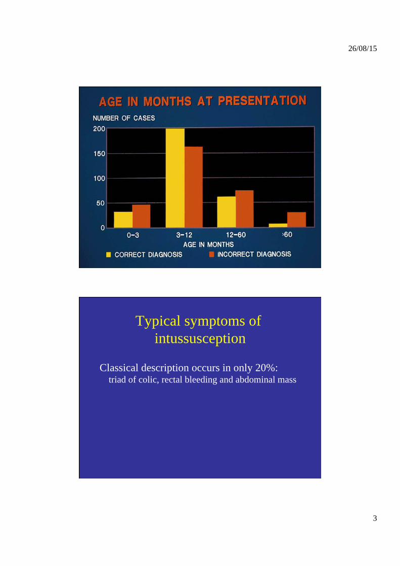
Intussusception can occur at any age • Most common between 3 months and 2 years • Most likely to be misdiagnosed outside this age-
range
26/08/15
3
Typical symptoms of intussusception
Classical description occurs in only 20%: triad of colic, rectal bleeding and abdominal mass
26/08/15
4
Typical symptoms of intussusception
Classical description occurs in only 20%: triad of colic, rectal bleeding and abdominal mass
Usual symptoms are :
- vomiting 90% - abdominal pain 85% - pallor - lethargy and listlessness
Vomiting
• Gastroenteritis unlikely if vomiting persists in absence of diarrhoea
• Initial vomiting reflex (autonomic)
• Later vomiting related to small bowel obstruction
26/08/15
5
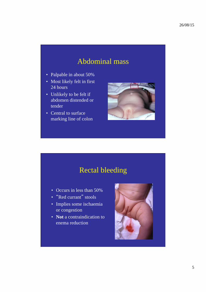
Abdominal mass • Palpable in about 50% • Most likely felt in first
24 hours • Unlikely to be felt if
abdomen distended or tender
• Central to surface marking line of colon
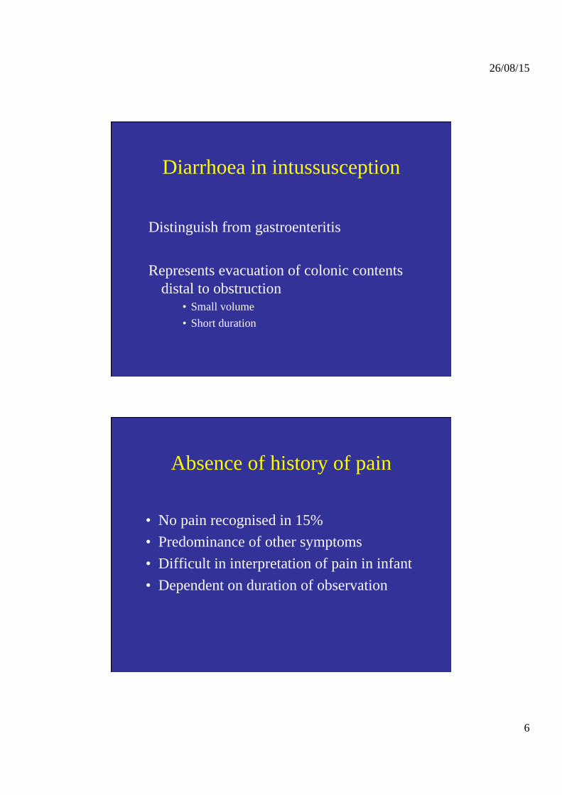
Rectal bleeding
• Occurs in less than 50% • “Red currant” stools • Implies some ischaemia
or congestion • Not a contraindication to
enema reduction
26/08/15
6
Diarrhoea in intussusception
Distinguish from gastroenteritis Represents evacuation of colonic contents
distal to obstruction • Small volume • Short duration
Absence of history of pain
• No pain recognised in 15% • Predominance of other symptoms • Difficult in interpretation of pain in infant • Dependent on duration of observation
26/08/15
7
Late presentation
• Unwell listless child, looks ill • Fever • Dehydration • Abdominal distension
Late presentation
• Unwell listless child, looks ill • Fever • Dehydration • Abdominal distension
Distinguish from other causes of sepsis
26/08/15
8
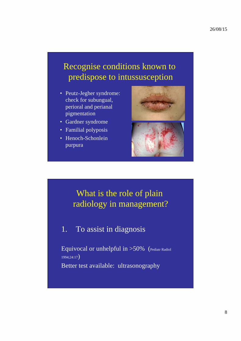
Recognise conditions known to predispose to intussusception
• Peutz-Jegher syndrome: check for subungual, perioral and perianal pigmentation
• Gardner syndrome • Familial polyposis • Henoch-Schonlein
purpura
What is the role of plain radiology in management?
1. To assist in diagnosis
Equivocal or unhelpful in >50% (Pediatr Radiol
1994;24:17) Better test available: ultrasonography
26/08/15
9
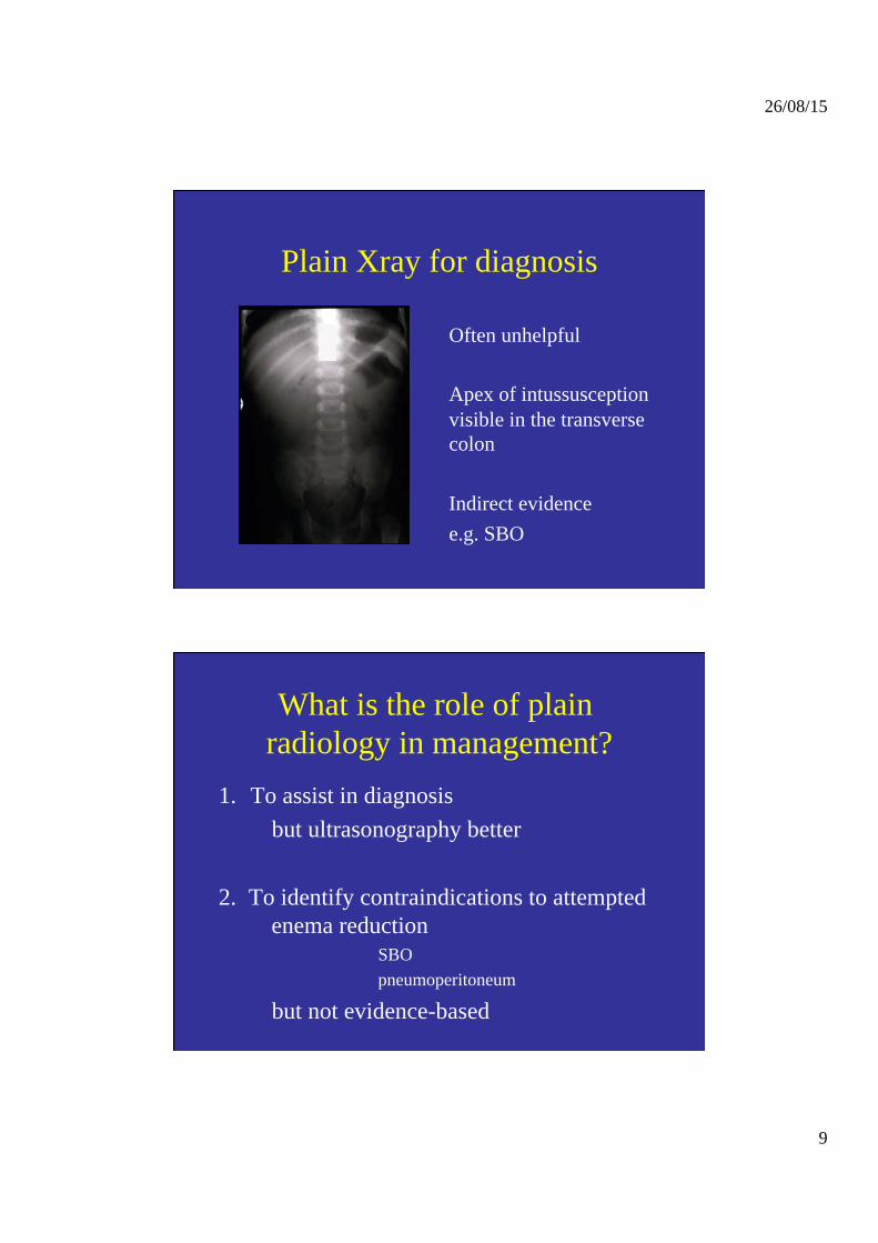
Plain Xray for diagnosis
Often unhelpful Apex of intussusception visible in the transverse colon Indirect evidence e.g. SBO
What is the role of plain radiology in management?
1. To assist in diagnosis but ultrasonography better
2. To identify contraindications to attempted
enema reduction SBO pneumoperitoneum
but not evidence-based
26/08/15
10
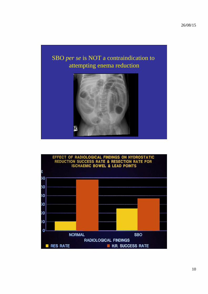
SBO per se is NOT a contraindication to attempting enema reduction
26/08/15
11
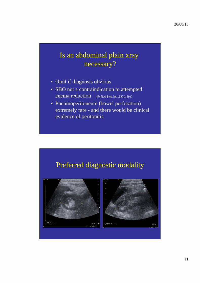
• Omit if diagnosis obvious • SBO not a contraindication to attempted
enema reduction (Pediatr Surg Int 1987;2:291) • Pneumoperitoneum (bowel perforation)
extremely rare - and there would be clinical evidence of peritonitis
Is an abdominal plain xray necessary?
Preferred diagnostic modality
26/08/15
12
How good is ultrasonography for diagnosis?
• Up to 100% sensitive and specific (Radiol 1994;191:781, Pediatr Surg Int 1998;14:158)
• Identifies pathological lesion at leadpoint (Pediatr Radiol 2000;30:594)
• Colour flow Doppler may predict reducibility (Radiol 1994;191:781)
When should ultrasonography be used for diagnosis?
• Not necessary if diagnosis extremely likely (Pediatr Surg Int 1998;14:157)
• Valuable when diagnosis uncertain - minimises radiation exposure
• Initial step when sonographic guided reduction planned
26/08/15
13
Options for treating intussusception?
• Barium enema • Gas (air or oxygen) enema • Hydrostatic reduction under ultrasound • Surgery: open or laparoscopic
• Which should be used when? – Depends on facilities/expertise, and clinical features
eg peritonitis or evidence of pathological leadpoint
What are the indications for attempting enema reduction?
All cases of confirmed intussusception -except:
1. clinical evidence of full thickness bowel necrosis (peritonitis or septicaemia)
2. pathological lesion at leadpoint identified or extremely likely e.g. Peutz Jegher syndrome
26/08/15
14
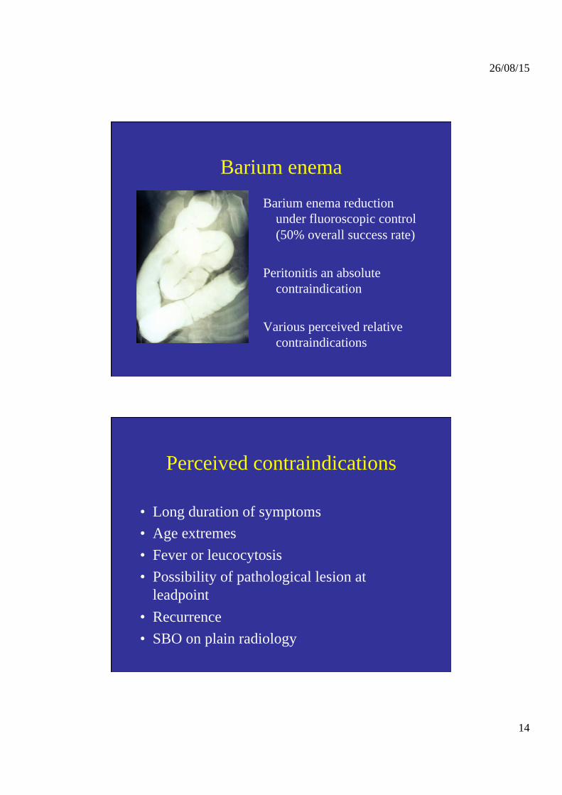
Barium enema Barium enema reduction
under fluoroscopic control (50% overall success rate)
Peritonitis an absolute
contraindication Various perceived relative
contraindications
Perceived contraindications
• Long duration of symptoms • Age extremes • Fever or leucocytosis • Possibility of pathological lesion at
leadpoint • Recurrence • SBO on plain radiology
26/08/15
15
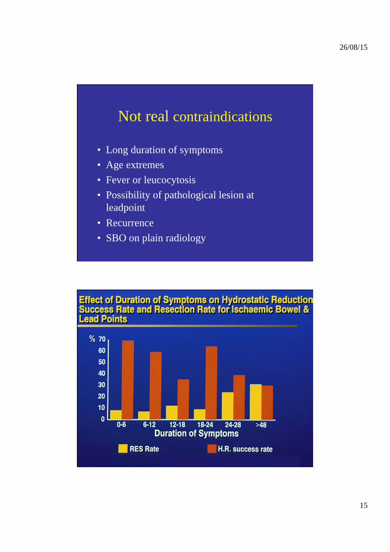
Not real contraindications
• Long duration of symptoms • Age extremes • Fever or leucocytosis • Possibility of pathological lesion at
leadpoint • Recurrence • SBO on plain radiology
26/08/15
16
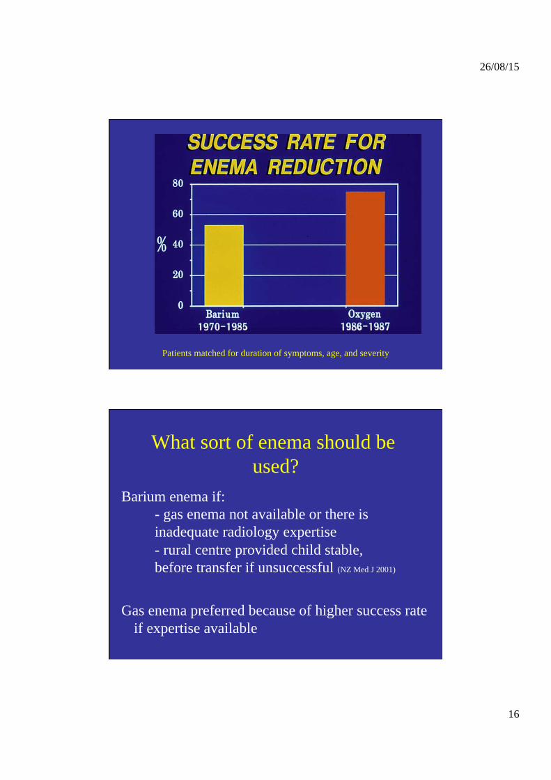
Patients matched for duration of symptoms, age, and severity
What sort of enema should be used?
Barium enema if: - gas enema not available or there is inadequate radiology expertise - rural centre provided child stable, before transfer if unsuccessful (NZ Med J 2001)
Gas enema preferred because of higher success rate if expertise available
26/08/15
17
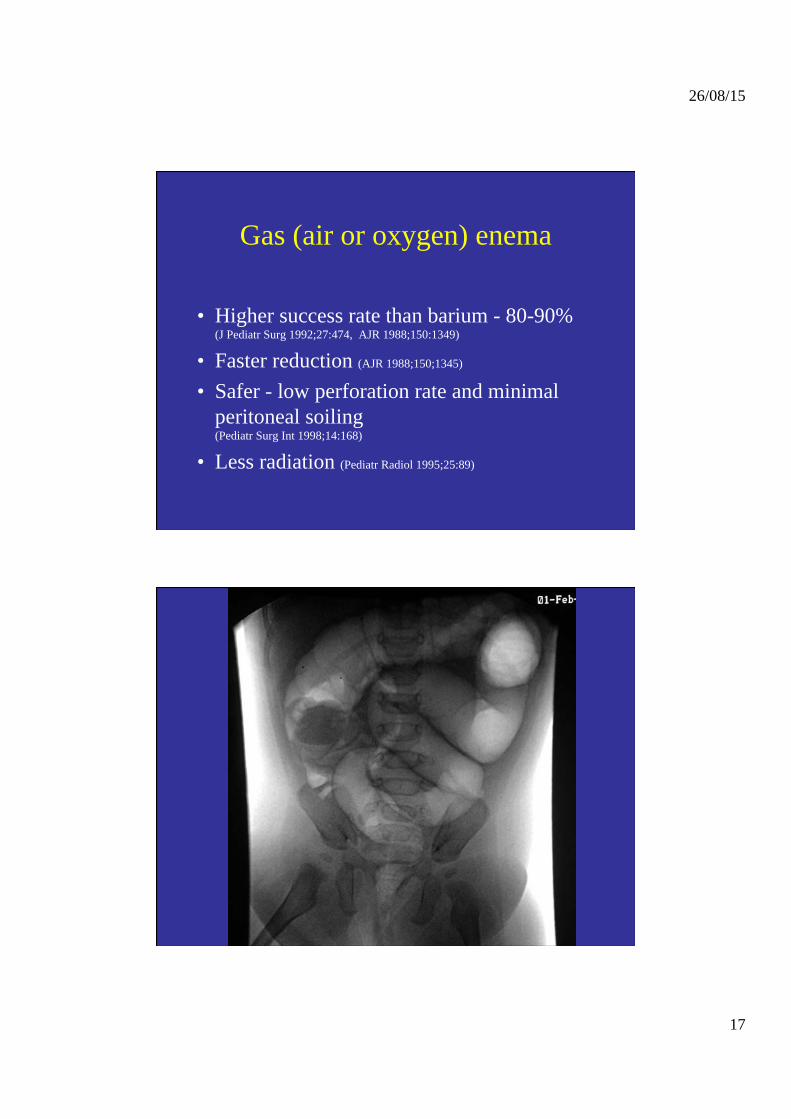
Gas (air or oxygen) enema
• Higher success rate than barium - 80-90% (J Pediatr Surg 1992;27:474, AJR 1988;150:1349)
• Faster reduction (AJR 1988;150;1345) • Safer - low perforation rate and minimal
peritoneal soiling (Pediatr Surg Int 1998;14:168)
• Less radiation (Pediatr Radiol 1995;25:89)
26/08/15
18
Ultrasonographic guided reduction
Follows straight on from diagnosis Limits radiation exposure Versatile:
with hydrostatic reduction (J Pediatr Surg 1999;34:1016) with gas enemas (Pediatr Radiol 2000;30:339, Radiol 2001;218:85)
Justification for delayed repeat gas enema
• First attempt fails in up to 25% • Most of these can be reduced manually or
already reduced at surgery (J Pediatr Surg 1994;29:588, Pediatr Surg Int 1999;15:214)
• Successful in >50% (J Pediatr Surg 1994;29:588) • Saves money and reduces hospital stay
(Aust NZ J Surg 1997;67:330)
26/08/15
19
Rationale for delayed repeat enemas
• Allows resolution of oedema after partial reduction
• Makes subsequent reduction easier • Requires at least 30 minutes
(J Pediatr Surg 1999;34:1016, J Pediatr Surg 1994;29:588) • 50% success rate with each repeat
(Pediatr Surg Int 1999;15:214)
When should a delayed repeat enema be used?
• Partial reduction with first (or preceding) attempt
• Child remains in good clinical condition • No evidence of pathological lesion at
leadpoint
26/08/15
20
When is surgery indicated?
1. Attempts at (repeated) enemas have failed 2. Full thickness bowel necrosis (peritonitis or
septicaemia) 3. Perforation during attempted reduction 4. Evidence of pathological lesion at leadpoint
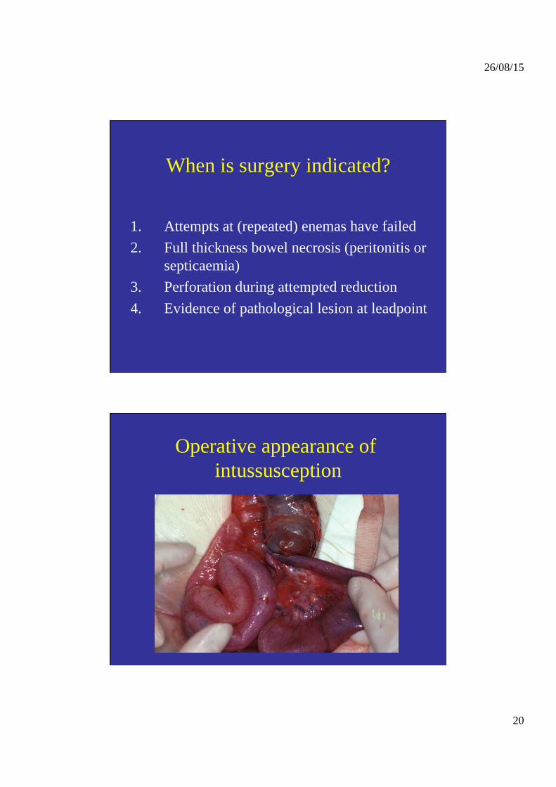
Operative appearance of intussusception
26/08/15
21
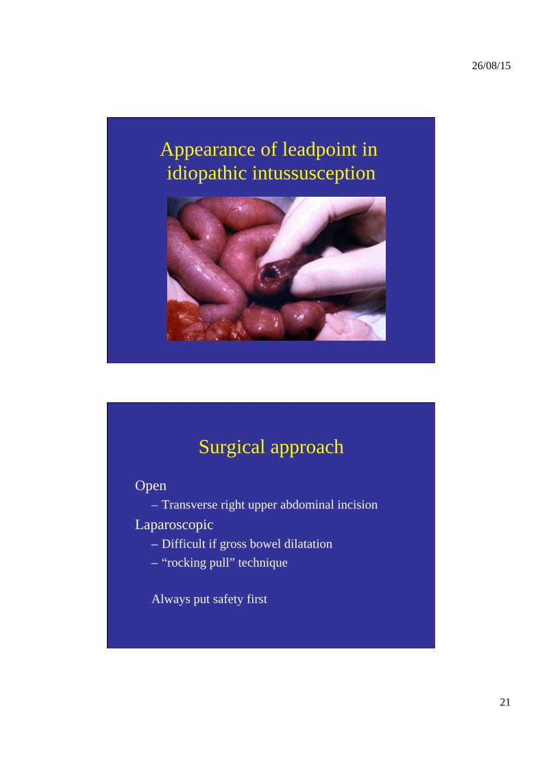
Appearance of leadpoint in idiopathic intussusception
Surgical approach
Open – Transverse right upper abdominal incision
Laparoscopic – Difficult if gross bowel dilatation – “rocking pull” technique
Always put safety first
26/08/15
22
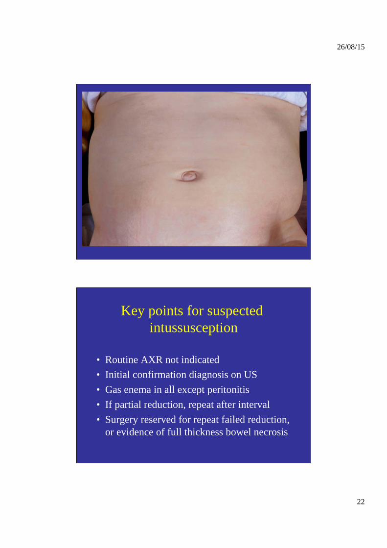
Key points for suspected intussusception
• Routine AXR not indicated • Initial confirmation diagnosis on US • Gas enema in all except peritonitis • If partial reduction, repeat after interval • Surgery reserved for repeat failed reduction,
or evidence of full thickness bowel necrosis
Recommended


![Learning Objectives Epidemiology - … Objectives ... • Barium enemaBarium enema ... Microsoft PowerPoint - Siddiqui handout w objectives,disclosure.ppt [Compatibility Mode]](https://img.pdfslide.us/doc/110x75/5ad44f597f8b9a6d708b6dd4/learning-objectives-epidemiology-objectives-barium-enemabarium-enema.jpg)