MICROSCOPY SPECIMENS
Introduction to Biological EvidenceKeiser University
Prof. Tim Seguin, MFS, CCSI, Ph.Dc

Feathers
Stained down feather of unknown origin. Visualized with an Olympus microscope. Image: Florida State University
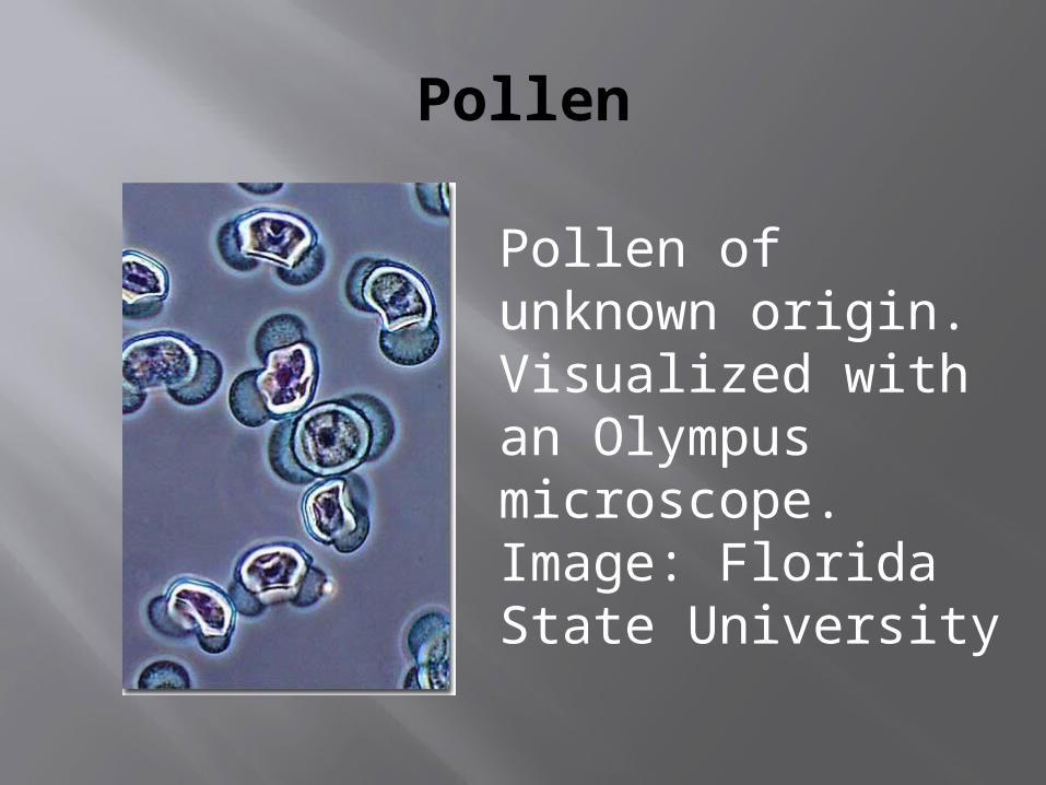
Pollen
Pollen of unknown origin. Visualized with an Olympus microscope. Image: Florida State University

Pollen
Prairie hollyhock pollen under scanning electron microscopy.
Image Credit: Dartmouth College/Charles Daghlian/NASA
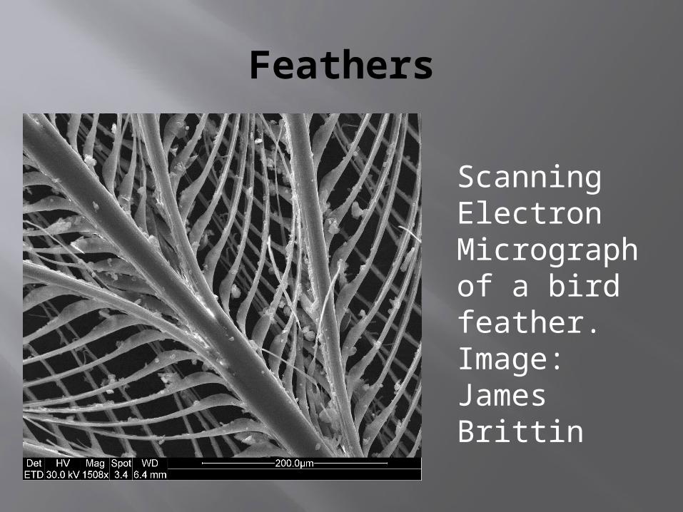
Feathers
Scanning Electron Micrograph of a bird feather. Image: James Brittin

Fur
Rabbit Fur visualized with a polarized light microscope. Image: Florida State University
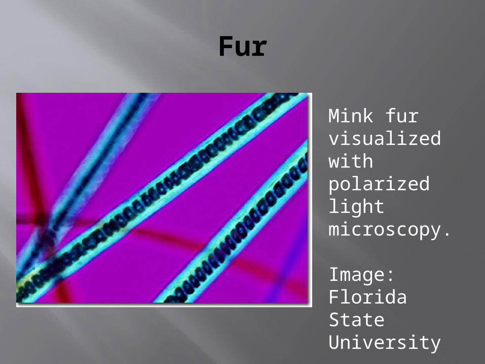
Fur
Mink fur visualized with polarized light microscopy.
Image: Florida State University
Human Hair
Human hair visualized with a polarizing microscope.
Image: Florida State University

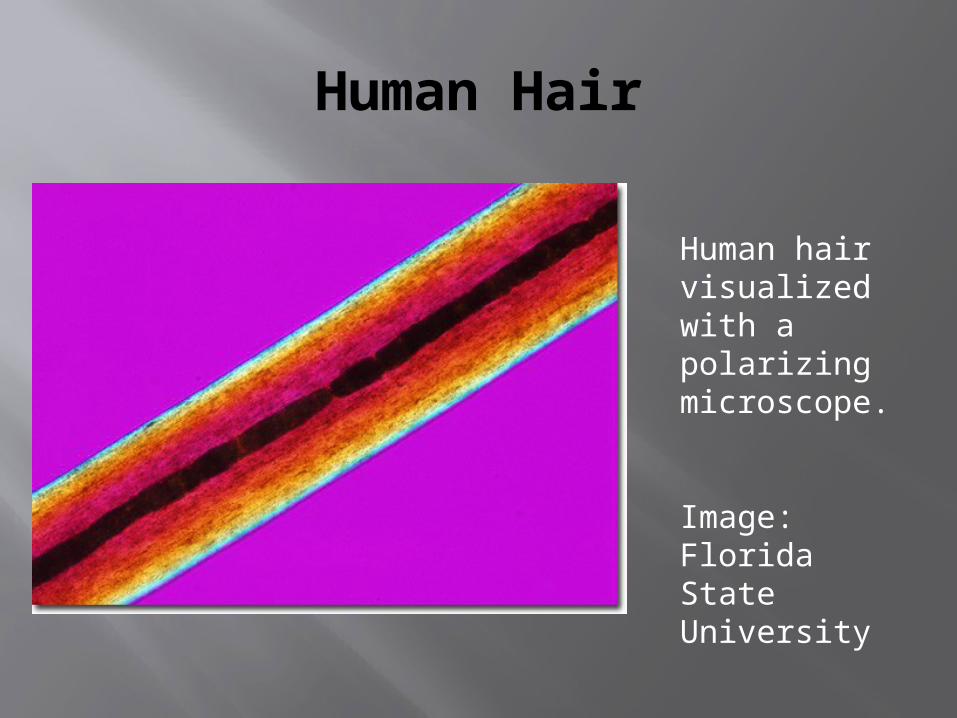
Human Hair
Human hair visualized with a polarizing microscope.
Image: Florida State University
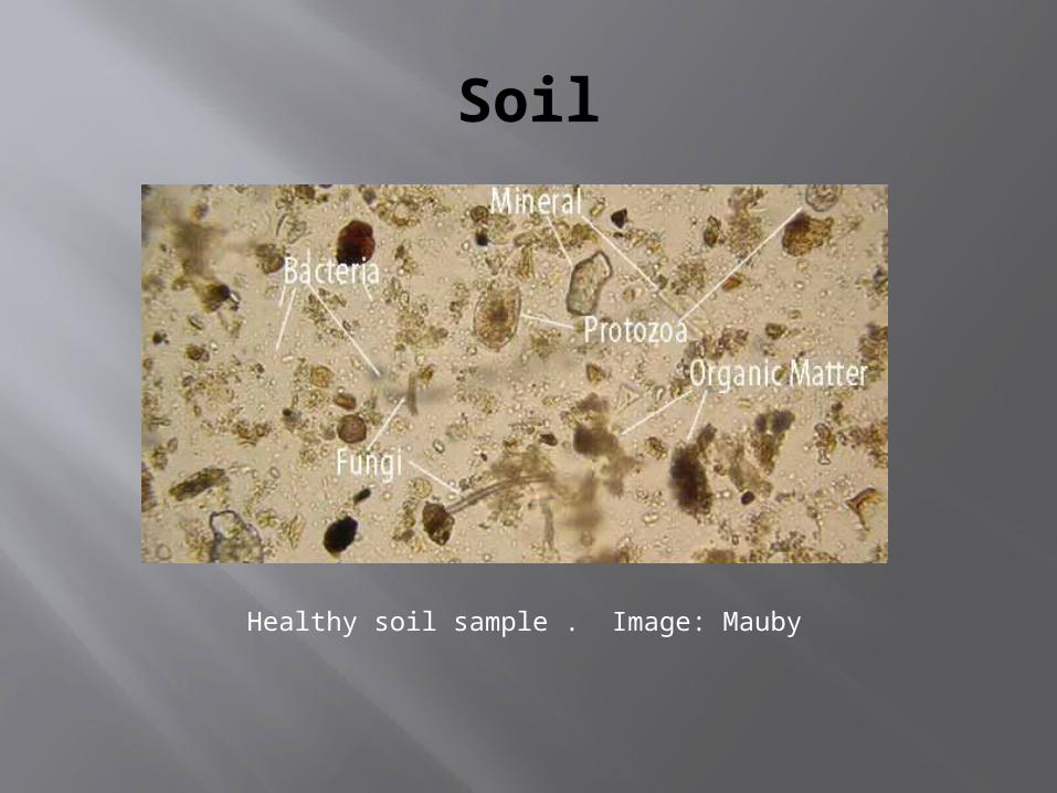
Soil
Healthy soil sample . Image: Mauby
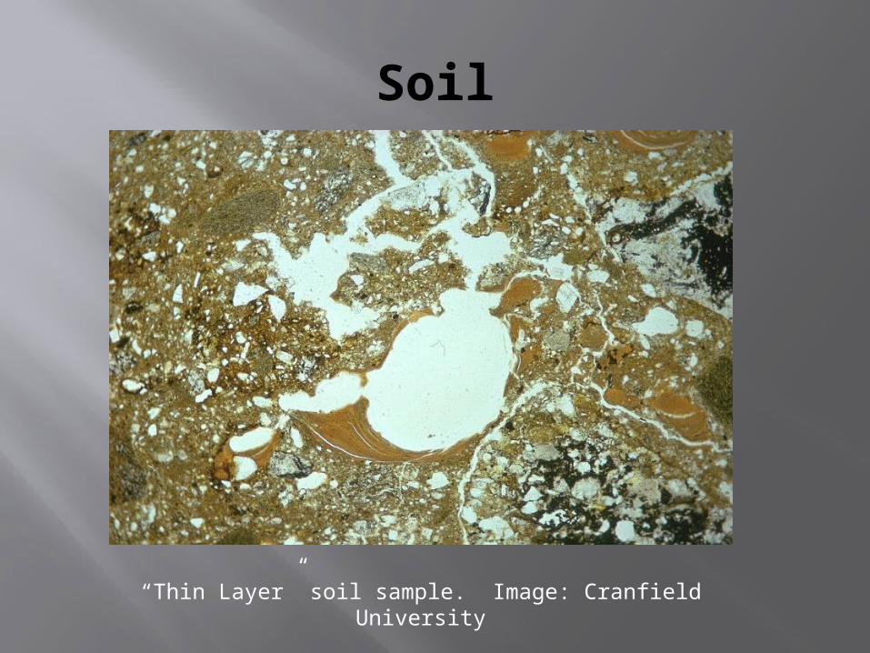
Soil
“Thin Layer” soil sample. Image: Cranfield University
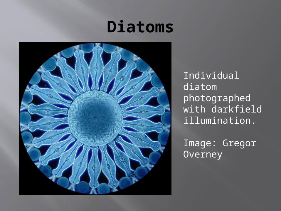
Diatoms
Individual diatom photographed with darkfield illumination.
Image: Gregor Overney

Diatoms
Group of diatoms living in an Antarctic ice sample. Image: NOAA
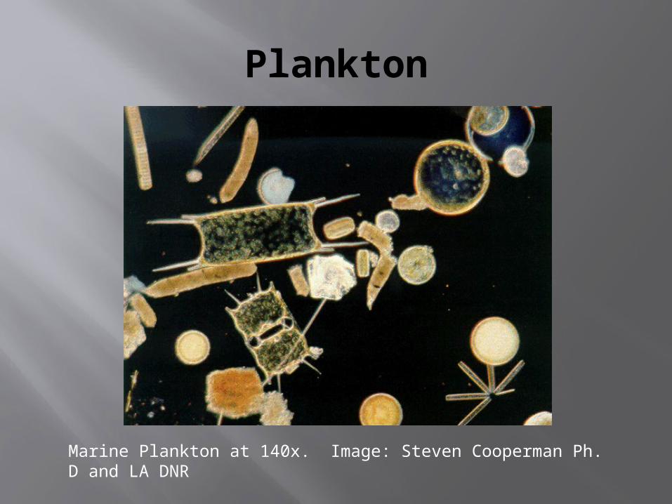
Plankton
Marine Plankton at 140x. Image: Steven Cooperman Ph. D and LA DNR
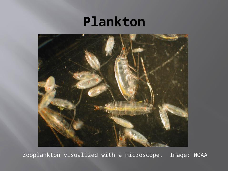
Plankton
Zooplankton visualized with a microscope. Image: NOAA
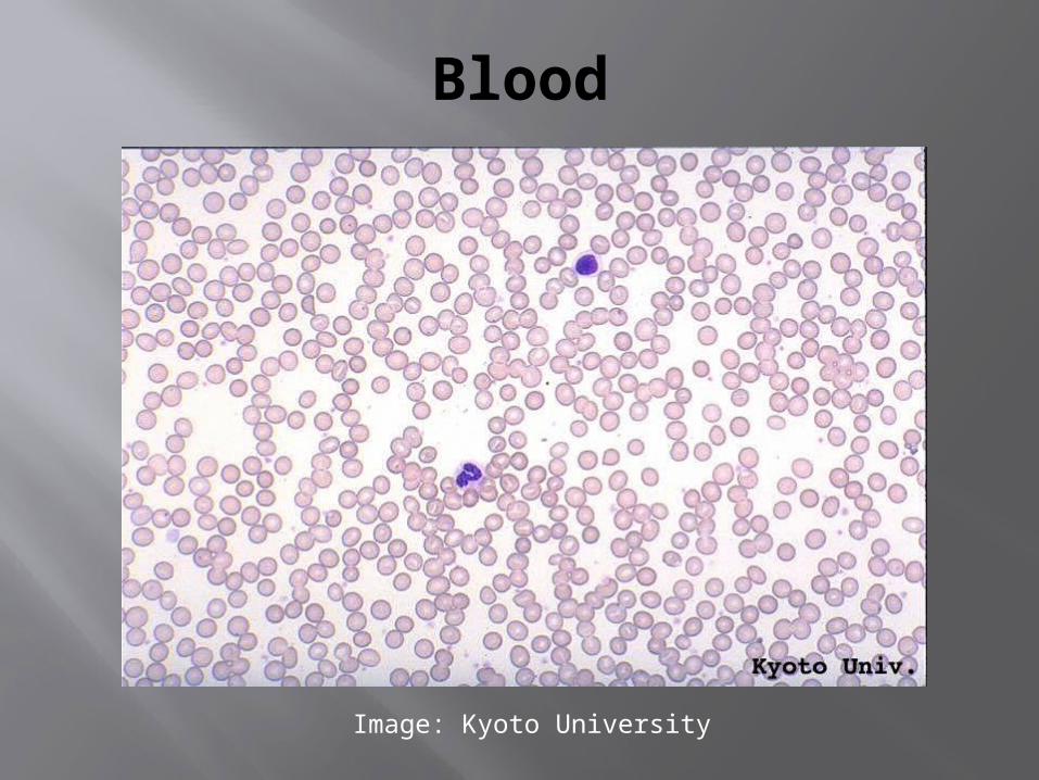
Blood
Image: Kyoto University
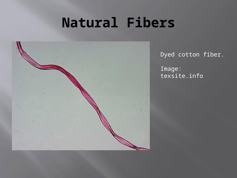
Natural Fibers
Dyed cotton fiber.
Image: texsite.info
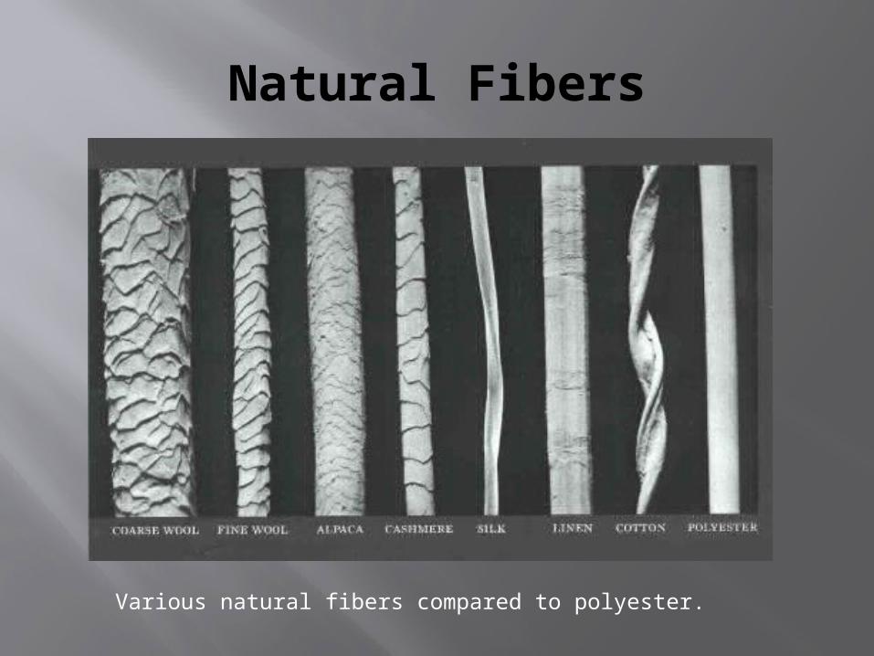
Natural Fibers
Various natural fibers compared to polyester.
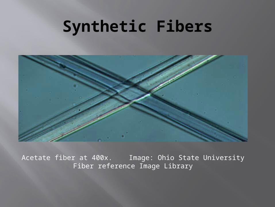
Synthetic Fibers
Acetate fiber at 400x. Image: Ohio State University Fiber reference Image Library
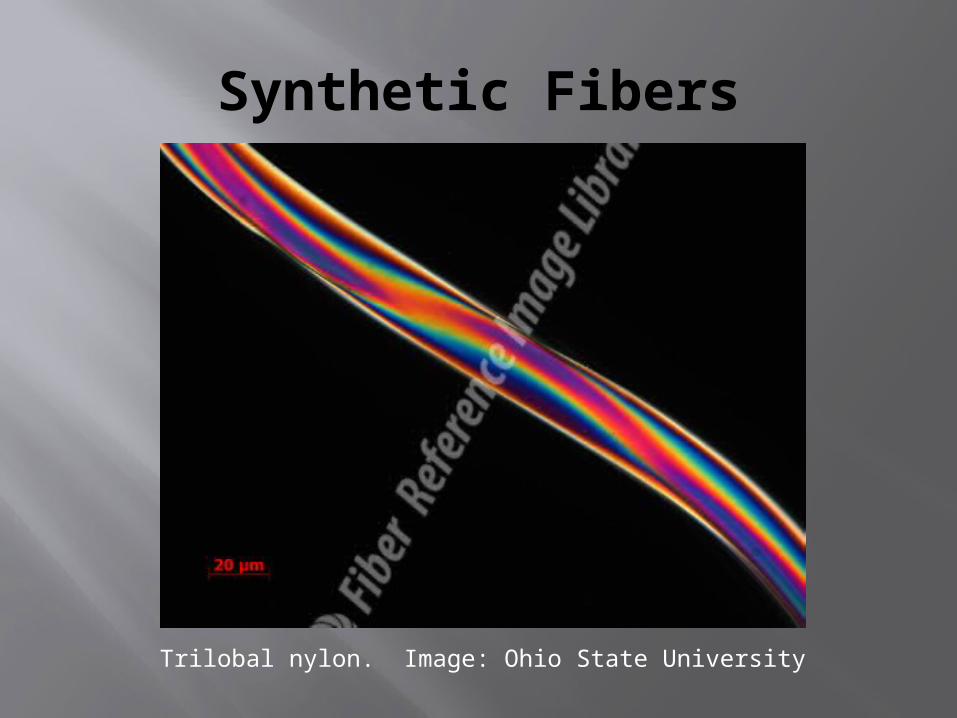
Synthetic Fibers
Trilobal nylon. Image: Ohio State University
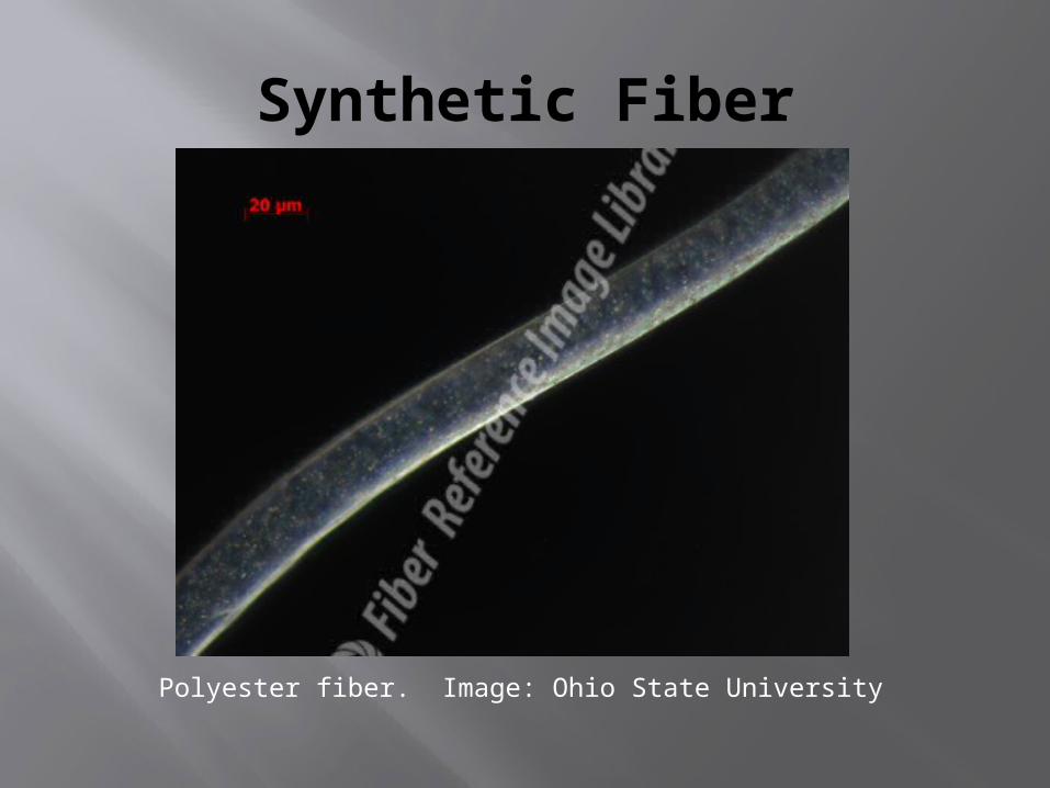
Synthetic Fiber
Polyester fiber. Image: Ohio State University
Recommended