Copyright 2009 John Wiley & Sons, Inc. 1
Chapter 12Nervous Tissue
Copyright 2009 John Wiley & Sons, Inc. 2
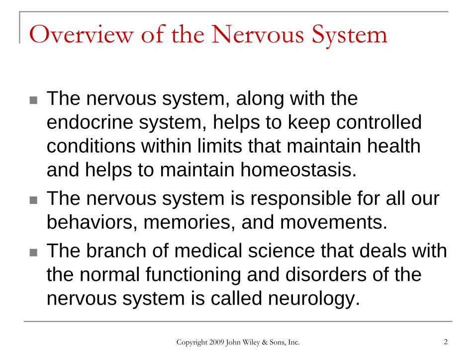
Overview of the Nervous System
The nervous system, along with the endocrine system, helps to keep controlled conditions within limits that maintain health and helps to maintain homeostasis.
The nervous system is responsible for all our behaviors, memories, and movements.
The branch of medical science that deals with the normal functioning and disorders of the nervous system is called neurology.
Copyright 2009 John Wiley & Sons, Inc. 3
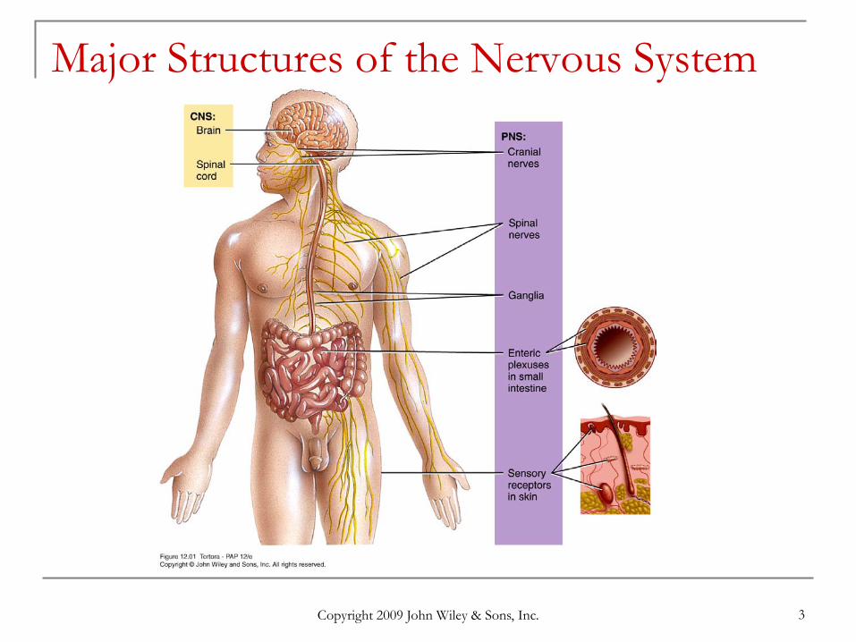
Major Structures of the Nervous System
Copyright 2009 John Wiley & Sons, Inc. 4
Overview of Major Structures Twelve pairs of cranial nerves. Thirty-one pairs of spinal nerves emerge from the spinal
cord. Ganglia, located outside the brain and spinal cord, are
small masses of nervous tissue, containing primarily cell bodies of neurons.
Enteric plexuses help regulate the digestive system. Sensory receptors are either parts of neurons or
specialized cells that monitor changes in the internal or external environment.
Copyright 2009 John Wiley & Sons, Inc. 5
Functions of Nervous System Sensory function: to sense changes in the internal and
external environment through sensory receptors. Sensory (afferent) neurons serve this function.
Integrative function: to analyze the sensory information, store some aspects, and make decisions regarding appropriate behaviors. Association or interneurons serve this function.
Motor function is to respond to stimuli by initiating action. Motor(efferent) neurons serve this function.
Copyright 2009 John Wiley & Sons, Inc. 6
Nervous System Divisions
Central nervous system (CNS) consists of the brain and spinal cord
Peripheral nervous system (PNS) consists of cranial and spinal nerves that contain
both sensory and motor fibers connects CNS to muscles, glands & all sensory
receptors
Copyright 2009 John Wiley & Sons, Inc. 7
Structure of a Multipolar Neuron
Copyright 2009 John Wiley & Sons, Inc. 8
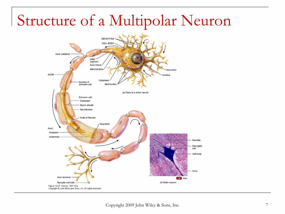
Histology of the Nervous System: Neurons Functional unit of nervous system Have capacity to produce action potentials
electrical excitability Cell body
single nucleus with prominent nucleolus Nissl bodies (chromatophilic substance)
rough ER & free ribosomes for protein synthesis neurofilaments give cell shape and support microtubules move material inside cell lipofuscin pigment clumps (harmless aging)
Cell processes = dendrites & axons
Copyright 2009 John Wiley & Sons, Inc. 9
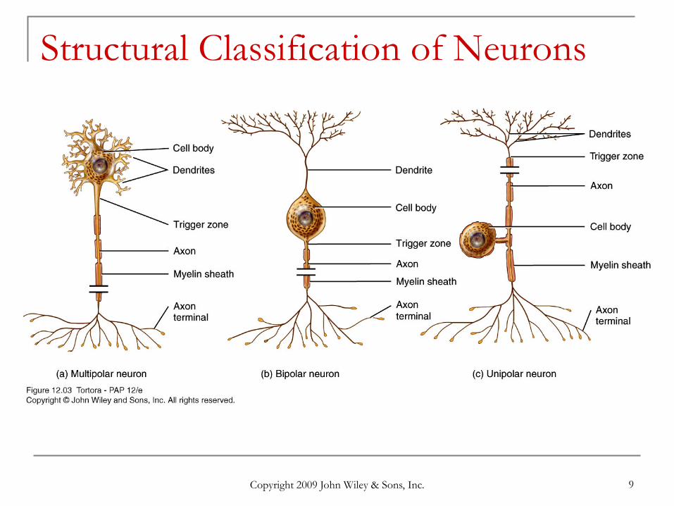
Structural Classification of Neurons
Copyright 2009 John Wiley & Sons, Inc. 10
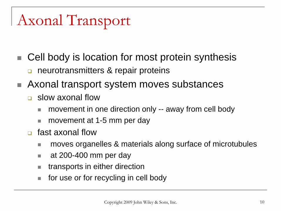
Axonal Transport
Cell body is location for most protein synthesis neurotransmitters & repair proteins
Axonal transport system moves substances slow axonal flow
movement in one direction only -- away from cell body movement at 1-5 mm per day
fast axonal flow moves organelles & materials along surface of microtubules at 200-400 mm per day transports in either direction for use or for recycling in cell body
Copyright 2009 John Wiley & Sons, Inc. 11
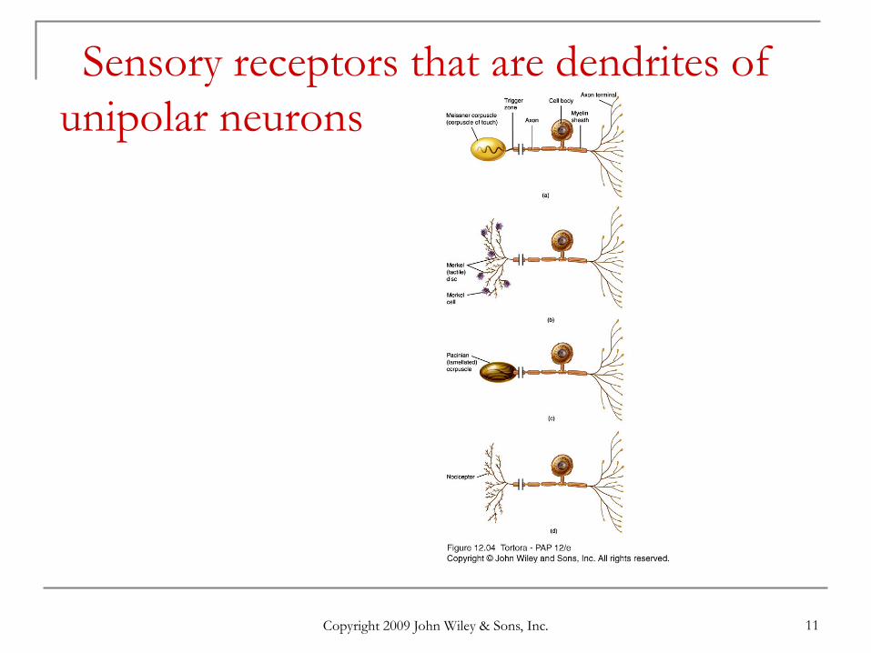
Sensory receptors that are dendrites of unipolar neurons
Copyright 2009 John Wiley & Sons, Inc. 12
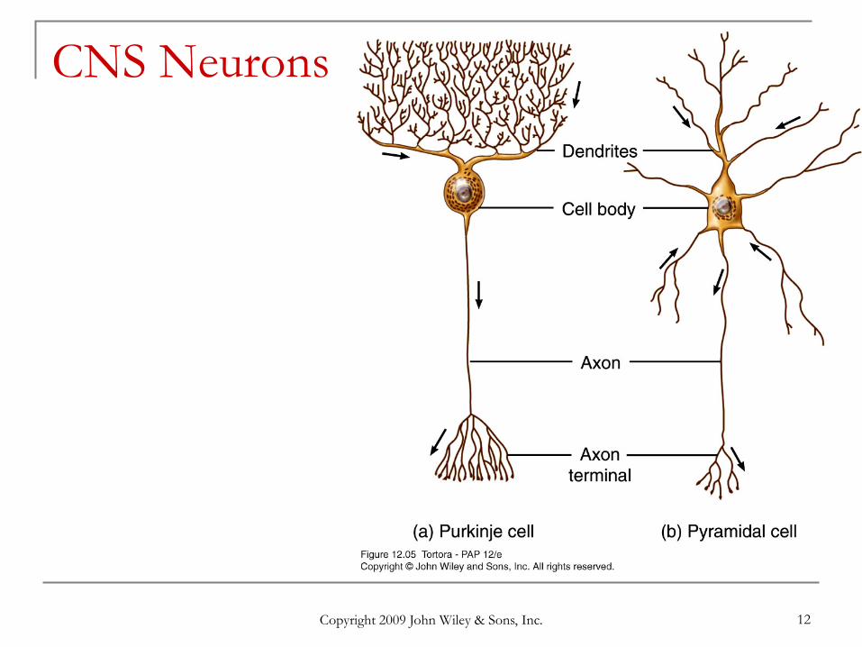
CNS Neurons
Copyright 2009 John Wiley & Sons, Inc. 13
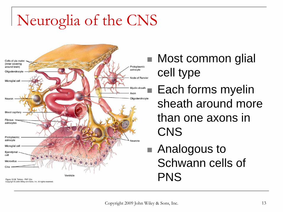
Neuroglia of the CNS
Most common glial cell type
Each forms myelin sheath around more than one axons in CNS
Analogous to Schwann cells of PNS
Copyright 2009 John Wiley & Sons, Inc. 14
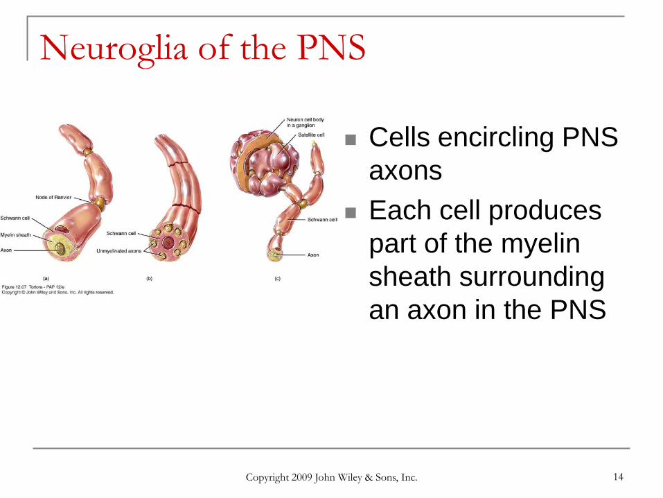
Neuroglia of the PNS
Cells encircling PNS axons
Each cell produces part of the myelin sheath surrounding an axon in the PNS
Copyright 2009 John Wiley & Sons, Inc. 15
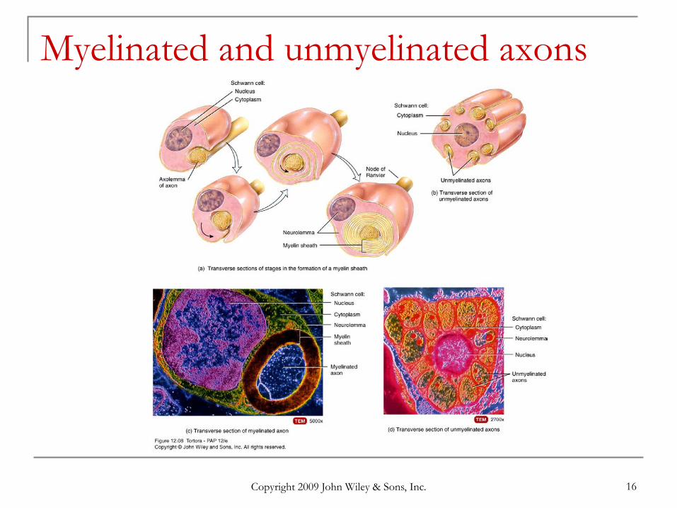
Myelinated and unmyelinated axons Schwann cells myelinate (wrap around) axons in the PNS during
fetal development Schwann cell cytoplasm & nucleus forms outermost layer of
neurolemma with inner portion being the myelin sheath Tube guides growing axons that are repairing themselves
Copyright 2009 John Wiley & Sons, Inc. 16
Myelinated and unmyelinated axons
Copyright 2009 John Wiley & Sons, Inc. 17
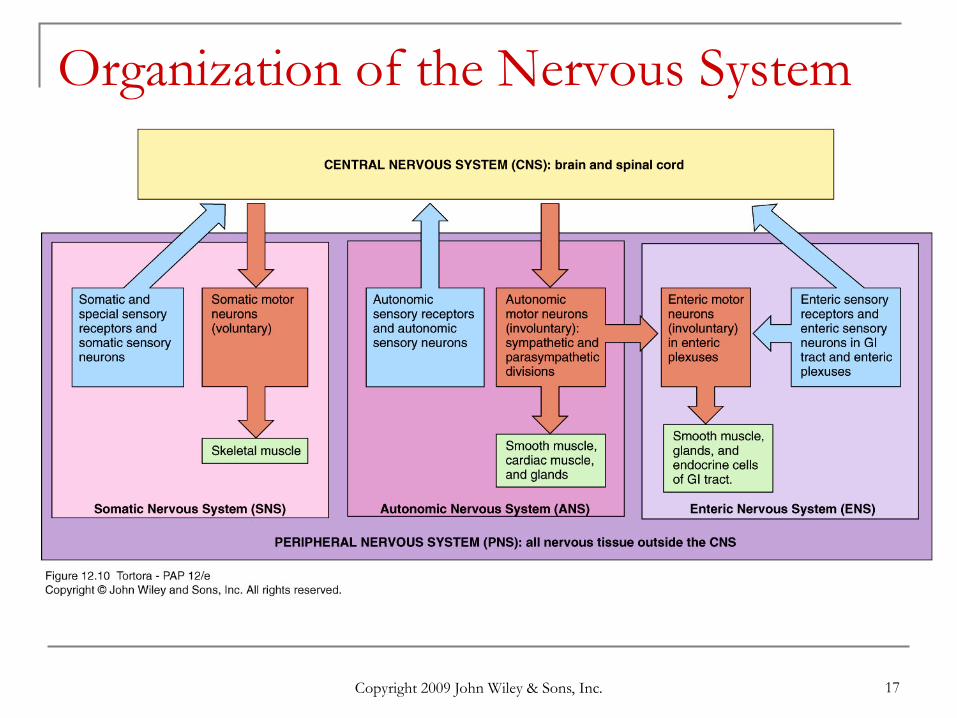
Organization of the Nervous System
Copyright 2009 John Wiley & Sons, Inc. 18
Subdivisions of the PNS Somatic (voluntary) nervous system (SNS)
neurons from cutaneous and special sensory receptors to the CNS
motor neurons to skeletal muscle tissue
Autonomic (involuntary) nervous systems sensory neurons from visceral organs to CNS motor neurons to smooth & cardiac muscle and glands
sympathetic division (speeds up heart rate) parasympathetic division (slow down heart rate)
Enteric nervous system (ENS) involuntary sensory & motor neurons control GI tract neurons function independently of ANS & CNS
Copyright 2009 John Wiley & Sons, Inc. 19
Electrical Signals in Neurons
Neurons are electrically excitable due to the voltage difference across their membrane
Communicate with 2 types of electric signals action potentials that can travel long distances graded potentials that are local membrane
changes only In living cells, a flow of ions occurs through
ion channels in the cell membrane

Copyright 2009 John Wiley & Sons, Inc. 20
Overview of Nervous System Functions
Copyright 2009 John Wiley & Sons, Inc. 21
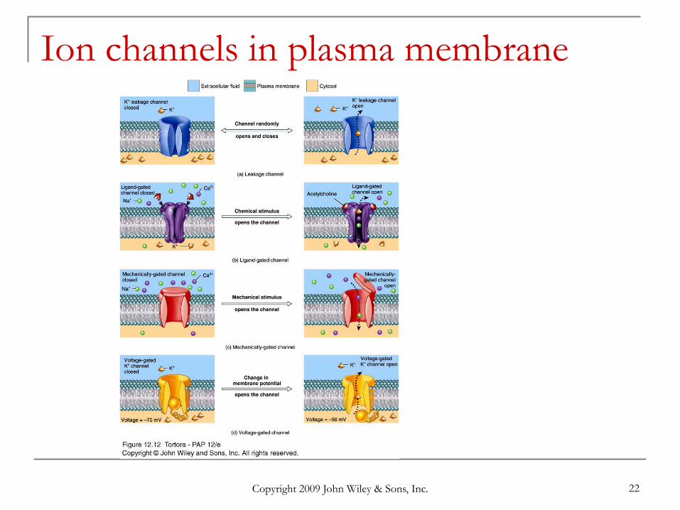
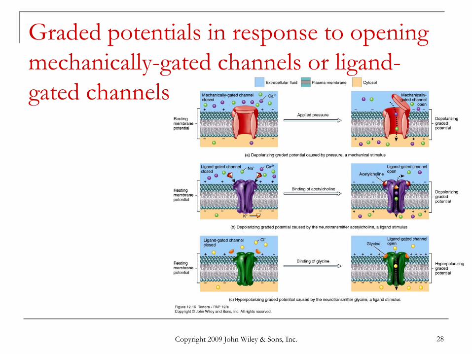
Types of Ion Channels Leakage (nongated) channels are always open
nerve cells have more K+ than Na+ leakage channels as a result, membrane permeability to K+ is higher explains resting membrane potential of -70mV in nerve
tissue Ligand-gated channels open and close in
response to a stimulus results in neuron excitability
Voltage-gated channels respond to a direct change in the membrane potential.
Mechanically gated ion channels respond to mechanical vibration or pressure.
Copyright 2009 John Wiley & Sons, Inc. 22
Ion channels in plasma membrane
Copyright 2009 John Wiley & Sons, Inc. 23
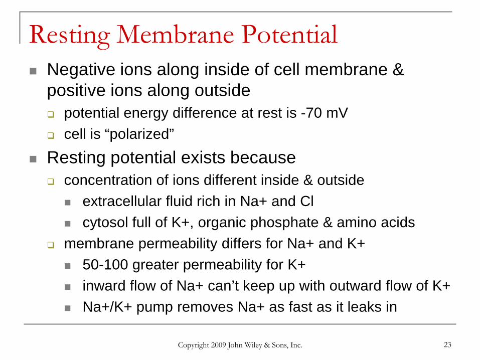
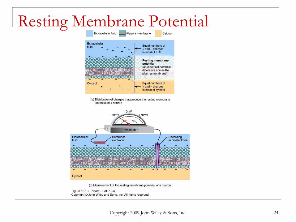
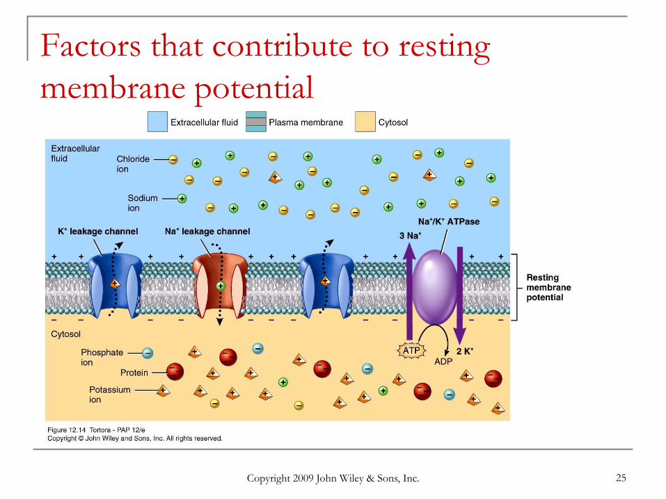
Resting Membrane Potential Negative ions along inside of cell membrane &
positive ions along outside potential energy difference at rest is -70 mV cell is “polarized”
Resting potential exists because concentration of ions different inside & outside
extracellular fluid rich in Na+ and Cl cytosol full of K+, organic phosphate & amino acids
membrane permeability differs for Na+ and K+ 50-100 greater permeability for K+ inward flow of Na+ can’t keep up with outward flow of K+ Na+/K+ pump removes Na+ as fast as it leaks in
Copyright 2009 John Wiley & Sons, Inc. 24
Resting Membrane Potential
Copyright 2009 John Wiley & Sons, Inc. 25
Factors that contribute to resting membrane potential
Copyright 2009 John Wiley & Sons, Inc. 26
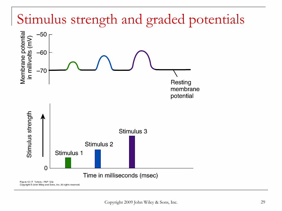
Graded Potentials
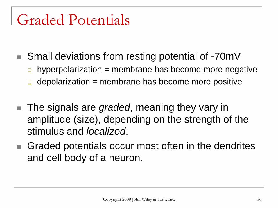
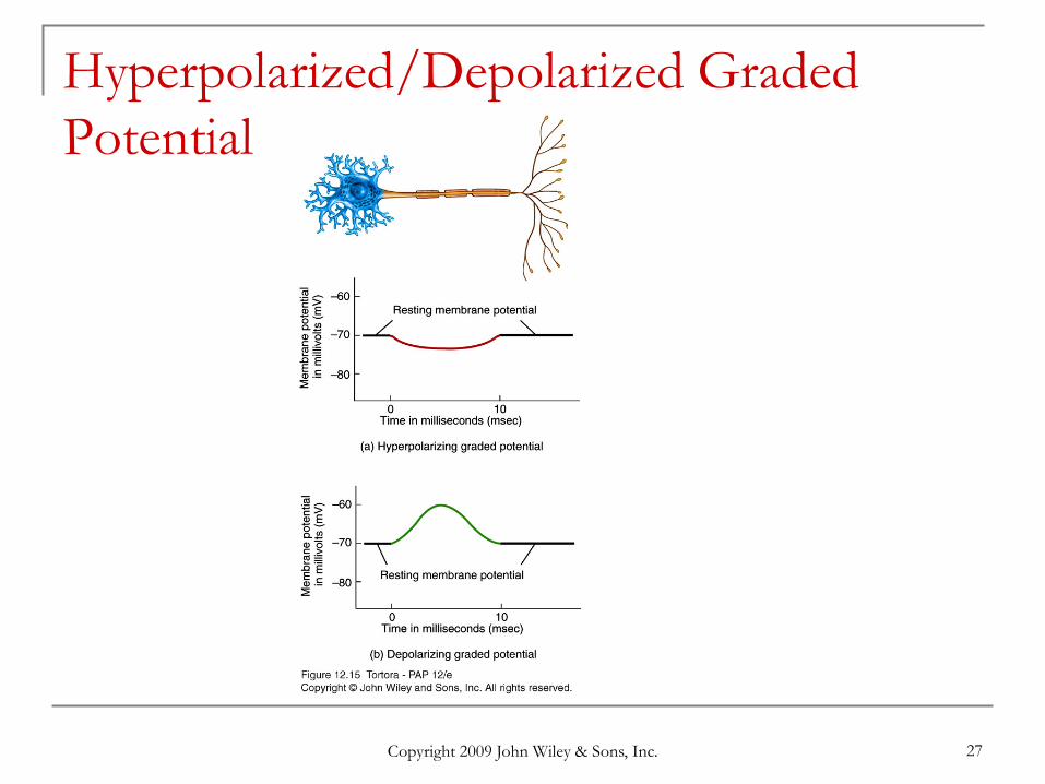
Small deviations from resting potential of -70mV hyperpolarization = membrane has become more negative depolarization = membrane has become more positive
The signals are graded, meaning they vary in amplitude (size), depending on the strength of the stimulus and localized.
Graded potentials occur most often in the dendrites and cell body of a neuron.
Copyright 2009 John Wiley & Sons, Inc. 27
Hyperpolarized/Depolarized Graded Potential
Copyright 2009 John Wiley & Sons, Inc. 28
Graded potentials in response to opening mechanically-gated channels or ligand-gated channels
Copyright 2009 John Wiley & Sons, Inc. 29
Stimulus strength and graded potentials
Copyright 2009 John Wiley & Sons, Inc. 30
Summation
Copyright 2009 John Wiley & Sons, Inc. 31
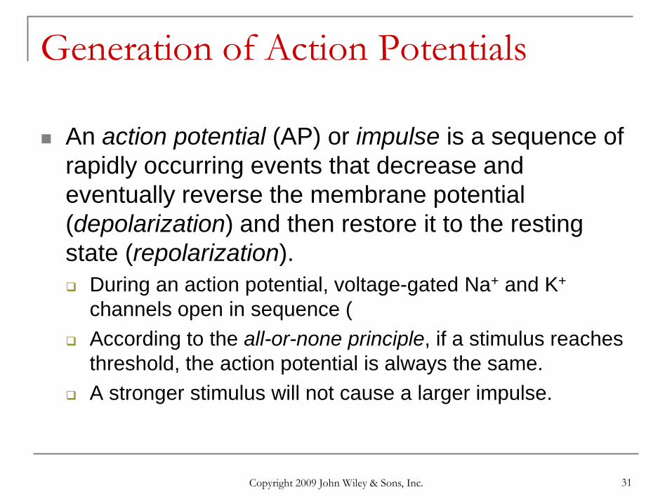
Generation of Action Potentials
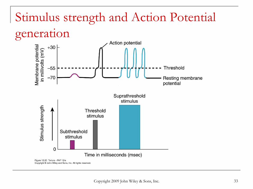
An action potential (AP) or impulse is a sequence of rapidly occurring events that decrease and eventually reverse the membrane potential (depolarization) and then restore it to the resting state (repolarization). During an action potential, voltage-gated Na+ and K+
channels open in sequence ( According to the all-or-none principle, if a stimulus reaches
threshold, the action potential is always the same. A stronger stimulus will not cause a larger impulse.
Copyright 2009 John Wiley & Sons, Inc. 32
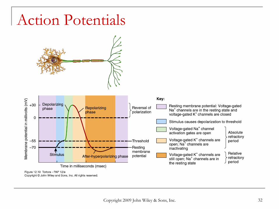
Action Potentials
Copyright 2009 John Wiley & Sons, Inc. 33
Stimulus strength and Action Potential generation

Copyright 2009 John Wiley & Sons, Inc. 34
Changes in ion flow during depolarizing and repolarizing phases of Action Potential
Copyright 2009 John Wiley & Sons, Inc. 35
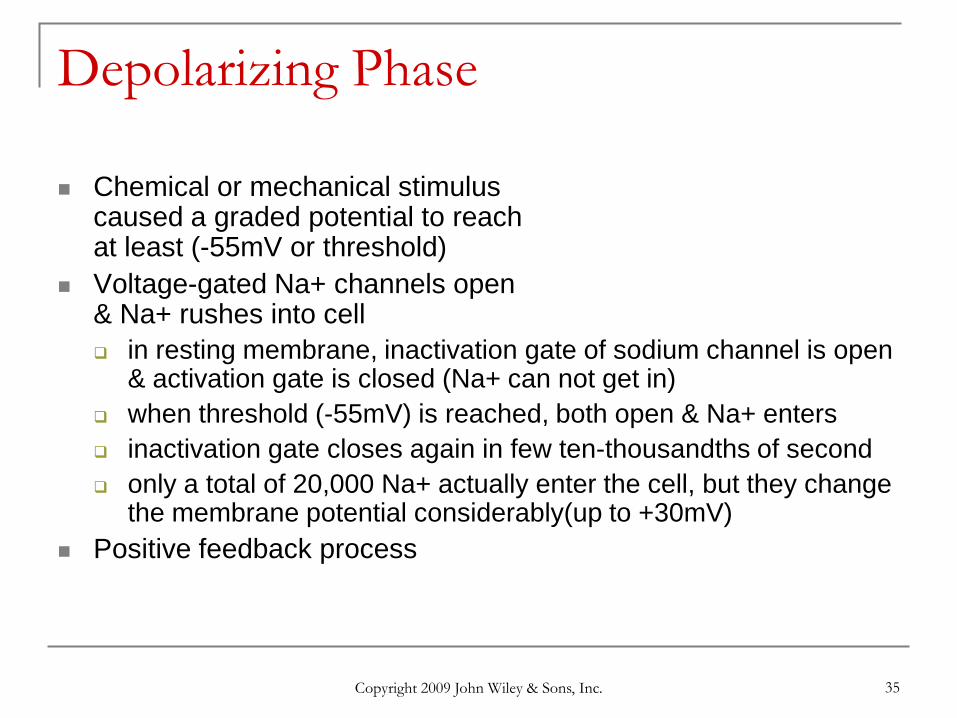
Depolarizing Phase
Chemical or mechanical stimuluscaused a graded potential to reachat least (-55mV or threshold)
Voltage-gated Na+ channels open& Na+ rushes into cell in resting membrane, inactivation gate of sodium channel is open
& activation gate is closed (Na+ can not get in) when threshold (-55mV) is reached, both open & Na+ enters inactivation gate closes again in few ten-thousandths of second only a total of 20,000 Na+ actually enter the cell, but they change
the membrane potential considerably(up to +30mV) Positive feedback process
Copyright 2009 John Wiley & Sons, Inc. 36
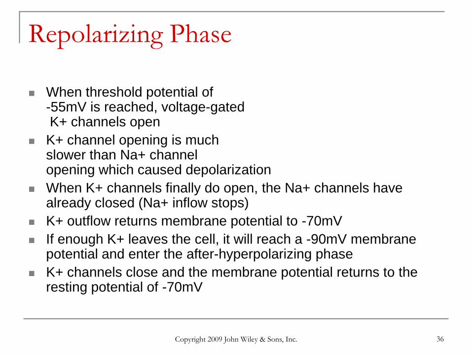
Repolarizing Phase
When threshold potential of-55mV is reached, voltage-gatedK+ channels open
K+ channel opening is muchslower than Na+ channelopening which caused depolarization
When K+ channels finally do open, the Na+ channels have already closed (Na+ inflow stops)
K+ outflow returns membrane potential to -70mV If enough K+ leaves the cell, it will reach a -90mV membrane
potential and enter the after-hyperpolarizing phase K+ channels close and the membrane potential returns to the
resting potential of -70mV
Copyright 2009 John Wiley & Sons, Inc. 37
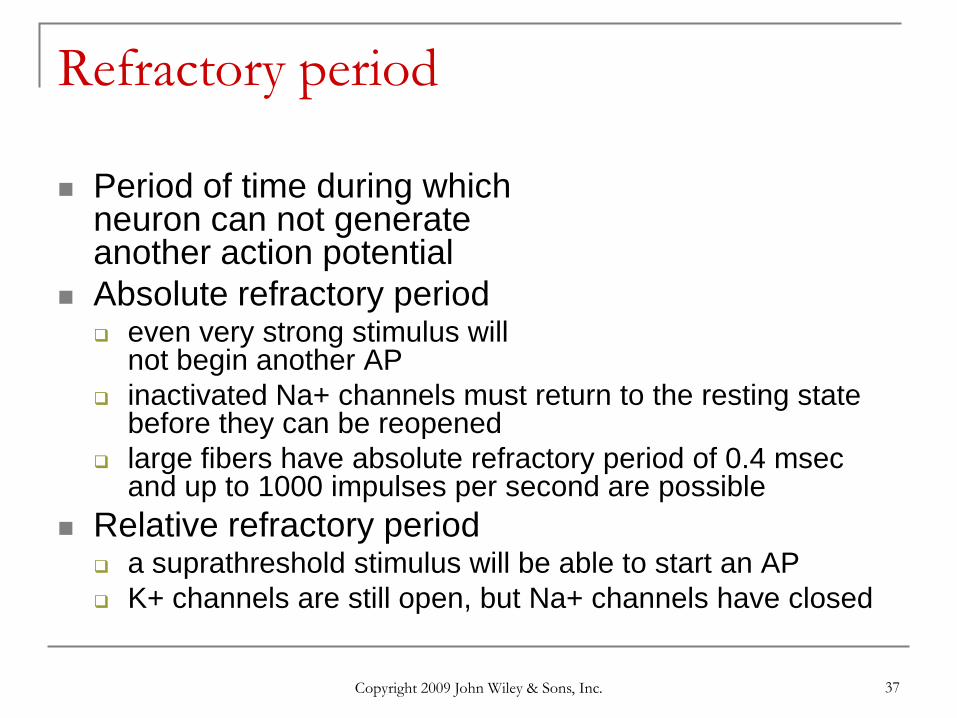
Refractory period
Period of time during whichneuron can not generateanother action potential
Absolute refractory period even very strong stimulus will
not begin another AP inactivated Na+ channels must return to the resting state
before they can be reopened large fibers have absolute refractory period of 0.4 msec
and up to 1000 impulses per second are possible Relative refractory period
a suprathreshold stimulus will be able to start an AP K+ channels are still open, but Na+ channels have closed
Copyright 2009 John Wiley & Sons, Inc. 38
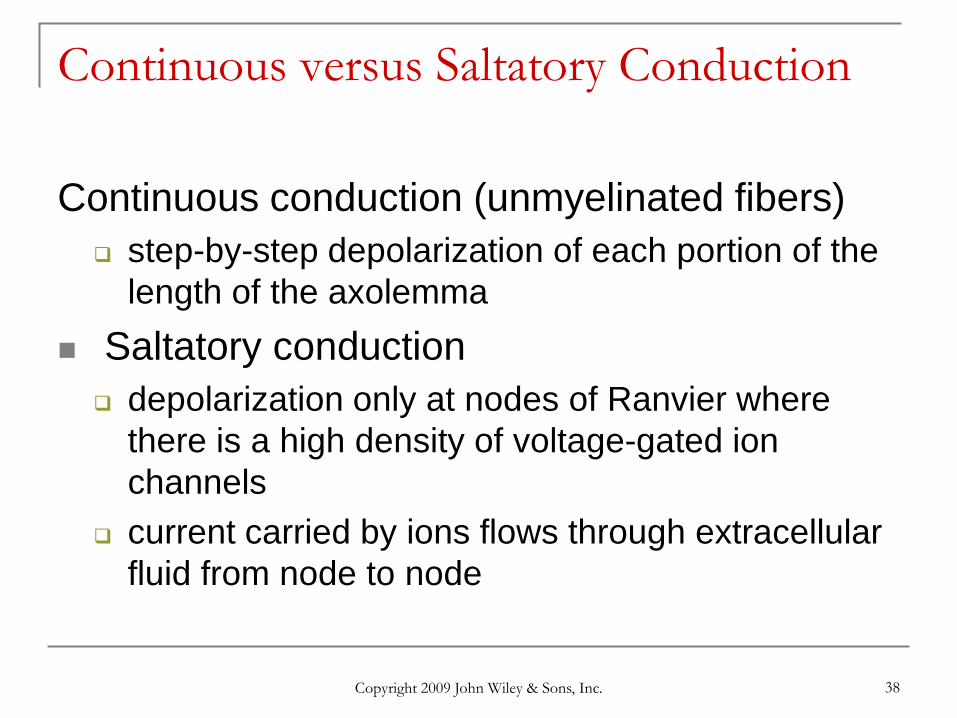
Continuous versus Saltatory Conduction
Continuous conduction (unmyelinated fibers) step-by-step depolarization of each portion of the
length of the axolemma Saltatory conduction depolarization only at nodes of Ranvier where
there is a high density of voltage-gated ion channels
current carried by ions flows through extracellular fluid from node to node
Copyright 2009 John Wiley & Sons, Inc. 39
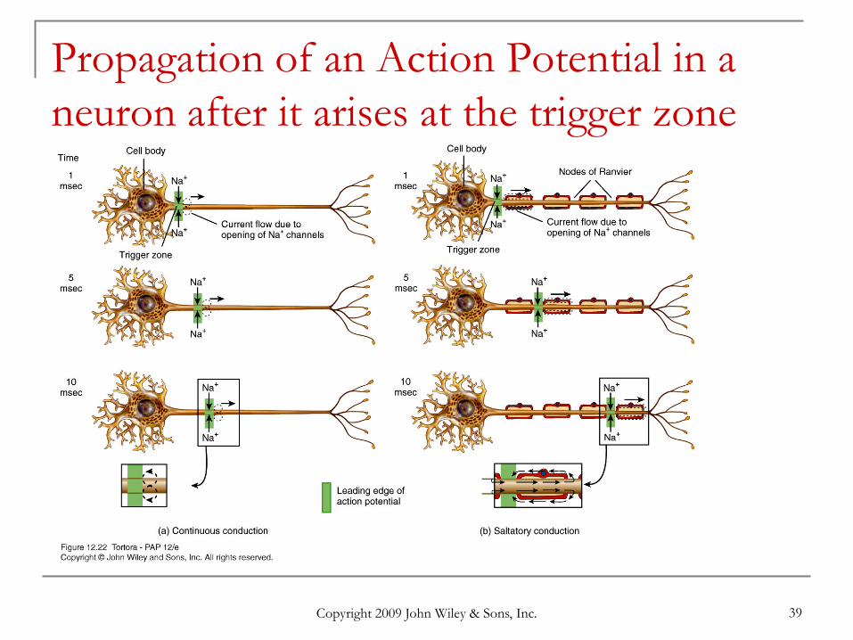
Propagation of an Action Potential in a neuron after it arises at the trigger zone
Copyright 2009 John Wiley & Sons, Inc. 40
Factors that affect speed of propagation
Amount of myelination
Axon diameter
Temperature
Copyright 2009 John Wiley & Sons, Inc. 41
Speed of impulse propagation The propagation speed of a nerve impulse is
not related to stimulus strength. larger, myelinated fibers conduct impulses faster
due to size & saltatory conduction Fiber types A fibers largest (5-20 microns & 130 m/sec)
myelinated somatic sensory & motor to skeletal muscle B fibers medium (2-3 microns & 15 m/sec)
myelinated visceral sensory & autonomic preganglionic C fibers smallest (.5-1.5 microns & 2 m/sec)
unmyelinated sensory & autonomic motor
Copyright 2009 John Wiley & Sons, Inc. 42
Signal Transmission at the Synapse
2 Types of synapses electrical
ionic current spreads to next cell through gap junctions faster, two-way transmission & capable of synchronizing
groups of neurons
chemical one-way information transfer from a presynaptic neuron
to a postsynaptic neuron axodendritic -- from axon to dendrite axosomatic -- from axon to cell body axoaxonic -- from axon to axon
Copyright 2009 John Wiley & Sons, Inc. 43
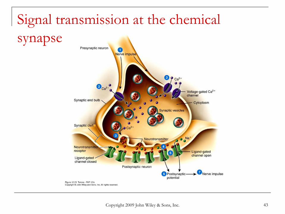
Signal transmission at the chemical synapse
Copyright 2009 John Wiley & Sons, Inc. 44
Chemical Synapses
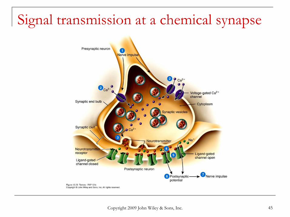
Action potential reaches end bulb and voltage-gated Ca+ 2 channels open
Ca+2 flows inward triggering release of neurotransmitter
Neurotransmitter crosses synaptic cleft & binding to ligand-gated receptors the more neurotransmitter released the greater the change
in potential of the postsynaptic cell
Synaptic delay is 0.5 msec One-way information transfer
Copyright 2009 John Wiley & Sons, Inc. 45
Signal transmission at a chemical synapse
Copyright 2009 John Wiley & Sons, Inc. 46
Excitory and Inhibitory Postsynaptic Potentials The effect of a neurotransmitter can be either
excitatory or inhibitory a depolarizing postsynaptic potential is called an EPSP
it results from the opening of ligand-gated Na+ channels the postsynaptic cell is more likely to reach threshold
an inhibitory postsynaptic potential is called an IPSP it results from the opening of ligand-gated Cl- or K+ channels it causes the postsynaptic cell to become more negative or
hyperpolarized the postsynaptic cell is less likely to reach threshold
Copyright 2009 John Wiley & Sons, Inc. 47
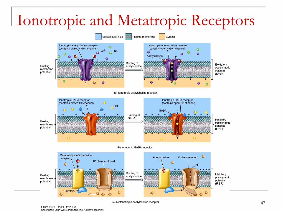
Ionotropic and Metatropic Receptors
Copyright 2009 John Wiley & Sons, Inc. 48
Removal of Neurotransmitter
Diffusion move down concentration gradient
Enzymatic degradation acetylcholinesterase
Uptake by neurons or glia cells neurotransmitter transporters Prozac = serotonin reuptake
inhibitor
Copyright 2009 John Wiley & Sons, Inc. 49
Three Possible Responses
Small EPSP occurs potential reaches -56 mV only
An impulse is generated threshold was reached membrane potential of at least -55 mV
IPSP occurs membrane hyperpolarized potential drops below -70 mV
Copyright 2009 John Wiley & Sons, Inc. 50
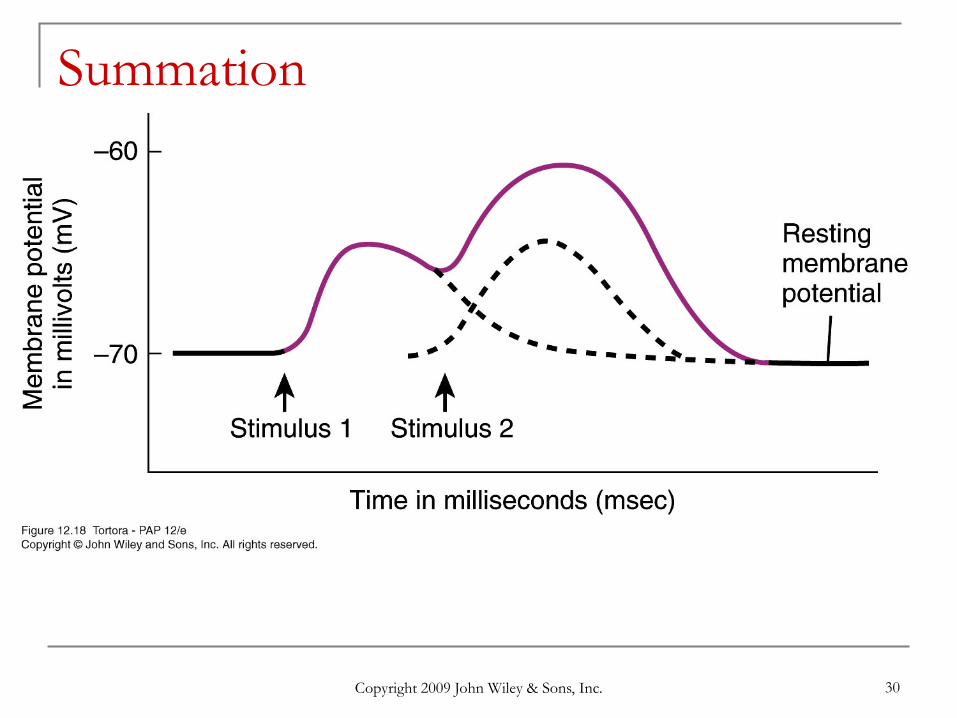
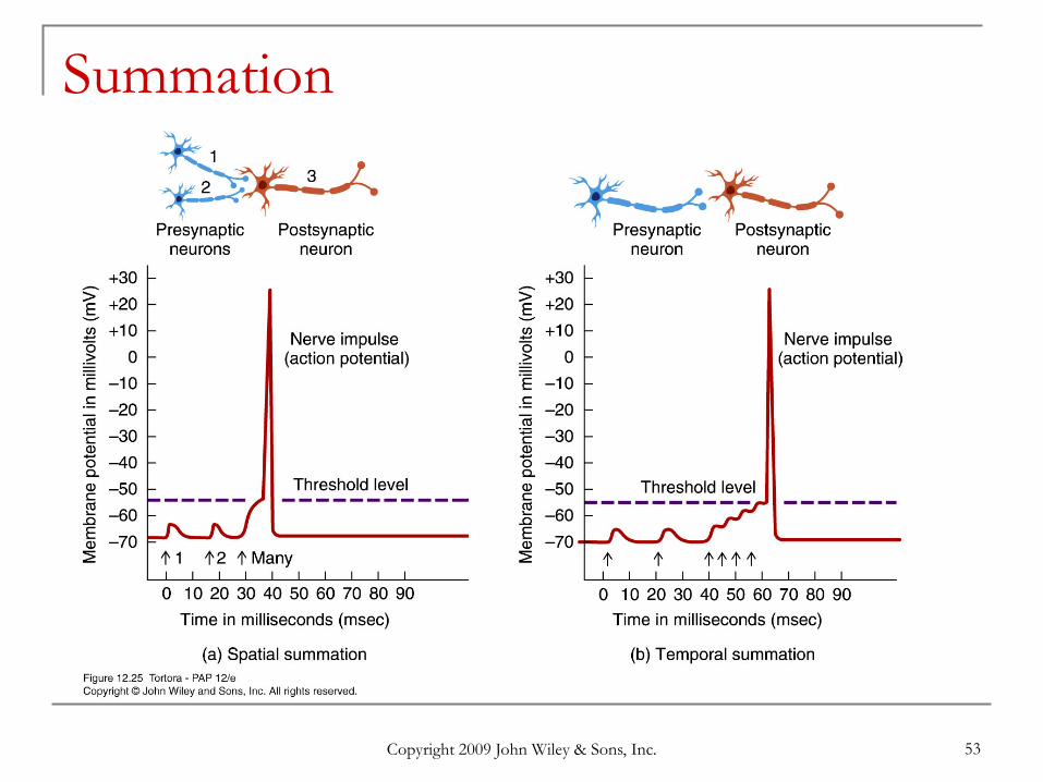
Summation
If several presynaptic end bulbs release their neurotransmitter at about the same time, the combined effect may generate a nerve impulse due to summation
Summation may be spatial or temporal.
Copyright 2009 John Wiley & Sons, Inc. 51
Spatial Summation
Summation of effects of neurotransmitters released from several end bulbs onto one neuron
Copyright 2009 John Wiley & Sons, Inc. 52
Temporal Summation
Summation of effect of neurotransmitters released from 2 or more firings of the same end bulb in rapid succession onto a second neuron
Copyright 2009 John Wiley & Sons, Inc. 53
Summation
Copyright 2009 John Wiley & Sons, Inc. 54
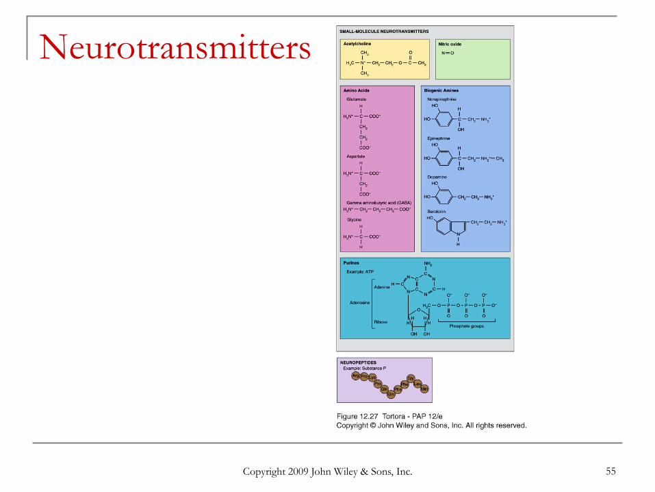
Neurotransmitters
Both excitatory and inhibitory neurotransmitters are present in the CNS and PNS; the same neurotransmitter may be excitatory in some locations and inhibitory in others.
Important neurotransmitters include acetylcholine, glutamate, aspartate, gamma aminobutyric acid, glycine, norepinephrine, epinephrine, and dopamine.
Copyright 2009 John Wiley & Sons, Inc. 55
Neurotransmitters
Copyright 2009 John Wiley & Sons, Inc. 56
Neurotransmitter Effects
Neurotransmitter effects can be modified synthesis can be stimulated or inhibited release can be blocked or enhanced removal can be stimulated or blocked receptor site can be blocked or activated
Agonist anything that enhances a transmitters effects
Antagonist anything that blocks the action of a neurotranmitter
Copyright 2009 John Wiley & Sons, Inc. 57
Small-Molecule Neurotransmitters Acetylcholine (ACh) released by many PNS neurons & some CNS excitatory on NMJ but inhibitory at others inactivated by acetylcholinesterase
Amino Acids glutamate released by nearly all excitatory neurons
in the brain ---- inactivated by glutamate specific transporters
GABA is inhibitory neurotransmitter for 1/3 of all brain synapses (Valium is a GABA agonist --enhancing its inhibitory effect)
Copyright 2009 John Wiley & Sons, Inc. 58
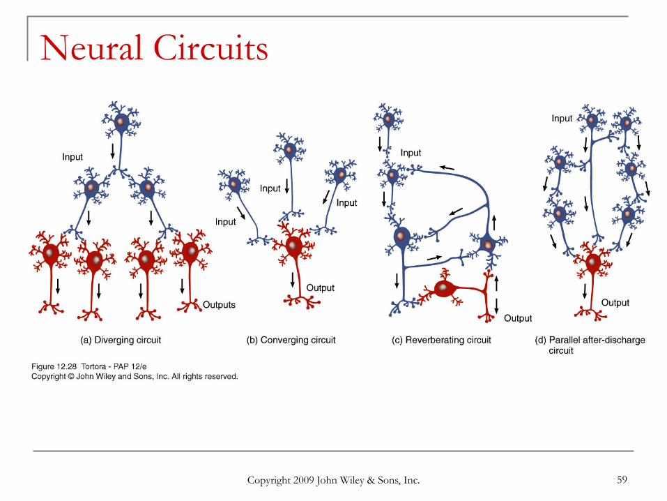
Neural Circuits
A neuronal network may contain thousands or even millions of neurons.
Neuronal circuits are involved in many important activities breathing short-term memory waking up
Copyright 2009 John Wiley & Sons, Inc. 59
Neural Circuits
Copyright 2009 John Wiley & Sons, Inc. 60
Regeneration & Repair
Plasticity maintained throughout life sprouting of new dendrites synthesis of new proteins changes in synaptic contacts with other neurons
Limited ability for regeneration (repair) PNS can repair damaged dendrites or axons CNS no repairs are possible
Copyright 2009 John Wiley & Sons, Inc. 61
Damage and Repair in the Peripheral Nervous System When there is damage to an axon, usually there are
changes, called chromatolysis, which occur in the cell body of the affected cell; this causes swelling of the cell body and peaks between 10 and 20 days after injury.
By the third to fifth day, degeneration of the distal portion of the neuronal process and myelin sheath (Wallerian degeneration) occurs; afterward, macrophages phagocytize the remains.
Retrograde degeneration of the proximal portion of the fiber extends only to the first neurofibral node.
Regeneration follows chromatolysis; synthesis of RNA and protein accelerates, favoring rebuilding of the axon and often taking several months.
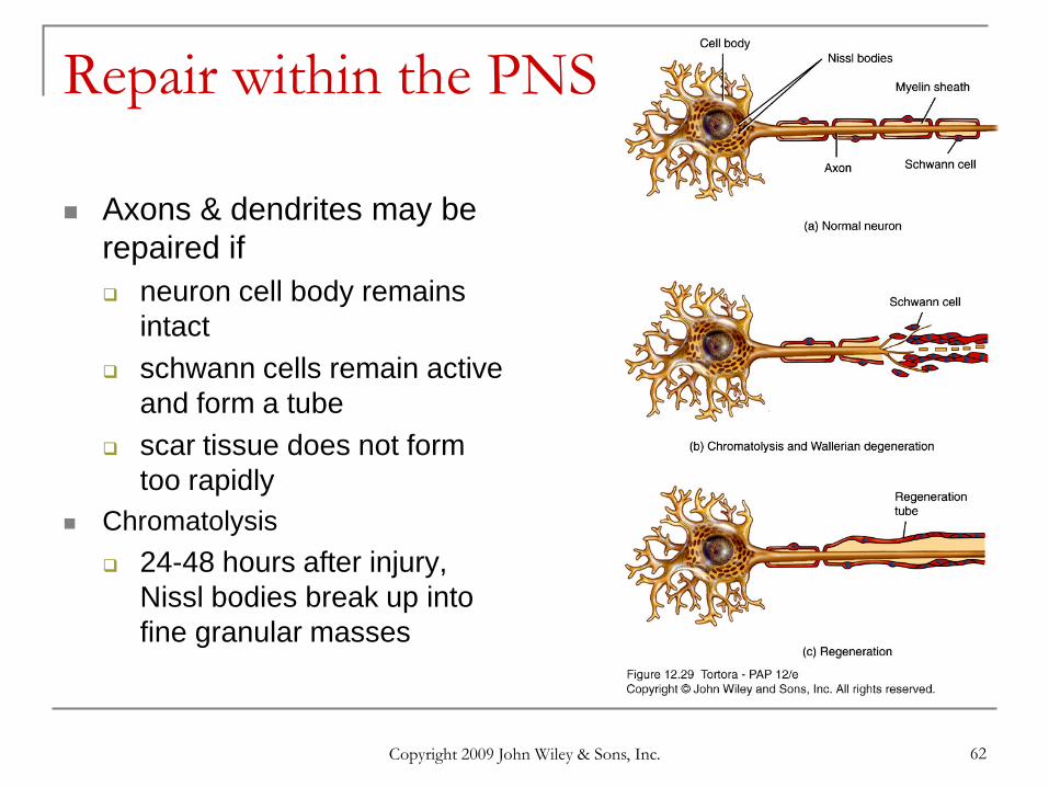
Copyright 2009 John Wiley & Sons, Inc. 62
Repair within the PNS
Axons & dendrites may be repaired if neuron cell body remains
intact schwann cells remain active
and form a tube scar tissue does not form
too rapidly Chromatolysis
24-48 hours after injury, Nissl bodies break up into fine granular masses
Copyright 2009 John Wiley & Sons, Inc. 63
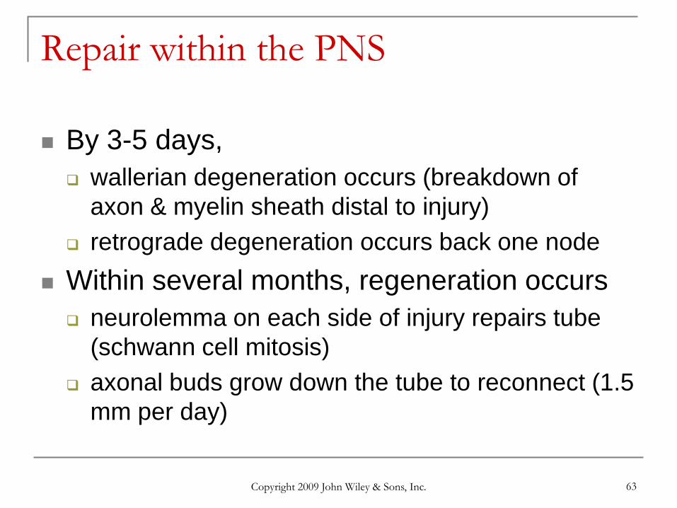
Repair within the PNS
By 3-5 days, wallerian degeneration occurs (breakdown of
axon & myelin sheath distal to injury) retrograde degeneration occurs back one node
Within several months, regeneration occurs neurolemma on each side of injury repairs tube
(schwann cell mitosis) axonal buds grow down the tube to reconnect (1.5
mm per day)
Copyright 2009 John Wiley & Sons, Inc. 64
Neurogenesis in the CNS Formation of new neurons from stem cells
was not thought to occur in humans There is a lack of neurogenesis in other
regions of the brain and spinal cord. Factors preventing neurogenesis in CNS inhibition by neuroglial cells, absence of growth
stimulating factors, lack of neurolemmas, and rapid formation of scar tissue

Copyright 2009 John Wiley & Sons, Inc. 65
Copyright 2009 John Wiley & Sons, Inc.All rights reserved. Reproduction or translation of this work beyond that permitted in section 117 of the 1976 United States Copyright Act without express permission of the copyright owner is unlawful. Request for further information should be addressed to the Permission Department, John Wiley & Sons, Inc. The purchaser may make back-up copies for his/her own use only and not for distribution or resale. The Publishers assumes no responsibility for errors, omissions, or damages caused by the use of theses programs or from the use of the information herein.
End of Chapter 12
Recommended