Embed Size (px)
Citation preview

Sections of the heart and bloodvessels

Tunica intima:Layer of the endothelial cells, underneath basal laminaSubendothelial layer: loose connective tissue with some smoothmuscle
Tunica media:Smooth muscle cells and elastic and reticular fibers, collagen typeI, proteoglycans
no characteristic or absent in veinsTunica adventitia:
mainly consists of connective tissue (longitudinally arranged collagenand elastic fibers, collagen type III)well-developed in veins (smooth muscle cells can be found)Vasa vasorum, vascular nerves
In muscular arteries internal elastic lamina (I. E. L.) separates the t. media from t. intima. Between the t. media and t. adventitia, theexternal elastic lamina (E. E. L.) is found.

T. intima:endothelial cells, basal laminasubendothelium: loose connective tissue, collagen and elastic fibers.
T. media:30-70 concentrically arranged elastic laminaesmooth muscle, some collagen fibers andfibrocytes.
T. adventitia:poorly developed, longitudinal collagen fibers, reticular fibers, fibrocytes.vasa vasorum, vascular nerves

- Increasing number of muscle cells, decreasing amount of fibers
T. intima: endothelial layer, basal lamina, subendothelial layer
I. E. L.: between t. intima and media, irregular, wavy lines(orcein and resorcin-fuchsin stain the lamina)
T. media: concentrically oriented smooth muscle cells intermingled withsome elastic and collagen fibers and fibrocytes
T. adventitia: E. E. L.: in larger arteries, longitudinal elastic fibers
poorly developed, collagen fibers, fibrocytes, fat cellsvasa vasorum, vascular nerves

1. Continuous / somatic capillariesbasal lamina is continuousSkeletal muscle, lungs, connective tissue, brain
2.a Fenestrated / visceral capillaries with diaphragmlarge feneastrae in the wall of endothelial cellstheir diamater is between 60-70 nmDiaphragm (macromolecular filter)Intense interchange of substances: resorptive epithelium of smallintestine, epihtelium of renal tubuli, epihtelium of the endocrineorgans
2.b Fenestrated capillaries without diaphragmBasal lamina is thickRenal glomeruli
3. Discontinuous sinusoidal capillariesEnlarged diameter, irregular shapemaybe with poresLiver, lymph node, spleen

- They have thinner wall, bigger lumen than the arteries have
T. intima: - endothel layer,basal laminasubendothelial layer
- at the border of the t. media, there is an increasingnumber of the elastic fibers
T. media: - thin layer- the smooth muscle cells are loosely situated,- collagen fibers and fibrocytes
T. adventitia: - the widest layer of the veins, rich of elastic fibers- longitudinal smooth muscle cells in the large veins- vasa vasorum (vessels of the vessels)

To study the structure of the wall of thedifferent blood vessels

20. Aorta (HE), 4X
Intima
Media
AdventitiaVasa vasorum
Subendothelium

20. Aorta (HE), 20X
Intima
Media
Endothelial cells
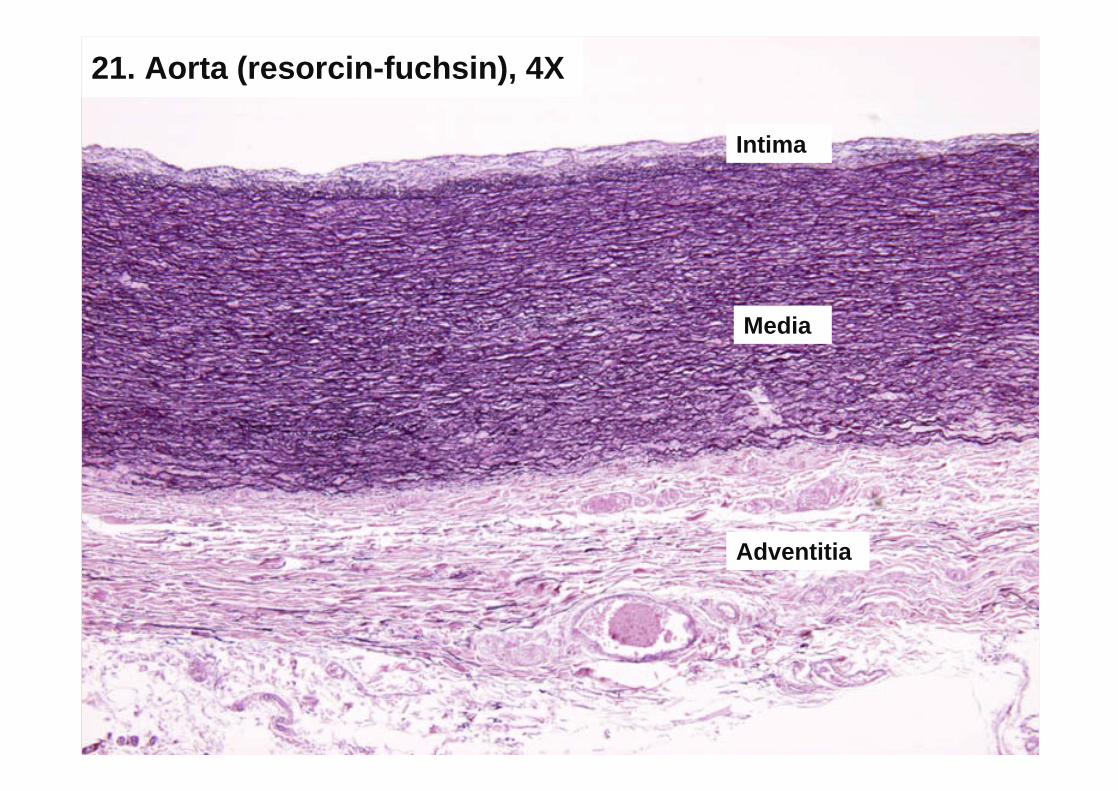
21. Aorta (resorcin-fuchsin), 4X
Intima
Media
Adventitia

21. Aorta (resorcin-fuchsin), 20X
Elastic laminas
Media
Intima

22. Artery-vein (HE), 4X
Artery
Vein

22. Artery-vein (HE), 20X
Intima
Media
Adventitia
I. E. L.
E. E. L.
Subendothelium
Smoothmuscle cells

22. Artery-vein (HE), 20X
Lumen of a vein
Intima
Media
Adventitia
Vasa Vasorum

23. Artery-vein (orcein), 4X
Vein
Artery

23. Artery-vein (orcein), 20X
Lumen of an arteryI. E.M.
Media
Adventitia
Intima
E. E. M.

23. Artery-vein (orcein), 40X
Intima
Media
Adventitia

Less prominent striationMorphological unit: branching / Y-shapemuscle cells (junctional complexes: gapjunction, intercalated disc / Eberth s line) Types: working muscle
propagating system

To study of the morphological unit of cardiacmuscle

15. Cardiac muscle (HE), 40X
Eberth s lines
capillary

16. Cardiac muscle (iron-hematoxilin), 40X
Eberth s lines
capillary