1
Biological Macromolecules & enzyme
Heba Al-Tamimi
2
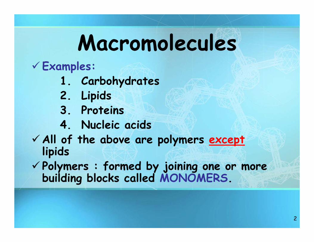
MacromoleculesExamples:
1. Carbohydrates2. Lipids3. Proteins4. Nucleic acids
All of the above are polymers exceptlipids
Polymers : formed by joining one or more building blocks called MONOMERS.

3
Dehydration Synthesis
• Polymers are formed by dehydration synthesis that involves removal of water.
HO H
HO HO HH
H2O
4
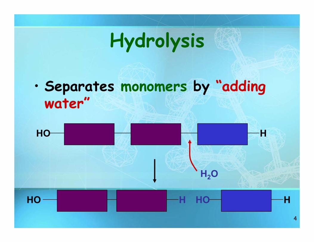
Hydrolysis
• Separates monomers by “adding water”
HO HO HH
HO H
H2O
5
Carbohydrates
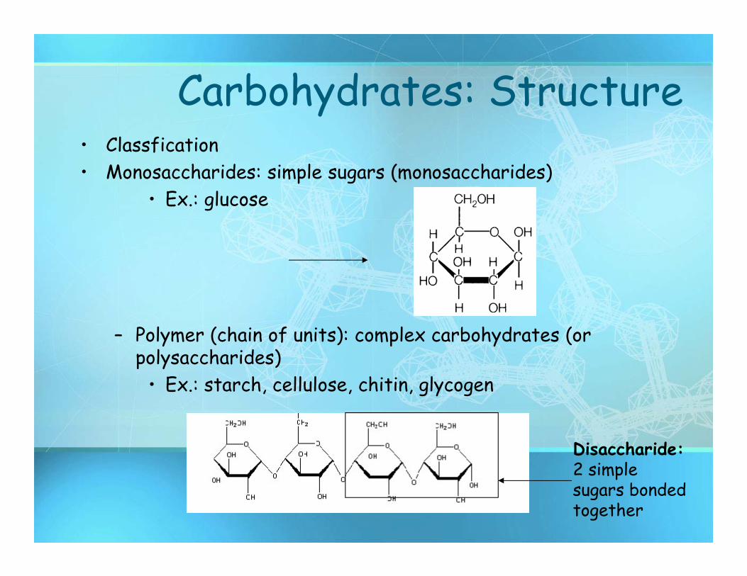
Carbohydrates: Structure• Classfication• Monosaccharides: simple sugars (monosaccharides)
• Ex.: glucose
– Polymer (chain of units): complex carbohydrates (or polysaccharides)
• Ex.: starch, cellulose, chitin, glycogen
Disaccharide: 2 simple sugars bonded together
7
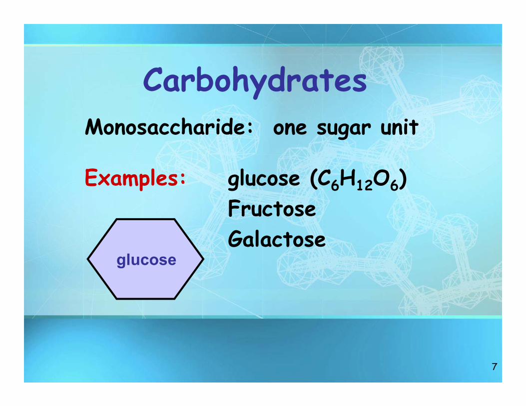
CarbohydratesMonosaccharide: one sugar unit
Examples: glucose (C6H12O6)FructoseGalactose
glucose
8
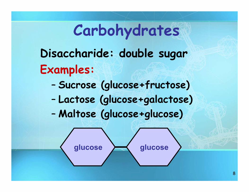
CarbohydratesDisaccharide: double sugar Examples:
– Sucrose (glucose+fructose)– Lactose (glucose+galactose)– Maltose (glucose+glucose)
glucoseglucose

9
CarbohydratesPolysaccharides: polymers composed of many sugar building blocks
Examples: starchglycogencellulose
glucoseglucose
glucoseglucose
glucoseglucose
glucoseglucose
cellulose
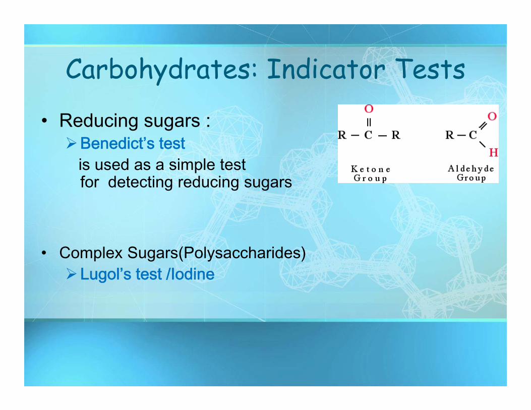
Carbohydrates: Indicator Tests
• Reducing sugars :Benedict’s test
is used as a simple test for detecting reducing sugars
• Complex Sugars(Polysaccharides)Lugol’s test /Iodine

5-1 Benedict’s solution
11
+ CuSO4/0H
-CooH
1-
2- reducing
Maltose
Glucose + Galucose
+ Cu2O(Red –brown precipitate)
Benedict’s solution
12
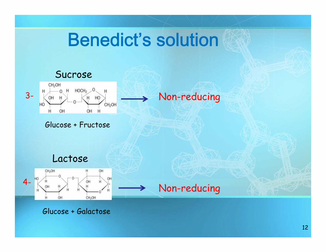
3- Non-reducing
4- Non-reducing
Sucrose
Lactose
Glucose + Galactose
Glucose + Fructose
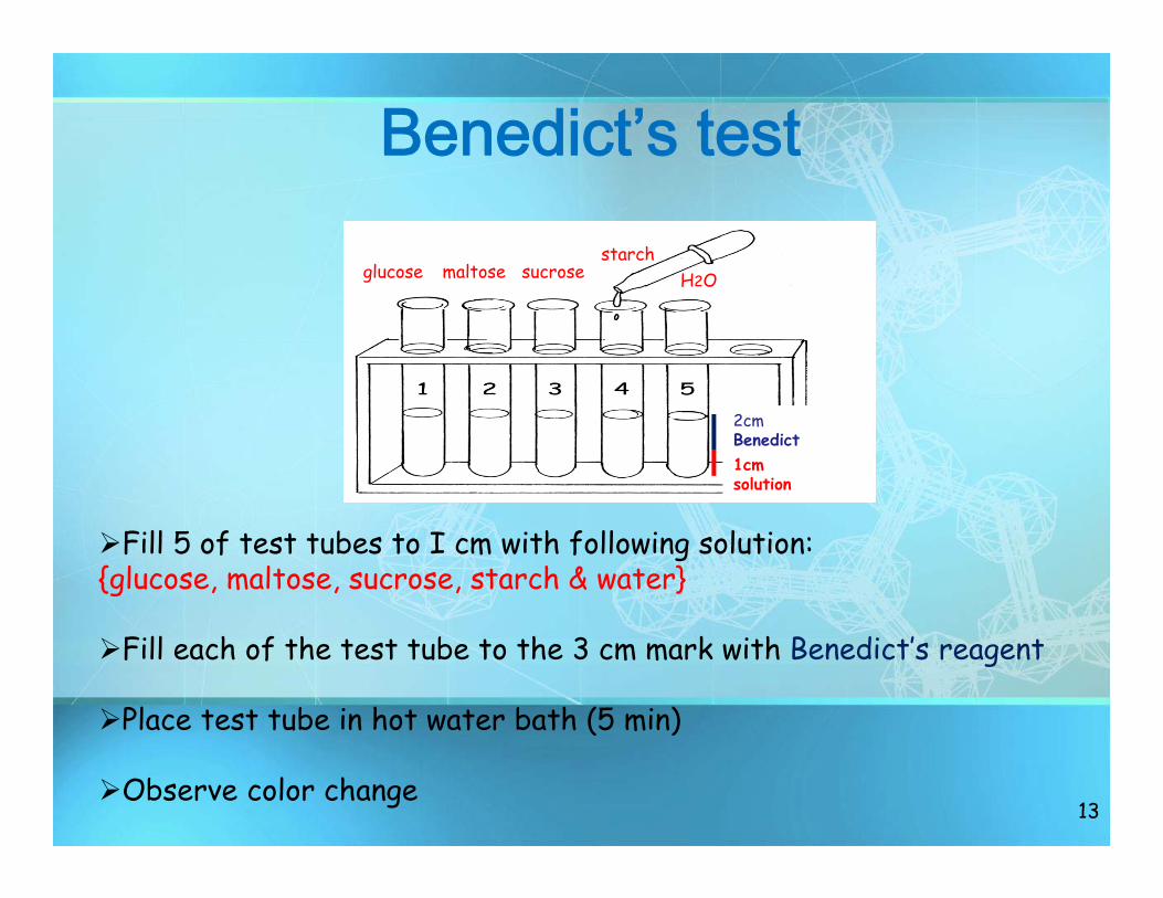
Benedict’s test
13
1cm solution
2cmBenedict
Fill 5 of test tubes to I cm with following solution: {glucose, maltose, sucrose, starch & water}
Fill each of the test tube to the 3 cm mark with Benedict’s reagent
Place test tube in hot water bath (5 min)
Observe color change
glucose maltose sucrosestarch
H2O
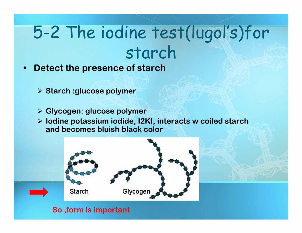
5-2 The iodine test(lugol’s)for starch
• Detect the presence of starch
Starch :glucose polymer
Glycogen: glucose polymer Iodine potassium iodide, I2KI, interacts w coiled starch
and becomes bluish black color
So ,form is important
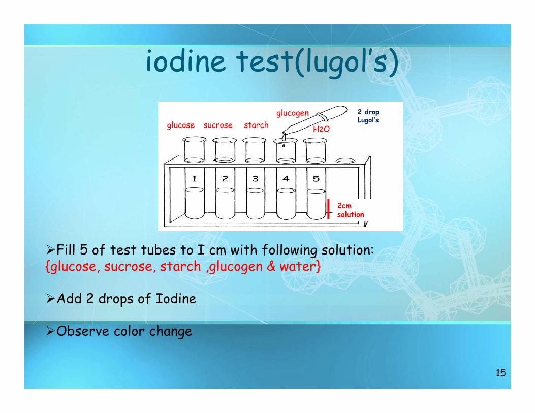
iodine test(lugol’s)
15
2cm solution
Fill 5 of test tubes to I cm with following solution: {glucose, sucrose, starch ,glucogen & water}
Add 2 drops of Iodine
Observe color change
glucose sucroseglucogen
starch H2O
2 dropLugol’s
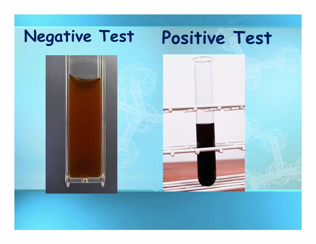
Negative Test Positive Test
17
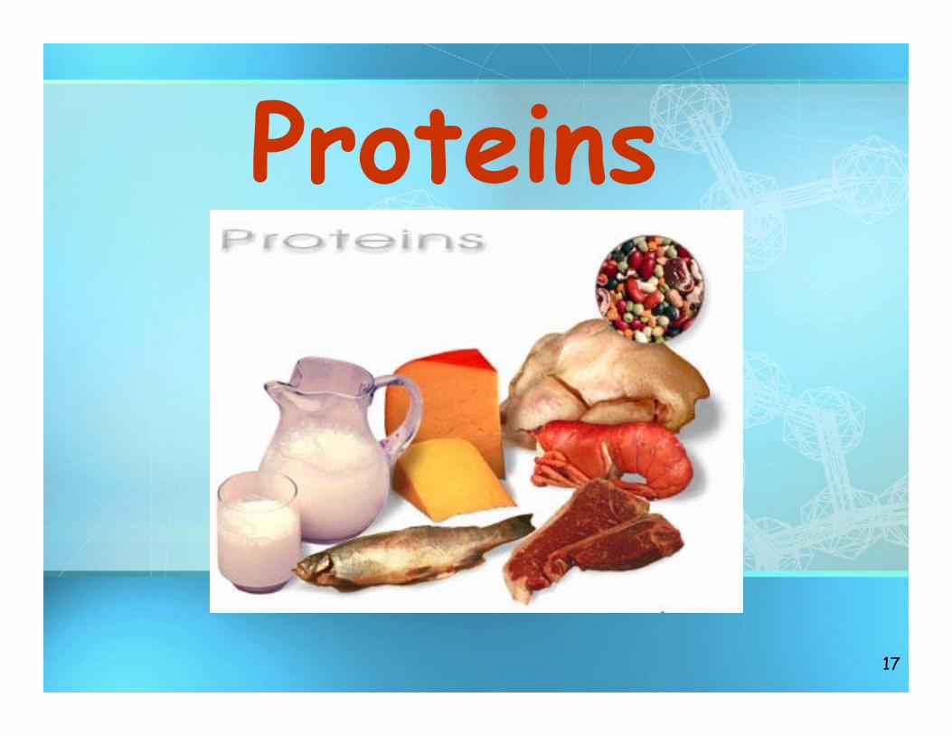
Proteins
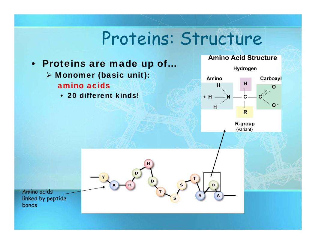
Proteins: Structure• Proteins are made up of…
Monomer (basic unit):amino acids• 20 different kinds!
Amino acids linked by peptide bonds
19
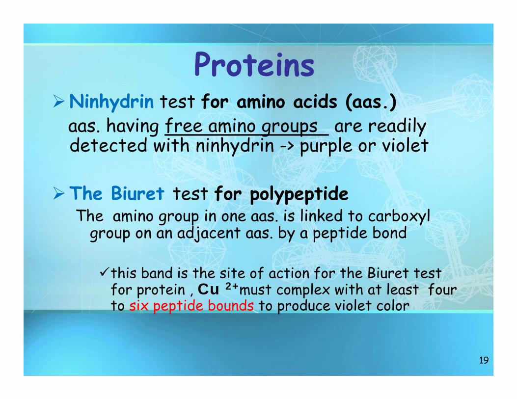
ProteinsNinhydrin test for amino acids (aas.)
aas. having free amino groups are readily detected with ninhydrin -> purple or violet
The Biuret test for polypeptideThe amino group in one aas. is linked to carboxyl
group on an adjacent aas. by a peptide bond
this band is the site of action for the Biuret test for protein , Cu 2+must complex with at least four to six peptide bounds to produce violet color
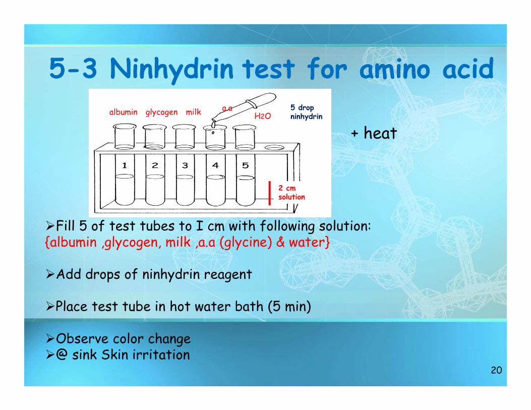
5-3 Ninhydrin test for amino acid
20
2 cm solution
Fill 5 of test tubes to I cm with following solution: {albumin ,glycogen, milk ,a.a (glycine) & water}
Add drops of ninhydrin reagent
Place test tube in hot water bath (5 min)
Observe color change@ sink Skin irritation
albumin glycogen milk a.aH2O
5 drop ninhydrin
+ heat
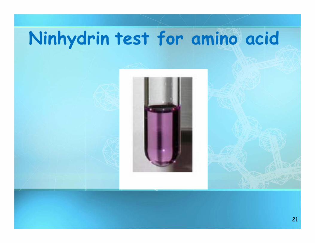
Ninhydrin test for amino acid
21
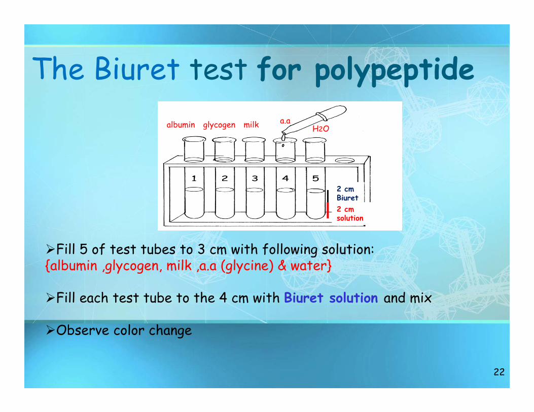
The Biuret test for polypeptide
22
2 cm solution
Fill 5 of test tubes to 3 cm with following solution: {albumin ,glycogen, milk ,a.a (glycine) & water}
Fill each test tube to the 4 cm with Biuret solution and mix
Observe color change
albumin glycogen milk a.aH2O
2 cm Biuret
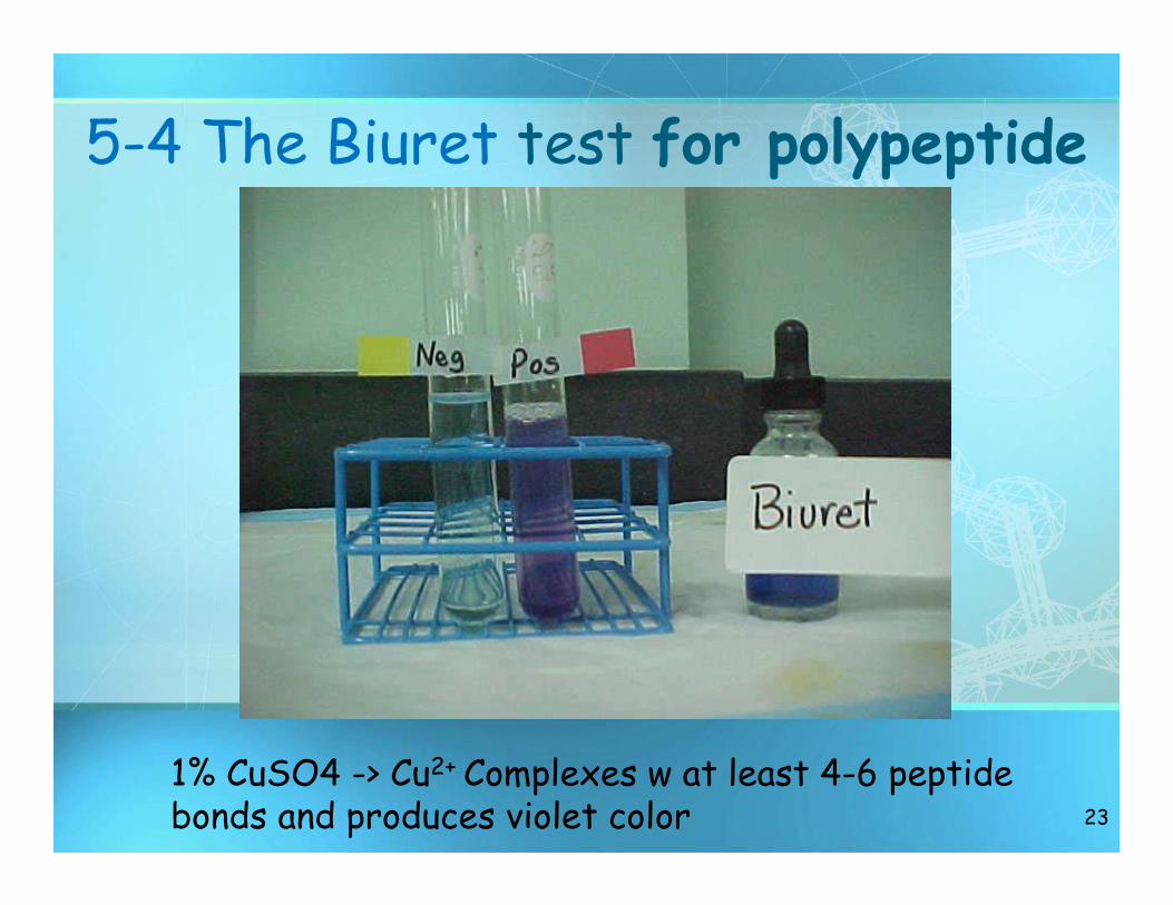
5-4 The Biuret test for polypeptide
23
1% CuSO4 -> Cu2+ Complexes w at least 4-6 peptide bonds and produces violet color
24
Lipids
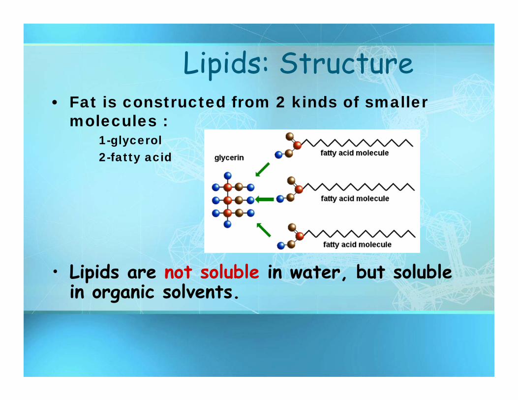
Lipids: Structure• Fat is constructed from 2 kinds of smaller
molecules :1-glycerol 2-fatty acid
• Lipids are not soluble in water, but soluble in organic solvents.
26
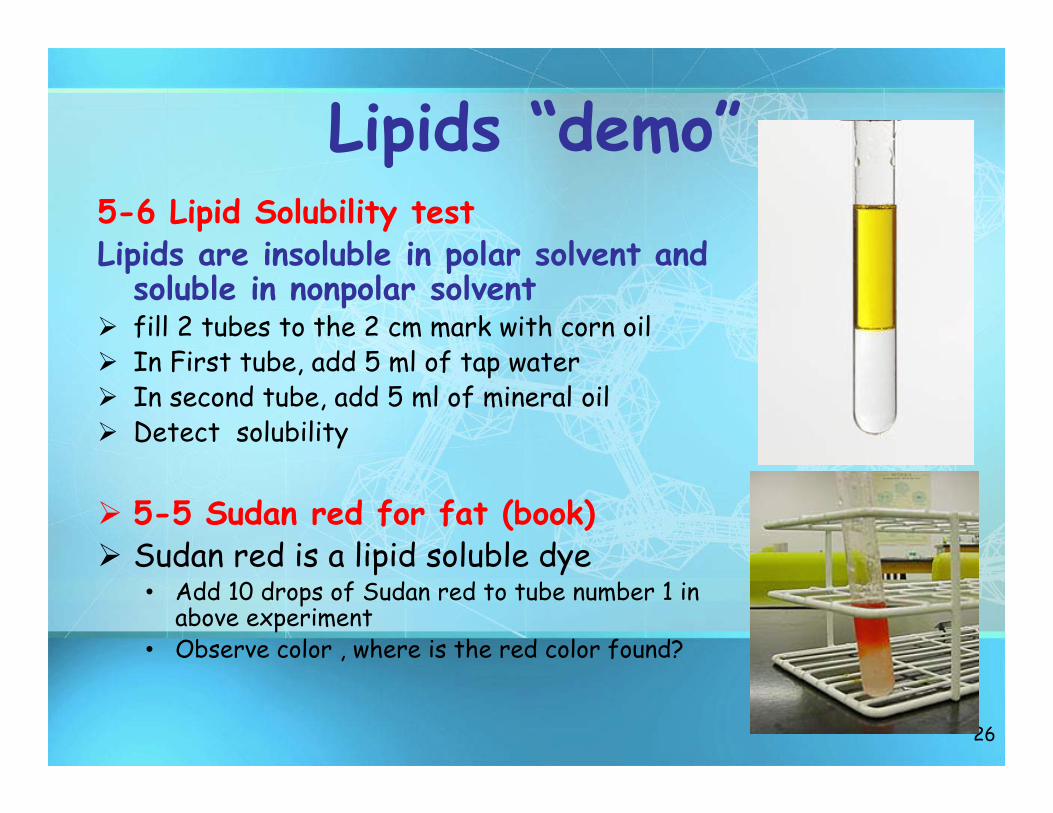
Lipids “demo”5-6 Lipid Solubility testLipids are insoluble in polar solvent and
soluble in nonpolar solvent fill 2 tubes to the 2 cm mark with corn oil In First tube, add 5 ml of tap water In second tube, add 5 ml of mineral oil Detect solubility
5-5 Sudan red for fat (book) Sudan red is a lipid soluble dye
• Add 10 drops of Sudan red to tube number 1 in above experiment
• Observe color , where is the red color found?
27
Nucleic Acids
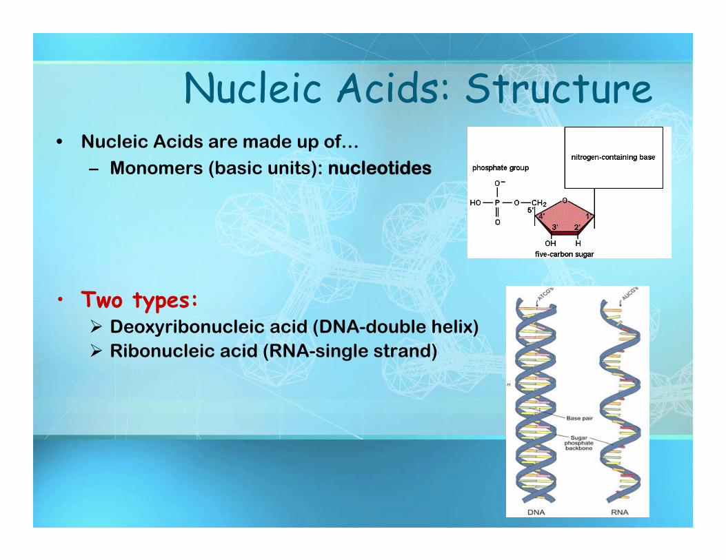
Nucleic Acids: Structure• Nucleic Acids are made up of…
– Monomers (basic units): nucleotides
• Two types: Deoxyribonucleic acid (DNA-double helix) Ribonucleic acid (RNA-single strand)
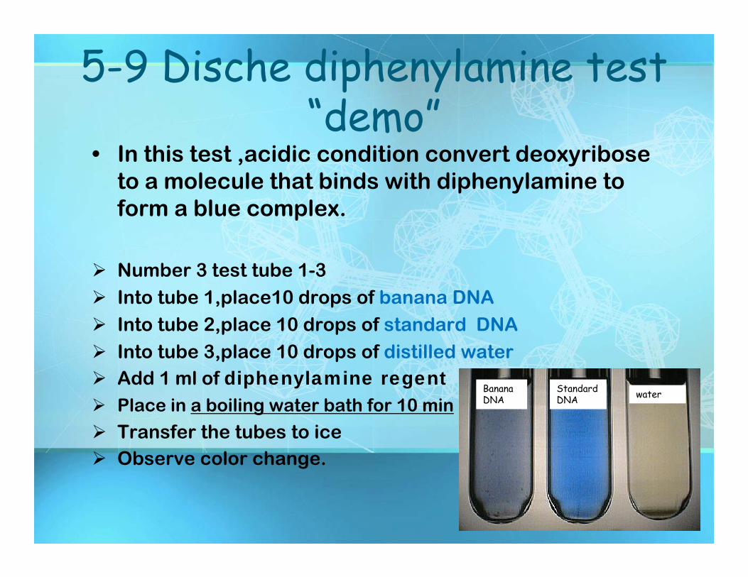
5-9 Dische diphenylamine test “demo”
• In this test ,acidic condition convert deoxyribose to a molecule that binds with diphenylamine to form a blue complex.
Number 3 test tube 1-3 Into tube 1,place10 drops of banana DNA Into tube 2,place 10 drops of standard DNA Into tube 3,place 10 drops of distilled water Add 1 ml of diphenylamine regent Place in a boiling water bath for 10 min
Transfer the tubes to ice Observe color change.
Banana DNA
Standard DNA water
Enzymes…
30
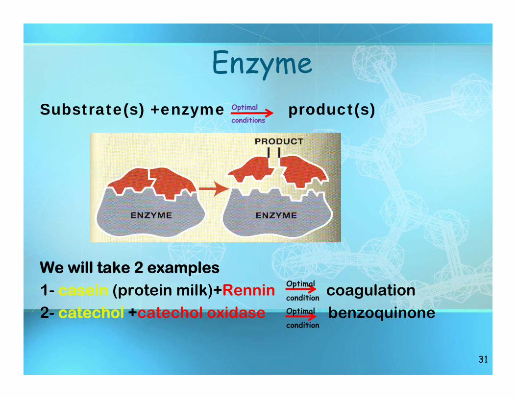
Enzyme Substrate(s) +enzyme product(s)
We will take 2 examples1- casein (protein milk)+Rennin coagulation2- catechol +catechol oxidase benzoquinone
31
Optimal
conditions
Optimal
conditionOptimal
condition
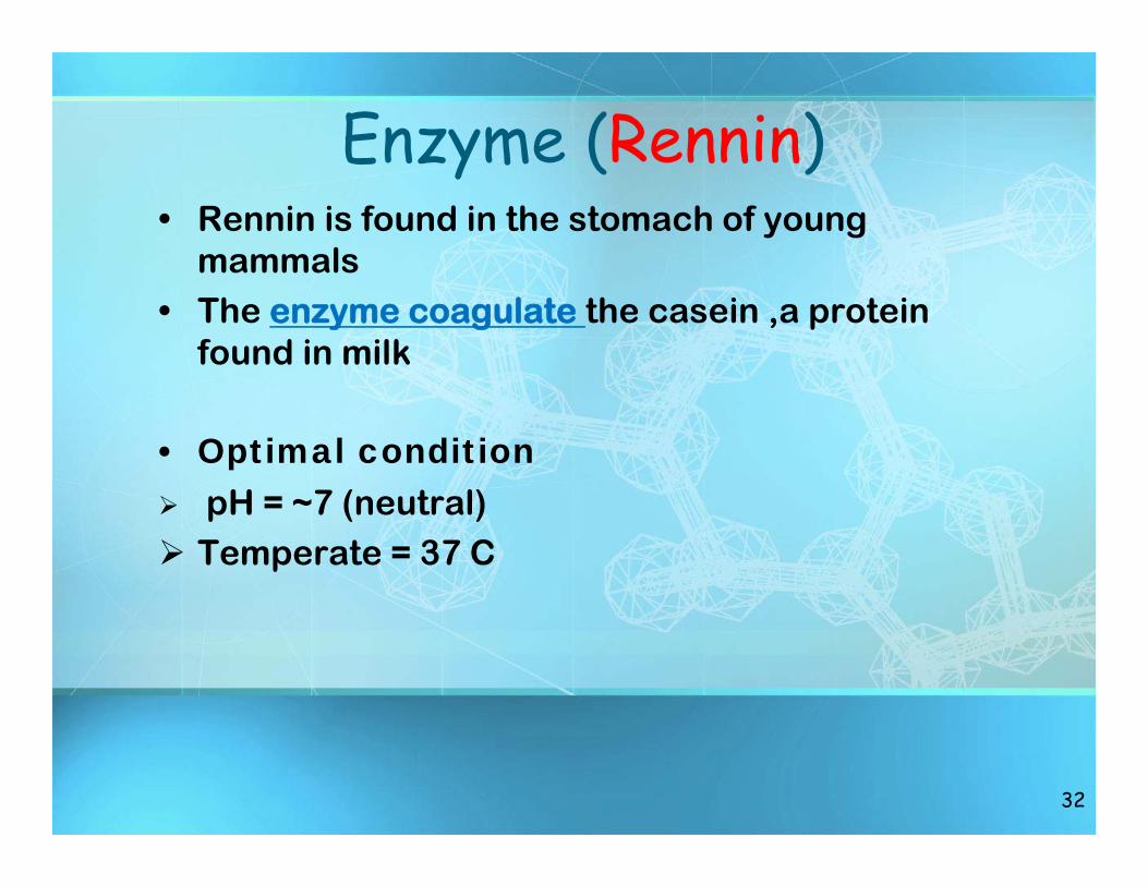
Enzyme (Rennin) • Rennin is found in the stomach of young
mammals• The enzyme coagulate the casein ,a protein
found in milk
• Optimal condition pH = ~7 (neutral) Temperate = 37 C
32
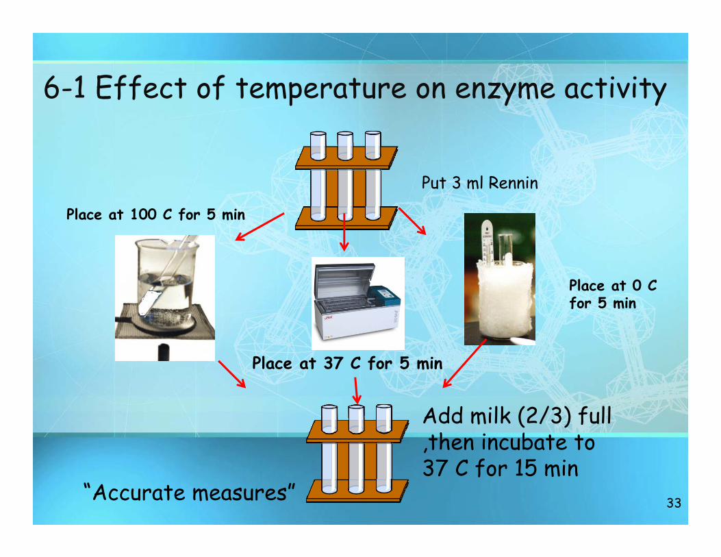
6-1 Effect of temperature on enzyme activity
33
Put 3 ml Rennin
Place at 0 C for 5 min
Place at 100 C for 5 min
Place at 37 C for 5 min
Add milk (2/3) full ,then incubate to 37 C for 15 min
“Accurate measures”
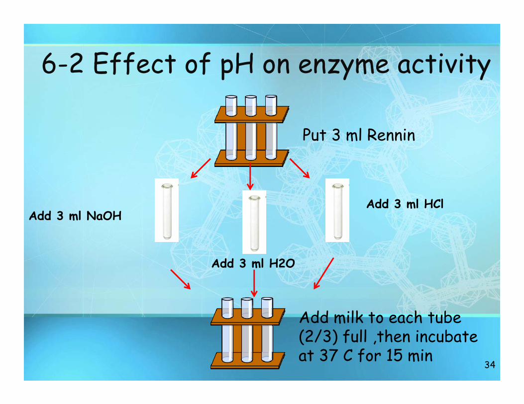
6-2 Effect of pH on enzyme activity
34
Put 3 ml Rennin
Add 3 ml HCl
Add 3 ml H2O
Add 3 ml NaOH
Add milk to each tube (2/3) full ,then incubate at 37 C for 15 min
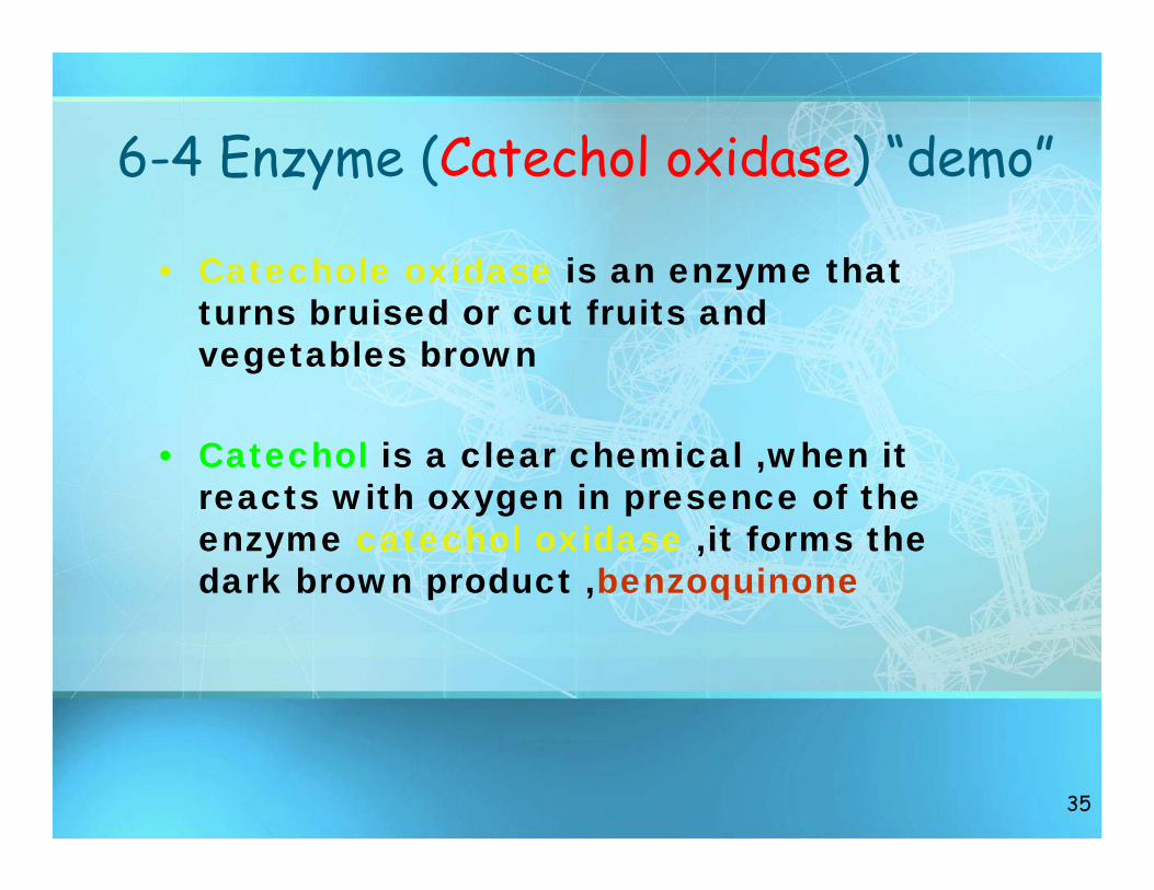
6-4 Enzyme (Catechol oxidase) “demo”
• Catechole oxidase is an enzyme that turns bruised or cut fruits and vegetables brown
• Catechol is a clear chemical ,when it reacts with oxygen in presence of the enzyme catechol oxidase ,it forms the dark brown product ,benzoquinone
35
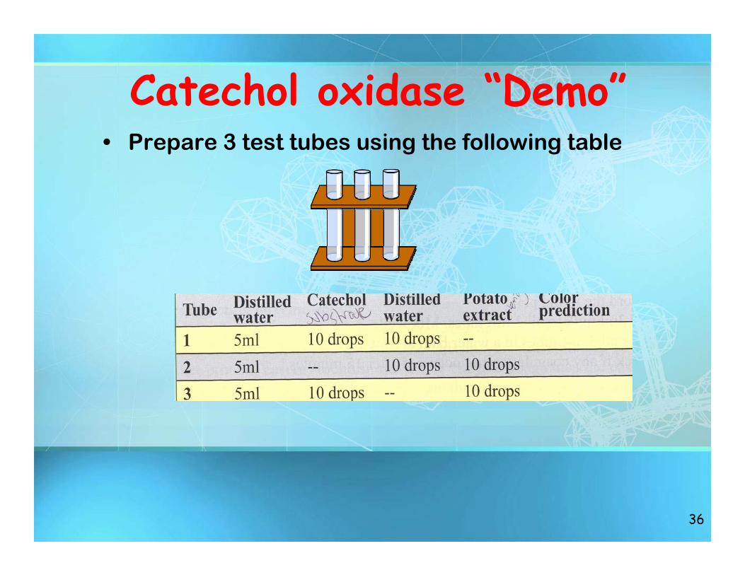
Catechol oxidase “Demo”• Prepare 3 test tubes using the following table
36
Go To Work……
37
Recommended