Bio-Osss
Collagen in the buccal gap atimmediate implants: a 6-month studyin the dog
Mauricio G. AraujoElena LinderJan Lindhe
Authors’ affiliations:Mauricio Araujo, Department of Dentistry, StateUniversity of Maringa, Parana, BrazilElena Linder, Jan Lindhe, Institute of Odontology,The Sahlgrenska Academy at the University ofGothenburg, Gothenburg, Sweden
Corresponding author:Mauricio AraujoRua Silva Jardim15/sala 0387013-010Maringa-Parana-BrazilTel./Fax: þ 55 44 3224 6444e-mail: [email protected]
Key words: biomaterial, dogs, extraction socket, grafting, implants
Abstract
Background: Following tooth extraction and immediate implant installation, the
edentulous site of the alveolar process undergoes substantial bone modeling and the ridge
dimensions are reduced.
Objective: The objective of the present experiment was to determine whether the process
of bone modeling following tooth extraction and immediate implant placement was
influenced by the placement of a xenogenic graft in the void that occurred between the
implant and the walls of the fresh extraction socket.
Material and methods: Five beagle dogs about 1 year old were used. The 4th premolar in
both quadrants of the mandible (4P4) were selected and used as experimental sites. The
premolars were hemi-sected and the distal roots removed and, subsequently, implants were
inserted in the distal sockets. In one side of the jaw, the marginal buccal-approximal void
that consistently occurred between the implant and the socket walls was grafted with
Bio-Osss Collagen while no grafting was performed in the contra-lateral sites. After 6
months of healing, biopsies from each experimental site were obtained and prepared for
histological analyses.
Results: The outline of the marginal hard tissue of the control sites was markedly different
from that of the grafted sites. Thus, while the buccal bone crest in the grafted sites was
comparatively thick and located at or close to the SLA border, the corresponding crest at the
control sites was thinner and located a varying distance below SLA border.
Conclusions: It was demonstrated that the placement of Bio-Osss Collagen in the void
between the implant and the buccal-approximal bone walls of fresh extraction sockets
modified the process of hard tissue healing, provided additional amounts of hard tissue at
the entrance of the previous socket and improved the level of marginal bone-to-implant
contact.
Following tooth extraction, the edentulous
site of the alveolar process will undergo
both quantitative and qualitative changes
(e.g., Pietrokovski & Massler 1967; Amler
1969; Schropp et al. 2003). Thus, during
healing, the bundle bone will gradually
disappear, the socket will be filled with
granulation tissue, provisional matrix and
woven bone that eventually will be re-
placed with trabecular bone and marrow
(e.g., Amler 1969; Evian et al. 1982; Ku-
boki et al. 1988; Cardaropoli et al. 2003;
Araujo & Lindhe 2005). Moreover, (i) the
walls of the socket will be markedly re-
duced both with respect to height and
width (Araujo et al. 2005, 2006) and (ii)
the dimensional changes will be more pro-
nounced in the buccal than in the lingual/
Date:Accepted 24 December 2009
To cite this article:Araujo MG, Linder E, Lindhe J. Bio-Oss
s
Collagen inthe buccal gap at immediate implants: a 6-month studyin the dog.Clin. Oral Impl. Res. 22, 2011; 1–8.doi: 10.1111/j.1600-0501.2010.01920.x
c� 2010 John Wiley & Sons A/S 1

palatal compartments of the extraction site
(Pietrokovski & Massler 1967; Pietro-
kovski et al. 2007). Although early case
reports indicated that implants placed in
fresh extraction sites may counteract post-
extractive bone modeling, later studies in
humans and experimental animals docu-
mented that such treatment would, in fact,
not influence the tissue alterations de-
scribed above (Botticelli et al. 2004; Araujo
et al. 2005, 2006; Blanco et al. 2008; Evans
& Chen 2008).
Different approaches have been advo-
cated to preserve or improve the dimension
and contour of the ridge following tooth
extraction including the use of various
graft or filler materials such as autografts,
allografts, synthetic graft, etc. and/or
barrier membranes (for a review, see
Botticelli 2006). One particular graft mate-
rial, comprised of deproteinized bovine
bone mineral has been widely used in
attempts to preserve the dimension of the
alveolar ridge following tooth extraction
(ridge preservation) as well as in angular
bone defects at teeth and in sinus augmen-
tation procedures (e.g., Hurzeler et al.
1997; Berglundh & Lindhe 1997; Piattelli
et al. 1999; Artzi et al. 2000, 2002; Yil-
dirim et al. 2000; Froum et al. 2002;
Carmagnola et al. 2003; Norton et al.
2003; Nevins et al. 2006). In some of the
studies referred to, grafting apparently had
a successful outcome, while in other re-
ports the benefits of such therapy were less
obvious.
In one recent animal experiment, it was
demonstrated that the placement of a xe-
nogenic graft comprised of bovine bone
combined with collagen from porcine ori-
gin in fresh extraction sockets of dogs
promoted de novo hard tissue formation,
in particular in the marginal portion of the
extraction site (Araujo et al. 2008). Here,
the dimension of hard tissue walls was
maintained and the profile of the ridge
was preserved. In a subsequent 2-week
study (Araujo et al. 2009), it was shown
that the early phases of hard tissue forma-
tion were altered in extraction sockets that
were grafted with Bio-Osss
Collagen im-
mediately after tooth removal. It appeared
that this modified wound healing and bone
modeling may have been influenced by the
presence of multinucleated cells that oc-
curred in tissues harboring the xenogenic
graft. Thus, in the grafted sites, substantial
amounts of newly formed bone could only
be detected in the apical portion of the
socket where the graft material was absent.
In the remaining portions of the grafted
sockets a mildly inflamed provisional
matrix surrounded the majority of the
Bio-Osss
particles, whose surface was fre-
quently, but not always, coated with mul-
tinucleated cells. In isolated areas of the
grafted sites, multinucleated cells were
absent and the foreign material was sur-
rounded by newly formed woven bone that
bridged adjacent granules of the xenogenic
particles.
The objective of the present experiment
was to determine whether the process of
bone modeling following tooth extraction
and immediate implant placement could be
influenced by the placement of Bio-Osss
Collagen in the void that occurred between
the implant and the walls of the fresh
extraction socket.
Material and methods
The ethical committee of the State Uni-
versity of Maringa approved the research
protocol. Five beagle dogs about 1 year old
and weighing between 10 and 12 kg each
were used. During surgical procedures, the
animals were anesthetized with intrave-
nously administered Ketamin (10%,
8 mg/kg, Agener Uniao, Sao Paulo, Brazil).
The fourth premolars in both quadrants
of the mandible (4P4) were selected and
used as experimental sites. The mesial
root canals were reamed and filled with
gutta-percha.
The fourth premolars were hemi-sected
and the distal roots were carefully removed
with the use of forceps (Fig. 1). The reci-
pient sites were prepared for implant in-
stallation according to the guidelines
provided by the manufacturer. Implants
(Straumanns
Dental Implant System;
Fig. 1. Clinical photograph illustrating the alveolar socket immediately after the extraction of the distal root of
the mandibular fourth premolar.
Fig. 2. Clinical photograph illustrating the position of the implants placed in the distal extraction socket of the
mandibular fourth premolar. Note that a void (gap) had been established between the implant and the buccal
and approximal bone walls.
Araujo et al �Xenogenic graft and immediate implants
2 | Clin. Oral Impl. Res. 22, 2011 / 1–8 c� 2010 John Wiley & Sons A/S
Standard Implant, 3.3 mm wide and 6 or
8 mm long; Straumann, Basel, Switzer-
land) were installed. Each implant was
placed in a lingual position in the socket.
Hence, a 1–2 mm wide and 3 mm deep
buccal-approximal void similar to a three-
wall bone defect was established between
the titanium rod and the inner bone
walls (Fig. 2). The marginal level of the
SLA-coated surface of all implants was,
following placement, located flush with
or slightly apical of the adjacent buccal
bone crest. On one side of the jaw, the
marginal buccal-approximal void that con-
sistently occurred between the implant and
the socket walls was grafted with Bio-Osss
Collagen (Geistlich Pharma AB, Wolhu-
sen, Switzerland), while no grafting was
performed in the contra-lateral sites
(Fig. 3).
Healing caps (Straumanns
Dental
Implant System) were adjusted to the im-
plants. The flaps and the wound margins
were replaced and secured to allow a semi-
submerged healing of the experimental
sites (Fig. 4). The sutures were removed
after 2 weeks. Every second day, the ani-
mals were exposed to mechanical plaque
removal. In addition, a clorhexidine (0.2%)
gel was placed every second day placed at
healing caps and adjacent teeth.
After 6 months of healing, the dogs
were euthanized with an overdose of
Ketamin and perfused with a fixative
containing a mixture of 5% glutaraldehyde
Fig. 3. Clinical photograph illustrating that the void between the implant and the bone walls had been filled
with the xenogenic graft.
Fig. 4. Clinical photograph of the occlusal aspect of the mandibular fourth premolar illustrating the mucosal
flaps that had been secured with interrupted sutures.
Fig. 5. Schematic drawing describing different land-
marks from which histometric measurements were
performed. IHC, level of the implant – healing cap
connection; SLA, marginal border of the rough im-
plant surface; C, marginal level of the bone crest; B/
I, marginal level of bone to implant contact; 1, 2 and
3 mm represent the levels ‘apical of SLA’ at which
the width of the buccal and lingual walls was
determined.
Fig. 6. Clinical photograph illustrating the implant sites after 6 months of healing. The peri-implant mucosa at
both the test and the control sites had normal texture and color and was free of signs of inflammation.
Araujo et al �Xenogenic graft and immediate implants
c� 2010 John Wiley & Sons A/S 3 | Clin. Oral Impl. Res. 22, 2011 / 1–8

and 4% formaldehyde (Karnovsky 1965).
The mandibles were removed and placed in
the fixative. Radiographs were obtained
from the implant locations and the position
of the ‘interproxmial’ bone was estimated.
Each experimental site, including the
mesial root and the distal socket area, was
dissected using a diamond saw (Exacts
Apparatebeau, Norderstedt, Hamburg,
Germany). The tissues were processed for
ground sectioning according to the meth-
ods described by Donath & Breuner (1982)
and Donath (1988). The samples were
dehydrated in increasing grades of ethanol
and infiltrated with Technovits
7200 VLC-
resin (Kulzer, Friedrichrsdorf, Germany),
polymerized and sectioned using a cutting
– grinding unit (Exacts
Apparatebeau).
From each biopsy unit, one buccal–lin-
gual section representing the central area of
the site was prepared. The sections were
reduced to a thickness of about 25 mm by
micro-grinding and polishing. The sections
were stained in Ladewig’s fibrin stain
(Donath 1993). The histological examina-
tions were performed in a Leitz DM-RBEs
microscope (Leica, Wetzlar, Germany)
equipped with an image system (Q-500
MCs
, Leica).
Histological examination
In the sections, linear measurements (mag-
nification � 16–50) were made between
the following landmarks (Fig. 5):
� (PM): margin of the peri-implant mu-
cosa.
� (aBE): apical cells of the barrier epithe-
lium.
� (SLA): the marginal termination of the
rough surface.
� (C): the crest of the buccal or the
lingual bone wall.
� (B/I): the most coronal point of contact
between bone and implant.
� (I/HC): the contact between the im-
plant and the healing cap.
The widths of the buccal and lingual
bone walls were determined by measuring
the distance between the buccal or the
lingual surface of the implant body and
the outer surface of the hard tissue wall.
The assessments were performed at the
SLA level as well as 1, 2 and 3 mm apical
of SLA.
Results
Healing following tooth extraction,
implant installation and grafting was un-
eventful in all dogs. At the 6-month
examination interval, the mucosa sur-
rounding all implants was virtually free of
clinical signs of inflammation (Fig. 6). The
margin of the peri-implant mucosa resided
at all 10 experimental sites on the healing
caps.
In the radiographs, it was observed that
the crest of the ‘interproximal’ bone at the
experimental sites was located at the level
of the cut distal furcation fornix of the
mesial root. Thus, there was no apparent
loss of bone at the ‘interproximal’ region
during the study period.
Fig. 7. Buccal–lingual section representing the test (a) and control (b) sites. B, buccal bone wall; I, implant; L,
lingual bone wall; PM, peri-implant mucosa. Ladewig’s fibrin staining; original magnifications � 16.
Fig. 8. Higher magnification of the buccal aspect of the peri-implant mucosa in Fig. 7. Test site (a), control site
(b). Note that the mucosa is rich in collagen fibers and contains few inflammatory cells. In addition, in the test
site (a) note the presence of a Bio-Osss
particle in the connective tissue lateral below the oral epithelium. CNT,
connective tissue; aBE, apical portion of the barrier epithelium; I, implant; OE, oral epithelium. Ladewig’s
fibrin staining; original magnifications � 50.
Araujo et al �Xenogenic graft and immediate implants
4 | Clin. Oral Impl. Res. 22, 2011 / 1–8 c� 2010 John Wiley & Sons A/S
The microscopic examination of the
ground sections revealed that all implants
were well-integrated with the surrounding
bone tissue that was comprised of miner-
alized bone and marrow (Fig. 7). Further-
more, the supracrestal soft tissue in all
10 implant sites (i) was rich in well-
oriented bundles of collagen fibers and
(ii) harbored only small inflammatory cell
infiltrations (Fig. 8a and b). The attach-
ment between the mucosa and the implant
was made up of a barrier epithelium that
was continuous in the apical direction,
with a zone of connective tissue attach-
ment that was in apparent direct contact
with the titanium cylinder. In the test
sites, Bio-Osss
particles were frequently
observed in the peri-implant mucosa
(Fig. 8a).
The outline of the marginal hard
tissue of the control sites was markedly
different from that of the test sites. Thus,
while the buccal bone crest in the test sites
was comparatively thick and located at or
close to the SLA level (SLA), the corre-
sponding crest at the control sites was
thinner and located at a varying distance
below SLA. Furthermore, in the test sites,
the marginal bone-to-implant contact (B/I)
was at the same level at the buccal and
lingual aspects, whereas in the control, the
lingual B/I was located more close to the
upper rim of the implant than its buccal
counterpart.
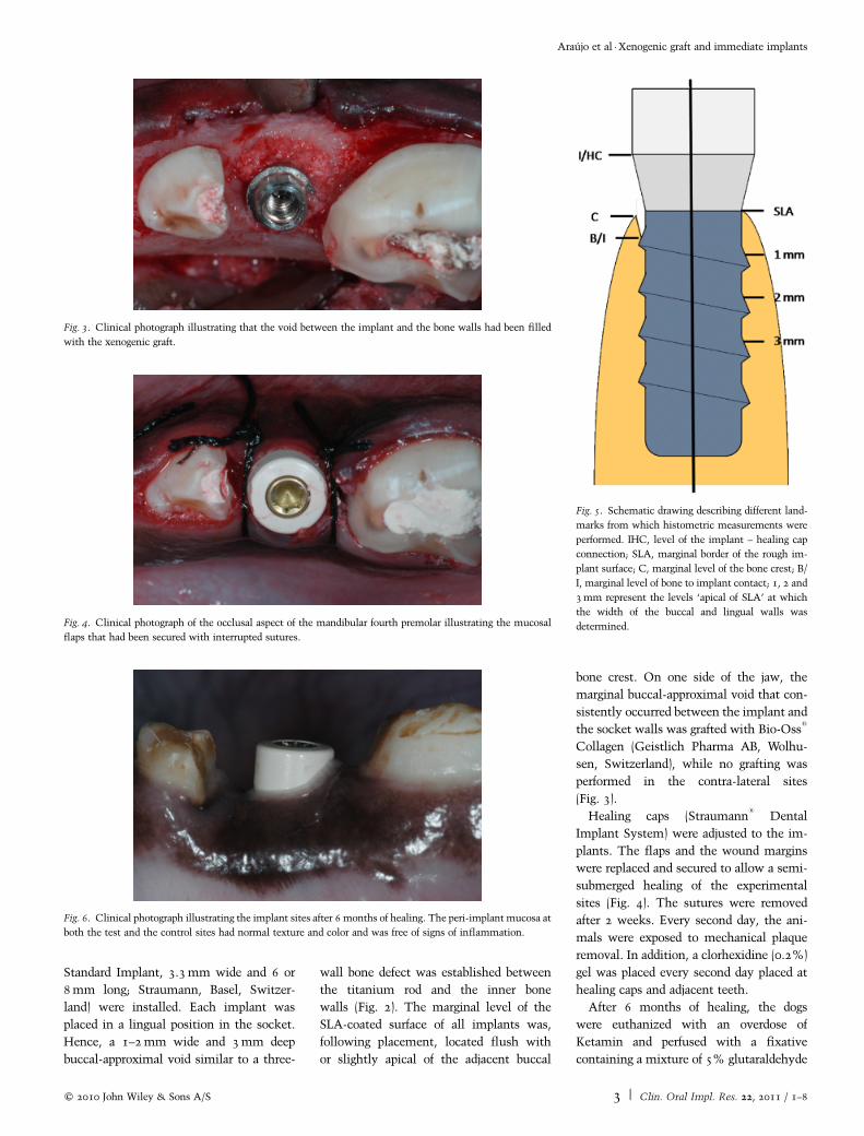
In the test sites, the buccal void that had
been grafted with Bio-Osss
Collagen fol-
lowing implant placement was, at sacri-
fice, filled with newly formed bone in
which a varying number of Bio-Osss
parti-
cles had been trapped (Fig. 9). This newly
formed hard tissue that was mainly com-
prised of woven and parallel-fibered bone
was, in four out of five test sites, contin-
uous with the old lamellar bone of the
buccal bone wall (Fig. 10).
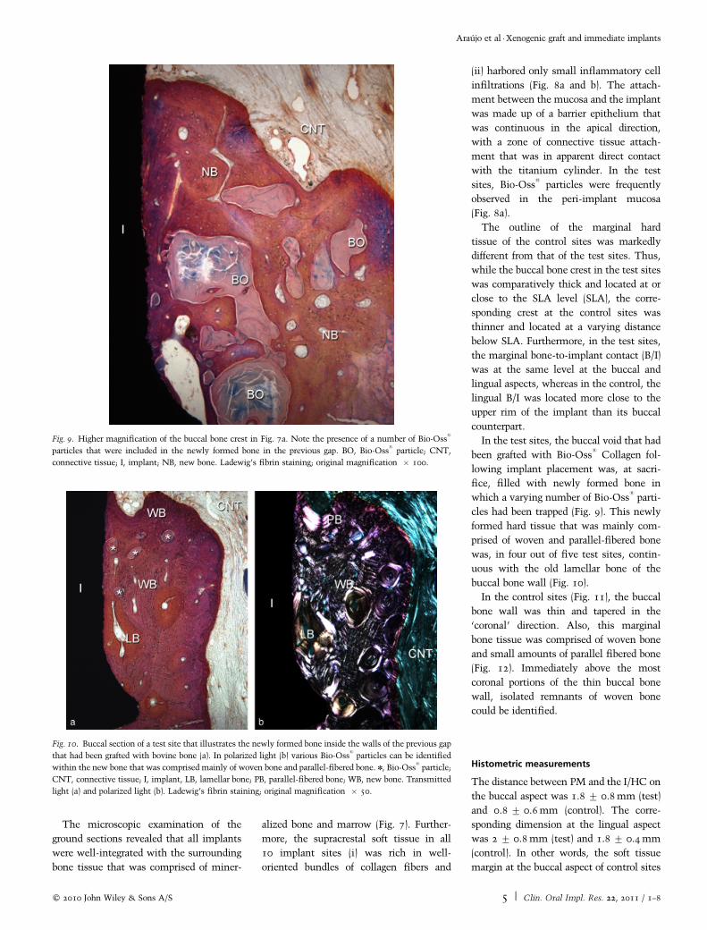
In the control sites (Fig. 11), the buccal
bone wall was thin and tapered in the
‘coronal’ direction. Also, this marginal
bone tissue was comprised of woven bone
and small amounts of parallel fibered bone
(Fig. 12). Immediately above the most
coronal portions of the thin buccal bone
wall, isolated remnants of woven bone
could be identified.
Histometric measurements
The distance between PM and the I/HC on
the buccal aspect was 1.8� 0.8 mm (test)
and 0.8� 0.6 mm (control). The corre-
sponding dimension at the lingual aspect
was 2� 0.8 mm (test) and 1.8� 0.4 mm
(control). In other words, the soft tissue
margin at the buccal aspect of control sites
Fig. 9. Higher magnification of the buccal bone crest in Fig. 7a. Note the presence of a number of Bio-Osss
particles that were included in the newly formed bone in the previous gap. BO, Bio-Osss
particle; CNT,
connective tissue; I, implant; NB, new bone. Ladewig’s fibrin staining; original magnification � 100.
Fig. 10. Buccal section of a test site that illustrates the newly formed bone inside the walls of the previous gap
that had been grafted with bovine bone (a). In polarized light (b) various Bio-Osss
particles can be identified
within the new bone that was comprised mainly of woven bone and parallel-fibered bone. n, Bio-Osss
particle;
CNT, connective tissue; I, implant, LB, lamellar bone; PB, parallel-fibered bone; WB, new bone. Transmitted
light (a) and polarized light (b). Ladewig’s fibrin staining; original magnification � 50.
Araujo et al �Xenogenic graft and immediate implants
c� 2010 John Wiley & Sons A/S 5 | Clin. Oral Impl. Res. 22, 2011 / 1–8
was located about 1 mm apical of that of
the test sites.
The length of the barrier epithelium
(PM–aBE; Table 1) varied between
2� 0.5 and 2.9� 0.7 mm, while the
zone of connective tissue attachment
(aBE–B/I) varied between 0.7� 0.4 and
1.9� 0.9 mm. The width of the buccal
soft tissue at the implant/healing cap level
(PM width; Table 1) was 1� 0.3 mm
in the test and 0.4� 0.4 mm in the
controls.
In both groups, the B/I level at the
buccal aspect was apical of the SLA
border (test: 0.1� 0.5 mm, control 1.3�0.7 mm). At the lingual aspects, the
corresponding bone level was at SLA
(Table 2). In both experimental groups,
the distances between the bone crest and
the B/I level at the buccal and lingual
aspects were, respectively, about 0.1 and
0.8 mm.
In both test and control sites (Table 3),
the thickness of the lingual bone wall
increased from 1.9 mm at the level of
SLA to about 3.6 mm at 3 mm apical of
SLA. The thickness of the marginal portion
of the buccal bone wall varied considerably
between the test and the control sites.
Thus, at SLA and at 1 mm apical of SLA,
there was virtually no bone at the control
sites, while at the corresponding levels at
the test sites the bone was 0.4� 0.6 and
1.1� 0.5 mm thick. Also, at more apical
levels, the buccal bone of the test sites was
markedly wider than that of the controls
(Table 3; Fig. 12).
Discussion
The present experiment demonstrated that
the placement of Bio-Osss
Collagen in the
void between an implant and the buccal-
approximal bone walls of a fresh extraction
socket (i) modified the process of hard
tissue healing, (ii) provided additional
amounts of hard tissue at the entrance of
the previous socket, (iii) improved the level
of marginal bone-to-implant contact and
(iv) prevented soft tissue recession.
The observation that following tooth
extraction and immediate implant place-
ment healing resulted in a substantial re-
duction of the dimension of the buccal
bone wall is in agreement with the findings
reported previously (Botticelli et al. 2004,
2006; Araujo et al. 2005, 2006; Blanco
et al. 2008). It was suggested (Araujo &
Lindhe 2005) that the reduction of the
buccal bone wall was in part related to (i)
the loss of bundle bone and (ii) the pre-
surgical thickness of the buccal bone tissue
(i.e. biotype). In the current experiment,
the reduction of the height of the buccal
bone wall was expressed as the distance
between the SLA border and the marginal
B/I and was estimated to be 1.3� 0.7 mm
long. This is an agreement with the find-
Fig. 11. Higher magnification of the buccal bone crest in Fig. 7b. Note that the buccal bone wall is thin and
tapered and in the coronal direction. CNT, connective tissue; B, bone; I, implant. Ladewig’s fibrin staining;
original magnification � 100.
Fig. 12. The buccal crest in a control site (a). In polarized light (b), it is observed that the newly formed bone is
comprised of woven bone, parallel-fibered bone and some lamellar bone. CNT, connective tissue; I, implant;
LB, lamellar bone; PB, parallel-fibered bone; WB, new bone. Transmitted light (a) and polarized light (b).
Ladewig’s fibrin staining; original magnification � 50.
Araujo et al �Xenogenic graft and immediate implants
6 | Clin. Oral Impl. Res. 22, 2011 / 1–8 c� 2010 John Wiley & Sons A/S
ings of Blanco et al. (2008) from a compar-
able study in the beagle dog. In previous
similar experiments from this laboratory
(i.e. tooth extraction and immediate im-
plant placement) (Araujo et al. 2005, 2006),
the buccal bone wall of premolar sites
during healing lost on average between 2
and 2.5 mm. In this context, it should be
realized that in the present study, implants
with a smaller diameter were used than in
the earlier experiments (3.3 vs. 4.1 mm),
and hence a larger void occurred between
the buccal bone wall and the endosseous
implant. In this buccal gap, healing re-
sulted in new bone formation coronal to
the receding buccal bone wall. This con-
clusion is substantiated by the observation
(Araujo et al. 2006) that the presence of a
large gap between the buccal wall and the
implant apparently promoted new bone
formation and enhanced the level of bone-
to-implant contact.
In the current experiment, single teeth
(roots) were gently removed to allow the
preservation of the attachment apparatus of
neighboring teeth. Hence, the level of the
‘interproximal’ bone between the implant
and the tooth underwent only minor
changes during the 6-month interval.
This is in agreement with data from a
clinical study by Schropp et al. (2003).
They studied soft and hard tissue changes
that occurred during a 12-month period
following single tooth extraction (premo-
lars and molars) in 46 subjects. The
authors reported that, while the width of
the edentulous socket site was markedly
reduced, only a minor change occurred at
the mesial and distal aspects of the extrac-
tion site. The fact that in the current study
almost no bone loss occurred in the ‘inter-
proximal’ region may also have reduced the
bone-level change that occurred at the
buccal and lingual aspects of the implant
as suggested by Botticelli et al. (2006)
The present study, also demonstrated
that the placement of a xenogenic graft in
the gap between the implant and the buccal
bone wall evidently modified the pattern of
hard tissue modeling. The new tissue that
formed in the gap region to a large extent
compensated for the hard tissue that was
lost following tooth extraction in the buc-
cal bone wall. This in agreement with data
previously presented showing that the pla-
cement of Bio-Osss
Collagen in fresh
extraction sockets may counteract post-
extraction ridge reductions (Araujo &
Lindhe 2009).
The newly formed hard tissue in the
grafted sites was comprised of woven and
parallel fibered bone that had become es-
tablished in close contact with the bioma-
terial. Thus, it may be argued that the
processes of modeling and remodeling at
the 6-month interval was incomplete and
that, hence further dimensional change
may occur during later phases of healing.
Moreover, several bone multicellular
units (BMUs; Frost 1964) were observed
in the center as well as in the periphery of
newly formed bone. Such BMUs were only
occasionally in direct contact with the Bio-
Osss
particles that seemed not to undergo
resorption.
The marginal portion of the buccal bone
was comprised of woven and parallel fi-
bered bone. This kind of immature bone is,
as a rule, replaced over time with mature
lamellar bone and marrow. It is not known,
however, whether such a remodeling will
take place in the marginal portion of bone
surrounding implants.
References
Amler, M.H. (1969) The time sequence of tissue
regeneration in human extraction wounds. Oral
Surgery Oral Medicine Oral Pathology 27: 309–
318.
Araujo, M.G. & Lindhe, J. (2009) Ridge preservation
with the use of Bio-Osss collagen. A 6 month
study in the dog. Clinical Oral Implants Re-
search 20: 433–440.
Araujo, M., Linder, E. & Lindhe, J. (2009) Effect of a
xenograft on early bone formation in extraction
sockets: an experimental study in dog. Clinical
Oral Implants Research 20: 1–6.
Araujo, M., Linder, E., Wennstrom, J. & Lindhe, J.
(2008) The influence of Bio-Oss collagen on
healing of an extraction socket: an experimental
study in the dog. The International Journal of
Periodontics & Restorative Dentistry 28: 123–
135.
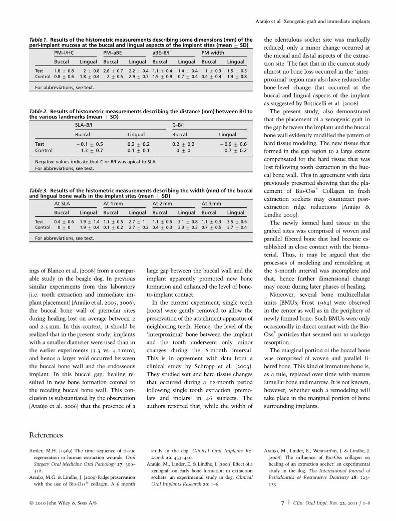
Table 1. Results of the histometric measurements describing some dimensions (mm) of theperi-implant mucosa at the buccal and lingual aspects of the implant sites (mean� SD)
PM–I/HC PM–aBE aBE–B/I PM width
Buccal Lingual Buccal Lingual Buccal Lingual Buccal Lingual
Test 1.8 � 0.8 2 � 0.8 2.6 � 0.7 2.2 � 0.4 1.1 � 0.4 1.4 � 0.4 1 � 0.3 1.5 � 0.5
Control 0.8 � 0.6 1.8 � 0.4 2 � 0.5 2.9 � 0.7 1.9 � 0.9 0.7 � 0.4 0.4 � 0.4 1.4 � 0.8
For abbreviations, see text.
Table 2. Results of histometric measurements describing the distance (mm) between B/I tothe various landmarks (mean� SD)
SLA–B/I C–B/I
Buccal Lingual Buccal Lingual
Test � 0.1 � 0.5 0.2 � 0.2 0.2 � 0.2 � 0.9 � 0.6Control � 1.3 � 0.7 0.1 � 0.1 0 � 0 � 0.7 � 0.2
Negative values indicate that C or B/I was apical to SLA.
For abbreviations, see text.
Table 3. Results of the histometric measurements describing the width (mm) of the buccaland lingual bone walls in the implant sites (mean� SD)
At SLA At 1 mm At 2 mm At 3 mm
Buccal Lingual Buccal Lingual Buccal Lingual Buccal Lingual
Test 0.4 � 0.6 1.9 � 1.4 1.1 � 0.5 2.7 � 1 1.1 � 0.5 3.1 � 0.8 1.1 � 0.3 3.5 � 0.6
Control 0 � 0 1.9 � 0.4 0.1 � 0.2 2.7 � 0.2 0.4 � 0.3 3.3 � 0.3 0.7 � 0.5 3.7 � 0.4
For abbreviations, see text.
Araujo et al �Xenogenic graft and immediate implants
c� 2010 John Wiley & Sons A/S 7 | Clin. Oral Impl. Res. 22, 2011 / 1–8

Araujo, M.G. & Lindhe, J. (2005) Dimensional ridge
alterations following tooth extraction. An experi-
mental study in the dog. Journal of Clinical
Periodontology 32: 212–218.
Araujo, M.G., Sukekava, F., Wennstrom, J.L. &
Lindhe, J. (2005) Ridge alterations following im-
plant placement in fresh extraction sockets. An
experimental study in the dog. Journal of Clinical
Periodontology 32: 645–652.
Araujo, M.G., Sukekava, F., Wennstrom, J.L. &
Lindhe, J. (2006) Tissue modeling following
implant placement in fresh extraction sockets.
Clinical Oral Implants Research 17: 615–624.
Artzi, Z., Nemocovsky, C., Tal, H. & Dayan, D.
(2002) Bovine-HA spongiosa blocks and immedi-
ate implant placement in sinus augmentation
procedures. Histopathological and histomorpho-
metric observations on different histological stain-
ings in 10 consecutive patients. Clinical Oral
Implants Research 13: 420–427.
Artzi, Z., Tal, H. & Dayan, D. (2000) Porous bovine
bone mineral in healing of human extraction sock-
ets. Part 1: histomorphometric evaluations at 9
months. Journal of Periodontology 72: 1015–1023.
Berglundh, T. & Lindhe, J. (1997) Healing around
implants placed in bone defects treated with Bio-
Oss. An experimental study in the dog. Clinical
Oral Implants Research 8: 117–124.
Blanco, J., Nunez, V., Aracil, L., Munoz, F. &
Ramos, I. (2008) Ridge alterations following im-
mediate implant placement in the dog: flap versus
flapless surgery. Journal of Clinical Perio-
dontology 35: 640–648.
Botticelli, D. (2006) Healing of marginal defects
around implants. Thesis, The Sahlgrenska Acad-
emy at Goteborg University, Sweden.
Botticelli, D., Berglundh, T. & Lindhe, J. (2004) The
influence of a biomaterial on the closure of a
marginal hard tissue defect adjacent to implants.
An experimental study in the dog. Clinical Oral
Implants Research 15: 285–292.
Botticelli, D., Persson, L.G., Lindhe, J. & Ber-
glundh, T. (2006) Bone tissue formation adjacent
to implants placed in fresh extraction sockets: an
experimental study in dogs. Clinical Oral Im-
plants Research 17: 351–358.
Cardaropoli, G., Araujo, M. & Lindhe, J. (2003)
Dynamic of bone tissue formation in tooth ex-
traction sites. An experimental study in dogs.
Journal of Clinical Periodontology 30: 809–818.
Carmagnola, D., Adriaens, P. & Berglundh, T.
(2003) Healing of human extraction sockets filled
with Bio-Oss. Clinical Oral Implants Research
14: 137–143.
Donath, K. (1988) Die Trenn-Dunnschliff-Technik
zur Herstellung histologische raparate von nicht
schneidbaren Geweben und Materialen. Der Pra-
parator 34: 97–206.
Donath, K. (1993) Preparation of Histological Sec-
tions (by the Cutting-Grinding Technique for
Hard Tissue and other Material not Suitable to
be Sectioned by Routine Methods) – Equipment
and Methodological Performance. Norderstedt,
Germany: EXAKT-Kulzer Publications.
Donath, K. & Breuner, G.A. (1982) A method for
the study of undecalcified bones and teeth with
attached soft tissues. The Sage-Schliff (sawing and
grinding) technique. Journal of Oral Pathology
11: 318–326.
Evans, C.D. & Chen, S.T. (2008) Esthetic outcomes
of immediate implant placements. Clinical Oral
Implants Research 19: 73–80.
Evian, C.I., Rosenberg, E.S., Coslet, J.G. & Corn, H.
(1982) The osteogenic activity of bone removed
from healing extraction sockets in humans. Jour-
nal of Periodontology 53: 81–85.
Frost, H.M. (1964) Mathematical Elements of La-
mellar Bone Remodeling. Springfield, IL: Charles
C. Thomas.
Froum, S., Cho, S.C., Rosenberg, F., Rohrer, M. &
Tarnow, D. (2002) Histological comparison of
healing extraction sockets implanted with bioac-
tive glass or demineralized freeze-dried bone allo-
graft: a pilot study. Journal of Periodontology 73:
94–102.
Hurzeler, M.B., Quinones, C.R., Kirsch, A., Gloker,
C., Schupbach, P., Strub, J.R. & Caffesse, R.G.
(1997) Maxillary sinus augmentation using differ-
ent grafting materials and dental implants in
monkeys. Part I. Evaluation of anorganic bovine-
derived bone matrix. Clinical Oral Implants Re-
search 8: 476–486.
Karnovsky, M.J. (1965) A formaldehyde -glutaralde-
hyde fixative of high osmolarity for use in electron
microscopy. The Journal of Cell Biology 27:
137A–138A.
Kuboki, Y., Hashimoto, F. & Ishibashi, K. (1988)
Time-dependent changes of collagen crosslinks in
the socket after tooth extraction in rabbits. Journal
of Dental Research 67: 944–948.
Nevins, M., Camelo, M., de Paoli, S., Friedland, B.,
Schenk, R.K., Parma-Benfenati, S., Simion, M.,
Tinti, C. & Wagenberg, B. (2006) A study of the
fate of the buccal wall of extraction sockets of
teeth with prominent roots. The International
Journal of Periodontics & Restorative Dentistry
26: 19–29.
Norton, M.R., Odell, E.W., Thompson, I.D. &
Cook, R.J. (2003) Efficacy of bovine bone mineral
for alveolar augmentation: a human histologic
study. Clinical Oral Implants Research 14:
775–783.
Piattelli, M., Favero, G., Scarano, A., Orsini, G. &
Piattelii, A. (1999) Bone reactions to anorganic
bovine bone (Bio-Oss) used in sinus augmentation
procedures: a histologic longterm report of
20 cases in humans. The International Journal
of Oral & Maxillofacial Implants 14: 835–840.
Pietrokovski, J. & Massler, M. (1967) Alveolar ridge
resorption following tooth extraction. The Journal
of Prosthetic Dentistry 17: 21–27.
Pietrokovski, J., Starinsky, R., Arensburg, B. &
Kaffe, I. (2007) Morphologic characteristics of
bony edentulous jaws. Journal of Prosthodontics
15: 141–147.
Schropp, L., Wenzel, A., Kostopoulos, L. & Karring,
T. (2003) Bone healing and soft tissue contour
changes following single-tooth extraction: a clin-
ical and radiographic 12-month prospective study.
The International Journal of Periodontics & Re-
storative Dentistry 23: 313–323.
Yildirim, M., Spiekermann, H., Biesterfeld, S. &
Edelhoff, D. (2000) Maxillary sinus augmentation
using xenogenic bone substitute material Bio-
Oss in combination with venous blood. A
histologic and histomorphometric study in
humans. Clinical Oral Implants Research 11:
217–229.
Araujo et al �Xenogenic graft and immediate implants
8 | Clin. Oral Impl. Res. 22, 2011 / 1–8 c� 2010 John Wiley & Sons A/S
Recommended