Embed Size (px)
Citation preview

Comparison of Efficacy of Buccal Fat Pad and Collagen Membrane in Surgical Management of Oral Submucous Fibrosis
International Journal of Preventive and Clinical Dental Research, July-September 2017;4(3):1-8 1
IJPCDR
Comparison of Efficacy of Buccal Fat Pad and Collagen Membrane in Surgical Management of Oral Submucous Fibrosis1Rajbir K Randhawa, 2Gagandeep S Randhawa, 3Saba Tiwari, 4KC Gupta, 5Anisha Maria, 6Mrinal Satpathy
IJPCDR
ORIGINAL RESEARCH10.5005/jp-journals-00000-0000
1,2Senior Lecturer, 3,6Assistant Professor, 4Professor, 5Principal and Head1,2Department of Oral and Maxillofacial and Reconstructive Surgery, Ahmedabad Dental College & Hospital, Ahmedabad Gujarat, India3Department of Oral and Maxillofacial and Reconstructive Surgery, L.N. Medical College and Research Centre, Bhopal Madhya Pradesh, India4Department of Oral and Maxillofacial and Reconstructive Surgery, Modern Dental College & Research Centre, Indore Madhya Pradesh, India5Department of Oral and Maxillofacial and Reconstructive Surgery, Rishiraj College of Dental Sciences & Research Centre Bhopal, Madhya Pradesh, India6Department of Oral and Maxillofacial and Reconstructive Surgery, People’s Dental Academy, Bhopal, Madhya Pradesh India
Corresponding Author:
ABSTRACTBackground and objectives: Oral submucous fibrosis (OSMF) is a chronic progressive premalignant condition, characterized by gradual trismus of mouth. The study was done to compare the efficacy of buccal fat pad (BFP) and collagen membrane as an interpositional material in surgical management of OSMF and also (1) to assess and compare the mouth opening achieved in both groups of patient; (2) the improvement in flexibility of buccal mucosa in both groups; (3) oral pain and burning sensation on intake of spicy food; (4) the rapidity in epithelialization of graft at the intraoral wound site.
Materials and methods: Thirty patients were randomly divided into 15 patients each in groups I and II respectively. Group I patients received BFP as the interpositioning material, whereas group II received xenogenous collagen membrane after bilateral excision of bands. Group I was compared with group II postop-eratively for mouth opening up to 6 months follow-up.
Results: Collagen membrane group showed better mouth opening postoperatively owing to their faster epithelialization rate and less wound contracture.
Conclusion: The collagen membrane (group II) proved to more efficient and the result was statistically significant as it showed better mouth opening postoperatively at 6 months, improve-ment in flexibility of buccal mucosa, reduction in postoperative pain and burning sensation, and faster epithelialization rate compared with BFP (group I).
Keywords: Buccal fat pad, Collagen membrane, Mouth opening, Oral submucous fibrosis.
How to cite this article: Randhawa RK, Randhawa GS, Tiwari S, Gupta KC, Maria A, Satpathy M. Comparison of Efficacy of Buccal Fat Pad and Collagen Membrane in Surgical Manage-ment of Oral Submucous Fibrosis. Int J Prev Clin Dent Res 2017;4(3):1-8.
Source of support: Nil
Conflict of interest: None
INTRODUCTION
In 1952, Schwartz described five Indian women from Kenya with a condition of the oral mucosa including the palate and pillars of the fauces, which he called “atrophia idiopathica (tropica) mucosae oris.” Later it was termed oral submucous fibrosis (OSMF).1
The OSMF is a disease with uncertain etiology, i.e., often encountered in practice in India. It is one of the precancerous conditions leading to carcinoma of cheek. It is caused by chewing irritants like tobacco, betel leaf with lime, and areca nut.2-5 These substances trigger the synthesis of collagen, tough fibrinous protein that stiff-ens the soft mucous membrane and muscles of the oral cavity. The tongue being highly vascular usually escapes. The mouth size shrinks in extreme cases; only a button size opening is left. There is scarring with atrophy of the mucous membrane and pain during swallowing, prevent-ing the patient from enjoying their meal. The atrophic mucous may ulcerate often, and subsequently may lead to malignancy.6,7
The frequency of malignant change in patients with OSMF ranges from 3 to 6%. Sixty-six patients with OSMF were followed up for a period of 17 years by Murti et al, who recorded a malignant transformation rate of 7.6%. With a longer follow-up of the same group, the malignant transformation rates could increase further.1
Various medical and surgical modalities have been advocated, but the present study is conducted with the aim of achieving results in terms of mouth opening after transecting the fibrous bands followed by grafting using buccal fat pad (BFP)/collagen and comparing their roles in achieving the same.8,9

Rajbir K Randhawa et al
2
MATERIAlS AND METHODS
The study compromising 30 subjects was carried out in the Department of Oral and Maxillofacial Surgery. Approval for this study was obtained from the ethical committee of college. Thirty patients were randomly divided into 15 patients each in groups I and II respectively. Group I patients received BFP as the interpositioning material, whereas group II received xenogenous collagen mem-brane after bilateral excision of bands.
Inclusion Criteria
• Thirty subjects with clinically and histologicallyproven bilateral OSMF.
• Patientsofagebetween20and50years,ofeithersexwith mouth opening less than 25 mm were treated surgically under general anesthesia.
• Patientswhohadgivenuptheirabusivehabitswereselected.
• Allpatientswereexplainedabouttheprocedureandinformed consent was obtained from each patient.
• Cases selected were in good health, none of thepatients presented with evidence of systemic disease, deficiencies, or frank oral infection.
• TheKhannaandAndrade10 classification for OSMF was followed.
Method of Study
Only Grade III and Grade IVa patients were included in the study. A detailed history was obtained from each patient with special reference to their habits and dura-tion, and the patients were asked to discontinue the habit before the procedure. Routine hemogram, urine examina-tion, and clinical examination were done to rule out any associated systemic diseases.
The local examination included distribution of fibrous bands, sites of involvement, and the interincisal distance (ID) between the incisal edges of the maxillary and mandibular central incisors, which were measured
using a simple ruler and expressed in millimeters. Also the patients were asked to blow the cheeks to check for flexibility of buccal mucosa, and presence of pain and burning sensation was noted.
Patients were clinically followed up postoperatively at third day, 1, 3 months, and 6 months. At above-men-tioned various intervals, mouth opening was measured visually with metal scale from incisal edges; subjective note of pain and burning sensation obtained; and flex-ibility of oral mucosa checked by asking the patients to blow the cheeks. Epithelialization was assessed visu-ally daily till complete epithelialization had occurred (Figs 1 and 2).
Materials used
Group I – Harvested autologous BFPGroup II – Xenogenous collagen membrane (5 × 5 cm)
marketedbyEUCAREPharmaceuticalsPrivateLimited.The collagen membranes come in varying dimension of 5 × 5, 10 × 10, and 25 × 25 cm, and its thickness is 0.6 mm. It is sterilized by ethylene oxide and gamma irradiation and is marketed in form-fill seal aluminum pouch packing containing a mixture of isopropyl alcohol and water; it
Figs 1A and B: Preoperative and postoperative blowing of cheeks
Figs 2A to C: Epithelialization of buccal fat pad within 4 weeks
A B C
A B

Comparison of Efficacy of Buccal Fat Pad and Collagen Membrane in Surgical Management of Oral Submucous Fibrosis
International Journal of Preventive and Clinical Dental Research, July-September 2017;4(3):1-8 3
IJPCDR
has a shelf life of over 5 years. Before use, it is washed with sterile normal saline.11-14
SURGICAl PROCEDURE
The surgery was performed under general anesthesia. The fibrous bands were palpated to assess the extent of the incision. The incisions were made bilaterally using no. 15 Bard Parker blade along each side of buccal mucosa at the level of occlusal plane away from the Stenson’s orifice. The incision extended posteriorly to pterygo-mandibular raphe (or) anterior pillar of the fauces and anteriorly as far as the angle of the mouth, depending upon the location of the fibrotic bands which restricted the mouth opening. The incision was carried out to the depth of the submucosal layer, and the wound created was further freed by manipulation using fingers until no restriction was felt. After release of fibrous bands, extrac-tion of all third molars was done.
The mouth was then forced to open using Heister’s mouth gag to an acceptable range of approximately 35 mm. The coronoid processes were approached from wound created and resected if a 35 mm mouth opening could not be achieved. A mouth opening of 35 mm as measured was considered to be the minimum acceptable opening in an adult.15-17 Then the mouth opening was measured from incisal edges intraoperatively.
In group I patients, following excision of fibrotic bands and forcible mouth opening, the BFP was
approached via posterosuperior margin of the created buccal defect, i.e., posterior to the zygomatic buttress. After blunt dissection through the submucosa, the BFP was teased out gently until significant amount was obtained to cover the defect without tension. It was found that technically BFP can be easily accessed and mobilized without undue stretching. Bilateral buccal defects of approximately 3 × 2 to 4 × 2.5 cm can be covered with BFP grafts without noticeable defects in the cheeks or mouth after hemostasis.18,19 The BFP was then secured in place with sutures using Vicryl 3-0, as standard in all patients (Fig. 3).
In group II patients, following excision of bands and forceful mouth opening intraoperatively, the raw wound was covered using collagen membrane. The material is available in thickness of 0.3 to 0.6 mm and in a range of dimensions. The size used for most of the cases in the present study was 0.6 mm × 5 cm × 5 cm. The material was reconstituted by immersion in normal saline for 5 minutes, and then cut with scissors to required shape, leaving a small overlap on the remaining mucous mem-brane. The graft was sutured with 3-0 Vicryl to attain close approximation to the underlying tissues.20-22
All patients received course of antibiotics, IV fluids for next 12 hours, anti-inflammatory drugs, and analgesics and cold sponging to cheeks for 48 hours and liquid diet for 5 days. Nasogastric feeding was given for 3 days and intensive physiotherapy was started within 48 hours
Figs 3A to F: (A) Preoperative mouth opening; (B) bilateral excision of bands; (C) intraoperative forceful opening; (D) BFP used as a graft for group I; (E) collagen membrane used as a graft for group II; and (F) postoperative opening at 6 months
A
D
B C
E F
Rajbir K Randhawa et al
4
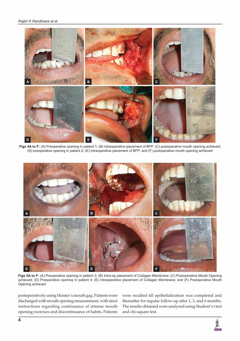
Figs 4A to F: (A) Preoperative opening in patient 1; (B) intraoperative placement of BFP; (C) postoperative mouth opening achieved; (D) preoperative opening in patient 2; (E) intraoperative placement of BFP; and (F) postoperative mouth opening achieved
Figs 5A to F: (A) Preoperative opening in patient 3; (B) Intra-op placement of Collagen Membrane; (C) Postoperative Mouth Opening achieved; (D) Preoperative opening in patient 4; (E) Intraoperative placement of Collagen Membrane; and (F) Postoperative Mouth Opening achieved
A
D
B C
E F
A
D
B C
E F
postoperatively using Heister’s mouth gag. Patients were discharged with mouth opening measurement, with strict instructions regarding continuance of intense mouth opening exercises and discontinuance of habits. Patients
were recalled till epithelialization was completed and thereafter for regular follow-up after 1, 3, and 6 months. The results obtained were analyzed using Student’s t test and chi-square test.

Comparison of Efficacy of Buccal Fat Pad and Collagen Membrane in Surgical Management of Oral Submucous Fibrosis
International Journal of Preventive and Clinical Dental Research, July-September 2017;4(3):1-8 5
IJPCDR
CASE SERIES
OBSERVATIONS AND RESUlTS
In the present study, 30 patients were taken up for evalua-tion of efficacy of BFP and collagen membrane in surgical management of OSMF.
The collagen membrane (group II) proved to be more efficient and the result was statistically significant as it showed better mouth opening postoperatively at 6 months, improvement in flexibility of buccal mucosa, reduction in postoperative pain and burning sensation, and faster epithelialization rate compared with BFP (group I).
Thirty patients were included in the study, with age group 20 to 50 years. Sex predilection favored predomi-nantly males.
Results of all the parameters are shown in Graphs 1 to 6.
DISCUSSION
Oral submucous fibrosis is a chronic, progressive pre-cancerous condition of oral mucosa, predominantly seen
in the Indian subcontinent. A progressive inability to open the mouth fully is an important feature in OSMF due to the formation of fibrous bands, especially in the buccal mucosa, posterior palate, and lips. The basic aim of any treatment modality is to relieve the symptoms that include burning sensation in the mouth, ulceration and
Graph 1: Habit distribution of the study group
Graph 2: Mouth opening at various stages
Graph 3: Improvement in flexibility of buccal mucosa Graph 4: Postoperative pain and burning sensation
Rajbir K Randhawa et al
6
stiffness of the oral mucosa, and progressive limitations in mouth opening, thereby tampering the functions like deglutition and speech.15,23
Relapse is a common complication that occurs after surgical release of the oral trismus caused by OSMF. Ini-tially surgeons aimed at surgical elimination of the fibrotic bands which showed further scar formation and reoccur-rence of trismus; hence, to prevent scar, they started using various interpositional graft material.24
Use of island palatal flap has limitation, such as its involvement with fibrosis and second molar tooth extrac-tion is required for flap to cover without tension.25 Bilat-eral palatal flaps leave a large raw area on palatal bones. Sometimes the defect created may be large and local flaps may not be able to cover the entire defect. A nasolabial flap is too short to cover the defect and causes visible scaring on the face.26 Tongue flaps have been used to cover the buccal defects but were found to be bulky and needed additional surgery for detachment. Bilateral tongue flaps can cause severe dysphagia and disarticulation and carry the risk of postoperative aspiration.27-29 Pindborg et al found an incidence of 38% tongue involvement in OSMF, which precludes its use. Bilateral free radial artery forearm free flaps require microvascular expertise, the flaps are hairy, and 40% of patients require secondary debulking procedures. Extraction of third molar tooth is required to avoid flap inclination between teeth.24,30-33
Buccal fat pad by virtue of its anatomic position and the ease with which it can be accessed and mobilized without causing any noticeable defect in the cheek or mouth was felt to be a reliable interposition material. The procedure, considering the anatomic proximity of the donor and the recipient site, is not a prolonged one. The graft can be approached through the same buccal incision, which was used to release the fibrosis. Should it fail, the consequences are not serious, as other options are open.
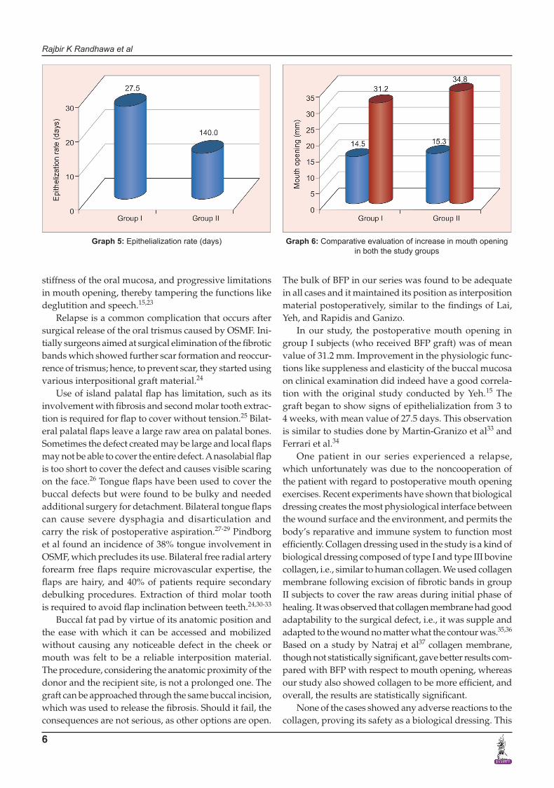
Graph 5: Epithelialization rate (days) Graph 6: Comparative evaluation of increase in mouth opening in both the study groups
The bulk of BFP in our series was found to be adequate in all cases and it maintained its position as interposition material postoperatively, similar to the findings of Lai, Yeh, and Rapidis and Ganizo.
In our study, the postoperative mouth opening in group I subjects (who received BFP graft) was of mean value of 31.2 mm. Improvement in the physiologic func-tions like suppleness and elasticity of the buccal mucosa on clinical examination did indeed have a good correla-tion with the original study conducted by Yeh.15 The graft began to show signs of epithelialization from 3 to 4 weeks, with mean value of 27.5 days. This observation is similar to studies done by Martin-Granizo et al33 and Ferrari et al.34
One patient in our series experienced a relapse, which unfortunately was due to the noncooperation of the patient with regard to postoperative mouth opening exercises. Recent experiments have shown that biological dressing creates the most physiological interface between the wound surface and the environment, and permits the body’s reparative and immune system to function most efficiently.Collagendressingusedinthestudyisakindofbiological dressing composed of type I and type III bovine collagen, i.e., similar to human collagen. We used collagen membrane following excision of fibrotic bands in group II subjects to cover the raw areas during initial phase of healing. It was observed that collagen membrane had good adaptability to the surgical defect, i.e., it was supple and adapted to the wound no matter what the contour was.35,36 Based on a study by Natraj et al37 collagen membrane, though not statistically significant, gave better results com-pared with BFP with respect to mouth opening, whereas our study also showed collagen to be more efficient, and overall, the results are statistically significant.
None of the cases showed any adverse reactions to the collagen, proving its safety as a biological dressing. This

Comparison of Efficacy of Buccal Fat Pad and Collagen Membrane in Surgical Management of Oral Submucous Fibrosis
International Journal of Preventive and Clinical Dental Research, July-September 2017;4(3):1-8 7
IJPCDR
result is in accordance with that of Mitchell.38 Granulation and epithelialization was good and wound contracture was far less comparatively. The appearance of grafted area was restored to normal texture in about 2 weeks. After 6 months follow-up, the postoperative mouth opening was of mean value of 34.8 mm. Because of the simple application and good tolerance of the membrane by oral tissues, collagen can be advocated as a temporary dressing material in orofacial region. It is an alternative to autolo-gous grafts rather than being a replacement of other grafts used in orofacial region and can be viewed as a satisfactory additional armamentarium to oral surgeons.37,39
Whatever the graft being used, the treatment should be coupled with cessation of betel quid/gutkha chewing and daily mouth opening exercises, and proper nutrition in order to manage both early and advanced stages of OSMF.
CONClUSION
The advantages of collagen over BFP were observed as follows:• Thematerialisreadilyavailableandeasilyreconsti-
tuted for simple chair-side technique.• Thecollagenmembraneremainedmoist,supple,and
intact when grafted.• Itwaseffectiveinpromotinghemostasis.• Itactedasatemporarycoveringmaterialonthesensi-
tive nerve endings of raw wounds, which reduced the postoperative pain.
• It acted as amechanical barrier preventing woundcontamination.
• Itappearstobesufficientlyrobusttowithstandmas-ticatory trauma.
• Thecollagenmembranedidnotevokeanyantigenicreactions.
• Itwasusefulininducinggranulationandepitheliali-zation and in preventing the degree of scarring and tissue contracture.No doubt, there are various variables affecting patient
compliance, both positively and negatively, including patient motivation, the nature and chronicity of the disease, treatment variables, and the quality of the patient–doctor relationship. Therefore, early and intensive postoperative rehabilitation is the most important factor in maintain-ing the ID. For this reason, psychologic preparation of the patient before surgery plays a significant role in the success of surgery. In turn, greater compliance positively affects mouth opening management and therefore, patient satisfaction with the outcome of treatment.40
REFERENCES
1. Rajendran R. Oral submucous fibrosis: etiology, pathogenesis, and future research. Bull World Health Organ 1994;72(6): 985-996.
2. Shah N, Sharma PP. Role of chewing and smoking habits in the etiology of oral submucous fibrosis (OSF): a case-control study. J Oral Pathol Med 1998 Nov;27(10):475-479.
3. Shah B, Lewis MAO, Bedi R. Oral submucous fibrosis in a 11-year-old Bangladeshi girl living in the United Kingdom. Br Dent J 2001 Aug;191(3):130-132.
4. Ariyawardana A, Athukorala ADS, Arulanandam A. Effect of betel chewing, tobacco smoking and alcohol consumption on oral submucous fibrosis: a case-control study in Sri Lanka. J Oral Pathol Med 2006 Apr;35(4):197-201.
5. Bathi RJ, Parveen S, Burde K. The role of gutka chewing in oral submucous fibrosis: a case-control study. Quintessence Int 2009 Jun;40(6):e19-e25.
6. Ramadass T, Manokaran G, Pushpala SM, Naryanan N, Kulkarni GN. Oral submucous fibrosis – new dimensions in surgery. Indian J Otolaryngol Head Neck Surg 2005 Apr;57(2):99-102.
7. Tilakaratne WM, Klinikowski MF, Saku T, Peters TJ, Warnaku-lasuriya S. Oral submucous fibrosis: review on aetiology and pathogenesis. Oral Oncol 2006 Jul;42(6):561-568.
8. Kumar A, Bagewadi A, Keluskar V, Singh M. Efficacy of lycopene in the management of oral submucous Fibrosis. Oral Surg Oral Med Oral Pathol Oral Radiol Endod 2007 Feb;103(2):207-213.
9. Surej KLK, Kurien NM, Sakkir N. Buccal fat pad reconstruc-tion for oral submucous fibrosis. Natl J Maxillofac Surg 2010 Jul-Dec;1(2):164-167.
10. Khanna JN, Andrade NN. Oral submucous fibrosis: a new concept in surgical management. Report of 100 cases. Int J Oral Maxillofac Surg 1995 Dec;24(6):433-439.
11. Schlegel AK, Möhler H, Busch F, Mehl A. Preclinical and clini-cal studies of a collagen membrane (Bio-Gide). Biomaterials 1997 Apr;18(7):535-538.
12. Güngörmüş M, Kaya O. Evaluation of the effect of heterolo-gous type i collagen on healing of bone defects. J Oral Maxil-lofac Surg 2002 May;60(5):541-545.
13. SculeanA,BerakdarM,ChiantellaGC,DonosN,ArweilerNB, Brecx M. Healing of intrabony defects following treatment with a bovine-derived xenograft and collagen membrane. A controlled clinical study. J Clin Periodontol 2003 Jan;30(1): 73-80.
14. Proussaefs P, Lozada J. The use of resorbable collagen membrane in conjunction with autogenous bone graft and inorganic bovine mineral for buccal/labial alveolar ridge augmentation: a pilot study. J Prosthet Dent 2003 Dec;90(6): 530-538.
15. YehCJ.Applicationofthebuccalfatpadtothesurgicaltreat-ment of oral submucous fibrosis. Int J Oral Maxillofac Surg 1996 Apr;25(2):130-133.
16. Jeevan Kumar KA, Brahmaji Rao J. Versatility of pedicled buccal fat pad in surgical management of oral submucous fibrosis – a study in 20 cases. Indian J Dent Adv 2009;1:5-11.
17. Rattan V. A simple technique for use of buccal pad of fat in temporomandibular joint reconstruction. J Oral Maxillofac Surg 2006 Sep;64(9):1447-1451.
18. Baumann A, Ewers R. Application of the buccal fat pad in oral reconstruction. J Oral Maxillofac Surg 2000 Apr;58(4): 389-392.
19. Singh J, Prasad K, Lalitha RM, Ranganath K. Buccal pad of fat and its applications in oral and maxillofacial surgery: a review of published literature 2004 to 2009. Oral Surg Oral Med Oral Pathol Oral Radiol Endod 2010 Dec;110(6):698-705.

Rajbir K Randhawa et al
8
20. BunyaratavejP,WangHL.Collagenmembranes:a review. J Periodontol 2001 Feb;72(2):215-229.
21. Omura S, Mizuki N, Horimoto S, Kawabe R, Fujita K. A newly developed collagen/silicone bilayer membrane as a mucosal substitute: a preliminary report. Br J Oral Maxillofac Surg 1997 Apr;35(2):85-91.
22. Sader R, Seitz O, Kuttenberger J. Resorbable collagen mem-brane in surgical repair of fistula following palatoplasty in nonsyndromic cleft palate. Int J Oral Maxillofac Surg 2010 May;39(5):497-499.
23. Lai DR, Chen HR, Lin LM, HuangYL, Tsai CC. Clinicalevaluation of different treatment methods for oral submucous fibrosis. A 10-year experience with 150 cases. J Oral Pathol Med 1995 Oct;24(9):402-406.
24. Amin MA, Bailey BM, Swinson B, Witherow H. Use of the buccal fat pad in the reconstruction and prosthetic rehabilita-tion of oncological maxillary defects. Br J Oral Maxillofac Surg 2005 Apr;43(2):148-154.
25. RapidisAD,AlexandridisCA,EleftheriadisE,AngelopoulosAP. The use of the buccal fat pad for reconstruction of oral defects: review of the literature and report of 15 cases. J Oral Maxillofac Surg 2000 Feb;58(2):158-163.
26. Maria A, Sharma Y, Kaur P. Use of nasolabial flap in the man-agement of oral submucous fibrosis – a clinical study. People’s J Sci Res 2011 Jan;4(1):28-30.
27. Mehrotra D, Pradhan R, Gupta S. Retrospective comparison of surgical treatment modalities in 100 patients with oral submucous fibrosis. Oral Surg Oral Med Oral Pathol Oral Radiol Endod 2009 Mar;107(3):e1-e10.
28. KrishnaPrasadL,ChakravarthyPS,SridharM,RamkumarS,Krishna S, Vivekanand SK. An evaluation of various surgical treatment modalities in the management of oral sub mucous fibrosis – a clinical study. J Orofac Sci 2009 Jan;1(2):17-20.
29. Gnanam A, Kannadasan K, Venkatachalapathy S, David J. Multimodal treatment options for oral submucous fibrosis. SRM Univ J Dent Sci 2010;1(1):26-29.
30. Shah A, Raj S, Rasaniya V, Patel S, Vakade M. Surgical man-agement of oral submucous fibrosis with the “Opus-5” diode laser. J Oral Laser Appl 2005 Jan;5(1):37-43.
31. Mokal NJ, Raje RS, Ranade SV, Prasad JS, Thatte RL. Release of oral submucous fibrosis and reconstruction using superficial temporal fascia flap and split skin graft—a new technique. Br J Plast Surg 2005 Dec;58(8):1055-1060.
32. LeeJT,ChengLF,ChenPR,WangCH,HsuH,ChienSH,WeiFC. Bipaddled radial forearm flap for the reconstruction of bilateral buccal defects in oral submucous fibrosis. Int J Oral Maxillofac Surg 2007 Jul;36(7):615-619.
33. Martin-GranizoR,NavalL,CastasA,GoizuetaC,RodriguezF, Monje F, Muñoz M, Diaz F. Use of buccal fat pad to repair intraoral defects: review of 30 cases. Br J Oral Maxillofac Surg 1997 Apr;135(2):81-84.
34. FerrariS,FerriA,BianchiB,CopelliC,MagriAS,SesennaE.A novel technique for cheek mucosa defect reconstruction using a pedicled buccal fat pad and buccinator myomucosal island flap. Oral Oncol 2009 Jan;45(1):59-62.
35. LinHJ,LinJC.Treatmentoforalsubmucousfibrosisbycol-lagenase: effects on oral opening and eating function. Oral Dis 2007 Jul;13(4):407-413.
36. HerfordAS,AkinL,CicciuM,MaioranaC,BoynePJ.Useof a porcine collagen matrix as an alternative to autogenous tissue for grafting oral soft tissue defects. J Oral Maxillofac Surg 2010 Jul;68(7):1463-1470.
37. Nataraj S, Guruprasad Y, Shetty JN. A comparative clinical evaluation of buccal fat pad and collagen in surgical man-agement of oral sub mucous fibrosis. Arch Dent Sci 2011 May;2(4):17-24.
38. Mitchell R. Treatment of fibrinolytic alveolitis by a collagen paste (formula k). A preliminary report. Int J Oral Maxillofac Surg 1986 Apr;15(2):127-133.
39. Rastogi S, Modi M, Sathian B. The efficacy of collagen mem-brane as a biodegradable wound dressing material for surgical defects of oral mucosa: a prospective study. J Oral Maxillofac Surg 2009 Aug;67(8):1600-1606.
40. HuangIY,WuCF,ShenYS,YangCF,ShiehTY,HsuHJ,ChenCH, Chen CM. Importance of patient’s cooperation in surgicaltreatment for oral submucous fibrosis. J Oral Maxillofac Surg 2008 Apr;66(4):699-703.