Basic Abdominal and Pelvic Imaging Concepts
David L. Smith, MD Assistant Professor of Radiology

Basic Imaging Concepts
Contrast Resolution
vs
Spacial Resolution
Spacial Resolution ...
...refers to the ability of the imaging modality to differentiate two closely-approximated objects.
Low spacial resolution techniques will be unable to differentiate between two objects that are
relatively close together.

Spacial Resolution
(The ability to see really small things)
X-ray > CT > US > MRI Modality Spacial resolution
X-ray < 1 mm
CT 1-2 mm
US 2-3 mm
MRI 3-4 mm

Contrast Resolution
The ability to distinguish differences in image intensity of adjacent structures of the basis of their
grayscale “color.”
CT MRI

Contrast Resolution
MRI > CT> US > X-ray
Abdominal Imaging Modalities
Radiography (aka KUB, plainfilm, x-ray)
Fluoroscopy (Fluoro)
Computed tomography (CT)
Magnetic resonance imaging (MRI)
Ultrasound (Sonography)
Different methods of looking at the same anatomy and the same pathology
Abdominal Imaging Modalities
Radiography (aka KUB, plainfilm, x-ray)
Fluoroscopy (Fluoro)
Computed tomography (CT)
Magnetic resonance imaging (MRI)
Ultrasound (Sonography)
REMEMBER: MRI CT x-ray!!!!!

With or without contrast???
Contrast is a substance administered into a patient's blood stream, GI tract, or other space which increases
that space's conspicuity on imaging.
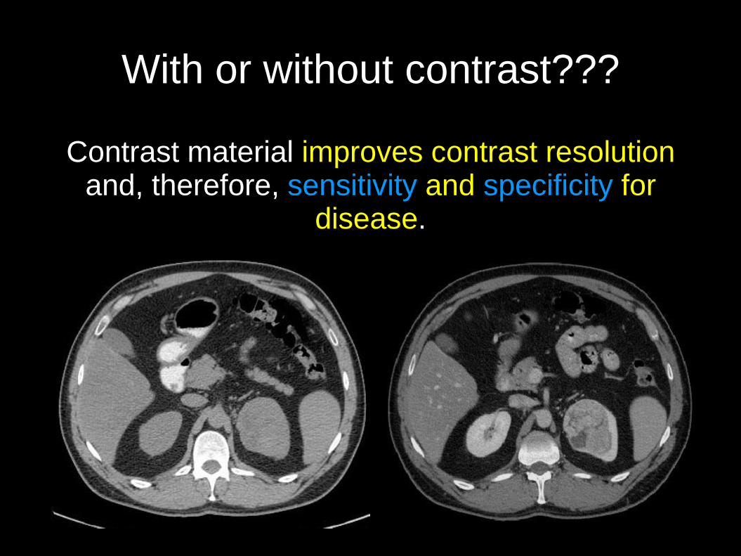
With or without contrast???
Contrast material improves contrast resolution and, therefore, sensitivity and specificity for
disease.
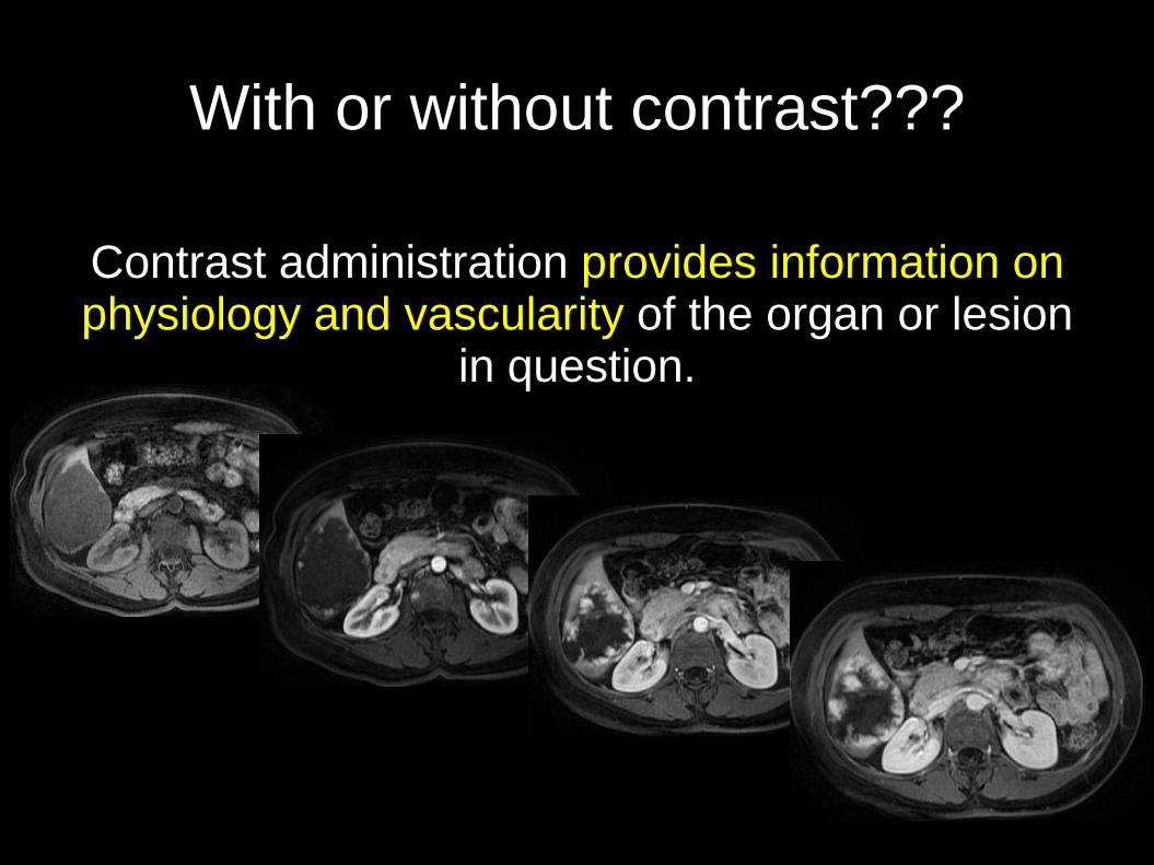
With or without contrast???
Contrast administration provides information on physiology and vascularity of the organ or lesion
in question.
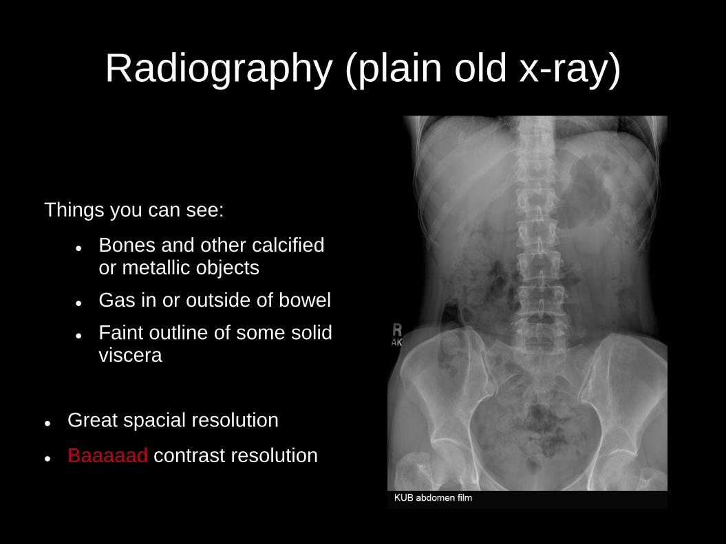
Radiography (plain old x-ray)
Things you can see:
Bones and other calcified or metallic objects
Gas in or outside of bowel
Faint outline of some solid viscera
Great spacial resolution
Baaaaad contrast resolution
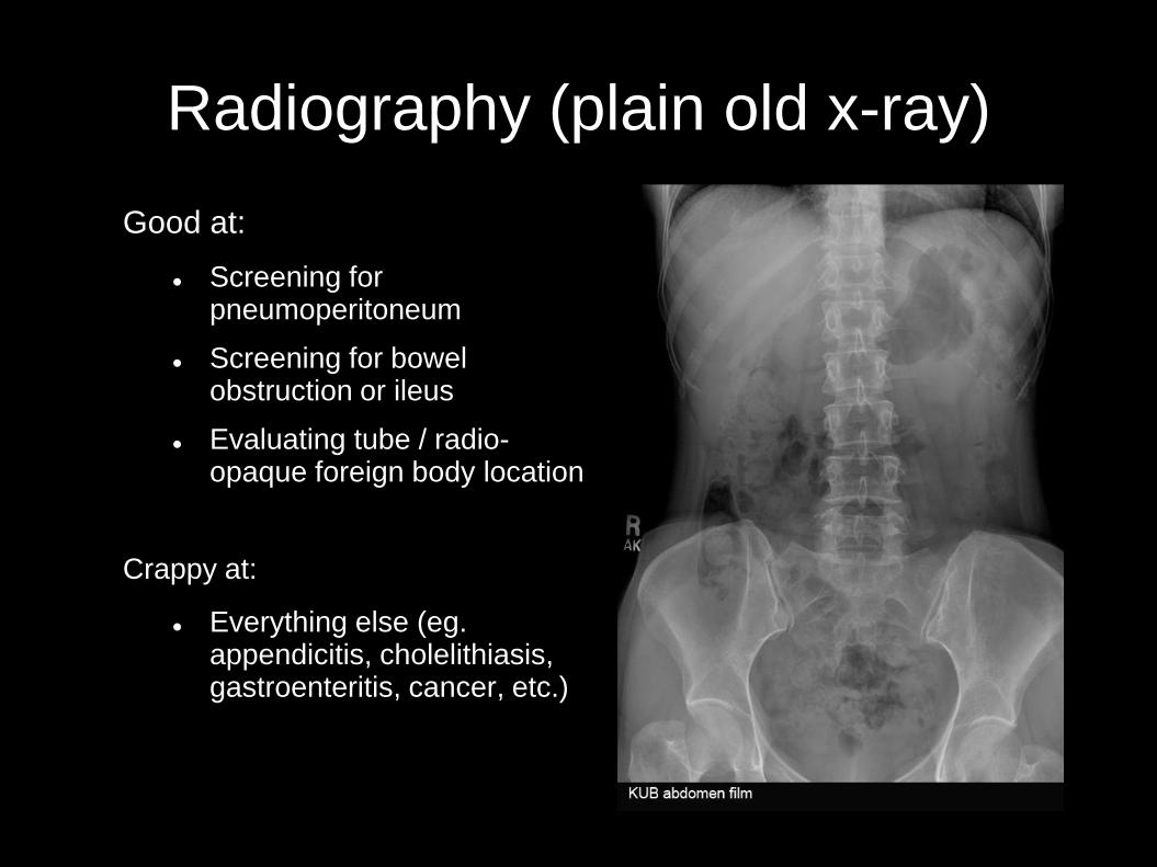
Radiography (plain old x-ray)
Good at:
Screening for pneumoperitoneum
Screening for bowel obstruction or ileus
Evaluating tube / radio-opaque foreign body location
Crappy at:
Everything else (eg. appendicitis, cholelithiasis, gastroenteritis, cancer, etc.)

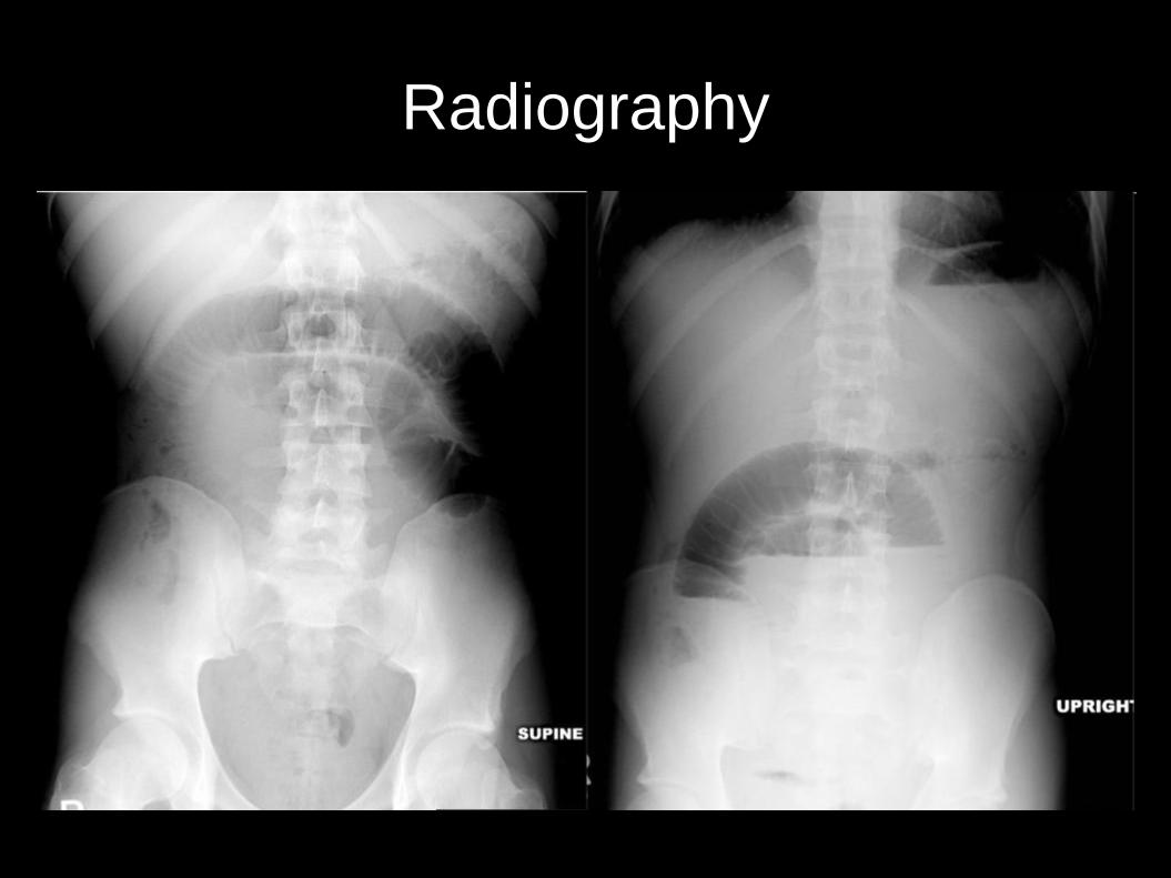
Radiograph variations
Positional
Decubitus→
Supine
Upright
After contrast administration
Intravenous pyelogram (IVP)
For tube placement verification

Radiography
Radiography

Radiography

Fluoroscopy
Like x-rays, but LIVE ON TV !!!!!
Contrast is administered to demonstrate the lumen
(inside) of the space we're interested in.
Provides anatomic and functional information.
To wit:

Routine fluoroscopic studies
Esophagram Upper GI Barium enema
mass, ulcer, reflux
mass, polyp
dysphagia, stricture

Fluoroscopy

Fluoroscopy
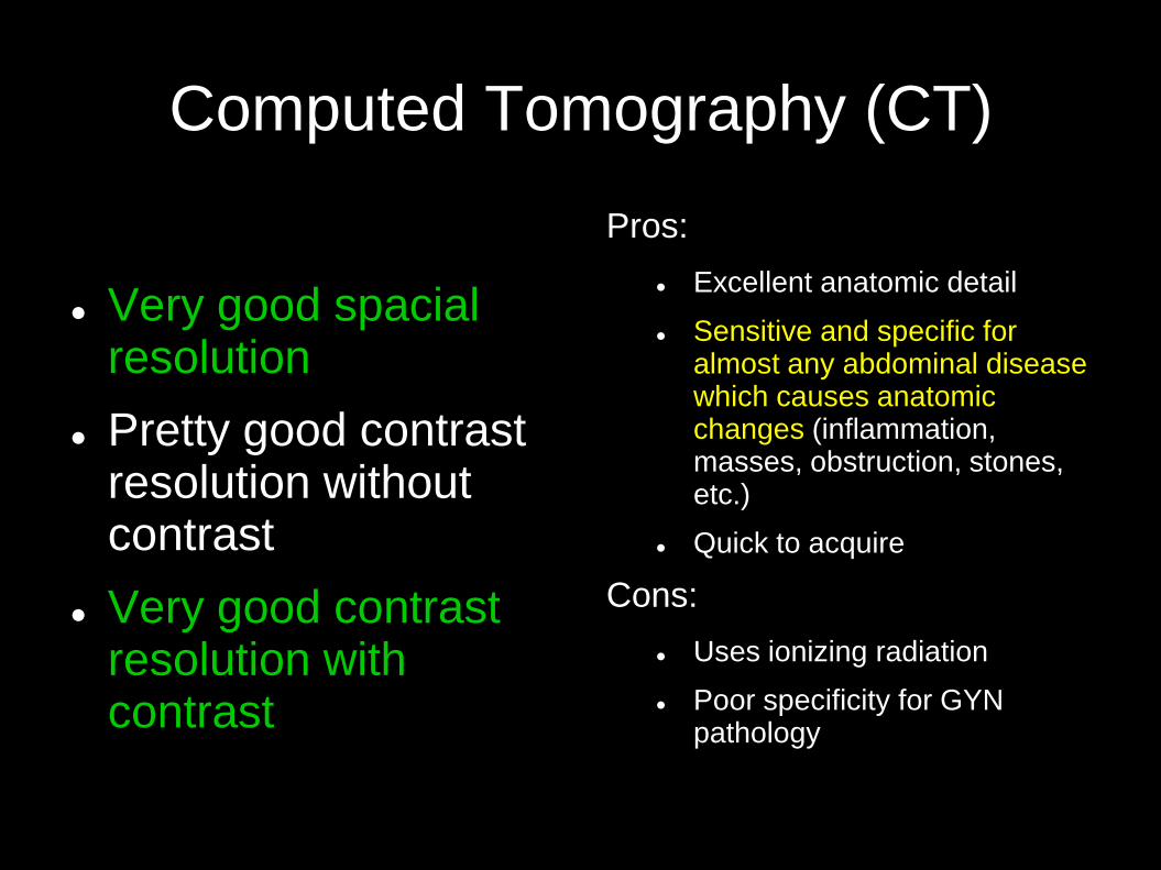
Computed Tomography (CT)
Very good spacial resolution
Pretty good contrast resolution without contrast
Very good contrast resolution with contrast
Pros:
Excellent anatomic detail
Sensitive and specific for almost any abdominal disease which causes anatomic changes (inflammation, masses, obstruction, stones, etc.)
Quick to acquire
Cons:
Uses ionizing radiation
Poor specificity for GYN pathology
Computed Tomography (CT)

Computed Tomography (CT)
Computed Tomography (CT)

Computed Tomography (CT)

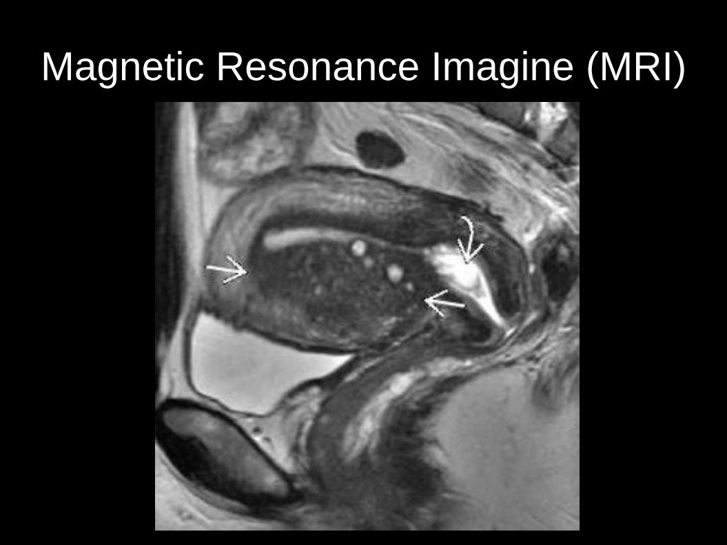
Magnetic Resonance Imaging (MRI)
Freaking amazing contrast resolution
Pretty good spacial resolution
Problem-solving technology
Pros
Excellent tissue characterization
Very sensitive and specific for soft tissue lesions, especially in solid organs
Excellent characterization of GYN pathology
Cons
Expensive
Long acquisition time
Quality depends on patient cooperation

Magnetic Resonance Imaging (MRI)
CT without T1WI in-phase T1WI out-of-phase

Magnetic Resonance Imaging (MRI)
Magnetic Resonance Imagine (MRI)

Magnetic Resonance Imagine (MRI)

Basic Abdominal and Pelvic Imaging Concepts
Remember the basics:
Resolution
Modalities (x-ray, Fluoro, CT, MRI, US)
With or without contrast?
If you don’t know what to do,
ask a radiologist !!!
Recommended