ATLAS OF Ultrasound-Guided Musculoskeletal Injections
Mala_FM_i-xxii.indd iMala_FM_i-xxii.indd i 08/11/13 1:58 PM08/11/13 1:58 PM

NOTICEMedicine is an ever-changing science. As new research and clinical experience broaden our knowledge, changes in treatment and drug therapy are required. The authors and the publisher of this work have checked with sources believed to be reliable in their efforts to provide information that is complete and generally in accord with the standards accepted at the time of publication. However, in view of the pos-sibility of human error or changes in medical sciences, neither the authors nor the publisher nor any other party who has been involved in the preparation or publication of this work warrants that the information contained herein is in every respect accurate or complete, and they disclaim all responsibil-ity for any errors or omissions or for the results obtained from use of the information contained in this work. Readers are encouraged to confirm the information contained herein with other sources. For example and in particular, readers are advised to check the product information sheet included in the package of each drug they plan to administer to be certain that the information contained in this work is accurate and that changes have not been made in the recommended dose or in the contraindications for administration. This recommendation is of particular importance in connection with the new or infrequently used drugs.
Mala_FM_i-xxii.indd iiMala_FM_i-xxii.indd ii 08/11/13 1:58 PM08/11/13 1:58 PM

ATLAS OF Ultrasound-Guided Musculoskeletal Injections
Gerard A. Malanga, MDClinical ProfessorDepartment of Physical Medicine and RehabilitationUMDNJ—New Jersey Medical SchoolNewark, New JerseyFounding PartnerNew Jersey Sports Medicine, LLCSummit, New Jersey
Kenneth R. Mautner, MDAssistant ProfessorDepartment of OrthopaedicsPhysical Medicine and RehabilitationEmory HealthcareAtlanta, Georgia
New York Chicago San Francisco Athens London Madrid
Mexico City Milan New Delhi Singapore Sydney Toronto
Mala_FM_i-xxii.indd iiiMala_FM_i-xxii.indd iii 08/11/13 1:58 PM08/11/13 1:58 PM

Atlas of Ultrasound-Guided Musculoskeletal Injections
Copyright © 2014 by McGraw-Hill Education. All rights reserved. Printed in China. Except as permitted under the United States Copyright Act of 1976, no part of this publication may be reproduced or distributed in any form or by any means, or stored in a data base or retrieval system, without the prior written permission of the publisher.
1 2 3 4 5 6 7 8 9 0 CTP/CTP 19 18 17 16 15 14
ISBN 978-0-07-176967-9MHID 0-07-176967-6
This book was set in Times by Thomson Digital.The editors were Brian Belval and Christina M. Thomas.The production supervisor was Catherine Saggese.Project management was provided by Ritu Joon, Thomson Digital.The text designer was Janice Bielawa.The cover designer was Thomas De Pierro.China Translation & Printing Services, Ltd. was printer and binder.
Library of Congress Cataloging-in-Publication Data
Atlas of ultrasound-guided musculoskeletal injections / [edited by] Gerard Malanga, Kenneth R. Mautner. p. ; cm. Includes bibliographical references and index. ISBN-13: 978-0-07-176967-9 (print book : alk. paper) ISBN-10: 0-07-176967-6 (print book : alk. paper) ISBN-13: 978-0-07-177204-4 (ebook) I. Malanga, Gerard A. II. Mautner, Kenneth R. [DNLM: 1. Musculoskeletal Diseases—ultrasonography—Atlases. 2. Injections—methods—Atlases. 3. Nerve Block—methods—Atlases. 4. Orthopedic Procedures—methods—Atlases. 5. Ultrasonography, Interventional—methods—Atlases. WE 17] RC925.7 616.7’07543—dc23 2013014104
McGraw-Hill Education books are available at special quantity discounts to use as premiums and sales promotions, or for use in corporate training programs. To contact a representative, please visit the Contact Us pages at www.mhprofessional.com.
Mala_FM_i-xxii.indd ivMala_FM_i-xxii.indd iv 08/11/13 1:58 PM08/11/13 1:58 PM

CONTENTS
Contributors .......................................................................................................................................................................................... xi
Foreword .............................................................................................................................................................................................xvii
Preface ..................................................................................................................................................................................................xix
Acknowledgments .............................................................................................................................................................................xxi
SECTION I Introduction 1
1. Introduction to Interventional Ultrasound ...................................................................................................................................................2Christopher J. Visco, MD
2. Ultrasound Physics for Interventional Procedures ....................................................................................................................................6Matthew D. Maxwell, MD / Nicholas H. Weber, DO / Gary P. Chimes, MD, PhD
3. Preparation and Setup for Musculoskeletal Ultrasound-Guided Procedures................................................................................14Paul H. Lento, MD
4. The Rationale and Evidence for Performing Ultrasound-Guided Injections ..................................................................................18Gerard A. Malanga, MD / Matthew Axtman, DO / Kenneth R. Mautner, MD
SECTION II Shoulder 23
5. Glenohumeral Joint Injection ..........................................................................................................................................................................24Troy Henning, DO
6. Acromioclavicular Joint Injection ...................................................................................................................................................................28Evan Peck, MD
7. Sternoclavicular Joint Injection ......................................................................................................................................................................33Evan Peck, MD
8. Subacromial-Subdeltoid Bursa Injection .....................................................................................................................................................36Gregory R. Saboeiro, MD
9. Biceps Tendon Sheath Injection .....................................................................................................................................................................40Sean N. Martin, DO / Joshua G. Hackel, MD, FAAFP
10. Subcoracoid Bursa Injection ............................................................................................................................................................................44Joshua G. Hackel, MD, FAAFP
11. Suprascapular Nerve Injection ........................................................................................................................................................................47Johan Michaud, MD, FRCPC
SECTION III Elbow 51
12. Elbow Joint Injection ..........................................................................................................................................................................................52Jonathan S. Halperin, MD
13. Common Extensor Tendon Peritendinous Injection ..............................................................................................................................57Scott Jeffery Primack, DO, FAAPMR, FACOPMR
14. Common Extensor Tendon Percutaneous Tenotomy ............................................................................................................................61John M. McShane, MD
15. Common Flexor Tendon Peritendinous Injection....................................................................................................................................65Scott Jeffery Primack, DO, FAAPMR, FACOPMR
16. Common Flexor Tendon Percutaneous Tenotomy .................................................................................................................................69Bradley D. Fullerton, MD
Mala_FM_i-xxii.indd vMala_FM_i-xxii.indd v 08/11/13 1:58 PM08/11/13 1:58 PM

vi ■ Contents
17. Distal Biceps Tendon and Bicipitoradial Bursa Injection .......................................................................................................................73Mederic M. Hall, MD
18. Distal Biceps Tendon Percutaneous Tenotomy ........................................................................................................................................78Mederic M. Hall, MD
19. Procedures of the Distal Triceps Tendon: Tendon Sheath and Percutaneous Tenotomy ........................................................82Jose A. Ramirez-Del Toro, MD
20. Ulnar Collateral Ligament Percutaneous Tenotomy ...............................................................................................................................86Joshua G. Hackel, MD, FAAFP
21. Olecranon Bursa Aspiration and Injection ..................................................................................................................................................89R. Amadeus Mason, MD
22. Deep Branch of the Radial Nerve Injection .................................................................................................................................................93Sean W. Mulvaney, MD
23. Ulnar Nerve Injection ..........................................................................................................................................................................................98Evan Peck, MD / Brian J. Shiple, DO
24. Median Nerve at the Pronator Teres Injection .......................................................................................................................................102Victor Ibrahim, MD / Adam D. Weglein, DO, DABMA
SECTION IV Hand and Wrist 107
25. Radiocarpal Joint Injection ............................................................................................................................................................................108Darryl Eugene Barnes, MD
26. Ganglion Cyst in the Wrist Aspiration and Injection ............................................................................................................................112B. Elizabeth Delasobera, MD / Garry Wai Keung Ho, MD, CAQSM / Thomas M. Howard, MD, FACSM
27. Distal Radial Ulnar Joint Injection ...............................................................................................................................................................115Darryl Eugene Barnes, MD
28. Scapholunate Joint Injection ........................................................................................................................................................................119Joseph J. Ruane, DO / Paul A. Cook, MD / Jeffrey A. Strakowski, MD
29. Carpal-Metacarpal Joint Injection ...............................................................................................................................................................122John FitzGerald, MD
30. Scaphotrapeziotrapezoidal Joint Injection .............................................................................................................................................125Kevin B. Dunn, MD, MS
31. Interphalangeal Joints Injection ..................................................................................................................................................................128Mark-Friedrich Berthold Hurdle, MD
32. First Extensor Compartment Injection: Abductor Pollicis Longus and Extensor Pollicis Brevis ...........................................131Ricardo J. Vasquez-Duarte, MD / Jackson Cohen, MD
33. Second Dorsal Compartment of Wrist Injection ....................................................................................................................................135Ricardo J. Vasquez-Duarte, MD / Jackson Cohen, MD
34. Intersection Syndrome of the First and Second Dorsal Compartments Injection ....................................................................138Bradly S. Goodman, MD / Prasanth Nuthakki, MD / Matthew Thomas Smith, MD / Srinivas Mallempati, MD
35. Third Dorsal Compartment of the Wrist Injection ................................................................................................................................141Sean N. Martin, DO
36. Distal Intersection Syndrome Injection ....................................................................................................................................................144Bradly S. Goodman, MD / Matthew Thomas Smith, MD / Prasanth Nuthakki, MD / Srinivas Mallempati, MD
37. Fourth Dorsal Compartment of the Wrist Injection .............................................................................................................................148Sean N. Martin, DO
38. Fifth Dorsal Compartment of the Wrist Injection ..................................................................................................................................152Todd P. Stitik, MD / Kambiz Nooryani, MD / Prathap Jayaram, MD / Asal Sepassi, MD
39. Sixth Dorsal Compartment of the Wrist Injection .................................................................................................................................155Todd P. Stitik, MD / Asal Sepassi, MD / Prathap Jayaram, MD / Kambiz Nooryani, MD
Mala_FM_i-xxii.indd viMala_FM_i-xxii.indd vi 08/11/13 1:58 PM08/11/13 1:58 PM

Contents ■ vii
40. Stenosing Tenosynovitis at the First Annular Pulley Injection .........................................................................................................158Jeffrey A. Strakowski, MD
41. Flexor Carpi Radialis Injection ......................................................................................................................................................................162Rebecca Ann Myers, MD / Jennifer K. Malcolm, DO / Mark Edward Lavallee, MD, CSCS, FACSM
42. Flexor Digitorum Superficialis and Profundus Tendon Sheath Injection .....................................................................................167Luis Baerga-Varela, MD
43. Carpal Tunnel Injection ...................................................................................................................................................................................171Jeffrey A. Strakowski, MD
44. Superficial Radial Nerve Injection ...............................................................................................................................................................176Paul D. Tortland, DO, FAOASM
SECTION V Pelvis 183
45. Sacroiliac Joint Injection .................................................................................................................................................................................184Mark-Friedrich Berthold Hurdle, MD
46. Hip Joint Injection .............................................................................................................................................................................................188Jerod A. Cottrill, DO
47. Hip Paralabral Cyst Aspiration and Injection ..........................................................................................................................................191Marko Bodor, MD / Sean Colio, MD
48. Pubic Symphysis Joint Injection ..................................................................................................................................................................195Ched Garten, MD
49. Piriformis Injection ...........................................................................................................................................................................................198Steve J. Wisniewski, MD / Jay Smith, MD
50. Obturator Internus Injection .........................................................................................................................................................................201Steve J. Wisniewski, MD / Jay Smith, MD
51. Ischial Bursa Peritendinous Injection .........................................................................................................................................................204Kimberly G. Harmon, MD
52. Hamstring Origin Percutaneous Needle Tenotomy .............................................................................................................................209Kimberly G. Harmon, MD
53. Greater Trochanteric Bursae Injection.......................................................................................................................................................213Marko Bodor, MD / John M. Lesher, MD, MPH
54. Gluteus Medius and Minimus Percutaneous Tenotomy ....................................................................................................................219Jon A. Jacobson, MD
55. Iliopsoas Bursa Peritendinous Injection ....................................................................................................................................................224Jon A. Jacobson, MD
56. Procedures of the Adductor Tendon: Tendon Sheath Injection and Percutaneous Tenotomy ..........................................228Henry A. Stiene, MD, FACSM
57. Quadriceps Hematoma Aspiration .............................................................................................................................................................233Robert Monaco, MD / Megan Groh Miller, MD
58. Sciatic Nerve Injection .....................................................................................................................................................................................236Joanne Borg Stein, MD
59. Femoral Nerve Injection .................................................................................................................................................................................239Danielle Aufiero, MD / Steven Sampson, DO
60. Obturator Nerve Injection ..............................................................................................................................................................................243Joanne Borg Stein, MD
61. Lateral Femoral Cutaneous Nerve Injection ............................................................................................................................................246Paul D. Tortland, DO, FAOASM
Mala_FM_i-xxii.indd viiMala_FM_i-xxii.indd vii 08/11/13 1:58 PM08/11/13 1:58 PM

viii ■ Contents
SECTION VI Knee 251
62. Intraarticular Injections of the Knee ...........................................................................................................................................................252John C. Cianca, MD
63. Gastrocnemius-Semimembranosus Bursa (Baker’s Cyst) Aspiration and Injection ..................................................................258Brandon J. Messerli, DO / Garrett S. Hyman, MD, MPH / R. Amadeus Mason, MD
64. Paramensical Cyst Aspiration and Injection ............................................................................................................................................263Jeffrey M. Payne, MD
65. Proximal Tibiofibular Joint Injection ..........................................................................................................................................................266Jeffrey M. Payne, MD
66. Distal Quadriceps Injection and Tenotomy .............................................................................................................................................269Ronald W. Hanson Jr, MD, CAQSM
67. Patellar Tendon Needle Tenotomy.............................................................................................................................................................272Joseph J. Albano, MD
68. Prepatellar Bursal Injection ...........................................................................................................................................................................277Joseph J. Albano, MD
69. Infrapatellar Bursa Injection ..........................................................................................................................................................................281Beth M. Weinman, DO / Kate E. Temme, MD / Megan L. Noon, MD / Anne Z. Hoch, DO
70. Distal Iliotibial Band: Peritendinous Injection and Percutaneous Tenotomy .............................................................................287Eugene Yousik Roh, MD / Michael Fredericson, MD
71. Popliteus Tendon: Tendon Sheath and Percutaneous Tenotomy ..................................................................................................292Brandee L. Waite, MD
72. Distal Biceps Femoris: Peritendinous Injection, Tenotomy, and Fenestration ...........................................................................296Robert Monaco, MD / Megan Groh Miller, MD
73. Procedures of the Distal Semimembranosus Tendon: Peritendinous and Percutaneous Tenotomy ...............................300Brandon J. Messerli, DO / Garrett S. Hyman, MD, MPH
74. Pes Anserine Bursa Injection .........................................................................................................................................................................303Jacob L. Sellon, MD / Jay Smith, MD
75. Tibial Collateral Ligament Bursa Injection ...............................................................................................................................................307Troy Henning, DO
76. Tibial Nerve Injection at Posterior Knee ...................................................................................................................................................310John L. Lin, MD
77. Common Peroneal Nerve Injection ............................................................................................................................................................316John L. Lin, MD
78. Saphenous Nerve Injection ...........................................................................................................................................................................320Joanne Borg Stein, MD
SECTION VII Foot and Ankle 325
79. Distal Tibiofibular Joint Injection ................................................................................................................................................................326Charles E. Garten II, MD
80. Tibiotalar Joint Injection .................................................................................................................................................................................329Kevin deWeber, MD, FAAFP, FACSM
81. Subtalar (Talocalcaneal) Joint Injection ....................................................................................................................................................333Kevin deWeber, MD, FAAFP, FACSM
82. Sinus Tarsi Injection .........................................................................................................................................................................................336Keith Hardy, MD
83. Talonavicular Joint Injection .........................................................................................................................................................................339Keith Hardy, MD
84. Tarsometatarsal (Lisfranc) Joint Injection ................................................................................................................................................343Arthur Jason De Luigi, DO, FAAPMR, DAPM
Mala_FM_i-xxii.indd viiiMala_FM_i-xxii.indd viii 08/11/13 1:58 PM08/11/13 1:58 PM

Contents ■ ix
85. Calcaneocuboid Injection ..............................................................................................................................................................................347Arthur Jason De Luigi, DO, FAAPMR, DAPM
86. Metatarsophalangeal Joint Injection .........................................................................................................................................................351Eric Robert Helm, MD / Nicholas H. Weber, DO / Megan Helen Cortazzo, MD
87. Interphalangeal Joint Injection ....................................................................................................................................................................354Eric Robert Helm, MD / Nicholas H. Weber, DO / Megan Helen Cortazzo, MD
88. Metatarsosesamoid Joint Injection ............................................................................................................................................................357Nicholas H. Weber, DO / Eric Robert Helm, MD / Megan Helen Cortazzo, MD
89. Tibialis Anterior Tendon Sheath and Tibialis Anterior Bursa Injection ..........................................................................................360Nelson A. Hager, MS, MD / Alfred C. Gellhorn, MD
90. Tendon Sheath Injection and Percutaneous Tenotomy of the Distal Peroneal Brevis Tendon ...........................................364Nelson A. Hager, MS, MD
91. Achilles Paratenon Injection .........................................................................................................................................................................367Luis Baerga-Varela, MD
92. Achilles Tendon Injection and Tenotomy ................................................................................................................................................372Bradley D. Fullerton, MD
93. Retrocalcaneal Bursa Injection .....................................................................................................................................................................378Mandy Huggins, MD / Gerard A. Malanga, MD
94. Retro-Achilles Bursa Injection .......................................................................................................................................................................380Mandy Huggins, MD / Gerard A. Malanga, MD
95. Tibialis Posterior Injection and Tenotomy ...............................................................................................................................................382Christopher J. Visco, MD
96. Flexor Hallucis Longus Tendon Sheath Injection ..................................................................................................................................387Johan Michaud, MD, FRCPC
97. Plantar Fascia Perifascial Injection ..............................................................................................................................................................392John C. Hill, DO, FACSM, FAAFP / Matthew Leiszler, MD
98. Plantar Fascia Intrafascial Injection ............................................................................................................................................................397John C. Hill, DO, FACSM, FAAFP / Matthew Leiszler, MD
99. Tibial Nerve Injection at the Ankle ..............................................................................................................................................................402John C. Hill, DO, FACSM, FAAFP / Matthew Leiszler, MD / Jay E. Bowen, DO
100. Saphenous Nerve Injection at the Ankle ..................................................................................................................................................407Amy X. Yin, MD / Joanne Borg Stein, MD
101. Sural Nerve Injection ........................................................................................................................................................................................410Rahul Naren Desai, MD / Jevon Simerly
102. Morton’s Neuroma Injection .........................................................................................................................................................................413Michael Goldin, MD / Brian J. Shiple, DO
SECTION VIII Special Procedures 419
103. Lavage and Aspiration of Rotator Cuff Calcific Tendinosis ................................................................................................................420Gregory R. Saboeiro, MD
104. Intrasheath Percutaneous Release of the First Annular Digital Pulley for Trigger Digits .......................................................425Jose Manuel Rojo Manaute, MD, PhD / Guillermo Emilio Rodríguez-Maruri, MD / Alberto Capa-Grasa, MD, PhD
105. Ultra-Minimally Invasive Carpal Tunnel Release ....................................................................................................................................430Jose Manuel Rojo Manaute, MD, PhD / Alberto Capa-Grasa, MD, PhD / Guillermo Emilio Rodríguez-Maruri, MD / Jay Smith, MD / Javier Vaquero Martín, MD, PhD
106. Percutaneous Tenotomy of the Common Extensor Tendon ............................................................................................................438Darryl Eugene Barnes, MD
Index ................................................................................................................................................................................................... 443
Mala_FM_i-xxii.indd ixMala_FM_i-xxii.indd ix 08/11/13 1:58 PM08/11/13 1:58 PM

CONTRIBUTORS
Joseph J. Albano, MDPrivate Practice of Regenerative and Sports MedicineTeam Physician for US Speedskating, Real Salt Lake Soccer,
US Military CyclingWestminster CollegeSalt Lake City, Utah
Danielle Aufiero, MDClinical Assistant ProfessorDepartment of Physical Medicine and RehabilitationWestern University of Health SciencesPomona, California
Matthew Axtman, DOSports Medicine FellowDepartment of OrthopaedicsEmory UniversityAtlanta, Georgia
Luis Baerga-Varela, MDAssistant ProfessorDepartment of Physical Medicine, Rehabilitation and Sports MedicineUniversity of Puerto RicoSan Juan, Puerto Rico
Darryl Eugene Barnes, MDConsultantDepartment of OrthopedicsMayo Clinic Health SystemAustin, Minnesota
Marko Bodor, MDAssistant ProfessorDepartment of Neurological SurgeryUniversity of California San FranciscoSan Francisco, California
Jay E. Bowen, DOAssistant ProfessorDepartment of Physical Medicine and RehabilitationUniversity of Medicine and Dentistry of New JerseyNewark, New Jersey
Alberto Capa-Grasa, MD, PhDAttending PhysicianPhysical Medicine and RehabilitationUniversity Hospital La PazAttending PhysicianSports Medicine and Physical EducationUniversity Hospital La PazMadrid, Spain
Gary P. Chimes, MD, PhDAssistant ProfessorDepartment of Physical Medicine and RehabilitationUniversity of Pittsburgh Medical CenterFellowship Director, Musculoskeletal Sports & Spine FellowshipDivision Chief of Physical Medicine and Rehabilitation, UPMC-EastPittsburgh, Pennsylvania
John C. Cianca, MDClinical Associate ProfessorBaylor College of MedicineDepartment of Physical Medicine and RehabilitationPrivate PracticeBaylor College of MedicineHouston, Texas
Jackson Cohen, MDResidentDepartment of Rehabilitation MedicineUniversity of Miami School of MedicineMiami, Florida
Sean Colio, MDSports Medicine PhysiatristSwedish Spine, Sports, and Musculoskeletal MedicineSwedish Medical GroupSeattle, Washington
Paul A. Cook, MDPhysicianHand and Microsurgery Associates, Inc.Columbus, Ohio
Megan Helen Cortazzo, MDAssistant ProfessorVice Chair of Outpatient ServicesDepartment of Physical Medicine and RehabilitationUniversity of Pittsburgh School of MedicineUniversity of Pittsburgh Medical CenterPittsburgh, Pennsylvania
Jerod A. Cottrill, DOPrivate PracticeDepartment of Physical Medicine and RehabilitationRebound OrthopedicsPortland, Oregon
Mala_FM_i-xxii.indd xiMala_FM_i-xxii.indd xi 08/11/13 1:58 PM08/11/13 1:58 PM
xii ■ Contributors
Alfred C. Gellhorn, MDClinical Assistant ProfessorDepartment of Rehabilitation MedicineUniversity of WashingtonSeattle, Washington
Michael Goldin, MDFellowDepartment of Physical Medicine and RehabilitationUniversity of Medicine and Dentistry of New JerseyNewark, New Jersey
Bradly S. Goodman, MDAssociate ProfessorDepartment of Physical Medicine and RehabilitationUniversity of Alabama at BirminghamBirmingham, AlabamaAssociate ProfessorDepartment of Physical Medicine and RehabilitationUniversity of Missouri at ColumbiaColumbia, Missouri
Joshua G. Hackel, MD, FAAFPAssociate ProfessorFlorida State School of MedicineTallahassee, FloridaTeam PhysicianDepartment of AthleticsUniversity of West FloridaPensacola, Florida
Nelson A. Hager, MS, MDAssociate Clinical ProfessorDepartment of Physical Medicine and RehabilitationUniversity of Washington School of MedicineMedical DirectorBone and Joint, Sports and Spine CenterUniversity of Washington Medical CenterSeattle, Washington
Mederic M. Hall, MDAssistant ProfessorDepartment of Orthopaedics and RehabilitationUniversity of Iowa Sports Medicine CenterIowa City, Iowa
Jonathan S. Halperin, MDClinical AssociateDivision of PhysiatrySharp Rees Stealy Medical GroupSan Diego, California
Ronald W. Hanson Jr, MD, CAQSMFellowship Director, Interventional OrthopedicsCenter for Regenerative MedicineThe Centeno-Schultz ClinicBroomfield, Colorado
B. Elizabeth Delasobera, MDSports Medicine FellowDepartment of Family MedicineFairfax Family Practice - VCUFairfax, VirginiaAttending PhysicianDepartment of Emergency MedicineGeorgetown University HospitalWashington, District of Columbia
Rahul Naren Desai, MDSports and Pain InterventionalistMusculoskeletal RadiologyEPIC ImagingPortland, Oregon
Kevin deWeber, MD, FAAFP, FACSMAssistant ProfessorDepartment of Family MedicineUniformed Services University of the Health SciencesBethesda, MarylandDirector, Military Sports Medicine FellowshipDepartment of Family MedicineFt. Belvoir Community HospitalFt. Belvoir, Virginia
Kevin B. Dunn, MD, MSNew Jersey Sports Medicine, LLCCedar Knolls, New Jersey
John FitzGerald, MDAssociate Clinical ProfessorDavid Geffen School of MedicineDepartment of Medicine and RheumatologyUniversity of California Los AngelesLos Angeles, California
Michael Fredericson, MDProfessor and Attending PhysicianDepartment of Orthopaedic SurgeryStanford University, Hospital and ClinicsStanford, California
Bradley D. Fullerton, MDMedical DirectorPrivate Practice PhysiatristDepartment of Physical Medicine and Rehabilitation Specialty Care Center Pediatric Spasticity ClinicDell Children’s Medical Center of Central TexasAustin, Texas
Charles E. Garten II, MDPrivate PracticeSports MedicineMyers Sports Medicine and Orthopaedic CenterAtlanta, Georgia
Ched Garten, MDMyers Sports Medicine and Orthopaedic CenterAtlanta, Georgia
Mala_FM_i-xxii.indd xiiMala_FM_i-xxii.indd xii 08/11/13 1:58 PM08/11/13 1:58 PM

Contributors ■ xiii
Keith Hardy, MDClinical InstructorDepartment of Rehabilitation MedicineUniversity of WashingtonSeattle, Washington
Kimberly G. Harmon, MDClinical ProfessorFamily Medicine and Orthopaedics and Sports MedicineUniversity of WashingtonTeam PhysicianUniversity of WashingtonSeattle, Washington
Eric Robert Helm, MDResidentDepartment of Physical Medicine and RehabilitationUniversity of Pittsburgh School of MedicinePittsburgh, Pennsylvania
Troy Henning, DOAssistant Professor and Attending PhysicianDepartment of Physical Medicine and RehabilitationUniversity of MichiganAnn Arbor, Michigan
John C. Hill, DO, FACSM, FAAFPProfessorDirector of Primary Care Sports Medicine FellowshipDepartment of Family MedicineUniversity of Colorado School of MedicineDenver, Colorado
Garry Wai Keung Ho, MD, CAQSMAssistant Program DirectorSports MedicineVCU-Fairfax Family Practice and Sports MedicineFairfax, VirginiaAssistant ProfessorFamily MedicineVirginia Commonwealth University School of MedicineRichmond, Virginia
Anne Z. Hoch, DOProfessorDepartment of Orthopaedic SurgeryMedical College of WisconsinMilwaukee, Wisconsin
Thomas M. Howard, MD, FACSMAssistant Clinical ProfessorDepartment of Family MedicineVCU School of MedicineRichmond, Virginia
Mandy Huggins, MDStaff PhysicianDepartment of OrthopaedicsBroward Health Physician GroupFt. Lauderdale, Florida
Mark-Friedrich Berthold Hurdle, MDAssistant ProfessorDepartment of Physical Medicine and RehabilitationMayo Clinic College of MedicineJacksonville, Florida
Garrett S. Hyman, MD, MPHClinical Assistant ProfessorDepartment of Rehabilitation MedicineUniversity of WashingtonSeattle, WashingtonConsulting PhysicianNorthwest Spine and Sports Physicians, PCKirkland, Washington
Victor Ibrahim, MDAssistant ProfessorDepartment of Rehabilitation MedicineGeorgetown University School of MedicineDirector, Ultrasound and Regenerative MedicineNational Rehabilitation HospitalWashington, District of Columbia
Jon A. Jacobson, MDProfessorDirector, Division of Musculoskeletal RadiologyDepartment of RadiologyUniversity of MichiganAnn Arbor, Michigan
Prathap Jayaram, MDResidentDepartment of Physical Medicine and RehabilitationBaylor College of Medicine/University of Houston AllianceHouston, Texas
Mark Edward Lavallee, MD, CSCS, FACSMAssociate Clinical ProfessorDepartment of Family MedicineIndiana University School of MedicineCo-DirectorSouth Bend-Notre Dame Sports Medicine Fellowship ProgramMemorial HospitalSouth Bend, Indiana
Matthew Leiszler, MDFellowPrimary Care Sports MedicineUniversity of ColoradoDenver, Colorado
Paul H. Lento, MDAssociate ProfessorDepartment of Physical Medicine and RehabilitationTemple University School of MedicineAttending PhysicianDepartment of Physical Medicine and RehabilitationTemple University HospitalPhiladelphia, Pennsylvania
Mala_FM_i-xxii.indd xiiiMala_FM_i-xxii.indd xiii 08/11/13 1:58 PM08/11/13 1:58 PM
xiv ■ Contributors
John M. Lesher, MD, MPHAdjunct Assistant ProfessorDepartment of Physical Medicine and RehabilitationUniversity of North CarolinaChapel Hill, North CarolinaAttending PhysicianDepartment of Physical Medicine and RehabilitationCarolina Neurosurgery and Spine AssociatesCharlotte, North Carolina
John L. Lin, MDAssistant ProfessorDepartment of RehabilitationEmory UniversityDirector of Post Acute Spinal Cord ServicesShepherd CenterAtlanta, Georgia
Arthur Jason De Luigi, DO, FAAPMR, DAPMAssistant ProfessorDepartment of Rehabilitation MedicineGeorgetown University School of MedicineDirector, Sports MedicineDepartment of Rehabilitation MedicineNational Rehabilitation Hospital/Georgetown University HospitalWashington, District of Columbia
Gerard A. Malanga, MDClinical ProfessorDepartment of Physical Medicine and RehabilitationUMDNJ—New Jersey Medical SchoolNewark, New JerseyFounding PartnerNew Jersey Sports Medicine, LLCSummit, New Jersey
Jennifer K. Malcolm, DOSports Medicine FellowSports Medicine InstituteSaint Joseph Regional Medical CenterMishawaka, IndianaMark Lavallee, MarylandDepartment of Sports MedicineMemorial HospitalSouth Bend, Indiana
Srinivas Mallempati, MDAttending PhysicianDepartment of Physical Medicine and RehabilitationSt. Vincents EastAttending PhysicianDepartment of Physical Medicine and RehabilitationTrinity Medical CenterBirmingham, Alabama
Sean N. Martin, DOFacultyFamily Medicine ResidencyHQ Air Armament CenterEglin Air Force Base, FloridaSports Medicine Physician
Javier Vaquero Martín, MD, PhDProfessorDepartment of Orthopaedic SurgeryUniversity of Gregorio MarañónMadrid, Spain
R. Amadeus Mason, MDAssistant ProfessorDepartment of OrthopaedicsEmory University School of MedicineAtlanta, Georgia
Kenneth R. Mautner, MDAssistant ProfessorDepartment of OrthopaedicsPhysical Medicine and RehabilitationEmory HealthcareAtlanta, Georgia
Matthew D. Maxwell, MDDepartment of Physical Medicine and RehabilitationUniversity of Pittsburgh Medical CenterPittsburgh, Pennsylvania
John M. McShane, MDMcShane Sports MedicineVillanova, Pennsylvania
Brandon J. Messerli, DOEvergreen Sport and Spine CenterDepartment of PhysiatryEvergreen Health Kirkland, Washington
Johan Michaud, MD, FRCPCAssociate Professor of PhysiatryCentre Hospitalier de l’Université de MontréalUltrasound ConsultantInstitut De Physiatrie du QuébecMontreal, QuébecCanada
Megan Groh Miller, MDDepartment of Primary Care Sports MedicineUMDNJ-Robert Wood JohnsonNew Brunswick, New Jersey
Robert Monaco, MDDirector of Sports MedicineDepartment of AthleticsRutgers UniversityPiscataway, New JerseyClinical Assistant ProfessorDepartment of Family MedicineUMDNJ-Robert Wood Johnson Medical SchoolNew Brunswick, New Jersey
Sean W. Mulvaney, MDAssistant ProfessorDepartment of Emergency and Military MedicineUniformed Services UniversityBethesda, Maryland
Mala_FM_i-xxii.indd xivMala_FM_i-xxii.indd xiv 08/11/13 1:58 PM08/11/13 1:58 PM

Contributors ■ xv
Rebecca Ann Myers, MDSports Medicine FellowSports MedicineUniversity of Notre DameSouth Bend, Indiana
Megan L. Noon, MDAssistant ProfessorDepartment of Orthopaedic Surgery, Physical Medicine
and RehabilitationMedical College of WisconsinMilwaukee, Wisconsin
Kambiz Nooryani, MDDepartment of Physical Medicine and RehabilitationNew Jersey Medical SchoolNewark, New Jersey
Prasanth Nuthakki, MDFellowInterventional PhysiatryAlabama Orthopedic Spine and Sports Medicine AssociatesBirmingham, Alabama
Jeffrey M. Payne, MDMayo Clinic Health SystemFaribault, Minnesota
Evan Peck, MDAssociate StaffSection of Sports MedicineDepartment of Orthopaedic SurgeryCleveland Clinic, FloridaWest Palm Beach and Weston, FloridaAffiliate Assistant ProfessorCharles E. Schmidt College of Medicine, Florida Atlantic UniversityBoca Raton, Florida
Scott Jeffery Primack, DO, FAAPMR, FACOPMRSenior Clinical InstructorDepartment of Physical Medicine and RehabilitationSchool of Public Health, University of Colorado School of HealthAurora, Colorado
Guillermo Emilio Rodríguez-Maruri, MDResidentDepartment of Physical Medicine and RehabilitationGregorio Marañón HospitalMadrid, Spain
Eugene Yousik Roh, MDClinical Assistant ProfessorDepartment of Orthopedics, Physical Medicine and RehabilitationStanford University Medical CenterRedwood City, California
Jose Manuel Rojo Manaute, MD, PhDDr. Prof. Javier Vaquero MartínDepartment of Orthopaedic SurgeryUniversity Hospital Gregorio MarañónMadrid, Spain
Joseph J. Ruane, DOAssociate Clinical ProfessorSports MedicineOhio University College of Osteopathic MedicineAthens, OhioMedical DirectorSpine, Sport & Joint CenterRiverside Methodist HospitalColumbus, Ohio
Gregory R. Saboeiro, MDAssociate ProfessorDepartment of RadiologyWeill Cornell Medical CenterAttending PhysicianDepartment of RadiologyHospital for Special SurgeryNew York, New York
Steven Sampson, DOClinical InstructorMedicineDavid Geffen School of Medicine at UCLALos Angeles, CaliforniaClinical Assistant ProfessorDepartment of Physical Medicine and RehabilitationWestern University of Health SciencesPomona, California
Jacob L. Sellon, MDSports Medicine FellowSports Medicine CenterDepartment of Physical Medicine and RehabilitationMayo ClinicRochester, Minnesota
Asal Sepassi, MDExternDepartment of Physical Medicine and RehabilitationUniversity of Medicine and DentistryNewark, New Jersey
Brian J. Shiple, DOAssistant Clinical ProfessorDepartment of Family MedicineTemple University School of MedicinePhiladelphia, Pennsylvania
Jevon SimerlyRestorePDXPortland, Oregon
Jay Smith, MDProfessorDepartment of Physical Medicine and RehabilitationMayo ClinicConsultantDepartments of Physical Medicine and Rehabilitation and RadiologyMayo ClinicRochester, Minnesota
Mala_FM_i-xxii.indd xvMala_FM_i-xxii.indd xv 08/11/13 1:58 PM08/11/13 1:58 PM

xvi ■ Contributors
Matthew Thomas Smith, MDSpine Health InstituteAltamonte, Florida
Joanne Borg Stein, MDAssociate ProfessorDepartment of Physical Medicine and RehabilitationHarvard Medical SchoolBoston, Massachusetts
Henry A. Stiene, MD, FACSMTeam PhysicianDepartment of Family MedicineBeacon Orthopaedics and Sports Medicine Xavier UniversityCincinnati, Ohio
Todd P. Stitik, MDProfessorDepartment of Physical Medicine and RehabilitationNew Jersey Medical SchoolDirector, Occupational/Musculoskeletal MedicinePhysical Medicine and RehabilitationNew Jersey Medical SchoolNewark, New Jersey
Jeffrey A. Strakowski, MDClinical Associate ProfessorDepartment of Physical Medicine and RehabilitationThe Ohio State University School of MedicineAssociate Director of Medical EducationDepartment of Physical Medicine and RehabilitationRiverside Methodist HospitalColumbus, Ohio
Kate E. Temme, MDClinical InstructorDepartment of Orthopaedic SurgeryMedical College of WisconsinMilwaukee, Wisconsin
Jose A. Ramirez-Del Toro, MDClinical InstructorDepartment of Physical Medicine and RehabilitationUniversity of Pittsburgh Medical Center (UPMC)Director of Sports MedicineCalifornia University of PennsylvaniaDirector of Sports Medicine and Spine CareThe Orthopedic Group, PCPittsburgh, Pennsylvania
Paul D. Tortland, DO, FAOASMMedical DirectorValley Sports Physicians and Orthopedic MedicineAvon, ConnecticutAssistant Clinical ProfessorDepartment of MedicineUniversity of Connecticut School of MedicineFarmington, Connecticut
Ricardo J. Vasquez-Duarte, MDAssistant ProfessorDepartment of Physical Medicine and RehabilitationUniversity of Miami Miller School of MedicineMiami, Florida
Christopher J. Visco, MDAssistant ProfessorDepartment of Rehabilitation and Regenerative MedicineColumbia University College of Physicians and SurgeonsAssistant Attending PhysicianDepartment of Rehabilitation and Regenerative MedicineNew York Presbyterian HospitalNew York, New York
Brandee L. Waite, MDAssistant ProfessorAssociate Director Sports Medicine FellowshipDepartment of Physical Medicine and RehabilitationUniversity of California Davis School of MedicineSacramento, California
Nicholas H. Weber, DOResident PhysicianDepartment of Physical Medicine and RehabilitationUniversity of Pittsburgh Medical CenterPittsburgh, Pennsylvania
Adam D. Weglein, DO, DABMAAssistant Clinical ProfessorDepartment of Family MedicineUniversity of Texas Houston Medical SchoolHouston, Texas
Beth M. Weinman, DOResident PhysicianDepartment of OrthopaedicsMedical College of WisconsinMilwaukee, Wisconsin
Steve J. Wisniewski, MDAssistant ProfessorDepartment of Physical Medicine and RehabilitationMayo ClinicRochester, Minnesota
Amy X. Yin, MDResident PhysicianDepartment of Physical Medicine and RehabilitationHarvard Medical School/Spaulding Rehabilitation HospitalBoston, Massachusetts
Mala_FM_i-xxii.indd xviMala_FM_i-xxii.indd xvi 08/11/13 1:58 PM08/11/13 1:58 PM

FOREWORD
This book is long overdue. Since I started using ultrasound
to inject hip joints in our sports medicine center in October
2003, the use of ultrasound to perform diagnostic and thera-
peutic interventional procedures has increased dramatically.
In 2003, ultrasound-guided musculoskeletal interventions
were primarily performed by radiologists, dedicated muscu-
loskeletal ultrasound (MSK US) courses emphasizing inter-
ventional procedures were sparse, and the peer-reviewed
literature contained perhaps a few dozen articles focusing on
interventional MSK US. Nearly a decade later, US-guided
musculoskeletal interventions are regularly performed by
not only radiologists but also physiatrists, family practitio-
ners, rheumatologists, anesthesiologists, orthopedic surgeons,
podiatrists, neurologists, and multiple other groups. Interven-
tional MSK US training has been integrated into many resi-
dency and fellowship training programs. Dedicated MSK US
courses are relatively easy to find, and many include specific
interventional training on cadavers. Finally, depending on the
journals you read (and my friends and colleagues know that
my list is long), there are one or more articles pertaining to
interventional MSK US published every month. Admittedly,
it is becoming increasingly difficult to keep up with the field.
Although I, like many others, have reflected upon these
past 10 years with both astonishment and a sense of accom-
plishment, I could not help but recognize that one important
task had not yet been completed. Every well-established field
has a reference text…a foundation…a “go to” resource. Yet,
no such text existed in the field of interventional MSK US.
Beginners in the field had no textbook from which to learn
the basics of interventional MSK US. More experienced
practitioners had no easily accessible reference to review
less frequently performed procedures or efficiently familiar-
ize themselves with new procedures. And finally, those who
might be considered “experts” in the field of interventional
MSK US by many had no place to share the wisdom they had
gained through years of experience (and many mishaps). Con-
sequently, I am extremely pleased that Drs. Gerry Malanga
and Kenneth Mautner decided to produce this multi-authored
textbook in which the “state of the art” of interventional MSK
US is presented.
Atlas of Ultrasound-guided Musculoskeletal Injections
provides a comprehensive overview of interventional MSK
US presented in a logical and user-friendly format. Whereas
the initial chapters cover the fundamentals of interventional
MSK US, the main body of the text dedicates one chapter
to each US-guided procedure. The format of each chapter
has been standardized for learning efficiency and includes
sections on key points, pertinent anatomy, common pathol-
ogy, US imaging, indications, and technique. Perhaps most
impressive is the vast spectrum of procedures covered in the
text, which ranges from basic US-guided joint injections to
perineural injections, tenotomies, and surgical procedures
such as percutaneous A1 pulley release. Consequently, this
text will appeal to practitioners from diverse backgrounds and
skill levels.
Of particular importance to me is that this text is authored
by experienced practitioners from multiple disciplines. In my
opinion the dramatic advances of the past decade have been a
direct result of the collaboration between enthusiastic practi-
tioners from multiple disciplines who shared a common goal
of improving patient care. Thus, Atlas of Ultrasound-guided
Musculoskeletal Injections appropriately provides a written
record of the field of interventional MSK US as told by many
of those who actively participated in its development. Thanks
to Gerry, Ken, and the multidisciplinary group of authors
who have dedicated their expertise, time, and effort to fill an
important gap in the field of interventional MSK US. In many
ways, the publication of this textbook was necessary to pro-
vide a foundation from which we can move into the future and
explore new methods to utilize US-guided interventions to
improve patient care. As I said, this textbook is long overdue.
Jay Smith, MD
Mayo Clinic
Rochester, Minnesota
Mala_FM_i-xxii.indd xviiMala_FM_i-xxii.indd xvii 08/11/13 1:58 PM08/11/13 1:58 PM

PREFACE
The use of diagnostic and interventional musculoskeletal
ultrasound has greatly enhanced our abilities in the diag-
nosis and treatment of a variety of musculoskeletal con-
ditions. Through our years of performing and teaching
ultrasound-guided injections, it became apparent that there
was no textbook to assist clinicians on how to properly
perform various ultrasound-guided injections. The medical
literature has increasingly demonstrated the superiority in
accuracy of ultrasound-guided injections when compared
to palpation-guided (“blind”) injections. Many clinicians
have experienced increased treatment options that can now
be offered to patients through the use of ultrasound-guided
injections.
As we write this book, the field of orthopedic medicine and
the way we practice are facing a paradigm shift. For years,
physicians of various specialties have used corticosteroid
injections as a mainstay of treatment for issues from tendini-
tis to arthritis. We have done this with limited proof of their
efficacy and significant evidence of the negative effects of
corticosteroids on tendons and chondral cartilage that may
result from this treatment. Over the past 5 years, orthobiolog-
ics treatments (i.e., the use of endogenous substances such as
platelet-rich plasma and bone marrow stem cells) have been
increasingly used and studied in treating soft tissue and car-
tilage disorders. The evidence for these treatments continues
to evolve. In the next 10 years, it is likely that the old para-
digm of treating supposed inflammation with steroid injec-
tions will continue to be replaced by using agents that will
promote tissue repair and modulation (inhibition) of degen-
erative inflammatory mediators. Many minimally invasive
techniques are being developed to deliver these orthobiolog-
ics, and ultrasound guidance is the predominate modality for
delivering these injections to the target tissue.
The focus of this textbook is to describe the techniques for
performing ultrasound-guided procedures without an empha-
sis on what is to be injected. Our goal is that each chapter
could be read independently describing the equipment for
the procedure, setup of the US machine, and approach to the
target area for each specific procedure. As an atlas, we hope
to cover almost every injection: joint, tendon, ligament, and
major nerves of the body that one may encounter in clini-
cal practice. We have also included some cutting-edge proce-
dures, such as ultrasound-guided carpal tunnel release, which
will likely expand in the years to come.
This book was a major collaborative effort among clini-
cians of various specialties who are considered to be leaders
in the field of musculoskeletal ultrasound. We selected chap-
ter authors based on current practice experience, as many of
these injections have never been described in the literature
and hence there are no studies to compare techniques. For
these chapters we have tried to provide the most efficient
way to reach the target tissue. We hope that this book will
challenge the readers, should they not agree with us, to set
up comparative studies that may further evaluate the effec-
tiveness of these procedures. Such studies are already being
carried out in various centers around the world. We hope
that the readers of this Atlas find it helpful in optimizing
the treatments that they offer to their patients providing a
state-of-the-art approach to the use of ultrasound guidance.
We look forward to your feedback to enhance this Atlas in
the future.
We would like to thank the multiple chapter authors for
their diligence in producing high-quality chapters. Special
thanks to Brian Belval who initially approached us regard-
ing the concept of this textbook. Many, many thanks to Sarah
M. Granlund, the Project Manager of this textbook. It is only
through her efforts in working with us as well as all the chap-
ter authors that this textbook has become a reality.
Sincerely,
Gerard A. Malanga, MD
Kenneth R. Mautner, MD
Mala_FM_i-xxii.indd xixMala_FM_i-xxii.indd xix 08/11/13 1:58 PM08/11/13 1:58 PM

ACKNOWLEDGMENTS
We would like to thank all the authors who contributed to this
book and all those who have advanced the field of Musculo-
skeletal Ultrasound over the past decade. We especially want
to acknowledge Keith Hardy, a friend and contributor to this
textbook, who recently passed away. Most importantly, none
of this could be possible without the loving support of our
wives and children.
Gerard A. Malanga, MD
Kenneth R. Mautner, MD
Mala_FM_i-xxii.indd xxiMala_FM_i-xxii.indd xxi 08/11/13 1:58 PM08/11/13 1:58 PM

33
KEY POINTS
CHAPTER 7
Evan Peck, MD
Sternoclavicular Joint Injection
■ Use a high-frequency linear array transducer.
■ Use a small-gauge (eg, 25) and relatively short (eg,
1.5-inch) needle.
■ Use caution to avoid passing the needle posterior to the
joint, where it may injure vital structures.
Pertinent Anatomy
The sternoclavicular joint (SCJ) is a diarthrodial joint con-
sisting of the articulation between the manubrium sterni, the
proximal clavicle, and the cartilage of the first rib (Figure 7-1).
The SCJ is stabilized by three major ligaments: the sternocla-
vicular ligament (consisting of the anterior, posterior, superior,
and inferior sternoclavicular ligaments), which attaches the
manubrium sterni to the clavicle; the costoclavicular ligament,
which attaches the cartilage of the first rib to the clavicle; and
the interclavicular ligament, which attaches the proximal end
of one clavicle to that of the other, and also attaches to the supe-
rior manubrium sterni. The joint contains an intraarticular disk.
Several vital structures are situated directly posterior to the
SCJ, including the subclavian vessels, trachea, and esophagus.
Common Pathology
The SCJ may be injured by a direct impact to the joint, or
indirectly from a blow to the shoulder. SCJ injuries may
be classified into sprains, partial ligamentous tears, and
FIGURE 7-1 ■ The sternoclavicular joint.
Sternal endof clavicle
Articular disc
Anterior sterno-clavicular ligament
Costo-clavicularor rhomeoidligament
Cartilage of1st rib
Inter-clavicularligament
Manubriumof sternum
complete ligamentous tears with resultant subluxations
and dislocations. Subluxations and dislocations of the SCJ
may be anterior or posterior. Posterior dislocations are of
increased concern because of the vital structures that exist
directly posterior to the joint. Osteoarthritis may also affect
the SCJ.
Ultrasound Imaging Findings
The SCJ is best sonographically visualized in its long axis, per-
pendicular to the joint line, using a high-frequency linear array
transducer, generally with a top frequency of no less than 10
MHz. The joint is typically easily located by direct palpation
prior to transducer placement. The intraarticular disk may be
visualized as a hypoechoic structure within the joint; increas-
ing the ultrasound’s gain setting may accentuate its appearance.
Pathology that may be visualized sonographically includes
cortical irregularities, widening or instability of the joint
(examined statically or dynamically), and joint effusion with
capsular distension.
Mala_Ch007_033-035.indd 33Mala_Ch007_033-035.indd 33 01/10/13 11:05 AM01/10/13 11:05 AM

34 ■ Chapter 7 / Sternoclavicular Joint Injection
Indications for Sternoclavicular Joint Injection
Injection of the SCJ may be considered for patients with recal-
citrant pain resulting from an SCJ-associated pain generator
that is unresponsive to appropriate activity modifications, oral
or topical medications, therapeutic modalities, therapeutic
exercises, and protection or bracing where indicated. In addi-
tion, SCJ injection may be used for diagnostic purposes if the
primary pain generator is uncertain based on history, physical
examination, and imaging findings.
Palpation-guided SCJ injection has been described, with
accuracy documented to be 78% in one study.1 Neither the
accuracy of ultrasound-guided SCJ injections, nor a com-
parison of clinical outcomes between palpation-guided and
ultrasound-guided SCJ injections, have been reported in the
literature.
Equipment
■ Needle: 25-gauge, 1.5-inch
■ Injectate: 0.5–1 mL of local anesthetic and 0.5–1 mL of
corticosteroid
■ Ultrasound machine with high-frequency linear-array
transducer
Author’s Preferred Technique
a. Patient position
i. Seated or supine
ii. Arm adducted in neutral rotation
b. Transducer position (Figure 7-2)
i. Anatomic sagittal oblique plane over the anterior
aspect of the SCJ
c. Needle orientation relative to the transducer (see
Figure 7-2)
i. In plane
d. Needle approach (Figure 7-3)
i. Anterior to posterior
e. Target
i. Anterior SCJ
f. Pearls/Pitfalls
i. Caution should be taken to avoid advancement of
the needle beyond the SCJ, where it may injure vital
structures.
FIGURE 7-2 ■ Transducer position and needle orientation for an anterior approach, transducer short-axis to joint, needle in-plane ul-trasound-guided SCJ injection. Sterile transducer cover not pictured.
FIGURE 7-3 ■ Ultrasound image for an anterior approach, trans-ducer short axis to joint, needle in-plane ultrasound-guided SCJ in-jection. Arrow corresponds to needle position during injection. Left, posterior; top, superficial. Note there are no bony landmarks for this injection approach.
Mala_Ch007_033-035.indd 34Mala_Ch007_033-035.indd 34 01/10/13 11:05 AM01/10/13 11:05 AM

Chapter 7 / Sternoclavicular Joint Injection ■ 35
Reference1. Weinberg AM, Pichler W, Grechenig S, et al. Frequency of suc-
cessful intra-articular puncture of the sternoclavicular joint: a cadaver study. Scand J Rheumatol. 2009;38(5):396–398.
Alternate Technique
a. Patient position
i. Seated or supine
ii. Arm adducted in neutral rotation
b. Transducer position (Figure 7-4)
i. Anatomic coronal oblique plane over the medial
aspect of the SCJ
c. Needle orientation relative to the transducer (see
Figure 7-4)
i. In plane
d. Needle approach (Figure 7-5)
i. Medial to lateral
e. Target
i. Medial SCJ
f. Pearls/Pitfalls
i. Caution should be taken to avoid advancement of
the needle beyond the SCJ, where it may injure vital
structures.
ii. An oblique stand-off technique may be necessary,
wherein additional sterile ultrasound gel is placed on
the patient’s skin over the SCJ and the manubrium
sterni, in order to maintain continuous visualization
of the needle while using this injection approach.
This is particularly true if a large step-off deformity
exists from SCJ injury.
FIGURE 7-5 ■ Ultrasound image for a medial approach, transducer long axis to joint, needle in-plane ultrasound-guided SCJ injection. Arrow corresponds to needle position during injection. Note oblique stand-off technique, with additional ultrasound gel stacked medially. This is visible in the upper right corner of the image. Left, lateral; top, superficial; C, clavicle; S, sternum.
FIGURE 7-4 ■ Transducer position and needle orientation for a medial approach, transducer long-axis to joint, needle in-plane ul-trasound-guided SCJ injection. Sterile transducer cover not pictured.
Mala_Ch007_033-035.indd 35Mala_Ch007_033-035.indd 35 01/10/13 11:05 AM01/10/13 11:05 AM

KEY POINTS
141
Sean N. Martin, DO
CHAPTER 35Third Dorsal Compartment of the Wrist Injection
Pertinent Anatomy
Located just ulnar (medial) to Lister’s tubercle, the third dor-
sal compartment of the wrist contains the extensor pollicis
longus (EPL) exclusively (Figure 35-1A and B). This tendon
contributes extension of the thumb and is accentuated during
sonography by actively resisting the patient while he or she
performs this motion.
Common Pathology
Pathology of the EPL tendon is often that of localized teno-
synovitis. This is most prominent as the thin tendon courses
next to Lister’s tubercle. Often, fluid is seen within the ten-
don sheath just proximal to the tubercle. Patients typically
describe pain localized to this location and, potentially, crepi-
tus with movement of their thumb. Less commonly, the EPL
tendon can become irritated immediately after its intersection
with the extensor carpi radialis longus tendon.1 Isolated par-
tial tears of the EPL tendon, either related to or independent
from tenosynovitis, can be seen on ultrasound imaging. One
predisposing factor to a tear is a previous distal radius fracture
adjacent to a particularly thin portion of the tendon. 1 It should
be noted, as well, that complete EPL rupture is a known com-
plication of distal radius fractures. Presence of rheumatoid
arthritis and use of corticosteroids (both locally and systemi-
cally) are also known risk factors for tendon rupture.2
■ Use a 23- to 25-gauge, 1.5-inch needle, distal to proxi-
mal, in plane.
■ Use a high-frequency linear array transducer.
■ Lister’s tubercle should serve as major landmark for
identifying this compartment.
■ Extensor pollicis longus tendon rupture is a known com-
plication following corticosteroid injection.
FIGURE 35-1 ■ A. Anatomy of the dorsal compartments of the wrist. B. Short axis view of the dorsal wrist. 3 EPL, extensor pol-licis longus.
65 4 2
1
3
A
635 4
2
1B
Mala_Ch035_141-143.indd 141Mala_Ch035_141-143.indd 141 08/10/13 9:59 AM08/10/13 9:59 AM

142 ■ Chapter 35 / Third Dorsal Compartment of the Wrist Injection
FIGURE 35-3 ■ Transverse view of EPL tendon with “hockey stick” probe. Arrow represents needle entry from radial to ulnar, in plane. 2nd, second dorsal compartment; 3rd, third dorsal compart-ment; 4th, fourth dorsal compartment; LT, Lister’s tubercle.
c. Needle orientation relative to the transducer (Figures 35-2
and 35-3)
i. In plane
d. Needle approach (see Figure 35-3)
i. Ulnar to radial or radial to ulnar
e. Target (see Figure 35-3)
i. Space between tendon sheath and tendon
f. Pearls and Pitfalls
i. Be sure to scan the area prior to injecting and use
power Doppler to avoid any nerves or vessels in the
area.
ii. Active patients should be counseled on reports of
rupture of the EPL tendon following corticosteroid
injection.3
Ultrasound Imaging Findings
The third compartment is most easily visualized in long and
short axis using a high-frequency linear array transducer at a
depth of less than 2 cm. Often it is helpful to float the trans-
ducer using a thick layer of ultrasound gel. This compart-
ment is most easily identified by first locating the prominent
Lister’s tubercle, which separates the third (ulnar aspect of
the tubercle) and second compartments (radial aspect of the
tubercle). This tubercle is seen as a hyperechoic prominence
of the dorsal radius. Correct identification of the EPL is most
reliably accomplished by visualization of the tendon at this
location as the tendon crosses the tendons of the second dor-
sal compartment (extensor carpi radialis longus and extensor
carpi radialis brevis) as it courses distally. The tendon is most
easily tracked using a short-axis view while remembering that
the tendon courses obliquely from medial to lateral. Imprecise
scanning through anisotropy may provide the false appear-
ance of a pathologic tendon, so it is imperative to not scan this
tendon obliquely. Distally, the EPL tendon becomes closely
approximated to the tendon of the extensor pollicis brevis just
prior to its insertion on the distal phalanx of the thumb.
Indications for Injections of the Third Dorsal Compartment
Ultrasound-guided corticosteroid injection of the third dorsal
compartment is typically reserved for localized tenosynovitis
that is unresponsive to more conservative measures. At the
time of publication, there is a void of studies on the efficacy
of this procedure through either palpation- or ultrasound-
guided technique.
Equipment
■ Needle: 23- to 25-gauge, 1.5-inch needle
■ Injectate: 0.5 mL of local anesthetic and 0.5 mL of an
injectable corticosteroid
■ High-frequency linear array transducer
Author’s Preferred Injection Technique
a. Patient position (Figure 35-2)
i. Seated with hand resting prone on a flat surface
b. Transducer position (see Figure 35-2)
i. Short axis to EPL tendon
FIGURE 35-2 ■ High-frequency linear array transducer is trans-verse to the compartment and needle approach is in plane.
Mala_Ch035_141-143.indd 142Mala_Ch035_141-143.indd 142 08/10/13 9:59 AM08/10/13 9:59 AM

Chapter 35 / Third Dorsal Compartment of the Wrist Injection ■ 143
3. Mills SP, Charalambous CP, Hayton MJ. Bilateral rupture of the extensor pollicis longus tendon in a professional goalkeeper following steroid injections for extensor tenosynovitis. Hand Surg. 1009;14(2-3):135–137.
Alternate Technique
a. Patient position (Figure 35-4)
i. Seated with hand resting prone on a flat surface
b. Transducer position (see Figure 35-4)
i. Long axis over EPL tendon
c. Needle orientation relative to the transducer (Figures 35-4
and 35-5)
i. In plane
d. Needle approach (see Figure 35-5)
i. Distal to proximal
e. Target (see Figure 35-5)
i. Space between tendon sheath and tendon
f. Pearls and Pitfalls
i. Short-axis scanning should also be used to confirm
both orientation of the needle toward the desired ten-
don and placement of the needle between the sheath
and the tendon.
ii. Active patients should be counseled on reports of
rupture of the EPL tendon following corticosteroid
injection.3
FIGURE 35-4 ■ High-frequency linear array transducer longitudi-nal to compartment; needle approach is in plane.
FIGURE 35-5 ■ Long-axis view of extensor pollicis longus (EPL) tendon with “hockey stick” probe. Arrow represents needle entry into tendon sheath from distal to proximal. 3rd, third dorsal com-partment.
References1. Bianchi S, Martinoli C. Ultrasound of the Musculoskeletal
System. New York: Springer; 2007.2. Björkman A, Jörgsholm P. Rupture of the extensor pollicis lon-
gus tendon: a study of aetiological factors. Scand J Plast Recon-str Surg Hand Surg. 2004;38(1):32–35.
Mala_Ch035_141-143.indd 143Mala_Ch035_141-143.indd 143 08/10/13 9:59 AM08/10/13 9:59 AM

KEY POINTS
303
Pes Anserine Bursa Injection CHAPTER 74
Jacob L. Sellon, MD / Jay Smith, MD
■ High-frequency linear array transducers can be used to
image and inject the bursa.
■ The transducer can be oriented short or long axis relative
to the pes anserine tendons.
■ The pes anserine bursa is located between the medial col-
lateral ligament and the pes anserine conjoint tendon.
■ Tilting the transducer will increase pes anserine tendon
conspicuity by creating anisotropy.
■ Avoid nearby neurovascular structures (eg, inferior
medial geniculate artery).
Pertinent Anatomy (Figure 74-1)
The pes anserinus is a tendinous confluence formed by the
sartorius, gracilis, and semitendinosus tendons as they insert
at a common point on the proximal anteromedial tibia. The
pes anserine bursa lies in the potential space between these
conjoint tendons and the underlying medial collateral liga-
ment (MCL) and medial tibia.
Common Pathology
Pes anserinus pain usually results from bursitis or tendinitis
and may be caused by repetitive overuse or direct trauma.
Pain in this region has been associated with knee osteoarthri-
tis, rheumatoid arthritis, diabetes mellitus, and obesity.1 The
diagnosis is usually based on clinical findings. Symptoms
may be triggered by ambulation, and in particular, rising from
a seated position or climbing stairs. The key finding on physi-
cal examination is tenderness in the region of the pes anseri-
nus. Swelling in this area may also be present.
Pes anserinebursa
FIGURE 74-1 ■ Anatomy of the pes anserine bursa.
Mala_Ch074_303-306.indd 303Mala_Ch074_303-306.indd 303 09/10/13 9:19 AM09/10/13 9:19 AM

304 ■ Chapter 74 / Pes Anserine Bursa Injection
Ultrasound Imaging Findings
The pes anserine tendons and bursa are usually best visual-
ized using a high-frequency linear array transducer. However,
a lower frequency probe may be preferable for patients with
large or edematous legs. The pes anserinus can be scanned
in both short (Figure 74-2) and long axes (Figure 74-3) rela-
tive to the tendons. Tilting the transducer to introduce tendon
anisotropy may increase the conspicuity of the pes anserine
tendons and facilitate differentiation of the tendons from the
underlying MCL (Figure 74-4). Although the sonographic
appearance of the pes anserine region is often normal in
patients presenting with clinical pes anserine tendinitis or bur-
sitis, one may occasionally visualize bursal fluid and/or ten-
don hypoechogenicity, either of which may be accompanied
by increased Doppler flow. The inferior medial geniculate
artery lies deep to the MCL and should not be misinterpreted
as bursal fluid or hypervascularity.
Indications for Pes Anserine Bursa Injection
Injection of the pes anserine bursa may be considered for
patients with recalcitrant pain unresponsive to rest, icing,
nonsteroidal antiinflammatory drugs, and physical therapy.
Using a cadaveric model, Finnoff and colleagues documented
a diagnostic accuracy rate (ie, injectate only located in bursa)
of 92% following sonographically guided pes anserine bursa
injections, compared to a 17% accuracy using a palpation-
guided technique. In comparison, the therapeutic accuracy
rates (ie, at least some injectate in the bursa) were 100% and
50%, respectively.2 Regarding clinical efficacy, Yoon and col-
leagues reported that ultrasound-guided pes anserine bursa
injections improved knee pain and function in patients with
knee osteoarthritis and concomitant pes anserine tendinitis/
bursitis.3 However, to date, no studies have directly compared
clinical outcomes of ultrasound-guided versus unguided
injections.
Equipment
■ Needle: 25-gauge, 1.5-inch needle
• Injectate
• 1–2 cc 1% lidocaine
• 1 cc corticosteroid
■ High-frequency linear array transducer
FIGURE 74-2 ■ Short-axis ultrasound image of pes anserine ten-dons (downward arrows) crossing superficial to the medial collateral ligament (MCL with diagonal arrow). The pes anserine bursa lies in the potential space between the pes anserine tendons and the MCL. Left, proximal; right, distal; top, superficial; bottom, deep.
FIGURE 74-3 ■ Long-axis ultrasound image of the gracilis tendon (downward arrow) crossing the underlying medial collateral liga-ment. The tendon appears dark due to anisotropy. Left, proximal; right, distal; top, superficial; bottom, deep.
FIGURE 74-4 ■ Same ultrasound image as Figure 74-2 after tilting transducer. Pes anserine tendons (downward arrows) are now aniso-tropic, increasing conspicuity.
Mala_Ch074_303-306.indd 304Mala_Ch074_303-306.indd 304 09/10/13 9:19 AM09/10/13 9:19 AM
Chapter 74 / Pes Anserine Bursa Injection ■ 305
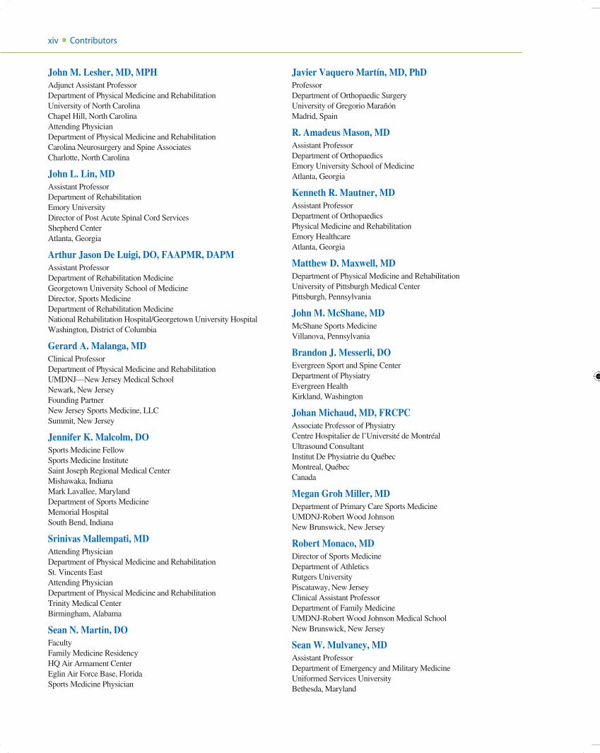
Author’s Preferred Technique (Figures 74-5, 74-6, and 74-7)
a. Patient position
i. Supine
ii. Hip externally rotated and knee slightly flexed
(rolled towel under knee)
b. Transducer position
i. Anatomic coronal plane (same plane as the MCL)
over the anterior fibers of the MCL
ii. Pes anserine tendons should be seen in an oblique
short-axis view as they cross the MCL
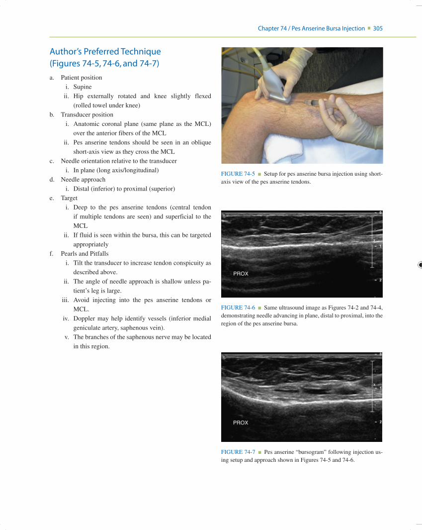
c. Needle orientation relative to the transducer
i. In plane (long axis/longitudinal)
d. Needle approach
i. Distal (inferior) to proximal (superior)
e. Target
i. Deep to the pes anserine tendons (central tendon
if multiple tendons are seen) and superficial to the
MCL
ii. If fluid is seen within the bursa, this can be targeted
appropriately
f. Pearls and Pitfalls
i. Tilt the transducer to increase tendon conspicuity as
described above.
ii. The angle of needle approach is shallow unless pa-
tient’s leg is large.
iii. Avoid injecting into the pes anserine tendons or
MCL.
iv. Doppler may help identify vessels (inferior medial
geniculate artery, saphenous vein).
v. The branches of the saphenous nerve may be located
in this region.
FIGURE 74-5 ■ Setup for pes anserine bursa injection using short-axis view of the pes anserine tendons.
FIGURE 74-6 ■ Same ultrasound image as Figures 74-2 and 74-4, demonstrating needle advancing in plane, distal to proximal, into the region of the pes anserine bursa.
FIGURE 74-7 ■ Pes anserine “bursogram” following injection us-ing setup and approach shown in Figures 74-5 and 74-6.
Mala_Ch074_303-306.indd 305Mala_Ch074_303-306.indd 305 09/10/13 9:19 AM09/10/13 9:19 AM

306 ■ Chapter 74 / Pes Anserine Bursa Injection
f. Pearls and Pitfalls
i. See “Pearls and Pitfalls” under “Author’s Preferred
Technique”
References1. Handy JR. Anserine bursitis: a brief review. South Med J
1997;90:376–377.2. Finnoff JT, Nutz DJ, Henning PT, Hollman JH, Smith J. Accu-
racy of ultrasound-guided versus unguided pes anserinus bursa injections. PM R 2010;2:732–739.
3. Yoon HS, Kim SE, Suh YR, Seo YI, Kim HA. Correlation between ultrasonographic findings and the response to cortico-steroid injection in pes anserinus tendinobursitis syndrome in knee osteoarthritis patients. J Korean Med Sci 2005;20:109–112.
Alternate Technique (Figures 74-8, 74-9, and 74-10)
a. Patient position
i. Supine
ii. Hip externally rotated and knee slightly flexed
(rolled towel under knee)
b. Transducer position
i. Start by finding the short-axis view of pes anserine
tendons as described above, and center gracilis ten-
don on the screen.
ii. Then rotate into long-axis view of central (graci-
lis) portion of conjoint tendon in anatomic coronal
oblique plane.
c. Needle orientation relative to the transducer
i. In plane (long axis/longitudinal)
d. Needle approach
i. Distal (inferior) to proximal (superior)
e. Target
i. Deep to the pes anserine tendons (central tendon
if multiple tendons are seen) and superficial to the
MCL.
ii. If fluid is seen within the bursa, this can be targeted
appropriately.
FIGURE 74-8 ■ Setup for pes anserine bursa injection using a long-axis view of the pes anserine tendons.
FIGURE 74-9 ■ Same ultrasound image as Figure 74-3, demon-strating needle advancing in plane, distal to proximal, into the region of the pes anserine bursa.
FIGURE 74-10 ■ Pes anserine “bursogram” following injection us-ing setup and approach shown in Figures 74-8 and 74-9.
Mala_Ch074_303-306.indd 306Mala_Ch074_303-306.indd 306 09/10/13 9:19 AM09/10/13 9:19 AM
Recommended