Embed Size (px)
Citation preview

The Essential Toolkit for Musculoskeletal Injections in Primary Care
Laurel Short, MSN, NP-c

Disclosure
I have no current affiliation or financial interest with any grantor or commercial interests that might have direct interest in the subject matter of the CE Program.

Objectives
• List the indications and precautions for muscular and joint injections
• Identify rationale for using injectable corticosteroids and local anesthetics
• Describe safety considerations and aseptic technique for musculoskeletal injections
• Review functional anatomy for the shoulder, elbow, wrist, hip, and knee
• Demonstrate injection technique guidelines and describe aftercare

Roadmap
• Didactic: general injection principles and safety
• 6 breakout sessions for specific injections-description/demonstration followed by practice time
• Review
Injections: Part of a Comprehensive Plan!
Injection
Rest
Rehab

General Principles
• Anatomic vs. trigger point injection
• Arthrocentesis = joint space aspiration
• Local anesthetic as diagnostic tool
– Followed by corticosteroid injection OR
– Combo corticosteroid + local anesthetic

Shopping List
Medications & Supplies

The DrugsCorticosteroids, Local Anesthetics, Viscosupplementation
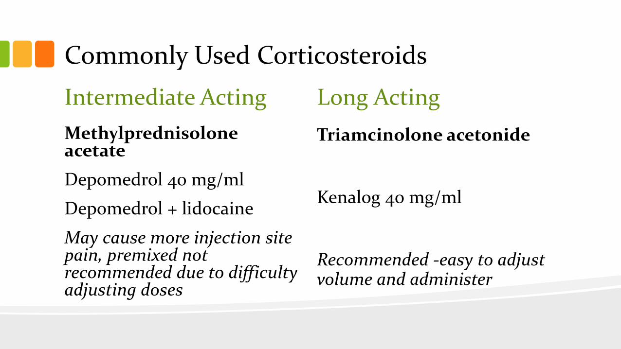
Commonly Used Corticosteroids
Intermediate Acting
Methylprednisolone acetate
Depomedrol 40 mg/ml
Depomedrol + lidocaine
May cause more injection site pain, premixed not recommended due to difficulty adjusting doses
Long Acting
Triamcinolone acetonide
Kenalog 40 mg/ml
Recommended -easy to adjust volume and administer

Corticosteroid Injection Indications
• Diagnostic AND/OR
• Therapeutic
– Suppressing inflammation/inflammatory “flares”
– Breaking up inflammatory damage-repair-damage cycle (?)
– Possible chondroprotective effect on cartilage metabolism or other process not related to inflammation
– Pain relief for tolerance of physical therapy

Local Anesthetics
• Work by causing a reversible conduction block along sensory nerve fibers
• Can make the procedure more comfortable
• Diagnostic tool
• Dilution- increased volume helps spread steroid to a larger surface area

Local Anesthetics
Short Acting
Lidocaine hydrochloride
0.5% 5mg/ml
1.0% 10mg/ml
2.0% 20mg/ml
Recommended. Acts rapidly, within seconds. Duration ~ 30 minutes.
Long Acting
Bupivacaine (Marcaine)
0.25% 2.5mg/ml
0.5% 5mg/ml
Slower onset, ~30 minutes for full effect. Duration >8 hours.

Viscosupplementation
• Osteoarthritis of the knee can be treated by lubricant injections
• OA = less lubrication and shock absorption within the joint
• In part related to less hyaluronic acid, part of synovial fluid
– HA molecules produce viscous solution that is both a lubricant and shock absorber

Viscosupplementation
• Indication- OA of knee– Failed conservative treatments (oral NSAIDs, cortisone
injection)
– Prolonging need for joint arthroplasty, or patients who are not good surgical candidates
• Proposed mechanisms of action– Cytokines/PGE inhibitor
– Inhibition of cartilage degredation
– Direct protective action on nociceptive endings

Hyaluronan (Orthovisc ®)
No avian protein allergy (not from rooster comb),
Largest molecule, Series of 3 injections
Hylan G-F 20 (Synvisc ® Synvisc-One ®)
Avian protein allergy
Uses formaldehyde & vinyl sulfone to increase molecular weight
Sodium Hyaluronate (Euflexxa ®, Gel-one®, Hyalgan ®, Supartz ®, Neovisc ®)
3 - 5 weekly injectionsCAUTION: avian protein allergy (eggs, feathers, poultry)

Platelet Rich Plasma (PRP)
Still considered experimental
• Patient’s whole blood (citrate dextrose as anticoagulant)
• Platelets spun down & activated by Thrombin & CaCl
• Thought to repair & regenerate cartilage, ligaments, muscle, tendons, and bone through cytokinens/growth factors
• Ultrasound guided injection, CPT Code 0232T

Additional SuppliesGloves
Gauze
Povidone-iodine swab
Alcohol wipe
Adhesive bandage
Ethyl chloride spray or ice (optional)
Needles & Syringes

Cheat SheetKnee:2cc 1 % Lidocaine 2cc 10mg Triamcinolone acetonide 25g x 1 ½” Needle
Shoulder:3cc 1 % Lidocaine 2cc 10mg Triamcinolone acetonide 25g x 1 ½” Needle
CTS, DEQ, Trigger Finger:1/2cc 1 % Lidocaine 1/4cc 40mg Triamcinolone acetonide 27g x 1/2” Needle
Elbow (Medial & Lateral), Pes Anserine Bursa, AC Joint:
1cc 1 % Lidocaine
1/2cc 40mg Triamcinolone acetonide
25g x 1 ½” Needle
Trigger Point Injection:
2cc 0.25 % Bupivacaine
2cc Sodium Chloride
25g x 1 ½” Needle
Trochanteric Bursa:
4cc 1 % Lidocaine
2cc 0.25% Bupivacaine
1cc 40mg Triamcinolone acetonide
21g x 2” Needle (or longer if needed)

Organize Team Members

Billing Codes: Aspiration/Injection
20610 Shoulder, Hip, or Knee
(20605 Wrist, Elbow, or Ankle)
20600 Fingers or Toes
20551 Tendon origin/insertion
20550 Tendon sheath, ligament
Trigger point injection
20552 (1-2) Muscles
20553 (3+) Muscles
64450 Occipital nerve block
Medications
Triamcinalone J3301
Dexamethasone J1100
(Bupivacaine J7799)
Ice pack applies 97010

GuidelinesKey Points to Assess Before Injecting

• Identify underlying etiology = good MSK exam
• Discuss risks & benefits with patient
• Knowledge of functional anatomy
• Avoid injecting an unstable joint (e.g. suspected rotator cuff or ACL tear)
• Avoid repeating injection too soon/too often- rule of thumb is no sooner than q 3 months

Potential Adverse Effects
• Postinjection flare (2-10%)
• Subcutaneous atrophy and/or skin depigmentation- more common with superficial injection
• Bleeding or bruising
• Steroid arthropathy – no real evidence for promotion of disease progression!
• Joint sepsis- rare
• Tendon rupture- minimized by good technique
• Facial flushing (1-5%)
• Hyperglycemia- usually <1 week
• Menstrual irregularity
• Decreased ESR/CRP levels
• Anaphylaxis- rare

Contraindications
Absolute
• Sepsis- local or systemic
• Fracture site
• Prosthetic joint
• Pediatric patients
• Bacteremia
• Allergy
• Uncontrolled bleeding disorder
Relative
• Diabetes
• Immunocompromised
• Large tendons (Achilles, infrapatellar)
• Sickle cell anemia
• Anticoagulation therapy-injection does not increase bleeding risk

Talking Points
• Gain patient confidence by discussing risks, benefits, additional recommendations
• Informed consent- verbal or written
• Steroids
– Serious side effects usually seen PO rather than injection due to less systemic absorption
– The body makes 20-30mg cortisone daily…we are using a small dose similar to your natural hormone
– You will better tolerate physical therapy/exercise when pain is controlled
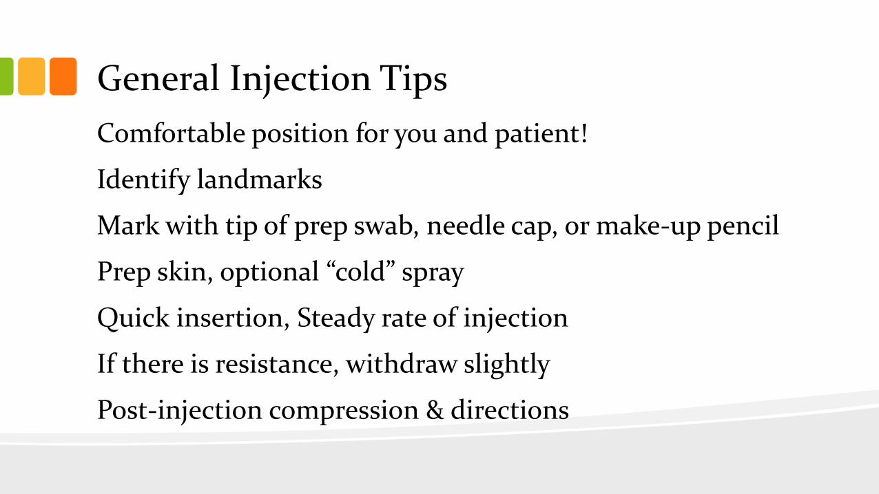
General Injection Tips
Comfortable position for you and patient!
Identify landmarks
Mark with tip of prep swab, needle cap, or make-up pencil
Prep skin, optional “cold” spray
Quick insertion, Steady rate of injection
If there is resistance, withdraw slightly
Post-injection compression & directions

What if it is not going as planned?

Post-injection Instructions
• Avoid excessive activity for 24-48 hours
• Gradual return to full activity
• Apply ice 3 x per day for 3 days (easy to remember)
• Ok to take NSAID/pain reliever
• Patient specific directions (DM, etc)
• Follow-up in 1-2 weeks, then rehab

Assess Outcomes-Feel like a hero!
• Follow-up within 2 weeks post-injection
• Consider physical therapy or a home exercise program to reduce risk of recurrence
• Additional modalities: ice/heat, oral or topical NSAID, essential oils, massage, yoga, exercise, PT
• If adequate improvement is not seen in 6-8 weeks, consider referral

Helpful Resources


Conference Adventures

Neck and ShoulderTrigger point and posterior shoulder injections

Shoulder Anatomy
• 3 Bones
scapula
clavicle
humerus
• Rotator cuff muscles (SITS)
Supraspinatus
Infraspinatus
Teres Minor
Subscapularis
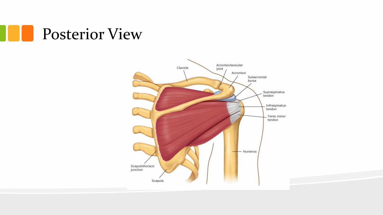
Posterior View

Shoulder Indications
• Tendinitis/tendinosis
• Impingement syndrome
• Bursitis
• Osteoarthritis

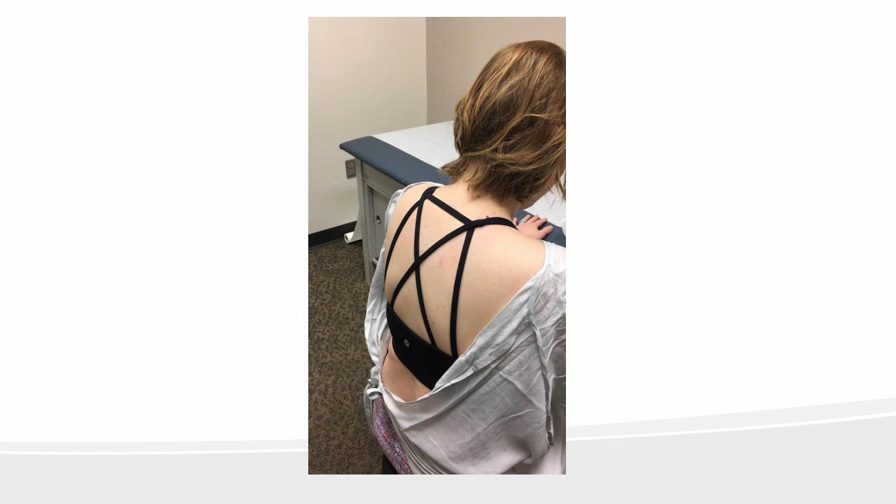
Subacromial Shoulder Injection
• Subacromial space
– Bursa is at anterior margin
– Size of a silver dollar
• Posterior approach
– Least pain receptors
– Biggest portal of entry
– Acromion slopes down in back- Angle up (about 15 °) with injection

Shoulder- Posterior Approach: 5ml syringe, 25 g 1.5” needle, 2cc 10mg Kenalog + 3cc 1% Lidocaine

Posterior Neck- Trigger Point Injections

Trigger Point Injections
• Focal, hyper-sensitive areas in tight areas of muscle
• Tender to palpation and can produce pain in a referral pattern
• May cause tension headaches, TMJ pain, regional pain, low back pain
• No steroid needed: 2ml normal saline + 2 ml anesthetic = 4ml total volume
• Inject 1ml to each trigger point, may see twitch response


ElbowLateral Epicondylitis, Medial Epicondylitis

Elbow Anatomy

3ml syringe, 25 g 1.5” needle, 1/2cc 40mg Kenalog + 1cc 1% Lidocaine
• Mostly Lateral– Tennis elbow
– Extensor-supinator. Occurs at the origin of the common extensor tendon. Anterior facet of the lateral epicondyle.
• Medial– Golfer’s elbow
– Flexor-pronator. Origin of the common flexor tendon. Anterior facet of the medial epicondyle.
– Careful of Cubital tunnel- ulnar nerve

Elbow Video

Wrist/HandDe Quervain’s tenosynovitis, Trigger finger

De Quervain’s Tenosynovitis
• Overuse injury of abductor pollicis longus & extensor pollicis brevis
• Pain at base of thumb & over radial styloid process
• Finklestein’s test

3ml syringe, 25 g 1.5” needle, 1/4cc 40mg Kenalog + 1/2cc 1% Lidocaine
• Thumb in slight flexion
• Feel gap between the 2 tendons
• Insert needle into the gap, then advance between the tendons
• Inject solution as a bolus


Trigger finger or trigger thumb
• May be acute or chronic
• Painful clicking and/or locking of finger or thumb
• May have painful, tender nodule at the base of the digit
• More common in those with diabetes

3ml syringe, 25 g 1.5” needle, 1/2cc 40mg Kenalog + 1cc 1% Lidocaine
• Position hand palm up
• Mark nodule at the A1 pulley
• Insert needle into the nodule
• Inject ½ solution into the nodule, then advance the needle slightly and inject the other ½ into the tendon sheath

On Your Mark, Get Set, Practice!

HipTrochanteric Bursitis

Greater Trochanteric Pain Syndrome
• Bursa- in line with pubis symphysis
• Rarely the primary issue! Key point- assess gait & strength
• Hip “rotator cuff”
– Abductor muscles: gluteus medius, minimus
– Abductor + external rotation: piriformis

Lateral hip • Tenderness over the greater trochanter
• Pain with sidelying, difficult to sleep
• Painful passive hip abduction/adduction
• May be chronic if underlying gait or muscle imbalance issues not addressed

5ml syringe, 18 g 2” needle, 1cc 40mg Kenalog + 4cc 1% Lidocaine
• Patient lies on unaffected side
• Upper leg extended
• Identify and mark point of maximum tenderness, over or near greater trochanter
• Insert needle perpendicular and advance to touch bone
• Pull back slightly, inject as bolus


KneeKnee joint, Pes anserine bursitis

Knee Joint
• Primary indication:
osteoarthritis
• Can be used for knee
strain and pain associated
with meniscus tear

Knee Anatomy
3 bones articulate:
Femur, tibia, patella
Main ligaments: lateral, medial, patellar, ACL, PCL
Meniscus: medial & lateral

5ml syringe, 25 g 1.5” needle, 2cc 10mg Kenalog+ 2cc 1% Lidocaine
• Patient sits or lies supine with knee flexed
• Identify and mark intersection of lateral and inferior joint lines
• Insert needle and advance slowly
• Injection solution as bolus


Pes Anserine bursitis
• Overuse injury-common in athletes
• Pain at the attachment site: medial side tibia just below joint line
• Combined tendon insertion

3ml syringe, 25 g 1.5” needle, 1/2cc 40mg Kenalog + 1cc 1% Lidocaine
• Patient sits or lies supine with knee flexed
• Identify and mark tender area over the bursa, if needed have patient flex knee against resistence
• Insert needle and advance to touch bone
• Pull back slightly and inject solution as bolus

Review and Documentation

Documentation ExamplesAfter verbal consent, under sterile conditions, I injected 2 ml of 10mg/ml Kenalog and 2 ml of 1% Xylocaine into the patient's left/right knee. The patient tolerated the injection well.
After verbal consent, under sterile conditions, I injected 1 ml of 40 mg/ml Kenalog and 4 ml of 1% Xylocaine into the patient's left/right trochanteric bursa. The patient tolerated the injection well.
After verbal consent, under sterile conditions, I injected 2ml of 10mg/ml Kenalog and 3 ml of 1% Xylocaine into the left/right shoulder subacromial space from a posterior approach. The patient tolerated the injection well.
After verbal consent, under sterile conditions, I injected 0.5 ml of 40mg/ml Kenalog and 1 ml of 1% Xylocaine into the left/right elbow at the lateral epicondyle. The patient tolerated the injection well.
After verbal consent, under sterile conditions, I injected 2ml of 0.25% Marcaine and 2ml of Sodium Chloride Saline Solution into the patient's (list muscles). The patient tolerated the injection well.
After verbal consent, under sterile conditions, I injected 0.25 ml of 40mg/ml Kenalog and 0.75 ml of 1% Lidocaine into the left/right thumb/finger A1 Pulley. The patient tolerated the injection well.
After verbal consent, under sterile conditions, I anesthetized the skin with 5ml of 1% Xylocaine and then cc's of clear yellow fluid was aspirated from the left/right knee and then injected 2ml of 10mg/ml Kenalog. The patient tolerated the procedure well.

Contact
Laurel Short
Kansas City Bone & Joint Clinic
Overland Park, KS
@Laurelontherun
Images/graphics: Unless otherwise noted, all images/graphics are from open sources or
property of Laurel Short
ReferencesFrontera, W. R., Silver, J. K., & Rizzo, T. D. (2014). Essentials of physical medicine and rehabilitation: Musculoskeletal disorders, pain, and rehabilitation (3rd ed.). London, United Kingdom: Saunders (W.B.) Co.
Holm, G. (2015). Arthrocentesis (Powerpoint slides).
Sarwark, J. F. (Ed.). (2014). Essentials of musculoskeletal care. United States: American Academy of Orthopaedic Surgeons.
Saunders, S., & Longworth, S. (2013). Injection techniques in Musculoskeletal medicine: A practical manual for Clinicians in primary and secondary care (4th ed.). London: Elsevier Health Sciences.