Embed Size (px)
Citation preview
1
Characterization of MazFsa, an endoribonuclease from Staphylococcus
aureus
Zhibiao Fu, Niles P. Donegan, Guido Memmi, Ambrose L. Cheung*
Department of Microbiology and Immunology, Dartmouth Medical School, Hanover, NH
03755, USA
*Corresponding author:
Department of Microbiology and Immunology, Dartmouth Medical School, Hanover, NH
03755
Phone: (603) 676-3350 ext 2. Fax: (603) 676-3355
E-mail: [email protected]
Running title: MazFsa, an endoribonuclease from S. aureus
Key words: Staphylococcus aureus, mazEFsa, toxin-antitoxin, endoribonuclease,
antibacterial therapy
ACCEPTED
Copyright © 2007, American Society for Microbiology and/or the Listed Authors/Institutions. All Rights Reserved.J. Bacteriol. doi:10.1128/JB.01272-07 JB Accepts, published online ahead of print on 12 October 2007
on July 5, 2018 by guesthttp://jb.asm
.org/D
ownloaded from
2
ABSTACT
The mazEF homologues of Staphylococcus aureus, designated mazEFsa, have been
shown to co-transcribe with the sigB operon under stress conditions. In this study, we
showed that MazEFsa, as with their E. coli counterparts, compose a toxin-antitoxin (TA)
module wherein MazFsa leads to rapid cell growth arrest and loss in viable c.f.u upon
overexpression. MazFsa is a novel sequence-specific endoribonuclease which cleaves
mRNA to inhibit protein synthesis. Using ctpA mRNA as the model substrate both in
vitro and in vivo, we demonstrated that MazFsa cleaves single-strand RNA preferentially
at the 5’ side of the first U or 3’ side of the second U residue within the consensus
sequences VUUV′ (where V and V′ are A, C, or G, and may or may not be identical).
Binding studies confirmed that the antitoxin MazEsa binds MazFsa to form a complex to
inhibit the endoribonuclease activity of MazFsa. Contrary to the system in E. coli,
exposure to selected antibiotics augmented mazEFsa transcription, akin to what one
would anticipate from the environmental stress response of the sigB system. These data
indicate that the mazEF system of S. aureus differs from the Gram negative counterparts
with respect to mRNA cleavage specificity and antibiotic stresses.
ACCEPTED
on July 5, 2018 by guesthttp://jb.asm
.org/D
ownloaded from
3
INTRODUCTION
Many bacteria have chromosomally encoded toxin-antitoxin (TA) loci in which
the toxin and antitoxin genes exist in an operon and are co-expressed together to form a
TA complex. The toxin is stable, whereas the antitoxin is a labile protein degraded in vivo
by host proteases (e.g. Clp or Lon in E. coli). Under conditions that preclude the
continuous synthesis of the antitoxin, the toxin can exert its toxic effect to inhibit cell
growth (7, 9, 12). There are at least eight typical TA families known in prokaryotes (12,
22). Among these, the MazEF and RelBE systems from E. coli have been the most
extensively studied (7, 9, 12). Structural studies have disclosed that the MazE-MazF
complex in E. coli consists of two MazF dimers and one MazE dimer in a hexameric
MazF2-MazE2-MazF2 configuration (17). In contrast, the RelBE complex from
Pyrococcus horikochi is a (RelE-RelB)2 tetramer (32).
Inhibition of protein synthesis by MazF in E. coli has been found to be
attributable to cleavage of cellular mRNA. More specifically, MazF in E. coli is a
sequence-specific endoribonuclease, cleaving mRNA at ACA sites independently of
ribosomes both in vitro and in vivo (6, 34). The cleavage occurs at the 5’-end of ACA
sequence to yield a 2’,3’-cyclic phosphate as part of the end product. The 2’-OH group of
the nucleotide preceding the ACA sequence is essential for MazF cleavage (37). In
contrast, the RelE toxin of E. coli was found to cleave mRNA positioned at the ribosomal
A-site both in vitro and in vivo (26). Cleavage occurs between the second and third bases
of A-site codon (UAH where H is usually G or A), with the cleavage efficiency
depending on the specific codon at the ribosomal A-site. For instance, UAG and UAA are
ACCEPTED
on July 5, 2018 by guesthttp://jb.asm
.org/D
ownloaded from
4
cleaved more efficiently than the UGA stop codon (26). The toxin systems from other
prokaryotes also appeared to represent sequence-specific endoribonucleases. The PemK
toxin from plasmid R100 in E. coli cleaves mRNA at UAH (where H is A, C or U) (36),
ChpBK cleaves at ACY (where Y is A, G or U) in a single strand RNA (38) while the
Bacillus subtilis MazF homolog EndoA cleaves mRNA at an UAC sequence (27).
Recently, two MazF homologs from Mycobacterium tuberculosis were also found to be
endoribonucleases. One of the MazF homologs from M. tuberculosis cleaves mRNA at
UAC triplets while the other homolog cleaves U-rich regions within mRNA (39).
In examining the sigB operon of S. aureus, Kullik et al. (18) noted that an ORF
immediately upstream of the sigB operon may encode a mazF homolog (designated pemK
homolog). Senn et al. (30) subsequently demonstrated that the sigB operon in S. aureus
strain COL comprises two additional ORFs (sa2059 and sa2058) in addition to rsbU,
rsbV, rsbW, and sigB. They also observed, as did Gertz et al. (13), that SA2058 and, to a
much lesser extent, SA2059 share some degree of homology with MazF and MazE of E.
coli, respectively. SA2059 and SA2058 are co-transcribed with the sigB operon under
stress conditions such as heat and high salt conditions (30). For brevity and consistency,
we propose to name SA2059 and SA2058 (designated as SA1873 and SAS067 in N315)
in COL as MazEsa and MazFsa, respectively, in S. aureus. Although it has been hinted
that the S. aureus MazEFsa may act as a TA module (30), there have been no
experimental data supporting this hypothesis. This confusion has been generated in part
as a consequence of a general lack of protein sequence similarity between MazEsa and its
E. coli counterpart.
ACCEPTED
on July 5, 2018 by guesthttp://jb.asm
.org/D
ownloaded from
5
In this study, we provided definitive evidence that MazEFsa is a TA module in S.
aureus, with MazFsa as the toxin. Our data demonstrated that MazFsa is a sequence-
specific endoribonuclease, which cleaves ctpA mRNA at a consensus U-rich sequence of
VUUV′ (where V and V′ are A, C, or G, and may or may not be identical) both in vivo
and in vitro. MazFsa showed high cellular toxicity in both E. coli and S. aureus upon
induction and inhibited protein synthesis in a cell-free system. Collectively, our results
suggest that activated MazEFsa TA module cleaves mRNA cleavage at a specific site
under stressful conditions to affect translation. This finding raises the possibility that
inhibition of MazEsa may represent a novel approach to antibacterial therapy for S.
aureus.
ACCEPTED
on July 5, 2018 by guesthttp://jb.asm
.org/D
ownloaded from
6
MATERIALS AND METHODS
Bacterial strains and culture conditions. We used E. coli strains DH5α and
BL21(DE3)pLysS and S. aureus strains Newman and 178RI (8) for these studies. S.
aureus 178RI carries an IPTG inducible T7 polymerase gene integrated into the geh locus
in the chromosome of RN4220. For transduction, phage 85 was used to produce phage
lysates of S. aureus 178RI. The phage lysate was then used to infect the
S. aureus
Newman as described (3) to obtain the S. aureus transductant ACL6094 carrying the T7
polymerase gene integrated into the chromosome in the Newman background. Cultures
were routinely grown in LB for E. coli and in 03GL or Trypticase soy broth (TSB) for S.
aureus with aeration at 37ºC. The media were supplemented with either ampicillin (Amp)
(70 µg/ml) or chloramphenicol (Cm) (10 µg/ml).
Construction of plasmids. The mazEsa (GenBank Accession Number Y16431) and
mazFsa (GenBank Accession Number Y07645) genes were amplified by PCR using S.
aureus Newman genomic DNA as a template and cloned into the NcoI and BamHI sites
of cloning vectors pCDF1 and pET14b (Novagen) in E. coli to make pCDF1-MazE(His)6
and pET14b-MazF(His)6 with the 6xHis tag at the N-terminus, respectively. The mazEsa
gene without the His tag was amplified by PCR and cloned into the NdeI/XhoI sites of
pETDuet1 (Novagen). An NcoI-BamHI digested DNA fragment from pET14b-
MazF(His)6 was then inserted to make pETDuet1-MazEF(His)6 with the 6x His tag only
at the MazFsa N-terminus. An NcoI-BamHI and an BglII-EcoRI digested DNA fragment
from pET14b-MazF(His)6 were further cloned into NcoI and BamHI digested pBAD22
(14) and BglII-EcoRI digested pG164 (8), respectively, to generate pBAD22-MazF(His)6
ACCEPTED
on July 5, 2018 by guesthttp://jb.asm
.org/D
ownloaded from
7
and pG164-MazF(His)6. The ctpA gene (encoding Carboxy-Terminal-Protease from S.
aureus, GenBank Accession Number NP_374534) was also amplified by PCR using S.
aureus Newman genomic DNA as the template and cloned into the NcoI and BamHI sites
of pET14b (Novagen) to produce pET14b-ctpA. A BglII-EcoRI digested fragment from
pET14b-ctpA was further cloned into BamHI and EcoRI digested pG164-MazF(His)6 to
generate pG164-MazF(His)6/ctpA. DNA techniques were performed according to
standard procedures (28).
Protein expression and purification. The MazE(His)6sa was expressed in E. coli
BL21(DE3)pLysS carrying the plasmid pCDF1-MazE(His)6 under IPTG induction (1
mM) for 4 hr. For MazF(His)6sa expression, the MazEsa and MazF(His)6sa genes were co-
expressed in E. coli BL21(DE3)pLysS harboring the plasmid pETDuet1-MazEF(His)6
after IPTG induction (1 mM) for 6 hr. The cells were harvested and subjected to lysis by
ultrasonication. MazE(His)6sa and MazE-MazF(His)6sa complex were purified with the
nickel-nitrilotriacetic acid resin affinity column (Novagen) according to the
manufacturer’s protocol. MazF(His)6sa was further purified from the MazE-MazF(His)6sa
complex as described previously (35). In brief, MazEsa was dissociated from
MazF(His)6sa in the purified MazE-MazF(His)6sa complex with 6 M guanidine HCl.
MazF(His)6sa was retrapped with the nickel-nitrilotriacetic acid resin affinity column,
eluted and refolded by stepwise dialysis as described previously (25).
Native PAGE. Various amounts of MazE(His)6sa and MazF(His)6sa were mixed in
binding buffer (50 mM Tris-HCl, pH 7.5, 5 mM MgCl2, 1 mM dithiothreitol, and 5%
glycerol) at 4ºC for 30 min and subjected to the native PAGE analysis in running buffer
ACCEPTED
on July 5, 2018 by guesthttp://jb.asm
.org/D
ownloaded from

8
containing 82.6 mM Tris-HCl (pH9.4) and 33 mM glycine as described previously (35).
The protein bands were visualized by staining with Coomassie Brilliant Blue.
Primer extension analyses. For in vitro primer extension, the ctpA mRNA was
transcribed from BamHI-linearized pET14b-ctpA plasmid, using the T7 large-scale
transcription kit (Promega) according to the manufacturer’s protocol. Five µg of ctpA
mRNA were partially digested with 15 pmol of MazFsa in a 20 µl reaction mixture
containing: 40 U of RNase inhibitor, 50 mM Tris-HCl (pH8.0), 50 mM NaCl, and 1 mM
DTT at 37ºC for 90 min. The digestion mixture was then extracted with phenol-
chloroform followed by ethanol precipitation to remove the proteins. Primer extension
(Promega Primer Extension Kit) analysis of the the digested mRNA was carried out with
labeled primers pEa d(GCTTGATCAGTTTTGTTTAAACCAC), pEb d(TGACCAT
GCCATCAATTGCAGC), pEc d(AGGACGAATGCCAGCACGTTCTG CTGG), and
pEd d(CTTCACTACCT CGTTGAACAGTTA), following the manufacturer’s protocol.
The primers were 5’-end labeled with [γ-32
P]ATP using T4 polynucleotide kinase. For in
vivo primer extension analysis of ctpA mRNA cleavage sites in E. coli, we isolated total
cellular RNA from E. coli BL21(DE3)pLysS cells harboring both pBAD22-MazF(His)6
and pET14b-ctpA. Cultures were induced with 1 mM IPTG to transcribe the ctpA mRNA
for 30 min. MazFsa was then induced by adding arabinose to a final concentration of
0.2%. For in vivo primer extension in S. aureus, 178RI harboring pG164-
MazF(His)6/ctpA was induced with 1 mM IPTG. The ctpA mRNA was co-transcribed
with the mazFsa mRNA. After induction, total cellular RNA from a 10 ml culture was
then extracted at the indicted time point as described previously (4). Trace DNA was
further removed by digestion with RNase-free DNase I (Roche) followed by extraction
ACCEPTED
on July 5, 2018 by guesthttp://jb.asm
.org/D
ownloaded from
9
with phenol-chloroform and ethanol precipitation to clean the RNA. Primer extension
was then carried out with different primers as described previously (16). The primer
extension product was analyzed on a 6% sequencing gel with the DNA sequencing ladder
prepared with the same primer running side by side followed by autoradiography.
Cleavage of synthetic RNAs by MazFsa. All RNAs were commercially synthesized (IDT,
Coralville, IA) and 5’-end labeled with [γ-32
P]ATP using T4 polynucleotide kinase. The
native sequence, 5’- UUGGCAAUUCAUAUCAAU-3’, corresponding to the sense RNA
was named AUUC, with AUUC as the target cleavage site. Seven other RNA substrates
with the center AUUC sequences changed to AGUC, AUGC, AUUU, UUUC, AUUG,
GUUC, and GUUG sequences were also synthesized and named as AGUC, AUGC,
AUUU, UUUC, AUUG, GUUC, and GUUG, respectively. The synthesized native DNA
sequence, 5’-TTGGCAATTCATATCAAT-3’, named as ATTC, was used as a control. A
19-base synthetic RNA (5’-UGCAAUUCAUAUGAAUUGU-3’) that can form hairpin
structure with the AUUC sequence located in the duplex region was named RB-1.
Another 19-base RNA (5’-UGCAAUUCAUAUCAAUAUG-3’), which cannot form the
hairpin structure, was named RB-2. An antisense RNA to the native AUUC RNA
sequence (5’-AUUGAUAUGAAUUGCCAA-3’), was named RB-3. The labeled RNA
substrates were digested with MazFsa at 37ºC for 30 min in a 10 µl of reaction mixture
containing 20 U of RNase inhibitor, 15 pmol of MazFsa, 1 pmol labeled RNA, and 10
mM Tris-HCl (pH 7.9). The formation of RNA-RNA duplex with the sense RNA AUUC
and RB-3, the antisense RNA, or MazFsa-mediated cleavage was analyzed as described
(36). Briefly, 1 pmol of labeled sense RNA was annealed with its antisense RNA in
different ratio combinations and incubated with 15 pmol of MazFsa at 37ºC for 30 min.
ACCEPTED
on July 5, 2018 by guesthttp://jb.asm
.org/D
ownloaded from
10
The reactions were stopped by adding loading buffer, and analyzed by separating on a
20% sequencing gel. The RNA ladder was prepared by partial alkaline hydrolysis
(Ambion) of the 5’-end labeled 18-base sense RNA, AUUC, according to the
manufacturer’s protocol.
Northern blot hybridization. Total RNA from S. aureus was prepared by using a Trizol
isolation kit (Invitrogen, CA) and a reciprocating shaker (4). For detection of specific
transcripts, gel-purified DNA probes were radiolabeled with [α-32
P]dCTP by use of a
random-primed DNA labeling kit (Roche Diagnostics GmbH), and hybridized under
aqueous-phase conditions at 65°C. The blots were subsequently washed with 2XSSC,
0.1% SDS twice at room temperature, 1XSSC, 0.1% SDS twice at 650C, and
autoradiographed as previously described (20).
Bacterial viability assay. Bacteria were stained with the membrane-permeable SYTO 9
and the membrane-impermeable propidium iododle using the LIVE/DEAD Baclight
Bacterial Viability Kit (Molecular Probes, Eugene, OR), and quantitated with
fluorescence microplate readers according to the manufacturer’s protocol. Bacteria with
intact cell membranes stain fluorescent green, whereas bacteria with damaged
membranes stain fluorescent red.
ACCEPTED
on July 5, 2018 by guesthttp://jb.asm
.org/D
ownloaded from
11
RESULTS
MazEFsa is a toxin-antitoxin (TA) module in S. aureus. A BLAST search of sa2058,
encoding a 120-residue protein in the COL genome identified this gene to have 20%
identity and 40% similarity to the E. coli MazF protein. The upstream gene sa2059,
which is co-transcribed with sa2058, encodes a 56-residue protein with only 12% identity
and 21% similarity to the E. coli MazE protein. We have named SA2058 and SA2059 as
MazFsa and MazEsa for brevity and clarity, which was referred by Mittenhuber (22) as
Orf136-s.a and Orf6-s.a (Fig.1A).
To examine if MazFsa and MazEsa function as a TA module, we determined
whether the MazFsa protein, when expressed independently, is toxic to bacterial cells.
For this purpose, the mazFsa gene was cloned into the vector pBAD with an arabinose
inducible promoter and the shuttle vector pG164 with an IPTG inducible promter to
generate pBAD-MazF(His)6 and pG164-MazF(His)6, respectively, as described in the
“Materials and methods”. The growth of S. aureus ACL6094 carrying the plasmid
pG164-MazF(His)6 with a T7-dependent promoter was inhibited on 03GL agar plates
supplemented with IPTG (1 mM) but not in the unsupplemented control (Fig. 1B). Time-
course studies were further carried out to characterize the toxicity of MazFsa. We found
that most of the cells with the mazFsa operon cloned into pG164 could not yield colonies
on nutrient agar plates after induction for 60 min while cells without induction showed
normal growth (Fig. 1C). Although the c.f.u counts were reduced 99.5% after 60 min
post-induction, fewer than 5% of the cells stained positively with propidium iodide
(Molecular Probes, Eugene, OR), a dye which binds “membrane-compromised” dead
ACCEPTED
on July 5, 2018 by guesthttp://jb.asm
.org/D
ownloaded from
12
bacteria (data not shown), thus indicating MazFsa expression mainly induced bacterial
stasis and hence a defect in replication. Similar results occurred in E. coli with the
plasmid pBAD-MazF(His)6 (data not shown). In contrast, E. coli and S. aureus cells with
mazEFsa cloned into pBAD and pG164, respectively, exhibited normal growth even
under respective induction (arabinose or IPTG) (data not shown). Together, these data
demonstrated that MazFsa is toxic to both E. coli and S. aureus and that this toxicity can
be reversed by co-expression of MazEsa with MazFsa.
To further characterize the MazEFsa TA module, we expressed and purified
MazF(His)6sa and MazE(His)6sa (both N-terminally tagged) (Fig. 1D) in E. coli as
described under “Materials and Methods”. MazE(His)6sa and MazF(His)6sa were mixed
together in a dose-dependent manner and subjected to native PAGE analysis. Despite the
noticed migration of MazE(His)6sa (pI 4.2), no obvious mobility was observed for
MazF(His)6sa alone (Fig. 1E, lane 2) presumably due to its basic pI (9.5), which
approaches the pH (9.4) of the running buffer used in native PAGE. Nevertheless, the
MazEFsa complex, appearing as a higher molecular weight species than the MazE(His)6sa
alone, was observed at the top of the gel (Fig. 1E, lanes 3-5). The quantity of the MazEFsa
complex rose with increasing concentrations of MazF(His)6sa while the amount of free
MazE(His)6sa at the bottom of the gel continued to diminish (Fig. 1E). These results
indicated that MazEsa and MazFsa, as a paired TA module, interact in vitro and possibly
in vivo.
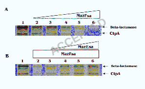
MazFsa inhibits protein synthesis in a cell-free system. We then examined the effect of
the purified MazFsa on protein synthesis in a cell-free system. The synthesis of the
ACCEPTED
on July 5, 2018 by guesthttp://jb.asm
.org/D
ownloaded from
13
truncated CtpA protein (~15 kDa), representing part of the carboxy terminal protease
from S. aureus, from the plasmid pET14b-ctpA was carried out at 37ºC for 1 hr using E.
coli T7 S30 extract system with and without MazFsa (Fig. 2A). The synthesis of CtpA
was inhibited with increasing concentrations of MazFsa, and was almost completely
blocked with 30 pmol added (Fig. 2A). Addition of MazEsa to the cell free system
containing MazFsa rescued CtpA synthesis in a dose-dependent manner (Fig. 2B). As
expected, the MazFsa protein also inhibited the synthesis of ß-lactamase from the bla
gene present in the pET14b-ctpA vector in E. coli (Fig. 2). Pre-incubation of the E. coli
cell-free protein synthesis system with MazFsa for 20 min at 37ºC prior to adding the
plasmid pET14b-ctpA and MazEsa did not have any significant effect on subsequent
CtpA synthesis (data not shown). These results suggest that the primary target for MazFsa
is not the ribosome, tRNA or other factors required for protein synthesis in this system,
but rather mRNA of the cell.
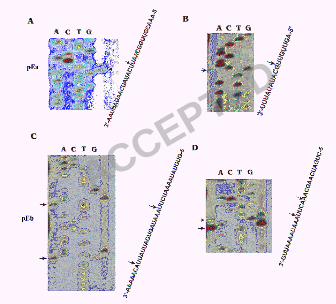
In vitro cleavage of ctpA mRNA by MazFsa. To determine whether MazFsa has
endoribonuclease activity, we prepared the ctpA mRNA using an in vitro transcription
system as described in the “Materials and Methods”. The ctpA mRNA was then incubated
with MazFsa in a dose and time-dependent manner. As shown in Fig. 3A, the ctpA mRNA
was cleaved into small fragments with 15 pmol of MazFsa in a time-dependent manner
while the addition of MazEsa inhibited the digestion of ctpA mRNA by MazFsa in a dose-
dependent fashion (Fig. 3B). These results demonstrate that MazFsa is an
endoribonuclease that cleaves mRNA to inhibit protein synthesis and MazEsa functions as
an antitoxin to counteract the endoribonuclease activity of MazFsa.
ACCEPTED
on July 5, 2018 by guesthttp://jb.asm
.org/D
ownloaded from
14
The ctpA mRNA was noted to be cleaved into distinct, but not smearing bands by
MazFsa (Fig. 3A), indicating that MazFsa may be a sequence-specific endoribonuclease.
To further map the cleavage site, we employed MazFsa to partially digest the ctpA mRNA
and then subjected the digest to primer extension, using four different DNA primers, pEa-
d, covering the experimental ctpA mRNA as described in the “Materials and Methods”.
To determine the cleavage sites, each primer extension product was analyzed on a 6%
sequencing gel with a DNA sequencing ladder prepared with the same primer (Fig. 4).
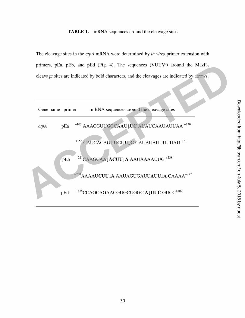
The cleavage sites in the ctpA mRNA as determined by primer extension studies are
shown in Table 1. Cleavages occurred preferentially in a U-rich region with a consensus
sequence of VUUV′ (where V and V′ are A, C, or G, and may or may not be identical) in
ctpA mRNA. The UU dinucleotides were found to be conserved among all cleavage sites.
However, the primary cleavages occur at either 5’-side of the first U or 3’-side of the
second U residue in the VUUV′ sequence, with most cleavages taking place 5’ of the first
U residue (Fig. 4, Table 1). However, not all of the VUUV′ sequences in the ctpA mRNA
were cleaved by MazFsa.
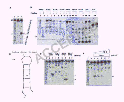
Cleavage specificity of MazFsa. To further define the specificity of cleavage sites, an 18-
base synthetic RNA (5’-UUGGCAAUUCAUAUCAAU-3’) with the AUUC sequence in
the center was used for the digestion with MazFsa. A clear cleavage was shown between
the A and U of the sequence (A↓UUC) (Fig. 5A). Seven additional 18-base synthetic
RNA substrates were synthesized with the AUUC sequence in the center being replaced
by AGUC, AUGC, AUUU, AUUG, GUUC, GUUG and UUUC to examine consensus
residues in the VUUV′ (where V and V′ are A, C, or G, and may or may not be identical)
ACCEPTED
on July 5, 2018 by guesthttp://jb.asm
.org/D
ownloaded from
15
(Fig. 5B). Our analyses showed that both U residues are essential for cleavage to occur;
alterations in any of the two U residues in the center of the consensus sequence
completely abolished the cleavage by MazFsa (Fig. 5B, lanes 4 and 6). The cleavage
efficiency reduced significantly if the first and the fourth residues were changed into U
(Fig. 5B, lanes 10 and 16). The fourth C residue can be changed to G without any
significant loss of cleavage efficiency (Fig. 5B, lane 12). The first A can be changed into
G with some degree of reduced cleavage efficiency (Fig. 5B, lanes 8 and 14). No
cleavage occurred with the corresponding single-strand DNA sequence (Fig. 5B, lane 18),
indicating that MazFsa specifically cleaves RNA. There was clear cleavage occurred with
the sequences AUUA and CUUA in the ctpA mRNA template, as shown in Fig. 4C and D.
These results confirm that MazFsa is an endoribonuclease that specifically cleaves the
consensus RNA sequence VUUV′.
There are other VUUV′ sequences present in the ctpA mRNA, but cleavage did
not occur with these sequences. We speculate that secondary structures of the substrate
may affect the cleavage by MazFsa. To test this, a highly purified 19-base synthetic RNA,
RB-1, which can form a hairpin structure with the AUUC sequence embedded within the
stem region (Fig. 5C), was digested with MazFsa. Cleavage was completely blocked with
this hairpin structure, whereas clear cleavage occurred in the purified RB-2, the synthetic
RNA without the hairpin structure that encompassed the AUUC sequence (Fig. 5D, lanes
5 and 7). We next examined the cleavage of MazFsa on the AUUC antisense RNA, RB3.
Although there is an AUUG sequence in the single strand AUUC antisense RNA ,
cleavage by MazFsa did not occur (Fig. 5D, lane 9), whereas the altered sense strand
RNA with the AUUG sequence (Fig. 5B, lane 12) was efficiently cleaved by MazFsa. The
ACCEPTED
on July 5, 2018 by guesthttp://jb.asm
.org/D
ownloaded from
16
reason for this discrepancy is unknown, but it is conceivable that the sequences adjacent
to VUUV′ may play a role in promoting cleavage. The cleavage by MazFsa was also
blocked when the sense RNA with the AUUC sequence annealed with its antisense strand
RNA, RB-3, to form an RNA-RNA duplex in a dose-dependent manner (Fig. 5E). These
results suggested that MazFsa cannot cleave the VUUV′ sequences in RNA-RNA duplex
and hence is only specific for single-strand RNA without any intramolecular basepairing
involving VUUV′.
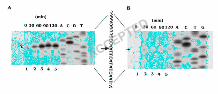
In vivo cleavage of ctpA RNA by MazFsa. To determine the MazFsa specific cleavage
sites in mRNA in vivo, primer extension analysis of ctpA mRNA was performed with
total RNA extracted from both E. coli and S. aureus carrying the corresponding plasmids
at various time points after induction as described in the “Materials and Methods”. A
clear in vivo cleavage site (A↓AUUC) was determined in E. coli with primer pEa, as
shown in Fig. 6A. A primer extension product appeared at 30 min after induction of
MazFsa, with subsequent time points showing cleavage in a time-dependent manner
(lanes 2-5). However, the effect of MazFsa in E. coli likely occurs within seconds of
initiation of the reaction as this extension product can almost be detected at time 0 min
(Fig. 6A, lane 1). In vivo cleavage was also detected in S. aureus 178RI carrying plasmid
pG164-MazF(His)6/ctpA with primer pEa (Fig. 6B), but the cleaved ctpA mRNA with the
extension product was faintly detected only after 30 min induction (Fig. 6B). We
speculate this may be due to a lower copy number of the co-transcribed ctpA mRNA.
Nevertheless, the cleavage recognition site in vivo (A↓AUUC) in both E. coli and S.
aureus (Fig. 6A and B) was found to be identical to the one in vitro (AAU↓UC) (Fig. 4A),
ACCEPTED
on July 5, 2018 by guesthttp://jb.asm
.org/D
ownloaded from
17
but the cleavage site was shifted two bases upstream. Collectively, these results indicated
that MazFsa recognizes the same site on ctpA mRNA both in vivo and in vitro, but the
exact cleavage site may differ by one to two bases between those in vivo and in vitro,
which could be due to trimming of the RNA ends by cellular RNases.
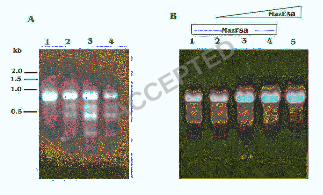
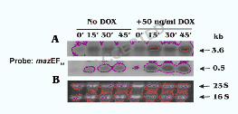
Environmental stress triggers increasing expression of the mazEFsa transcript. The
mazEFsa operon is located upstream of the sigB operon and is co-transcribed as a 3.6 kb
transcript (Fig. 1A). This genetic arrangement suggests that mazEFsa may be related to
environmental stresses. We thus examined the transcription of mazEFsa upon exposure to
antibiotics. Increased expression of a 3.6-kb and a 0.5-kb transcripts, corresponding to the
co-transcript with sigB operon and the transcript of mazEFsa operon alone (Fig. 1A),
respectively, was detected with the mazEFsa probe upon exposure to doxycycline (DOX)
for 45 min (Fig. 7). The increase of both transcripts was also found with exposure to sub-
MIC concentrations of other antibiotics, e.g., erythromycin and penicillin (unpublished
data). Interestingly, no reduction in c.f.u counts was observed with exposure to antibiotic
at sub-MIC concentrations (data not shown). Similarly, we also found brief exposure of
S. aureus cells to heat (48°C) activated transcription of the mazEFsa promoter
(unpublished data), thus confirming the finding of Senn et al. who also demonstrated
increased transcription of mazEFsa with sigB upon heat stress treatment (30). This is
different from the mazEF system in E. coli wherein brief exposure of antibiotic or heat
disrupts mazEF transcription and translation, thus leading to proteolysis of the labile
MazE and hence unleashing the endoribonuclease activity of MazF (1, 15, 29). This
mode of action of MazF in E. coli has been termed programmed cell death by a group of
ACCEPTED
on July 5, 2018 by guesthttp://jb.asm
.org/D
ownloaded from
18
investigator (9), but this claim has been disputed by studies presented by Tsilibaris et al.
(33).
DISCUSSION
Staphylococcus aureus is a major opportunistic pathogen that is a leading cause of
nosocomial infections associated with surgical wounds and indwelling medical devices.
Despite antimicrobial therapy, the morbidity and mortality associated with S. aureus
infections remain high, due in part to the organism's ability to develop resistance to
antibiotics including that to vancomycin (5, 20). In searching for antimicrobial targets
within the stress-induced operon, we noticed, as did Kullik et al. (18), that the ORF
(sa2058) upstream of rsbU, the first gene within the established sigB operon, shares
sequence similarity with mazF of E. coli. Given that mazE and mazF in prokaryotes are
often co-transcribed, we were puzzled with the functional identity of these two ORFs
since sa2059, directly upstream of sa2058, has little homology with mazE of E. coli.
In S. aureus, sa2059 and sa2058 have been shown to co-transcribe with the sigB
operon, particularly under stressful conditions (30). Given that both mazEF and the sigB
operons are modulated under stress and that sa2058 is homologous with mazF of E. coli,
it is reasonable to speculate that SA2058 and SA2059 may represent a MazEF–like
system in S. aureus despite a lack of supporting experimental data. In this study, we
demonstrate that MazEFsa of S. aureus is a TA module wherein MazFsa is the toxin and
MazEsa is the antitoxin that binds MazFsa to inhibit its toxicity. Our data showed that
MazFsa is toxic to both E. coli and S. aureus after induction for its expression (Fig. 1). It
ACCEPTED
on July 5, 2018 by guesthttp://jb.asm
.org/D
ownloaded from
19
inhibits protein synthesis in a cell-free system by cleaving mRNA susbstrates. MazEsa, on
the other hand, inhibits the toxicity of MazFsa by preventing cleavage of the target mRNA
and hence releases the inhibition in protein synthesis by MazFsa (Fig. 2 and 3). This
inhibition was due to the formation of MazEsa/MazFsa complex (Fig. 1D), which prevents
the free form of MazFsa from cleaving the target RNA (Fig. 3B). However, the exact
stoichiometry by which the C-terminal arm of MazEsa mimics the similarly charged
sugar-phosphate backbone of RNA to inhibit MazFsa toxin activity by occupying the
RNA binding site on the MazFsa toxin as described for E. coli (19) will require further
detailed crystal structure studies of the MazE/MazFsa complex.
In S. aureus and some Gram-positive bacteria (e.g. B. subtilis and L.
monocytogenes), mazEF homologues are located immediately upstream of the sigB
operon, which encodes σB, the main alternative transcription factor involved in stress
response of many Gram positive bacteria, and a series of anti-sigma factors to control the
concentration of free σB (10, 13, 27, 30). In contrast, the mazEF genes in E. coli are
located downstream of the relA gene. relA encodes a synthase for ppGpp and is up-
regulated in response to uncharged tRNA at the ribosomal A-site during amino acid
starvation and other stressful conditions including antibiotic exposure (1, 15, 29). It was
shown that overproduction of ppGpp (by overproducing RelA′, a truncated version of
ppGpp synthetase I of E. coli) in a strain derived from MC4100 represses expression
from the mazEF promoter. These authors then suggested that physiological conditions
that confer increased levels of ppGpp would reduce synthesis of MazE antitoxin, hence
enabling degradation of the more labile MazE antitoxin by the ClpPA protease system
and unleashing the toxic effect of MazF to execute programmed cell death (PCD) (1).
ACCEPTED
on July 5, 2018 by guesthttp://jb.asm
.org/D
ownloaded from
20
However, Christensen et al. (6) investigated that the transcription pattern of mazEF
during amino acid starvation induced by serine hydroxamate (SHT) was stimulated
strongly by amino acid starvation and this stimulation depended on Lon. No TA-locus
dependent cell killing was observed during this amino acid starvation. Penersen et al. (25)
also showed that the toxicity of MazF in E. coli can be rescued by the antitoxin MazE,
expressed within 6 hr after MazF induction. They further proposed that MazF does not
mediate cell killing but rather induce a bacteriostatic condition. Both studies have shed
doubt on the notion of PCD proposed by Aizenman et al (1). Indeed, even with the
overproduction of MazF, E. coli cells can retain transcriptional and translational
competence for 4 days despite their growth arrest (31). Although Sat et al. (29) and
Hazan et al. (15) suggested PCD mediated by mazEF from E. coli upon exposure to some
antibiotics, controversial results were presented for the same PCD experiments by
Tsilibaris et al. (33), thus the physiological roles of the toxin proteins remain under
debate.
Our results demonstrated that the expression of the mazEFsa transcripts was up-
regulated (Fig. 7) when the culture was exposed to sub MIC levels of some antibiotics,
with no great loss of cell viability. Given the divergent structural arrangement between E.
coli and S. aureus with respect to MazEF and stress operon, the regulation of the S.
aureus MazEFsa TA module in response to stress warrants additional investigation
(manuscript in preparation). Pedersen et al. (25) reported that RelE induced cell stasis
exhibited increased sensitivity towards environmental stresses, e.g. heat shock, oxygen
radicals and osmotic stress. The above studies have led to the suggestion that TA
complexes might constitute a novel approach toward the potential development of a new
ACCEPTED
on July 5, 2018 by guesthttp://jb.asm
.org/D
ownloaded from
21
class of antimicrobial compounds, which activate or mimic bacterial toxins. Compounds
could function through several different mechanisms, such as preventing or reducing the
association between a given TA pair, or manipulating the signaling pathway that leads to
toxin activation (9, 12, 21).
As with E. coli MazF, the MazFsa of S. aureus was also found to be a ribosome-
independent endoribonuclease, but with very different sequence specificity compared
with other MazF homologs. In particular, it cleaves the RNA substrate in a U-rich region
with the consensus sequences VUUV′ (where V and V′ are A, C, or G, and may or may
not be identical) as demonstrated both in vivo and in vitro (Fig. 4 and 5). Most commonly,
the cleavage sites reside in the 5’-end of the first U residue and at the 3’-end of the
second U (Fig. 4; Table 1). Importantly, the two U residues are essential for the MazFsa
cleavage since replacement of either U residue abolishes the cleavage while the V and V′
residues can be A, C and G. When V or V′ residues were changed to U, the cleavage
efficiency was significantly reduced (Fig. 5). Previously, the MazF of E. coli was
demonstrated to cleave RNA substrates specifically at the 5’-end of ACA sequences (34).
Similarly, two MazF homologs from Mycobacterium tuberculosis were also found to
cleave at UAC triplets and (U/C)U↓(A/U)C(U/C) in the mRNA, respectively (39).
Another PemK family toxin EndoA from Bacillus subtilis, which shares homology with
MazF of E. coli, was also shown to cleave at a UAC sequence (27). These results suggest
that the cleavage sites of different MazF homologs in prokaryotes can differ. In particular,
MazFsa is the first example of a toxin that cleaves most commonly at the 5’ or 3’ end an
invariant UU residue with a consensus sequence of VUUV′ (where V and V′ are A, C, or
G, and may or may not be identical). How various toxins contribute to bacterial cell
ACCEPTED
on July 5, 2018 by guesthttp://jb.asm
.org/D
ownloaded from

22
physiology and metabolism in response to stress by cleaving mRNAs at specific sites is
of general interest and mertis further studies.
As there are other VUUV′ sequences within the ctpA mRNA that are not
amenable to cleavage, we thus investigated the role of secondary structure of RNA
including stem loop structure and RNA duplex in mRNA cleavage by MazFsa. Our data
clearly showed that the VUUV′ sequence can be cleaved as part of a loop, but not as part
of the stem where the VUUV′ sequence may form a partial RNA duplex (Fig. 5C and D).
To confirm this, we incubated MazFsa with a perfect RNA-RNA duplex where the
antisense RNA was complimentary to the sense RNA strand that is amenable to cleavage
under in vitro conditions. As predicted from the stem-loop study and in concordance
with the data from the MazF of E. coli (37), MazFsa can only cleave the single strand
RNA strand at the predicted VUUV′ site, but not the perfect RNA-RNA duplex (Fig. 5E).
Curiously, the MazFsa toxin cannot cleave the complementary antisense RNA strand with
the 5’-GAAUUG-3’ sequence where the first four bases is complementary to the AUUC
consensus sequence in the sense strand and the last four nucleotides constitute the
putative AUUG cleavage site (see AUUC antisense RB-3 in Materials and Methods). The
difference in cleavage between the sense and the AUUC antisense strand is not entirely
clear, but it may be due to secondary structure or that the adjacent sequence may have
contributed to recognition of the putative site. Our data also demonstrated that MazFsa
recognizes the same site on ctpA mRNA both in vivo and in vitro, but the exact cleavage
site differed by one to two bases (Fig. 6). These differences may be due to changes in the
buffering environment, which could affect the folding of ctpA mRNA substrate. It is also
quite possible that there may be another protein interacting with the MazFsa besides just
ACCEPTED
on July 5, 2018 by guesthttp://jb.asm
.org/D
ownloaded from
23
MazEsa, which could change the conformation of this endoribonuclease, or that the RNA
ends may be trimmed by RNases in vivo.
Recently, Moritz and Hergenrother showed that the mazEF TA system was found
to be ubiquitous among plasmids obtained from vancomycin-resistant Enterococci (21).
Consistent with the early discovery of TA system in plasmids (11, 24), they propose that
the MazEF system functions to stabilize the plasmid in Enterococcus species. Since the
vanA gene, the critical component of vancomycin resistance in Enterococci (21), resides
on the same plasmid as that of the mazEF genes in over 90% of the strains, this raises the
possibility that TA systems may also serve to maintain the vancomycin-resistant gene in
Enterococcus species. Given that gene transfer has been shown to occur between
Staphylococci and Enterococci, it remains to be seen if MazEFsa system plays an
important role in maintenance of antibiotics resistance genes in S. aureus.
Although the MazFsa toxin shares sequence similarity to its counterparts in E. coli
and B. subtilis, the antitoxin MazEsa was found to be homologous to MazE-like
molecules only in S. epidermis, S. hemolyticus and S. saprophyticus, but not to other
paralogs in Gram positive species (e.g. YdcD in B. subtilis). Studies in another TA
module called yefM/yoeB in Streptococcus pneumoniae showed that the toxicity of YoeB
could be reverted by its cognate antitoxin YefM, but not by the YefM homolog from E.
coli (23). The above findings clearly indicate that antitoxins are different between
species within the same TA systems while the toxins are more homologous.
Collectively, our findings indicate that the MazFsa of S. aureus differs in cleavage
specificity from its E. coli counterpart. Based on the arrangement of mazEF together
with the sigB operon as a single transcription unit and that the sigB operon is a known
ACCEPTED
on July 5, 2018 by guesthttp://jb.asm
.org/D
ownloaded from
24
stress induced transcription unit, we speculate that the toxic effect of MazFsa of S. aureus
in response to stress likely diverges from that of E. coli. Finally, genomic mining reveals
that MazE may be unique in staphylococcal species. Accordingly, we predict that a
successful anti-MazE strategy will be active against other staphylococcal species as well.
ACKNOWLEDGEMENTS
We thank Dr. Eric Brown and Dr. Todd Black for providing S. aureus strain SA178RI
and plasmid pG164. This work was supported by research grants AI47441 (to A.L.C.)
from NIH.
ACCEPTED
on July 5, 2018 by guesthttp://jb.asm
.org/D
ownloaded from
25
REFERENCES
1. Aizenman, E., Engelberg-Kulka, H., and Glaser, G. 1996. An Escherichia coli
chromosomal "addiction module" regulated by guanosine [corrected] 3',5'-
bispyrophosphate: a model for programmed bacterial cell death. Proc. Natl. Acad. Sci.
USA 93:6059-6063.
2. Amitai, S., Yassin, Y., and Engelberg-Kulka, H. 2004. MazF-mediated cell death in
Escherichia coli: a point of no return. J. Bacteriol. 186:8295-8300.
3.Cheung A.L., Koomey J.M., Butler C.A., Projan S.J., Fischetti V.A. 1992.
Regulation of exoprotein expression in Staphylococcus aureus by a locus (sar) distinct
from agr. Proc. Natl. Acad. Sci. USA 89:6462-6.
4. Cheung, A.L., Eberhardt, K.J., and Fischetti, V.A. 1994. A method to isolate RNA
from gram-positive bacteria and mycobacteria. Anal. Biochem. 222:511-514.
5. Cheung, A.L., Bayer, A.S., Zhang, G., Gresham, H., and Xiong, Y.Q. 2004.
Regulation of virulence determinants in vitro and in vivo in Staphylococcus aureus.
FEMS Immunol. Med. Microbiol. 40:1-9.
6. Christensen, S.K., Pedersen, K., Hansen, F.G., and Gerdes, K. 2003. Toxin-
antitoxin loci as stress-response-elements: ChpAK/MazF and ChpBK cleave translated
RNAs and are counteracted by tmRNA. J. Mol. Biol. 332:809-19.
7. Condon, C. 2006. Shutdown decay of mRNA. Mol. Microbiol. 61:573-583.
8. D'Elia, M.A., Pereira, M.P., Chung, Y.S., Zhao, W., Chau, A., Kenney, T.J.,
Sulavik, M.C., Black, T.A., and Brown, E.D. 2006. Lesions in teichoic acid
biosynthesis in Staphylococcus aureus lead to a lethal gain of function in the otherwise
dispensable pathway. J. Bacteriol. 188:4183-4189.
ACCEPTED
on July 5, 2018 by guesthttp://jb.asm
.org/D
ownloaded from
26
9. Engelberg-Kulka, H., Hazan, R., and Amitai, S. 2005. mazEF: a chromosomal
toxin-antitoxin module that triggers programmed cell death in bacteria. J. Cell Sci.
118:4327-4332.
10. Ferreira, A., Gray, M., Wiedmann, M., and Boor, K.J. 2004. Comparative
genomic analysis of the sigB operon in Listeria monocytogenes and in other Gram-
positive bacteria. Curr. Microbiol. 48:39-46.
11. Gerdes, K., Rasmussen, P.B., and Molin, S. 1986. Unique type of plasmid
maintenance function: postsegregational killing of plasmid free cells. Proc. Natl. Acad.
Sci. USA 83:3116-3120.
12. Gerdes, K., Christensen, S.K., and Lobner-Olesen, A. 2005. Prokaryotic toxin-
antitoxin stress response loci. Nat. Rev. Microbiol. 3:371-82.
13. Gertz, S., Engelmann, S., Schmid, R., Ohlsen, K., Hacker, J., and Hecker, M.
1999. Regulation of sigmaB-dependent transcription of sigB and asp23 in two different
Staphylococcus aureus strains. Mol. Gen. Genet. 261:558-566.
14. Guzman, L.M., Belin, D., Carson, M.J., and Beckwith, J. 1995. Tight regulation,
modulation, and high-level expression by vectors containing the arabinose PBAD
promoter. J. Bacteriol. 177:4121-4130.
15. Hazan, R., Sat, B., and Engelberg-Kulka, H. 2004. Escherichia coli mazEF-
mediated cell death is triggered by various stressful conditions. J. Bacteriol. 186:3663-
3639.
16. Huang, L., Tsui, P., and Freundlich, M. 1992. Positive and negative control of
ompB transcription in Escherichia coli by cyclic AMP and the cyclic AMP receptor
protein. J. Bacteriol. 174:664-670.
ACCEPTED
on July 5, 2018 by guesthttp://jb.asm
.org/D
ownloaded from
27
17. Kamada, K., Hanaoka, F., and Burley, S.K. 2003. Crystal structure of the
MazE/MazF complex: molecular bases of antidote-toxin recognition. Mol. Cell 11:875-
884.
18. Kullik, I., Giachino, P., and Fuchs, T. 1998. Deletion of the alternative sigma factor
sigmaB in Staphylococcus aureus reveals its function as a global regulator of virulence
genes. J. Bacteriol. 180:4814-4820.
19. Li, G.Y., Zhang, Y., Chan, M.C., Mal, T.K., Hoeflich, K.P., Inouye, M., and
Ikura, M. 2006. Characterization of dual substrate binding sites in the homodimeric
structure of Escherichia coli mRNA interferase MazF. J. Mol. Biol. 357:139-150.
20. Manna, A.C., and Cheung, A.L. 2006. Expression of SarX, a negative regulator of
agr and exoprotein synthesis, is activated by MgrA in Staphylococcus aureus.
J. Bacteriol. 188:4288-4299.
21. Moritz, E.M., and Hergenrother, P.J. 2007. Toxin-antitoxin systems are ubiquitous
and plasmid-encoded in vancomycin-resistant enterococci. Proc. Natl. Acad. Sci. USA
104:311-316.
22. Mittenhuber, G. 1999. Occurrence of mazEF-like antitoxin/toxin systems in bacteria.
J. Mol. Microbiol. Biotechnol. 1:295-302.
23. Nieto, C., Cherny, I., Khoo, S.K., de Lacoba, M.G., Chan, W.T., Yeo, C.C., Gazit,
E., and Espinosa, M. 2007.The yefM-yoeB toxin-antoxin systems of E. coli and S.
pneumoniae: Functional and structural correlation. J. Bacteriol. 189:1266-1278.
24. Ogura, T., and Hirage, S. 1983. Mini-F plasmid genes that couple host cell division
to plasmid proliferation. Proc. Natl. Acad. Sci. USA 80:4784-4788.
ACCEPTED
on July 5, 2018 by guesthttp://jb.asm
.org/D
ownloaded from
28
25. Pedersen, K., Christensen, S.K., and Gerdes, K. 2002. Rapid induction and
reversal of a bacteriostatic condition by controlled expression of toxins and antitoxins.
Mol. Microbiol. 45:501-510.
26. Pedersen, K., Zavialov, A.V., Pavlov, M.Y., Elf, J., Gerdes, K., and Ehrenberg,
M. 2003. The bacterial toxin RelE displays codon-specific cleavage of mRNAs in the
ribosomal A site. Cell 112:131-140.
27. Pellegrini, O., Mathy, N., Gogos, A., Shapiro, L., and Condon, C. 2005. The
Bacillus subtilis ydcDE operon encodes an endoribonuclease of the MazF/PemK family
and its inhibitor. Mol. Microbiol. 56:1139-1148.
28. Sambrook, J., Fritsch, E.F., and Maniatis, T. 1989. Molecular Cloning: A
Laboratory Manual, second ed., Cold Spring Harbor Laboratory Press, Cold Spring
Harbor, NY.
29. Sat, B., Hazan, R., Fisher, T., Khaner, H., Glaser, G., and Engelberg-Kulka, H.
2001. Programmed cell death in Escherichia coli: some antibiotics can trigger mazEF
lethality. J. Bacteriol. 183:2041-2045.
30. Senn, M.M., Giachino, P., Homerova, D., Steinhuber, A., Strassner, J.,
Kormanec, J., Fluckiger, U., Berger-Bachi, B., and Bischoff, M. 2005. Molecular
analysis and organization of the sigmaB operon in Staphylococcus aureus. J. Bacteriol.
187:8006-8019.
31. Suzuki, M., Zhang, J., Liu, M., Woychik, N.A., and Inouye, M. 2005. Single
protein production in living cells facilitated by an mRNA interferase. Mol. Cell 18:253-
261.
ACCEPTED
on July 5, 2018 by guesthttp://jb.asm
.org/D
ownloaded from
29
32. Takagi, H., Kakuta, Y., Okada, T., Yao, M., Tanaka, I., and Kimura, M. 2005.
Crystal structure of archaeal toxin-antitoxin RelE-RelB complex with implications for
toxin activity and antitoxin effects. Nat. Struct. Mol. Biol. 12:327-331.
33. Tsilibaris, V., Maenhaut-Michel, G., Mine, N., and Van Melderen, L. 2007. What
is the benefit for E. coli to have multiple toxin-antitoxin systems in their genomes?
J. Bacteriol. (In press).
34. Zhang, Y., Zhang, J., Hoeflich, K.P., Ikura, M., Qing, G., and Inouye, M.
2003.MazF cleaves cellular mRNAs specifically at ACA to block protein synthesis in
Escherichia coli. Mol. Cell 12:913-923.
35. Zhang, J., Zhang, Y., and Inouye, M. 2003. Characterization of the interactions
within the mazEF addiction module of Escherichia coli. J. Biol. Chem. 278:32300-
32306.
36. Zhang, J., Zhang, Y., Zhu, L., Suzuki, M., and Inouye, M. 2004. Interference of
mRNA function by sequence-specific endoribonuclease PemK. J. Biol. Chem.
279:20678-206784.
37. Zhang, Y., Zhang, J., Hara, H., Kato, I., and Inouye, M. 2005. Insights into the
mRNA cleavage mechanism by MazF, an mRNA interferase. J. Biol. Chem. 280:3143-
3150.
38. Zhang, Y., Zhu, L., Zhang, J., and Inouye, M. 2005. Characterization of ChpBK,
an mRNA interferase from Escherichia coli. J. Biol. Chem. 280:26080-26088.
39. Zhu, L., Zhang, Y., the, J.S., Zhang, J., Connell, N., Rubin, H., and Inouye, M.
2006. Characterization of mRNA interferases from Mycobacterium tuberculosis. J.
Biol. Chem.281:18638-18643.
ACCEPTED
on July 5, 2018 by guesthttp://jb.asm
.org/D
ownloaded from
30
TABLE 1. mRNA sequences around the cleavage sites
The cleavage sites in the ctpA mRNA were determined by in vitro primer extension with
primers, pEa, pEb, and pEd (Fig. 4). The sequences (VUUV′) around the MazFsa
cleavage sites are indicated by bold characters, and the cleavages are indicated by arrows.
Gene name primer mRNA sequences around the cleavage sites
ctpA pEa +103
AAACGUUGGCAAU↓UC AUAUCAAUAUUAA +130
+156
CAUCACAGUUGUU↓G CAUAUAUUUUUAU+181
pEb +221
CAAGCAA↓ACUU↓A AAUAAAAUUG +238
+250
AAAAUCUU↓A AAUAGUGAUUAUU↓A CAAAA+277
pEd +475
CCAGCAGAACGUGCUGGC A↓UUC GUCC+502
ACCEPTED
on July 5, 2018 by guesthttp://jb.asm
.org/D
ownloaded from
31
FIGURE LEGENDS
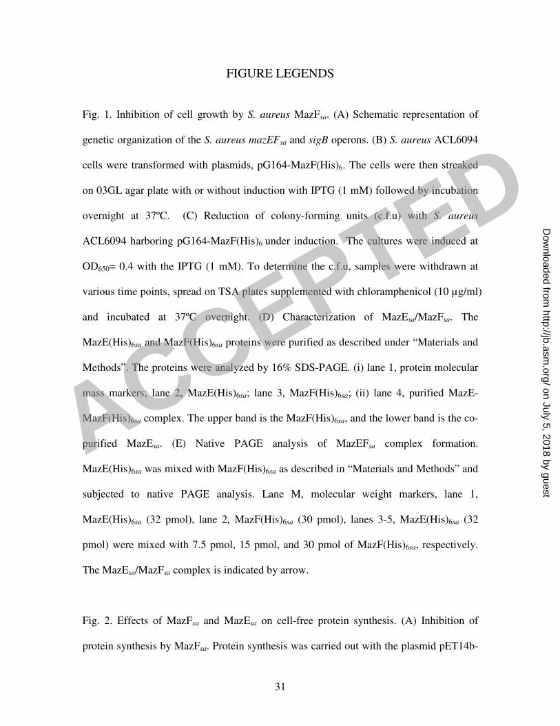
Fig. 1. Inhibition of cell growth by S. aureus MazFsa. (A) Schematic representation of
genetic organization of the S. aureus mazEFsa and sigB operons. (B) S. aureus ACL6094
cells were transformed with plasmids, pG164-MazF(His)6. The cells were then streaked
on 03GL agar plate with or without induction with IPTG (1 mM) followed by incubation
overnight at 37ºC. (C) Reduction of colony-forming units (c.f.u) with S. aureus
ACL6094 harboring pG164-MazF(His)6 under induction. The cultures were induced at
OD650= 0.4 with the IPTG (1 mM). To determine the c.f.u, samples were withdrawn at
various time points, spread on TSA plates supplemented with chloramphenicol (10 µg/ml)
and incubated at 37ºC overnight. (D) Characterization of MazEsa/MazFsa. The
MazE(His)6sa and MazF(His)6sa proteins were purified as described under “Materials and
Methods”. The proteins were analyzed by 16% SDS-PAGE. (i) lane 1, protein molecular
mass markers; lane 2, MazE(His)6sa; lane 3, MazF(His)6sa; (ii) lane 4, purified MazE-
MazF(His)6sa complex. The upper band is the MazF(His)6sa, and the lower band is the co-
purified MazEsa. (E) Native PAGE analysis of MazEFsa complex formation.
MazE(His)6sa was mixed with MazF(His)6sa as described in “Materials and Methods” and
subjected to native PAGE analysis. Lane M, molecular weight markers, lane 1,
MazE(His)6sa (32 pmol), lane 2, MazF(His)6sa (30 pmol), lanes 3-5, MazE(His)6sa (32
pmol) were mixed with 7.5 pmol, 15 pmol, and 30 pmol of MazF(His)6sa, respectively.
The MazEsa/MazFsa complex is indicated by arrow.
Fig. 2. Effects of MazFsa and MazEsa on cell-free protein synthesis. (A) Inhibition of
protein synthesis by MazFsa. Protein synthesis was carried out with the plasmid pET14b-
ACCEPTED
on July 5, 2018 by guesthttp://jb.asm
.org/D
ownloaded from
32
ctpA in E. coli T7 S30 extract system at 37ºC for 1 hr. Lane 1, control without MazFsa;
lanes 2-6, varying amounts of MazFsa were added at 3.75 pmol, 7.5 pmol, 11.25 pmol, 15
pmol, and 30 pmol, respectively. (B) Protein synthesis was rescued by the addition of
MazEsa. Lane 1, control without MazFsa; lane 2, 15 pmol of MazFsa; lanes 3-6, 8 pmol,
16 pmol, 32 pmol, and 64 pmol of MazEsa were added together with 15 pmol of MazFsa,
respectively. The synthesis of ß-lactamase and CtpA was indicated by arrows.
Fig. 3. Endoribonuclease activity of MazFsa. Five µg of ctpA mRNA were digested with
15 pmol of MazFsa as described in “Materials and Methods”. (A) Cleavage of ctpA
mRNA by MazFsa. Lane 1, control without MazFsa, lanes 2-4, mRNA substrates were
digested for 60 min, 90 min, and 120 min, respectively. (B) Inhibition of cleavage with
the addition of MazEsa. Lane 1, mRNA with MazFsa, lanes 2-4, mRNA substrates
digested by MazFsa together with 4 pmol, 8 pmol, and 16 pmol of MazEsa, lane 5, mRNA
with 32 pmol of MazEsa.
Fig. 4. In vitro primer extension analysis of the MazFsa cleavage sites in the ctpA mRNA.
Primer extension was carried out as described in “Materials and Methods”. Each primer
extension product was analyzed on a 6% sequencing gel with the DNA sequencing ladder
prepared with the same primer. (A) and (B) Cleavage sites in ctpA mRNA detected with
primer pEa. (C) and (D) Cleavage sites in ctpA mRNA detected with primer pEb. The
RNA sequences complementary to the DNA sequence ladder are shown to the right of
each figure and corresponding cleavage sites are indicated by arrows.
ACCEPTED
on July 5, 2018 by guesthttp://jb.asm
.org/D
ownloaded from
33
Fig. 5. Cleavage of synthetic RNA by MazFsa. All RNA substrates labeled at the 5’-end
with [γ-32
P]ATP were digested with MazFsa and subjected to analysis in a 20%
sequencing gel running with the RNA ladder by alkaline hydrolysis as described in
“Materials and Methods”. (A) Cleavage of a synthetic 18-base RNA, AUUC. Lane 1,
RNA ladder generated by alkaline hydrolysis; lane 2, RNA substrate without the addition
of MazFsa; lane 3, RNA substrate digested by15 pmol of MazFsa. The corresponding
RNA sequence is shown to the right. The cleavage product and site were indicated by
arrows. (B) Cleavage specificity of MazFsa with synthetic 18-base RNA substrates. Seven
RNA substrates with the center AUUC sequence were changed to AGUC, AUGC,
GUUG, UUUC, AUUG, GUUC, and AUUU sequences and named correspondingly. The
ATTC indicated the same length of DNA substrate. Lanes 1, 3, 5, 7, 9, 11, 13, 15, and 17
with no MazFsa. Lanes 2, 4, 6, 8, 10, 12, 14, 16 and 18 with 15 pmol of MazFsa added.
The background in the absence of MazF was attributed to impurities or incomplete
synthesis of the full length RNA substrates. Nevertheless, cleavage can be seen with
MazFsa, as indicated by arrows. (C) Predicted secondary structure formed by RB-1 (5’-
UGCAAUUCAUAUGAAUUGU-3’) using the RNA secondary prediction website
(http://www.genebee.msu.su/services/rna2_reduced.html). (D) Cleavage of highly
purified synthetic RNA substrates with different secondary structures. An 18-base sense
RNA, AUUC, and AUUC antisense RNA RB-3, were digested separately by MazFsa. The
19-base RNAs, RB-1 and RB-2 (RB-1 variant), were also digested with MazFsa. Lanes 2,
4, 6, and 8 with no MazFsa addition. Lanes 3, 5, 7, and 9 with 15 pmol of MazFsa added.
The cleavage products are indicated by arrows. (E) Effects of the RNA-RNA duplex
formation on cleavage by MazFsa. Lane 1, RNA ladder generated by alkaline hydrolysis;
ACCEPTED
on July 5, 2018 by guesthttp://jb.asm
.org/D
ownloaded from
34
lane 2, labeled sense RNA alone; lane 3, labeled sense RNA digested with 15 pmol of
MazFsa; lanes 4-7, the labeled 18-base sense RNA, AUUC, was annealed with AUUC
antisense RNA RB-3, in ratios of 1: 0.2, 1:0.4, 1:0.8, and 1:1 as indicated and then
digested with 15 pmol of MazFsa at 37ºC for 30 min.
Fig. 6. In vivo cleavage of the ctpA mRNA after induction for MazFsa expression. The
expression of MazFsa in E. coli BL21(DE3)pLysS carrying plasmids, pET14b-ctpA and
pBAD-MazF(His)6, was induced by arabinose (0.2%) (A). The expression of MazFsa in S.
aureus 178RI carrying plasmid, pG164-MazF(His)6/ctpA was induced by 1 mM IPTG
(B). Total cellular RNAs were extracted at the indicated time points. Primer extension
was then carried out as described in “Materials and Methods”. The DNA sequencing
ladder was prepared using the same primer. The cleavage sites in the ctpA mRNA were
indicated by arrows. (A) and (B) represent in vivo primer extension with primer pEa from
E. coli and S. aureus, respectively.
Fig. 7. Doxycycline (DOX) stress induced the increasing expression of mazEFsa
transcripts. The S. aureus Newman culture was treated with 50 ng/ml DOX at OD650 =
1.0. Total cellular RNAs were extracted at the indicated time points. Northern blot
analysis was carried out with the mazEFsa probe as described in “Materials and Methods”.
(A) Transcripts detected with the probe. (B) 23S and 16S served as the internal loading
control.
ACCEPTED
on July 5, 2018 by guesthttp://jb.asm
.org/D
ownloaded from


![Zhibiao Zhao arXiv:0801.1599v2 [q-fin.ST] 20 Mar 2008 · Parametric and nonparametric models and methods in financial econometrics∗,† Zhibiao Zhao Department of Statistics Pennsylvania](https://img.pdfslide.us/doc/110x75/5eb5ad6f8eb1025587244fa3/zhibiao-zhao-arxiv08011599v2-q-finst-20-mar-2008-parametric-and-nonparametric.jpg)