Embed Size (px)
Citation preview

AN ABSTRACT OF THE THESIS OF
OMAR AHMED ARAKJI for the DOCTOR OF PHILOSOPHY (Name) (Degree)
in Food Science and Technology presented on January 22, 1968 (Major) (Date)
Title: THE ISOLATION AND CHARACTERIZATION OF THE PECTIC
ENZYMES OF THE MC FARLIN CRANBERRY
Abstract approved: __JI Dr. H. Y/Yang 7"
Cranberries are processed mainly for the production of jelly,
sauce and juice. The content of pectic substances in the cranberry
gives the fruit a desirable property for processing such products.
Pectic enzymes which catalyze the hydrolysis of pectic substances
may affect the consistency of these products.
The concern of this study was to isolate and characterize the
pectic enzymes that may be found in the cranberry fruit. The
methods utilized in the preparation of enzyme extracts were by (1)
the preparation of acetone powder (2) the preparation of acetone
powder in the presence of polyethylene glycol and (3) by extraction
in the presence of polyvinylpyrr olid one. The following conclusions
were made:
(1) Cranberry protein extracts were found to exhibit poly-
galacturonase activity.
(2) The cranberry polygalacturonase extract obtained by the
use of polyethylene glycol exhibited a 40. 3 percent loss in
viscosity over one percent pectave solution in citrate buffer
at pH 5. 0 and 300C during the initial hour of the reaction.
A 22. 1 and 8. 6 percent loss in viscosity was found when
the polyvinylpyrrolidone extract and the acetone powder
extract were used as the source of enzyme respectively.
(3) Cranberry polygalacturonase may be classified as an
endo- type polygalacturonase which catalyzes a random
hydrolysis of both low and high methoxyl pectic substances.
(4) Maximum activity of the cranberry polygalacturonase was
found to be at pH 5. 0.
(5) Sodium chloride concentration up to 0. 6 M showed no
significant effect on the polygalacturonase activity in a
citrate buffer at pH 5.0.
(6) The cranberry polygalacturonase was inactivated when
exposed to 100oC for 35 minutes at pH 5.0.
(7) Cranberry proteins were found to possess low pectin
esterase activity. Optimum activity of cranberry pectin
esterase occurred at pH 7. 5 with sodium chloride concen-
tration at 0. 15M.
(8) Cranberry pectin esterase was inactivated when exposed
to 100°C for five minutes.

The Isolation and Characterization of the Pectic Enzymes of the McFarlin Cranberry
by
Omar Ahmed Arakji
A THESIS
submitted to
Oregon State University
in partial fulfillment of the requirements for the
degree of
Doctor of Philosophy
June 1968

APPROVED:
Associate Professor of Food $cien6e and^Technology in charge of major
Head of Department of Food Sci%m:e and Technology
Dean of Graduate School
Date thesis is presented January 22, 1968
Typed by Gwendolyn Hans en for Omar Ahmed Arakji

ACKNOWLEDGMENT
The author wishes to thank Dr. Hoya Y. Yang for his help and
guidance during the course of this study and the preparation of this
thesis.
Sincere appreciation is expressed to Dr. Harold W. Schultz,
Dr. Robert F. Cain and Dr. Morris W. Montgomery for their valuable
suggestions and encouragement.
Acknowledgment is due to Ocean Spray Cranberries, Inc. for
supporting this study by funds and supply of cranberries.
Thanks are also extended to Miss Sherry Sheets for typing the
draft copies of this thesis.
Last but not least, the continuous encouragement of my parents,
understanding and patience of my wife are greatly acknowledged.
TABLE OF CONTENTS
Page
INTRODUCTION 1
LITERATURE REVIEW 3
Pectic Substances 3 Chemical Constitution 3 Nomenclature 5
Pectic Substances 5 Protopectin 5 Pectinic Acids 6 Pectin 6 Pectic Acid 6
Classification of Pectic Enzymes 6 Polygalacturonases (PCs) 8
Mechanism of Action and Specificity 8 Endopolymethylgalacturonase (Endo-PMG) 10 Endopolygalacturonase (Endo-PG) 10 Exopolymethylgalacturonase (Exo-PMG) 11 Exopolygalacturonase (Exo-PG) 11
Preparation and Purification 12 Methods of Determination 13 Effect of External Factors on PG Activity 14
Effect of pH 14 Effect of Temperature 15 Activation, Inhibition and Inactivation 15
Occurence of Polygalacturonase 17 Previous Work on Cranberry Polygalacturonase 17
Pectin Esterase 18 Action and Specifity 18 Determination of Pectin Esterase Activity 19 Preparation and Purification 19 Effects of External Factors on Activity 20
Effect of pH 20 Effect of Temperature 20 Activation, Inhibition, and Inactivation 21
Occurance of Pectin Esterase 22 Pectin Transeliminase (PTE) 23 Protopectinase 24
The Role and Importance of Pectic Enzymes on the Texture of Fruits and Vegetables 25

Page
MATERIALS AND METHODS 29
Source of the Fruit 29 Extraction and Preparation of Cranberry Pectins 30
Preparation of Alcohol Insoluble Solids (AJS) 30 Isolation of the Water Soluble Pectins 30 Isolation of Sodium Hexametaphosphate (Calgon) Soluble Pectins 31
Methoxyl Content and Equivalent Weight of Pectic Substances 32 Equivalent Weight of Pectic Substances 32 Methoxyl Content of Pectic Substances 33 Preparation of the Insoluble Residue of the Alcohol Insoluble Solids 33
Extraction and Preparation of Cranberry Proteins 34 Preparation of Acetone Powder 34 Preparation of Acetone Powder in Presence of Polyethylene Glycol (PEG) 34
Preparation of Cranberry Proteins in Presence of Polyvinyl Pyrrolidone (PVP) 35
Precipitation and Dialysis of Cranberry Proteins 35 Nitrogen Determinations of Cranberry Protein Extracts 36
Viscometric Determinations to Characterize Cranberry Polygalacturonase 37
Determination of pH Optimum for the Polygalacturonase Activity 38
Effect of Cranberry PG on Different Pectic Substances 38 Effect of Salt Concentration on PG Activity 39 Effect of Heat on Polygalacturonase Activity 39
Action of Cranberry Proteins on the Insoluble Residue of the Cranberry AIS 40
Determination of Pectin Esterase in Cranberries 40 Preparation of Cranberry PE 41 Determination of the Optimum pH 41 Effect of Salt Concentration on PE Activity 42
Transeliminase Activity of Cranberry Proteins 42 Fractionation of Cranberry Proteins on DEAE Cellulose Column Chromatography 43
RESULTS AND DISCUSSION 45
Analysis of Substrates 45 Extraction, Precipitation and Dialysis of Cranberry Proteins 46

Page
Determination of Cranberry Polygalacturonase Activity 47 Effect of Extraction Procedure on PG Activity 47 pH Optimum for the Cranberry Polygalacturonase Activity 51
Effect of Salt Concentration on Polygalacturonase Activity 51
Polygalacturonase Activity of Cranberry Proteins on Different Pectic Substances 53
Effect of Cranberry Protein Concentration on Polygalacturonase Activity 60
Heat Inactivation of Cranberry Polygalacturonase 60 Chromatographic Separation of Cranberry Proteins on DEAE Cellulose Column 65
Effect of Cranberry Proteins on the Insoluble Residue of Cranberries and Alcohol Insoluble Solids 68
Pectin Transeliminase in Cranberry Proteins 70 Pectin Esterase in the Cranberry 70
pH Optimum of Pectin Esterase 70 Pectin Esterase Activity 72 Influence of Salt on Pectin Esterase Activity 73 Heat Inactivation of Pectin Esterase 75
DISCUSSION 77
SUMMARY AND CONCLUSION 80
BIBLIOGRAPHY 82

LIST OF TABLES
Table Page
1. Methoxyl content and equivalent weight values of citrus and cranberry pectic substances 46
2. Action of cranberry enzyme extracts at pH 5. 0 and 30CC. One percent sodium polypectate was used as substrate (means of five determinations). 49
3. Effect of NaCl Cone, on the Pectinolytic Activity of Cranberry Proteins (means of five determinations) 53
4. Percent drop in viscosity of one percent substrate solutions after 20 hrs. at 30oC. 59
5. Effect of heat on cranberry polygalacturonase 62
6. The pectinolytic activity of DEAE cellulose column chromatography fractions in one percent sodium polypectate at pH 5.0 and 300C 68
7. Effect of cranberry proteins on the insoluble residue of the AIS 69
8. Effect of pH on the activity of cranberry pectin esterase 72
9. Effect of NaCl on cranberry pectin esterase activity at pH 7. 5 73
10. Influence of heat on cranberry pectin esterase 76

LIST OF FIGURES
Figure Page
1. Schematic diagram of partially esterified poly- galacturonic acid. 4
2. Mode of action of pectic enzymes. 9
3. Mechanism of pectintranseliminase action. 24
4. Percent loss in viscosity of one percent sodium poly- pectate solution at pH 5.0 and 30oC, using cranberry polygalacturonase prepared by three different methods. 50
5. Effect of pH on cranberry polygalacturonase activity on sodium polypectate at 30"C. 52
6. Percent loss in viscosity, of one percent sodium polypectate solution at different pH and 30°C. 52
7. Change in viscosity of sodium polypectate (Sunkist 6042) solution due to the action of cranberry poly- galacturonase at pH 5.0 and 30oC. 55
8. Change in viscosity of citrus pectin (Sunkist 3442) due to the action of cranberry polygalacturonase at pH 5.0 and 30oC. 56
9. Change in viscosity of cranberry water soluble pectin due to the action of cranberry poly- galacturonase at pH 5. 0 and 300C. 57
10. Change in viscosity of cranberry calgon soluble pectin due to the action of cranberry polygalacturonase at pH 5.0 and 30°C. 58
11. The relation between cranberry protein concentration and its polygalacturonase activity on one percent citrus polypectate solution at pH 5.0 and 30oC. 61
12. The relation between different enzyme concentrations of cranberry polygalacturonase and their initial rate of activity on one percent polypectate solution at pH 5. 0 and 30 0C. 63

Figure Page
13. Thermal death rate curve for the cranberry PG activity at pH 5. 0 on sodium polypectate. 64
14. Chromatography of cranberry protein dialysate on DEAE cellulose column and the polygalacturonase activity of the fractions at pH 5. 0 and 30°C. 66
15. Sodium chloride concentration in eluates from DEAE cellulose column. 67
16. Activity of PE of cranberries at different pH values, using citrus pectin as substrate. 71
17. Cranberry PE activity on one percent citrus pectin solution (0. 15M NaCl) at pH 7. 5 and 30oC. 74

THE ISOLATION AND CHARACTERIZATION OF THE PECTIC ENZYMES OF THE MC FARLIN CRANBERRY
INTRODUCTION
The texture of fresh and processed fruits and vegetables is of
considerable value to the food processor and the consumer. The
presence of pectic substances in the plant cell plays an important
role in maintaining rigidity to plant tissues and a certain consistency
to fruit purees and juices. Pectic enzymes act on the polymer con-
stituting the pectin molecule to produce smaller fragments, thus
disrupting the rigidity of the tissue or the consistency of the juice or
puree. These enzymes are called polygalacturonase, pectic esterase,
and pectin transeliminase. They may be inherent in the plant tissue
or may be produced by different types of microorganisms. The
desirability of the cranberry fruit lies in its high content of pectin,
which facilitates the production of cranberry sauce and jelly. Pectic
enzymes from the cranberry could bring about a catalytic effect on
the hydrolysis of the pectins present. As a result, the nature of the
product will be affected.
In spite of the undesirable action of pectic enzymes on the
texture of processed plant tissue, this same property is of beneficial
use in the preparation of fruit juices. The pectic enzymes may be
added to pureed fruits to facilitate the expression of a higher yield

of juice. This is accomplished by the ability of the enzymes to act
on the middle lamella of the fruit cell to cause the disruption of the
cell wall. Pectic enzymes are also used to clarify wines and fruit
juices.
This study was initiated to determine and characterize the
nature of the pectic enzymes of the McFarlin cranberries. The study
involves extraction of the enzymes by different methods followed by
determining their activity under different conditions.

LITERATURE REVIEW
The following discussion presents a review about pectic enzymes
and their distribution in nature. A general classification of the
enzymes, their properties, nnode of action, methods of preparation,
and determination will be included. The review will also include
a brief presentation on the chemical constitution of the substrates,
the pectic substances, as well as definitions of their various types.
Pectic Substances
Chemical Constitution
Pectic substances are polymers whose major units are made of
galacturonic acids, linked by a-1, 4-glycosidic linkages. Approxi-
mately two thirds of the carboxylic acid groups on the polymer are
esterified with methanol (Reed, 1966). Rhamnose, arabinose,
galactose, and traces of other sugars are found to be accompanying
the pectin molecule or linked as side groups to the main chain
(McCready and Gee, I960; Jansen et al. , 1949). However, enzymic
analysis using partially reduced pectins indicated that the main chain
probably does not contain sugars other than anhydrogalacturonic
acid (Solms and Deuel, 1955).

— o
OH COOH
Figure 1. Schematic diagram of partially esterified polygalacturonic acid.
The degree of esterification represents the number of
esterified carboxyl groups. When all carboxyl groups in pure
polygalacturonic acids are all esterified, the methoxyl content is
16. 32 percent and the degree of esterification is 100 percent
(Doesburg, 1965).
Measurements of the molecular weights of pectic substances is
made by osmotic pressure determinations, viscometric measure-
ments and sedimentation velocity. The molecular weights from
different sources range from 10,000 to 20,000 (Deuel and Stutz, 1958;
Joslyn, 1962).
Both hydroxyl groups on carbon atoms 1 and 4 of the D-
galacturonic acid molecule are in axial position making the pectin
molecule atrans-a-1, 4-polysaccharide (Eliezen and Hyman, 1957),
thereby hindering the free rotation at the glycosidic linkages and
restricting flexibility. On the other hand, the secondary hydroxyl
groups on carbon atoms 2 and 3, and the carboxyl group on carbon
5 are in equatorial position, making them easily accessible (Deuel

and Stutz, 1958). Historical and recent developments in extracting
and characterizing the properties of pectic substances are reviewed
by Kertesz (1952), Duel and Stutz (1958), Doesberg (1965), and
Joslyn (1962).
Nomenclature
Due to past irregularity in the nomenclature of pectic sub-
stances, in 1944 the American Chemical Society adopted the follow-
ing definitions (Baker et al. , 1944).
Pectic Substances
Pectic substances is a group designation for those complex
colloidal carbohydrate derivatives which occur in, or are prepared
from, plants and contain a large proportion of anhydrogalacturonic
acid units which are thought to exist in a chain-like combination. The
carboxyl groups of polygalacturonic acids may be partly esterified
by methyl groups and partly or completely neutralized by one or
more bases.
Protopectin
The term protopectin is applied to the water-insoluble parent
pectic substance which occurs in plants and which upon restricted
hydrolysis, yields pectinic acids.

Pectinic Acids
The term pectinic acids is used for colloidal polygalacturonic
acids containing more than a negligible proportion of methyl ester
groups. Pectinic acids, under suitable conditions, are capable of
forming gels (jellies) with sugar and acid or, if suitably low in
methoxyl content, with certain metalic ions. The salts of pectinic
acids are either normal or acid pectinates.
Pectin
The general term pectin (or pectins) designates those water
soluble pectinic acids of varying methyl ester content and degree of
neutralization which are capable of forming gels with sugar and acid
under suitable conditions.
Pectic Acid
The term pectic acid is applied to pectic substances mostly
composed of colloidal polygalacturonic acids and essentially free
from methyl ester groups. The salts of pectic acid are either nor-
mal or acid pectates.
Classification of Pectic Enzymes
The importance of pectic enzymes in the food industry is of
considerable value. In the limited observations on the formation of

pectic substances derived through the incorporation of various
isotope-tagged compounds and groupings into pectins, Kertesz (I960)
stated that we know little about the mechanism of pectin formation
in plants and nothing about the enzymes responsible for their
synthesis, especially those which partake in polymer formation.
However, the information available concerning the enzymes which
act on pectic substances is large. Demain and Phaff (1957), and
Deuel and Stutz (1958) reviewed the literature of these enzymes.
The two main groups of pectic enzymes have been described.
1. Depolymerizing enzymes which split the a-1, 4-glycosidic
bonds of pectic acid and pectinic acid. Enzymes respon-
sible for hydrolyzing these bonds are referred to as poly-
galacturonases (PGs). These enzymes belong to the group
called hydrolases because of their mechanism of action.
Another enzyme causing depolymerization of pectic sub-
stances is called transeliminase. This enzyme is a
depolymerase yet not a hydrolase. The mechanism of its
action will be discussed under the heading Pectin
Trans eliminase.
2. The pectinesterases which act on pectins and pectinic acids
by decreasing their degree of esterification and liberating
equivalent amount of methanol and free carboxyl groups
(Doesberg, 1965), The enzyme protopectinase is thought

to be responsible for the solubilization of the water
insoluble pectic substance, the protopectin. However,
there is no adequate indicative proof for the existence of
protopectinase as a distinct enzyme. This will be dis-
cussed further under the heading Protopectinase.
Polygalacturonases (PGs)
Synonyms used in the literature include pectinase, pectolase,
pectin glycosidase, and pectin depolymerase.
Mechanism of Action and Specificity
Polygalacturonases are regarded as hydrolases. A double
replacement mechanism for the reaction is thought to be operative.
According to this mechanism, the substrate is first attacked by a
nucleophilic group of the enzyme , and subsequently the enzyme-
substrate intermediate is attacked by the nucleophilic water
molecules (Deuel and Stutz, 1958).
Many observations show that different polygalacturonases
were found to act differently on pectic substances with different
degrees of methylation and polymerization, and the extent of
hydrolysis differ from one polygalacturonase to the other. Demain
and Phaff (1957) based their classification of polygalacturonases
on the substrate attacked, and the optimum pH values of the different

PCs. The latter criteria is not a popular one and is used only to a
small extent. From the former criteria, it had been found that most
of the pectic enzymes prefer either pectin or pectic acid as sub-
strates (Demain and Phaff, 195 7). The following schematic
diagram suggests the possibilities of pectic substances break down
by various types of polygalacturonases (Demain and Phaff, 1957).
Exo-PMG
^—Pectin- PE
Endo-PMG Endo-PG/
-^—Pectic acid 5»—
Intermediate uronides
Digalacturonic acid
Galacturonic acid
Exo-PG
Figure 2. Mode of action of pectic enzymes
Random degradation of the pectin polymer is the most common
type of breakdown encountered. However, a terminal mechanism has
also been found during which galacturonic acid is liberated at the
start of the reaction. A random type split is characterized by rapid
lowering of viscosity and a slow increase in reducing power, while
terminal attack causes very slow viscosity changes. The prefix
"endo" has been applied to those enzymes carrying out random
hydrolysis since the numerous bonds of the inner part of the chain
are broken in preference to the few terminal linkages. Similarly
"exo" refers to the enzymes causing terminal hydrolysis (Demain
and Phaff, 1957). The following,groups of polygalacturonases were

10
described by Demain and Phaff (1957):
Endopolymethylgalacturonase (Endo-PMG). This enzyme
attacks pectin without previous deesterification, however, it is
incapable of complete hydrolysis, not more than 26% of the total
glycosidic bonds (Joslyn, 1962). As a result of this enzymatic
hydrolysis, the hydrolysate probably contains uronides of 4 or 5
residues per molecule.
On the basis of optimum pH values of the enzyme, it was found
that endo-PMG is of two different types, one that causes partial
hydrolysis of pectins at pH 5.0 - 6. 0 and called endo-PMG I, and the
other causes the hydrolysis of pectin at pH 8.0 - 9.0 and referred
to as endo-PMG II.
Endopolygalacturonase (Endo-PG). This enzyme carries out
a random hydrolysis of pectic acid to the 70 percent level resulting
in a mixture of digalacturonic acid and galacturonic acids (Demain
and Phaff, 1957). Endo (1964 a, b, c) purified endopolygalacturonase
from Coniothyrium diplodiella by ultra-centrifugation and free
boundary electrophoresis. Three types of endo-polygalacturonases
were identified (I, II, III). They, differ by their degree of hydrolysis
of pectic acids as well as their sedimentation coefficients, 2. 68,
3. 35, and 3. 05 respectively. Optimum pH range was found to be
between four and six. The action of endopolygalacturonase is very
limited. The enzyme hydrolyzes pectic acid at least 5000 times as

11
fast as it does starch, maltose, sucrose, and carboxymethyl-
cellulose (Ldneweaver et al. , 1949).
Exopolymethylgalacturonase (Exo-PMG). This enzynne
hydrolyzes pectic substances only from one end of the chain molecule.
They preferentially attack pectins with high degree of methylation
(Deuel and Stutz, 1958). The breakdown of the pectin was found to
go to completion. Matus (as cited by Demain and Phaff, 195 7) in a
study to differentiate between the two enzymes of fungal origin that
hydrolyzed pectic acids and pectins from one end of the chain, found
that as the degree of esterification was increased, the ratio of the
enzymatic activity, in terms of liberated reducing groups to
enzymatic activity in terms of viscosity also increased. The highly
esterified compounds appeared to be attacked from the one end of
the chain while random hydrolysis was the mechanism of pectic
acid attack.
Exopolygalacturonase (Exo-PG). Compared to Exo-PMG, this
enzyme attacks the pectic acid polymers and causes hydrolysis from
the terminal end of the chain. Hydrolysis goes to completion by the
liberation of galacturonic acid. Obtaining the enzyme from
Aspergillus niger, Saito (1955) found evidence of the terminal attack
by both paper chromatography studies as well as by comparing results
of reducing group liberation and viscosity reduction. Endo (1964)
extracting the enzyme from Coniothyrium diplodiella and purifying it

12
on DEAE cellulose and by column zone electrophoresis, found that
it was most active in the pH range of 4. 0 - 4. 5 and that it was stable
between the pH values of 3. 0 and 6. 0'. Effect of temperature on
exopolygalacturonase revealed that the enzyme was stable up to 550C
after which the activity te/nded to decrease. Endo (1964) also found
that pectin was little affected by the same enzyme.
Preparation and Purification
Most of the polygalacturonases prepared for commercial use
are obtained from microorganisms, mainly molds. Kertesz (1955)
discussed the methods employed in the preparation and partial
purification of these enzymes. However most of these preparations
contained a mixture of polygalacturonases as well as pectinesterase.
Pectinesterase can be inactivated from these preparations by allow-
ing the crude enzyme extract to stand at pH 3. 0 then adjusting the
pH value to five. Ion exchange methods may also be used (Kertesz,
1951). Endo (1963) using column chromatography was able to
fractionate different polygalacturonasesobtained from Coniothyrium
diplodiella.
Stahmann (1963) reviewed several factors that make plant
proteins unstable and difficult to deal with. The isolation of enzymes
from plant tissue causes the dessembling of cellular components
which brings about several chemical changes. Some plant tissues

13
contain a high concentration of tannins and that these polyphenols
complex with proteins and cause their precipitation (Stahmann, 1963).
Loomis and Battaile (1966) reviewed the various problems
caused by the interaction of plant phenolic compounds and proteins.
Phenols combine with proteins in two fundamentally different ways,
reversibly by hydrogen bonding, and irreversibly by oxidation of
quinones followed by covalent condensations of the quinones with
reactive groups on the protein molecule. Loomis (1968) discussed
the principles of enzyme isolation from phenol containing tissues by
the use of phenol complexing agents namely polyvinylpyrrolidone and
polyethylene glycol. Any means of binding tannins (flavolans) and the
higher molecular weight polyphenols, is considered an important
extraction aid in isolating enzymes from plants containing these sub-
stances (Badran and Jones, 1965).
Methods of Determination
PG activity was formerly measured by determining the
amount of substrate left unchanged. This method involves the
precipitation and determination of the remaining polymers of the
pectic substance. The drawbacks of this procedure lie in the time
consuming nature of the steps involved.
The two methods most widely used in determining the activity
of polygalacturonases employ (1) the measurements of reducing

14
groups found and (2) the change in physical properties. Poly-
galacturonase catalyzes the hydrolysis of the a-1, 4-glycosidic
linkages in pectic polyurosides. Every fissure of such a bondage
produces a reducing group which was previously engaged in the for-
mation of the polymer. The hypiodite method described by Willstatter
and Scherdel is used for this determination (Jansen and MacDonnell,
1945).
Viscosity changes have been extensively used in the determina-
tion of pectin-polygalacturonase activity. After about one fourth of
all glycosidic linkages have been hydrolyzed, the viscosity scarcely
changes. The major viscosity changes occur very early in the
reaction. For this reason, the measurements of viscosity changes
is a very sensitive indication of even traces of pectin poly-
galacturonase action (Kertesz, 1951). A random type split of the
a-1, 4-glycosidic bondage is characterized by a rapid lowering of
viscosity and a slow increase in reducing power, while terminal
attack causes very slow viscosity changes, although reducing group
formation may take place at the very same ratio (Demain and Phaff,
1957).
Effect of External Factors on PG Activity
Effect of pH. pH values for the different polygalacturonase
have been used by Demain and Phaff (1957) as a minor criterion to

15
differentiate them. The literature contains data which indicates
optimum ranges from pH 3.0 to 5.0 (Kertesz, 1951). However a few
extremes have been reported for polygalacturonases extracted from
several types of bacteria which possess optimum activity at pH values
between 7. 0 and 8. 0, while that of Penicillium chrysoqenum is
rapidly inactivated at such pH values (Kertesz, 1951).
Effect of Temperature. The temperature optimum of poly-
galacturonase is greatly dependent on experimental conditions,
especially the length of heating time. When short periods of heating
are used the temperature optimum appears to be much higher than
when longer periods of exposure are applied (Kertesz, 1951).
Tomatoes may contain an unusually heat resistant pectinolytic factor,
with 20 percent of the original activity remaining after a heat treat-
ment of one hour at 100°C in extracts saturated with salt (McCollach
and Kertesz, 1948). Fungal pectinases in the form of dry powder
are quite resistant to heat (Kertesz, 1951). Purified poly-
galacturonases of fungal sources were found to be somewhat more
sensitive to temperature than corresponding crude preparations.
Optimum temperatures are in the range of 45 to 55"C (Rahmann and
Joslyn, 1953),
Activation, Inhibition and Inactivation. Because of the known
activating effect of alkali chlorides on the deesterifying enzyme, it
is difficult to say whether the observed activation of pectin

16
polygalacturonase action as reported by Pallmann (cited by Kertesz,
1951) was direct or caused by enhancement of the simultaneous
deesterification which in turn facilitated pectin polygalacturonase
action. Since there are several complicating factors not entirely
eliminated in the above studies, as well as complications that may
arise by the effect of the cations on the substrate, these reports
should be regarded with reservation (Kertesz, 1951; Schubert, 1954).
Rahman and Joslyn (1953) studied the inhibitory effect of
mercurous chloride, sodium floride, iodoacetic acid, and sulfur
dioxide on polygalacturonase. No apparent inhibition was found.
Alkali sulfonate was found to be an effective inhibitor for polygalac-
turonase in Pectinol M added to cherries at pH 3. 0 to 4. 0 (Yang _et al. ,
I960; Steele and Yang, I960). The inactivation effect of the
detergent alkali sulfonate is much less effective on polygalacturonase
than on pectin methyl esterase (Kertesz, 1951).
Hathaway and Seakins (1958) using viscometric measurements
showed that tannins exert a powerful inactivation on the early stages
of pectinolysis. Also, a pectinase inhibition was found in sweet
potatoes and grape leaves by Uritani and Stahmann (1961), and by
Bell and Etchells (1958) respectively. The inhibiting substance was
stable to heat, nondialyzable and could not be completely precipitated
with acetone or concentrated ammonium sulfate. Later work revealed
that the inhibitor is a tannin-like substance (Porter et al. , 1961).

17
Exposure of polygalacturonase to high acidity as well as low
acidity decreases its activity. At pH 0. 6 and 250C a solution of poly-
galacturonase loses 90 percent of its activity in 20 minutes. When
present in 0. 5N NaOH, the enzyme was completely inactivated at
20oC in 40 minutes. (Kertesz, 1951).
Occurence of Polygalacturonase
Polygalacturonases, of the various types classified earlier,
are found in fungi, yeasts, bacteria, and plants (Reed, 1966). The
tomato and avacado polygalacturonases were found to have their
activity qualitatively similar to that of fungal preparations. Plant
polygalacturonases have been studied and found in cucumber vines
(Bell et al. , 1955). The enzyme was also found in beans, radishes,
carrots, and cherries by Konovalova and by Ozawa (as cited by
Reed, 1966).
Previous Work on Cranberry Polygalacturonase
Hobson (1962) extracted the proteins from cranberries with
2.25 grams of a 10:1 (w/w) mixture of sodium chloried and disodium
salt of ethylene diamine tetra acetic acid. The substrate used for
the polygalacturonase assay was Wichmann pectic acid (Newbold and
Joslyn, 1952). The procedure used was the colorimetric method
based on that of Willaman and Davidson (1924). No activity of the
18
enzyme was detected. Patterson et al. (1967) in studying the effect
of bruising a cranberry, found that softening results from endogenous
enzymatic degradation of pectic substances in the cell wall. No
enzymatic activity was found in unbruised tissues. The method
employed was the cup-plate method developed by Dingle et jal. (1953).
Pectin Esterase
Action and Specifity
This enzyme catalyzes the hydrolysis of the methyl ester groups
of pectinic acids and pectin liberating equivalent amounts of methanol
(Demain and Phaff, 1957). PE is a highly specific enzyme which
saponifies almost exclusively the methyl ester groups of pectic sub-
stances (Deuel and Stutz, 1958). Matus, as cited by Demain and
Phaff (1957), in order to characterize the different polygalacturonases,
used glycol esters of pectic acid to make sure that PE does not effect
the structure of the substrate because of its inability to deesterify
glycol esters. MacDonnell et al. (1950) found that methyl esters of
polymanuronic acid were not attacked. The hydrolysis of methyl
esters of pectic acids proceeds linearly along the chain molecule as
successive methoxyl groups are split off (Schultz et al. , 1945; Solms
and Deuel, 1954).

19
Determination of Pectin Esterase Activity
The two methods that are used to determine the activity of
pectin esterase make use of either the methanol liberated or the
carboxyl group exposed upon the deesterification of the methylester
on the pectin molecule. Determining the amount of methanol
liberated is a laborious procedure. The methanol should be distilled
and measured either colorimetrically or by gas liquid chromato-
graphy. For every methanol molecule liberated one carboxylic
group will become available. The activity of pectin methyl esterase
is conventionally determined by measuring the commensurate increase
in acidity. This is usually accomplished by continuous titration with
a standard alkali while maintaining the pH of the substrate-enzyme
mixture at a constant value (Somogyi and Romani, 1964).
Preparation and Purification
Plant pectin esterases are strongly adsorbed on the water-
insoluble cellular constituents, and therefore plant juices expressed
directly or after freezing contain only a fraction of the enzyme
present in the tissue (Kertesz, 1955). The enzyme can be easily
separated from the water insoluble cellular tissues by the use of
salt solutions, raising the tissue macerate pH above five, the addi-
tion of soluble pectin to the extracting medium or a combination of

20
these procedures (Kertesz, 1951; Kertesz, 1955; Jans en and Jang,
I960). During dialysis of the extracted enzymes, they may
precipitate. However, they may be redissolved in a small quantity
of salt solution (Kertesz, 1955). The precipitation during dialysis
does not apply to all the pectin esterases obtained from different
sources (McColloch et al. , 1946).
Effects of External Factors on Activity
Effect of pH. The pH optimum for pectin esterase of higher
plants is influenced by the concentration and kinds of cations present
(Kertesz, 1955). In salt-free solutions, the pectin methyl esterase
activity is nearly zero at pH 4. 0 and it increases rapidly as the pH
is raised to 8.0 (McColloch and Kertesz, 1947), The activity of
pectin esterase at pH 5. 7 was found to be much higher in the
presence of 0.2 M cations. However, cations have no effect on the
activity between pH 7. 0 - 8.0, its chief usefulness is to counteract
adverse pH conditions (Holden, 1946; Lineweaver and Ballou, 1945;
McColloch and Kertesz, 1947). The cation activation of pectin
esterase is to reverse the inhibition of the anionic carboxyl group.
Thus cations appear to prevent the formation of enzyme-carboxyl
complexes (Lineweaver and Ballou, 1945).
Effect of Temperature. The PE of higher plants is compara-
tively heat resistant. Above 60oC the enzyme is gradually inactivated.
21
At pH 4. 0 the enzyme solution had to be heated to about 80°C for
inactivation (Kertesz, 1939). The pectin esterase of molds is much
more sensitive to heat than that of higher plants (McColloch and
Kertesz, 1948). Mold pectin esterase can be completely inactivated
at temperatures which do not noticeably affect the activity of the
tomato enzyme. Atkins and Rouse (1953, 1954) discussed the time
tennperature relationships for the inactivation of pectin esterases in
citrus juices. Kohn (1953), found that pectin esterase is more
sensitive to heat than polygalacturonase in studying the pectic
enzymes in the clarification of apple juice.
Activation, Inhibition, and Inactivation. The activation of PE
by cations have been mentioned earlier. Moreover, salt activation
of this enzyme falls into two classes with respect to the value of the
cation component. With bivalent cations and pH 6.0, maximum
activation is produced in the neighborhood of 0. 03 M. At higher con-
centrations, under the same pH the activity is suppressed. Mono-
valent cations produced maximum activation at pH 6. 0 in 0. 1 M
concentrations and do not suppress activity below 1. 0 M concentra-
tion (Kertesz, 1951).
The presence of salt in the enzyme-substrate systems prevents
the inhibition of the enzyme by the free carboxyl groups present on
the pectinic acid substrate as well as those formed during the
reaction by the formation of cation-carboxyl complexes (Kertesz,

22
1951; Lineweaver and Ballou, 1945). The pectin methyl esterase of
higher plants is unusually resistant to the effect of chemical agents
such as formaldehyde, iodine, iodoacetic acid, cyanide, and
mercurial compounds. However, synthetic detergents inactivate
the plant PE irreversibly, but not the fungal PE (Doesburg, 1965;
Rahman and Joslyn, 1953).
Sucrose exhibits inhibitory effect on pectin methyl esterase at
levels that delay gelation (above 13% sucrose)(Chang ^t al. , 1965).
Pollard et al. (1958) in an investigation of pectin changes in
cider fermentation found that tannins from the apple tissue inhibits
the activity of pectin methyl esterase at varying degrees.
Occurance of Pectin Esterase
The enzyme occurs commonly in roots, stems, leaves, and
fruits of many higher plants and is also produced by microorganisms.
MacDonnell et al. (I960) in comparing the pectin esterases extracted
from alfalfa, tomato, orange and fungi found that they differ
quantitatively. Pectin methyl esterase was found in bananas
(Hultin and Levine, 1965; Hultin et al. , 1966), cherries, apples,
pears (Reed, 1966). Pectin esterases were also found in Erwinia
carotovora and Pseusomonas pumicola (Mills, 1949). Fungal pectin
esterase was detected by Reid (1950). The PE found in Penicillium
chrysogenium was an inducible enzyme, the formation of "which was

23
stimulated by pectic substances (Phaff, 1947). Several observations
indicated that the enzyme PE is closely associated with the insoluble
fruit tissue and its extraction is facilitated by the presence of cations
and or neutral pH or soluble pectins (Janssen and Jang, I960; Polland
and Kieser, 1951).
The concentration of pectin methyl esterase in plants was found
to change at different stages of maturity (Reed, 1966).
Pectin Transeliminase (PTE)
Albersheim ^t jal. (I960) showed that commercial pectic
enzymes contained a fraction that attacks pectinic acid to yield
products which possess a double bond between carbon four and carbon
five of the galacturonic acid unit, and absorbs ultraviolet light with
a maximum at 235 mfA.. Albersheim and Killias (1962) describe the
procedure for the purification of the enzyme. The enzyme acts only
at the non-reducing end of uronide chains and cleaves unsaturated
dimers. The following reaction shows the mechanism of PTE.
(See Figure 3) Extracting the enzyme from Bacillus polymyxa,
Nagel and Vaughn (1961) found that degradation of trigalacturonate
yields an unsaturated dimer and a monogalacturonate. Also a
degradation of a tetramer yields an unsaturated trimer and a mono-
galacturonate, while at a slower rate, a normal dimer and an altered
dimer are produced (Anderson, 1963).

24
PTE
OH /C^ H CO ^ ^O H^CO O
3 ^C H
,OH + OH
H OH
Figure 3. Mechanism of pectintranseliminase action
The optimum pH of the pectin transeliminase was shown to be
between 5. 1 and 5. 3. Heating for a period of 20 minutes at 60"C
resulted in a loss of 24 percent of the original activity (Albersheim
et al. , I960).
Pectin trans eliminase was detected in the following organisms:
Clostridium multifermentans (MacMillan and Vaughn, 1964),
Erwinia aroideae (Kertesz, 1955), Xanthomonas campestris (Starr
and Nasuno, 1963) and in Pseudomonads (Preiss and Ashwell, 1963 a,
b).
Protopectinase
The term protopectinase is applied to the enzyme which
hydrolyzes or solvates protopectins to produce water soluble pectic

25
substances. Sloep (as cited by Doesberg, 1965) stated that the
softening of fruits from Mespilus germanica was thought to be due
to this enzyme. However, Roelfsen (1954) attributes this softening
to enzymes other than a distinct protopectinase. Reed (1950)
attributes the solubilization of protopectin to the combined action of
pectin ester ase and polygalacturonase that are bound to the cell tissue.
The Role and Importance of Pectic Enzymes on the Texture of Fruits and Vegetables
Pectic substances play an essential role in the desirable con-
sistency of processed fruits and vegetables. The content of these
substances and their properties which include solubilization and
jellying is largely dependent on the action of pectic enzymes
(McColloch and Kertesz, 1949). McCready and McComb (1954)
investigated and characterized the changes that pectic substances
undergo during ripening and found that the degree of methylation in
peaches and pears drops from 86 percent in the unripe fruit to 40 per-
cent after ripening. No polygalacturonase was detected in unripe
fruits, but in ripe pears and avocadoes PG exhibited a 0. 001 and
0.0 35 millimole of bonds split/hr/gram of fruit respectively. This
suggests that pectic enzymes come in contact with the pectic

26
substances during ripening and hydrolyzes them, thus becoming less
effective in maintaining a firm structure in the fruits and become less
important in contributing to the consistency of processed foods.
Studies on the influence of ripening on the quality and quantity
of pectins have been demonstrated in apples (Woodmansee j2t al. ,
1959), clingstone and freestone peaches (Postmayr et al. , 1956),
tomatoes (Kertesz, 1938), and Bartlett pears (Dome et al. , 1956).
These studies revealed that a significant increase in soluble solids
during ripening occurred and that it was thought to be partly due to
the conversion of insoluble protopectin to soluble pectins. There is
little doubt that the transformation of protopectin to soluble pectin
is enzymatically catalyzed (Reed, 1966). However, the fact that the
enzyme protopectinase, which is defined as the enzyme responsible
for the solubilization of protopectin, have not been isolated, Joslyn
(1962) suggested that protopectinase and polygalacturonase are
identical and that the solubilization of pectic substances is brought
about by polygalacturonase.
In studying the effect of pectin methyl esterase in snap beans,
Van Buren et al. (1962) found that the enzyme converts pectins to
pectic acids which in the presence of cations forms pectates and
consequently an increased firmness of the tissue was observed. At
moderate blanching temperatures, the activity, of the pectin esterase
increases, resulting in an increased content in pectate which serve

27
as an intercellular cement.
The softening of canned apricots was studied by Luh and
Dastur (1966), who explained the loss in firmness to the conversion
of protopectin to water soluble pectin due to either enzymatic
degradation or chemical hydrolysis.
The softening of cucumbers have been studies by Etchells et al.
(1955) and found that a number of fungi (Mucor, Fusanium,
Aspergillus) were growing on the cucumber flower and suspected
them to be a primary source of the enzyme.
Steele and Yang (I960) found that "Cat's^Claws" disease of
Bing and Lambert cherries contained a large concentration of
polygalacturonase, which caused rapid softening of brined cherries
when 10 percent by count of the infected cherries were brined with
firm Bing cherries.
Other than the deteriorating effect of pectic enzymes, Schilt
(as cited by Doesberg, 1965) reviewed the use of pectic enzymes for
industrial, analytical, and pharmaceutical purposes. Pectin methyl
esterase has been used in the production of low methoxyl pectins
(Hill et al. , 1949). Polygalacturonases are applied in fruit juice
manufacturing. The enzyme when added to the milled pulp, facilitates
pressing the juice from the fruits.
Polygalacturonases are widely used in the clarification of fruit
juices and wines. Endo (1965 a, b, c, d, e) studied the classification

28
of apple juice by the joint action of polygalacturonase and pectin
esterase. The mechanism of clarification was thought to be by
solubilizing the insoluble pectin bound to the suspended particles,
followed by a decrease in viscosity of soluble pectin, which results
in the flocculation of suspended particles. For the clarification and
pressing of fruit juices, it is desirable to produce preparations of
pectinolytic enzymes which are balanced in their content of pectin
esterases and polygalacturonases (Doesberg, 1965).
29
MATERIALS AND METHODS
This study was undertaken primarily to investigate the pectic
enzymes in cranberries, namely polygalacturonase, pectin esterase,
protopectinase, and transeliminase.
Phenolic compounds, namely tannins, are abundant in the
nature cranberry and upon maceration of the berries for enzymes
extraction, these compounds may react with the proteins reversibly
by hydrogen bonding or irreversibly by oxidation thus affecting the
activity of the enzymes. The methods used to avoid this phenomenon
were by precipitating the proteins from the berries with acetone,
combination of acetone and polyethylene glycol (Badran and Jones,
1965) and by extraction in the presence of polyvinyl pyrrolidone
(Loomis, 1968),
The above extracted proteins were suspended in water,
precipitated with (NH ) SO , dialyzed against distilled water and
then used to test the activity of the pectic enzymes.
Source of the Fruit
The McFarlin cranberries, Vaccinium macrocarpon, used for
this study were grown in Markham, Washington. The ripe harvested
berries were placed in polyethylene bags, sealed in tin containers,
frozen and stored at a temperature of -200C.

30
Extraction and Preparation of Cranberry Pectins
Preparation of Alcohol Insoluble Solids (AIS)
Frozen cranberries were blended in a Waring blender for 30
seconds with sufficient (95%) ethanol making the final concentration
to be 70% alcohol (w/v). To inactivate the enzymes, the slurry was
heated to 80.0oC (1770F) for 30 minutes. Hereafter the slurry was
blended in a Waring blender for one minute and kept at 80"C for 30
minutes. After immediate cooling to room temperature the slurry
was filtered through a nylon cloth, washed several times with 70 per-
cent alcohol and the solids air dried. The air dried solids were
suspended in ethanol to make the final concentration 60 percent
alcohol (w/v). The suspension was allowed to stand overnight for
20 hours. The suspended slurry was stirred, then filtered on a
Bucchner funnel, resuspended in acetone to make the final concentra-
tion 50 percent (w/v) and allowed to stand overnight. Thereafter,
the suspension was filtered by suction through a Bucchner funnel,
washed with acetone, and air dried.
Isolation of the Water Soluble Pectins
Twenty grams of the AIS were dispersed in 800 mis of water
and stirred for 20 hours. Toluene was added at a one percent level
as a bacterio-static agent. The suspension was then filtered through

31
a nylon cloth and the filtrate centrifuged at 1475 x G for ten minutes.
This procedure was repeated six times until a negative anthrone test
(Helbert and Brown, 1957) was evident. The supernatants were
mixed and the water soluble pectins were precipitated by slow addi-
tion of 95 percent alcohol acidified with one percent hydrochloric
acid until a final concentration of 60 percent alcohol was reached.
The mixture was allowed to stand for 20 hours after which it was
centrifuged. The precipitated pectin was washed twice with 70 per-
cent alcohol and recentrifuged. The precipitated fraction was
washed twice with acetone and air dried followed by drying at 60"C
in vacuum oven over 29 inch gauge for 24 hours. The dried water
soluble pectins were ground in a Wiley hammermill (screen 60).
The powder was used as substrates in the viscometric studies to
determine the polygalacturonase activity of the cranberry.
Isolation of Sodium Hexametaphosphate (Calgon) Soluble Pectins
The residue left after isolating the water soluble pectins was
suspended in 800 mis of water containing 0. 4 percent of hexameta-
phosphate. The suspension, with toluene added at the one percent
level as a bacteriostatic agent, was stirred for 20 hours after which
it was filtered through a nylon cloth followed by centrifugation at
1475 x G for ten minutes. This extraction procedure was repeated
twice. Ethyl alcohol was added slowly to the supernatants to

32
accomplish the precipitation of the pectins until a final concentration
of 60 percent alcohol was reached. The ethanol used was acidified
with one percent concentrated HC1. The suspended material was
allowed to stand overnight before centrifuging at 1475 x G for ten
minutes. The precipitate was washed twice with 70 percent alcohol
followed by washing with acetone and then air dried. The air dried
hexametaphosphate(calgon)soluble pectins were dried at 60CC in
a vacuum oven under 29 inches of vacuum, thereafter the dried
material was ground in a Wiley hanrvmermill (screen 60). This
ground material was used as a substrate to determine the viscometric
changes due to the cranberry polygalacturonase.
Methoxyl Content and Equivalent Weight of Pectic Substances
Equivalent Weight of Pectic Substances
The method employed for this determination was similar to
that described by Owens ^t al. (1952). To an accurately weighed
sample (approximately 0. 500 grams) of the pectic substance, five
mis of ethanol were added to enhance the solubilization of the pectin.
One hundred mis of carbon dioxide free water were added and the
solutions titrated slowly with 0. 0497N NaOH to pH 7. 5 using a
/- • u / J i TX T^ ^ 1000 x wt. of sample (grams) Corning pH meter (model 7). Eq. wt. = ^ ; . ^ iITT—* . 6 r ' M N x mls 0f NaOH
The equivalent weight can be defined as the number of grams of

33
pure polygalacturonic acids which corresponds with an equivalent of
free carboxyl groups.
Methoxyl Content of Pectic Substances
The method employed was that of Owens et al. (1952). To the
neutralized solution used for equivalent weight determination, 25 mis
of 0.2496N NaOH were added. The solution was allowed to stir for
30 minutes before an equivalent amount of standard hydrochloric
acid (0. 254N) was added from a buret. The solution was finally
slowly titrated with 0. 0497N NaOH to pH 7. 5 end point. Percent of'
N x mis of NaOH x 3. 1 Methoxyl Content = ■wt. of Sample (grams)
Preparation of the Insoluble Residue of the Alcohol Insoluble Solids
After extracting the hexametaphosphate soluble pectins, the
residue was suspended in distilled water, filtered through a nylon
cloth to remove the hexametaphosphate. This procedure was repeated
four times. The washed residue was then suspended in acetone, air
dried followed by drying at 60oC in a vacuum oven under 29 inches
of vacuum. The dried material was ground in a Wiley hammermill
(screen 60) and stored in a screw cap jar to be used as substrate in
studying the action of cranberry proteins.

34
Extraction and Preparation of Cranberry Proteins
Preparation of Acetone Powder
With all equipment pre-cooled at -20oC, a 100 grams of washed
frozen cranberries were converted to pulp in a Waring blender at full
speed for three minutes in the presence of acetone (-200C), one to
five w/v. The slurry was filtered by suction through Whatman No. 1
filter paper. The cake was then washed several times with 200 ml
portions of cold acetone after which it was allowed to dry in a
dessicator over concentrated H-SO, under vacuum for 20 hours at 2 4
room temperature. The acetone powder was stored in a tight flask
at -20°C. The yield was 4. 33 grams per 100 grams of frozen tissue.
Preparation of Acetone Powder in Presence of Polyethylene Glycol (PEG)
The method employed was similar to that of Badran and Jones
(1965) modified as follows: 100 grams of frozen cranberries were
added to 100 mis of two percent polyethylene glycol. Cold acetone
(-200C) was also added in the ratio of one to five (suspension:acetone).
The mixture was blended in a Waring blender for three minutes at
full speed. The suspension was filtered by suction through Whatman
No. 1 filter paper. The cake was then washed with several volumes
of cold acetone then dried over concentrated H_SC> under vacuum at 2 4

35
room temperature for 20 hours. The powder was stored at -20oC.
Preparation of Cranberry Proteins in Presence of Polyvinylpyrr olid one (PVP)
The method employed was the modified procedure developed by
Loomis (1968). One hundred grams of frozen cranberries were
ground with a mortar and pestle using liquid nitrogen to arrest the
biochemical reactions during the process and facilitate grinding.
The liquid nitrogen frozen powder was suspended in citrate buffer
pH 5.0 containing 100 grams of polyvinylpyrrolidone. The buffer
was prepared by mixing 0. 52 grams of citric acid and 8. 68 grams of
sodium citrate then diluting to 1000 mis. This suspension was stirred
with a magnetic stirrer for 30 minutes then filtered through a nylon
cloth. The residue was reextracted with 300 mis of buffer. The
combined filtrates were centrifuged in a Surval centrifuge at
27,000 x G to remove the particulate matter. From the clear
supernatant, the cranberry proteins were precipitated with ammonium
sulfate (75 percent saturation).
Precipitation and Dialysis of Cranberry Proteins
Four grams of the acetone powder prepared were suspended
in cold 800 mis of 0. 15M NaCl solution and stirred gently for 30
minutes. The suspension was squeezed through a nylon cloth

36
followed by filtration through Whatman No. 1 filter paper. This
procedure was repeated by resuspending the residue in 200 mis of
0. 15M NaCl and the filtrates combined.
The solubilized proteins were precipitated by the slow addition
of ammonium sulfate until a 75 percent saturation was reached. The
suspension was allowed to stand for 18 hours. Having a density
lower than that of the 75 percent saturated ammonium sulfate solu-
tion, the salted out proteins were skimmed off the top and dissolved
in 40 mis of cold distilled water. The protein solution was dialyzed
against distilled water in a cellophane tubing for 20 hours. All
steps were carried out at 4°C.
The protein dialysate was used as the source of enzyme to
determine the pectinolytic properties of cranberry proteins.
Nitrogen Determinations of Cranberry Protein Extracts
The micro-Kjeldahl method used for nitrogen determination
was similar to that of the AOAC (I960). Two mis of the protein
dialysate were pipetted into a 2. 1 x 20 cm test tube. Two grams of
K SO , 40 mgms of HgO, and two mis of concentrated H_SO were
added. The mixture was digested for one hour after the solution
became clear and then allowed to cool. Two mis of H_0 were
added to dissolve the solids. The contents were transferred into a
micro-Kjeldahl distillation apparatus. Eight mis of 50 percent NaOH

37
were added. The distillates were trapped in five mis of four percent
boric acid solution containing two drops of bromocresol green and
methyl red indicator, then titrated with a standardized HC1 solution.
Viscometric Determinations to Characterize Cranberry Polygalacturonase
The procedure employed in this study was similar to that of
Bell ^t jal. (1955). A 1.2 percent solution of the pectic substance in
a sodium hydroxide-citric acid buffer (pH 5) was prepared. The
buffer was made by mixing two grams of NaOH and five grams of
citric acid in 1500 mis of distilled water and adjusting the pH to five
using a Corning pH meter (Model 7). Ten mis of this solution were
placed in an Oswald viscometer and placed in a water bath main-
tained at 30 C. Two mis of cranberry dialysates were added to the
ten mis pectic substance solution making a final concentration of one
percent. Flow time readings were made initially, and at different
intervals for a period of 20 hours. The percent loss in viscosity
was calculated as follows:
A-B ——-rr x 100 = percent loss in viscosity A-W '
A - Initial time of drainage B - Time of drainage at end of period W - Flow time of water
Bell et al. (1955) established a table to convert the percent loss in
38
viscosity to pectinolytic units. With 100 units of activity equaling
50 percent loss in viscosity of a one percent pectate-pectinolytic
enzyme solution at 30oC and pH 5.0 for 20 hours. When the percent
loss in viscosity values are plotted against the log of the concentra-
tion or of the time, the data are nearly linear except below the ten
percent level. The relative viscosity was calculated as follows:
nl dltl — = with n-.d , t corresponding to viscosity, density, and n2 d2t2 1 1 1
flow time of the sample and n?, d_, t_ represent the values for
distilled water.
Determination of pH Optimum for the Polygalacturonase Activity
Sodium hydroxide-citric acid buffers having different values
of pH were prepared from the previously discussed buffer prepara-
tion by adjusting the values with dilute solutions of sodium hydroxide
and citric acid as necessary. Solutions of the pectic substances were
made using the buffers with different pH values and viscometric
changes were determined at 30oC using the protein dialysate
preparation described previously as an enzyme source.
Effect of Cranberry PG on Different Pectic Substances
Four different types of pectic substances were used as sub-
strate on which the activity of cranberry polygalacturonase was

39
determined. Two of the substrates were of citrus origin obtained
from Sunkist Growers Inc. , Sodium polypectate No. 6024 and Citrus
Pectin No. 3442. The remaining two were the water soluble and the
hexametaphosphate soluble pectins extracted from the cranberries.
Solutions of 1. 2 percent of the four different substrates were pre-
pared in sodium hydroxide : citric acid buffer pH 5. 0. Using two mis
of the cranberry protein dialysate as the enzyme source and ten mis
of the substrate, viscosity determinations were made initially and at
intervals over a period of 20 hours. The substrate-enzyme mixtures
were maintained at 30oC.
Effect of Salt Concentration on PG Activity
Using 0.2, 0.4, and 0. 6M NaCl pH 5 buffers, a 1. 2 percent
Sunkist polypectate solution "was prepared. With these solutions as
substrates and two mis of the protein dialysate as enzyme, vis co-
metric changes were made to determine the effect of NaCl on the
pectinolytic activity of cranberry proteins initially and after 20 hours
at 30oC.
Effect of Heat on Polygalacturonase Activity
Aliquotes of the protein dialysate adjusted to pH 5. 0 with citrate
buffer were placed in test tubes and heated at 1000C for the following
periods of time: 5, 10, 15, 25, 35, and 45 minutes. Immediately

40
after the lapse of each period, one tube was removed and immersed
in crushed ice. These heat treated samples were used to study the
effect of heat on the polygalacturonase activity in cranberry proteins.
The viscometric procedure as described earlier was used to deter-
mine the effect of heat on cranberry polygalacturonase.
Action of Cranberry Proteins on the Insoluble Residue of the Cranberry AIS
The method used was described by Gizis (1964). Three grams
of the cranberry insoluble residue powder were suspended in 150 mis
of citric acid - NaOH buffer pH 5.0. To half the suspension, four
mis of cranberry protein dialysate were added while four mis of
buffer were added to the other. The samples were placed in a
shaker-incubator at an adjusted temperature of 300C. Aliquotes
were removed, filtered through Whatman No. 1 filter paper at initial
and different time intervals to determine the concentration of soluble
pectates present. The method employed was similar to that of
Dubois ^t al. (1956). One ml of the filtrate was diluted to 25 mis
with distilled water in a volumetric flask. One ml of the diluted
sample was analyzed for the soluble carbohydrates present.
Determination of Pectin Esterase in Cranberries
The assay method used was described by Kertesz (1955). Fifty
mis of one percent pectin solution nnade 0. 15M NaCl, then adjusted to

41
pH 7. 5, were placed in a 100 ml beaker attached to a constant
temperature water bath. Five mis of enzymic solution was added
to the beaker and the pH immediately readjusted to pH 7. 5 and the
time noted. The solution was maintained at pH 7. 5 by titration with
a 0.05M NaOH.
The activity was expressed in micro equivalents of ester
hydrolyzed per gram of acetone powder per hour.
Preparation of Cranberry PE
A 0. 25 grams of acetone powder whose method of preparation
was described in an earlier section were dispersed in ten mis of
0. 15M NaCl and stirred continuously for fifteen minutes. The
dispersion was centrifuged at 25, 000 x G for 15 minutes and the
supernatant was used to determine the activity of pectinesterase.
Determination of the Optimum pH
To determine the pH optimum for cranberry pectin esterase,
the assay was carried by measuring the number of carboxyl groups
liberated as a result of the deesterification of the methyl ester on
the pectin molecule by pectin esterase. The pH values 5. 0, 6. 0,
7. 0, 7. 5, and 8. 0 in the enzyme-substrate system were maintained
by continuous titration with a standardized NaOH solution. A one

42
percent Sunkist Pectin (3442) was used for substrate and maintained
at 30oC.
Effect of Salt Concentration on PE Activity
NaCl was added to 50 mis of one percent Sunkist pectin (3442)
solutions making a final concentration of 0. 15 and 0. 30M. One
sample contained no NaCl. These were used as a substrate to
determine the effect of salt on PE activity at pH 7. 5 and 30"C.
Transeliminase Activity, of Cranberry Proteins
The procedure followed was described by Albersheim et al.
(I960). The activity was followed by measuring the changes in light
absorption on a Beckman DB spectrophotometer at 235 mjj. of a 0. 5
percent pectin solution (Sunkist 3442) at a pH of 5. 2 in the presence
of the enzyme. The dialysate of the ammonium sulfate precipitate
was used as the enzyme source. The buffer was a 0. 1M citrate-
phosphate prepared by mixing equivalent amounts of Na HPO and
citric acid then adjusting the pH to a value of 5. 2. A unit of pectin
transeliminase was defined as the amount of enzyme in 0. 1 mis of
protein dialysate which will cause the absorption of light at 235 m^ji
by 2.0 ml of a 0. 5 percent citrus pectin solution contained in a one cm
cuvette to be increased by one optical density unit in one minute.

43
Fractionation of Cranberry Proteins on DEAE Cellulose Column Chromatography
Twenty-five grams of DEAE cellulose obtained from Eastman
Organic Chemicals were dispersed in 500 mis of IN NaOH and
stirred for 30 minutes. The suspension was filtered through a
Buchner funnel and washed with IN NaOH until the filtrates were
colorless. Sufficient 1 N NaOH was added to make the suspension
strongly acidic. This was then filtered and washed with distilled
water until all the acid was removed.
The DEAE cellulose was then resuspended in IN NaOH solu-
tion, filtered, and washed free of alkali with water before being
adjusted to the pH of the selected buffer (Peterson, 1962). The
cellulose was then placed on a Biichner funnel and washed several
times with the starting buffer pH 5. 0 prepared by diluting five grams
of citric acid and two grams of NaOH and making the volume to
1500 mis. The washed DEAE cellulose was resuspended in the same
buffer solution, stirred then allowed to settle thus making possible
the removal of the fine particles by siphoning. This procedure was
repeated several times until the supernatant solution was almost
clear. The suspension was placed in a two liter bottle with a nozzle
on the lower rim. Then the suspension was diluted with the starting
buffer making a final concentration of about 60 mis per gram of
cellulose and continually stirred on a magnetic stirrer. The

44
suspension was then allowed to drip into a glass chromatographic
column (2 x 50 cm) at the bottom of which a compressed glasswool
plug was placed topped by a stainless steel screen (80 mesh) then
filled with the starting buffer. The cellulose was allowed to settle
by gravity. The column was equilibrated overnight with the starting
buffer at 4°C, then mounted on a fraction collector equipped with ten
ml volumetric siphon. Eighteen mis of protein dialysate (Method of
preparation was discussed earlier) were carefully added to the
column and a sodium chloride gradient elution was started. The
elution gradient was prepared by placing 50 mis of 0. 01, 0. 05, 0. 10,
0. 30, and 1. 0M NaCl citric acid, NaOH buffer solution pH 5. 0 in the
chambers of aBuchler Varigrad in an increasing order. The rate of
elution was 60 mis per hour. The gradient was then followed by the
addition of 200 mis of 1. 0M NaCl to the last chamber. Absorbancy
of the fractions was measured on a Beckman DB spectrophotometer
at 280 1141. The fractions were then tested for the polygalacturonase
activity by the viscometric method. Sodium chloride concentration
in the fractions was determined by the Chromate Indicator method
(Rich, 1963). The column used was 2 x 27 cm.

45
RESULTS AND DISCUSSION
This research was designed to determine the nature and
activity of the pectinolytic enzymes as extracted from the McFarlin
cranberry. Since the method of extracting the enzymes affect their
activity, three methods of extraction were investigated. The
extracted enzyme preparations were further fractionated on a DEAE
cellulose column chromatography and the polygalacturonase activity
of each fraction was determined. Viscosity changes caused by the
cranberry polygalacturonase were determined on citrus pectin,
citrus polypectate as well as on both water soluble pectins and
hexametaphosphate soluble pectins extracted from the cranberries.
Analysis of Substrates
The four types of pectic substances, their methoxyl content,
and equivalent weight values used for substrates in this study are
shown in Table 1.
Table 1 shows that a difference exists between citrus and
cranberry pectins, both in methoxyl content and equivalent weight
values. There is considerable variation in the methods used to
extract pectic substances from fruits. The procedure used and the
stage of maturity of the fruit have a large effect on the physical and
chemical properties of the pectin molecules.

46
Table 1. Methoxyl content and equivalent weight values of citrus and cranberry pectic substances
Substrate Methoxyl Content % Equivalent Weight
Sunkist Pectin 8.74 1529.9 3442
Sodium Polypectate 1.22 8383.8 6024
Cranberry Water 5.86 2109.1 Soluble Pectins
Cranberry Calgon 4. 50 296. 7 Soluble Pectins
Extraction, Precipitation and Dialysis of Cranberry Proteins
Three different methods for extracting the cranberry proteins
were used. The first method was by the preparation of cranberry
acetone powder at -20CC. The second procedure was similar to that
reported by Badran and Jones (1965) in which an acetone powder of
the cranberries was prepared in the presence of polyethylene glycol
at -20"C. The third method employed was similar to that of Loomis
and Battaile (1966) in which polyvinylpyrrolydone was utilized. The
protein in the latter method was extracted in citrate buffer pH 5.0.
In the first two methods, the protein was solubilized in water from
the prepared powder. The three extracted protein fractions were
precipitated by ammonium sulfate at 75 percent saturation then
dialyzed against distilled water for 20 hours. The dialysate was

47
used to study the polygalacturonase activity.
Determination of Cranberry Polygalacturonase Activity
Effect of Extraction Procedure on PG Activity
Following the viscometric changes of a one percent pectic sub-
stance in a sodium hydroxide-citric acid buffer at pH 5. 0 and 30°C
proved to be a sensitive method in determining the activity of poly-
galacturonase in cranberry proteins. The determination of the num-
ber of reducing groups formed as a result of polygalacturonase
activity was found to be very slow and was abandoned for the former
viscometric method. The viscometric method is particularly sensi-
tive to detect endo-polygalacturonases activities which attack the
pectin molecule at random. In an enzyme-substrate system, pectin
solutions show a large decrease in viscosity and a small increase in
reducing groups during the initial stage of degradation (Demain and
Phaff, 1957; Deuel and Stutz, 1958).
Table 2 shows the loss in viscosity of a 1.0 percent sodium
polypectate in NaOH-citric acid buffer (pH 5.0) at 30CC for a period
of 20 hours using the three different types of protein extracts as
enzyme sources. The same table also shows that the proteins
extracted by acetone in conjunction with polyethylene glycol has a
higher activity than the proteins extracted either by acetone alone

48
or by the method which employed polyvinyl pyrrolidone. Non-
enzymatic hydrolysis of the substrate was evident in the blank
samples where no protein was added to the system.
The amount of nitrogen per two milliliters of dialysate obtained
from the method in which PVP was employed was higher than the
dialysate obtained when acetone-polyethylene glycol was used in the
preparation. The lowest value was obtained from the acetone powder
dialysed extract.
The initial rates presented in Table 2, when conditions of the
substrate-enzyme systems are more uniform and better known, gave
a better concept of the different activities for the three different
enzyme extracts. The initial rate corresponds to the change occurring
in the first hour of the reaction. The activity of the acetone-poly-
ethylene extracted proteins show approximately 4. 7 and 1.8 fold
increase over the acetone powder and polyvinyl pyrrolidone extracted
proteins respectively.
Figure 4 shows the course of action of the three different
enzyme extracts on one percent sodium polypectate solution at pH
5.0 and 30oC. After a loss of 40-60 percent of the viscosity, the
rate of the reaction tended to decrease slowly until no significant
change in the rate of viscosity change was observed.

Table 2. Action of cranberry enzyme extracts at pH 5.0 and 30oC. One percent sodium polypectate was used as substrate (means of five determinations).
Method for % loss in viscosity Initial rate mg N2/2 ml Enzyme Extraction Enzyme Added Blank Difference % loss/hour Dialysate
Acetone Powder 69.0 7.4 61.6 8.6 0.154
Acetone - PEG
PVP
94. 5 7.4 87. 1 40. 3 0.191
88.0 7.9 81. 1 22. 1 0.214
ifc.

90 r
80
70
•a 60 -
B
o u
SO
40
30 -
20 "
10 .
7 9 Time in hours
Figure 4. Percent loss in viscosity of one percent sodium polypectate solution at pH 5. 0 and 30°C, using cranberry polygalacturonase prepared by three different methods. U1
o

51
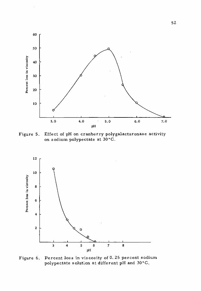
pH Optimum for the Cranberry Polygalacturonase Activity
Studies to find the pH optimum were determined using 0. 25 per-
cent sodium polype eta te solution at 30 "C and maintaining constant
pH values of 3, 4, 4. 5, 5, 5. 5, 6, and 7. Two milliliters of one to
two dilution of the protein dialysate prepared by acetone and poly-
ethylene glycol were used for an enzyme source.
Figure 5 shows the percent loss in viscosity of the substrate
due to the pectinolytic action of cranberry proteins over a period of
one hour at the different pH values. The highest activity was
observed at pH 5.0. Moreover, at pH values below five, the activity
decreased slowly while at pH values above 5. 0, the activity of the
enzyme dropped sharply with almost complete loss in activity at
pH 7.0.
The use of a one percent solution of the substrate to deter-
mine pH optimum was abandoned because of the jellying effect at pH
values below four. Figure 6 shows that pectate molecules were
subjected to hydrolysis in the absence of enzyme preparation and this
effect increased as the pH value decreased.
Effect of Salt Concentration on Polygalacturonase Activity
Kertesz (1952) reported that salt may have some influence on
some polygalacturonase preparations. The effect of sodium chloride
52
Figure 5. Effect of pH on cranberry polygalacturonase activity on sodium polypectate at 30°C.
o u
> a
o i—*
c <u 2
BL,
12 r
10
8 -
pH
Figure 6. Percent loss in viscosity of 0. 25 percent sodium polypectate solution at different pH and 30"C.

53
on the cranberry polygalacturonase was determined by using 0.2,
0. 4, and 0. 6M of the salt in the enzyme-substrate system. Table 3
shows the percent loss in viscosity of one percent sodium pectate
solutions at pH 5.0 and 30°C using different salt concentrations for
a period of 20 hours. The data on Table 3 indicate that sodium
chloride influence on the cranberry pectinolytic activity was very
small. Initial rate values corresponding to the change in viscosity
during the first hour of the reaction were found to be insignificant.
The sodium ion concentration in the. buffer used was 0. 033M.
Table 3. Effect of NaCl Cone, on the Pectinolytic Activity of Cranberry Proteins (means of five determinations)
NaCl Added % loss in Viscosity Initial Standard (Moles/liter) over a 20 hr period Rate Deviation
0.00M 87.1
0.20M 85.8
0.40M 85.7
0.60M 85.7
Polygalacturonase Activity of Cranberry Proteins on Different Pectic Substances
The activity of cranberry proteins extracted by the method
employing acetone and polyethylene glycol was determined using the
following four different substrates, sodium polypectate (Sunkist
6042), citrus pectin (Sunkist 3442), cranberry water soluble pectin
40. 3 0. 22
39.7 0.98
39.2 1.04
39.5 1.46
54
and cranberry sodium hexametaphosphate (calgon) soluble pectins.
Figures 7, 8, 9, and 10 show the course of action of cranberry
polygalacturonic enzyme on the four different substrates by deter-
mining the change in viscosity of a one percent substrate concentra-
tion over a period of 20 hours, at pH 5. 0 and 30"C. Two milliliters
of cranberry protein dialysate corresponding to 0. 19 milligrams
nitrogen was used as the enzyme source. A citric acid - NaOH
buffer solution pH 5. 0 was used in the preparation of the enzyme-
substrate system. The system contained toluene at the one percent
level as a bacteriostatic agent.
Table 4 shows that the percent loss in viscosity over a period
of 20 hours using cranberry protein dialysate as enzyme source and
one percent solutions of the four different substrates at pH 5. 0 and
30oC.
The catalytic effect of cranberry proteins exhibited a decrease
in viscosity on the four different substrates used. The activity of
the polygalacturonase was slightly higher on citrus sodium poly-
pectate than citrus pectin. This may be due to the lower degree of
methoxyl content in the former substrate.
Also, a slightly higher activity of the enzyme was exhibited on
the cranberry calgon soluble pectins over that of the water soluble
pectins. The calgon soluble pectins possess a lower methoxyl
content. A significant difference in the catalytic effect of cranberry

10 Time in hours
14 20
Figure 7. Change in viscosity of sodium polypectate (Sunkist 6042) solution due to the action of cranberry polygalacturonase at pH 5.0 and 30oC.

25.00 r
22.50 -J
20.00 -
17.50
a H 15.00 y a
.§■ 12.50 o o
10.00
7.50
5.00 -
8 10 Time in hours
Figure 8. Change in viscosity of citrus pectin (Sunkist 3442) due to the action of cranberry polygalacturonase at pH 5. 0 and 30oC.

4.00
3.90
3.80
3.70
3.60
a S 3.50 (- <a o a
I" 3. 40 -
3.30 -
3.20 -
3.10
3.00 2 4 6 8 10 12 14 16 18
Time in hours Figure 9. Change in viscosity of cranberry water soluble pectin due to the action of
cranberry polygalacturonase at pH 5. 0 and 30° C.
20

28.00 r
26.00 -
a C <u o
24.00 -
22..00 -
20.00 .
8 10 Time in hours
Figure 10. Change in viscosity of cranberry calgon soluble pectin due to the action of cranberry polygalacturonase at pH 5. 0 and 300C. oo

59
proteins was evident between cranberry and citrus pectins. The
pectinolytic enzyme showed a higher activity rate when the latter was
used for substrate. This may be ascribed to the difference in the
chemical structure of both substrates. Three of the four blank
samples, where no protein was added, exhibited approximately
similar losses in viscosity. However, citrus pectin blank sample
showed a lower loss than the other three substrates. This may be
attributed to the high methoxyl content of the citrus pectin which may
interfere by decreasing the rate of acid hydrolysis (Gizis, 1964).
Table 4. Percent drop in viscosity of one percent substrate solutions after 20 hrs. at 300C
Substrate
Percent Loss in Viscosity (Means of Five Determinations)
Protein Dialysate Standard Added Blank Difference Deviation
Sodium Polypectate 94. 5 (Sunkist 6042)
Citrus Pectin 86.1 (Sunkist 3442)
Cranberry Water 38.2 Soluble Pectins
Cranberry Calgon 42. 0 Soluble Pectins
7.4 87.1
3.7 82.1
8.0
8.2
30. 2
33.8
0. 22
0. 42
0. 17
0. 17

60
Effect of Cranberry Protein Concentration on Polygalacturonase Activity
This study was conducted to find the relationship between the
enzyme concentration and the course of activity of its pectinolytic
property. The different protein concentrations used in this study
-2 corresponded to 19.1, 9.55, 4.77, and 2.38 x 10 milligrams of
nitrogen. One percent sodium polypectate solution was used as sub-
strate at pH 5. 0 and 30°C. Figure 11 shows the course of action of
the different protein concentrations over a period of 20 hours. Figure
12 shows the relationship between protein concentration against the
initial rates exhibited by the four different concentrations used. The
initial rate corresponds to the change occurring during the first hour
of the reaction. When the rate of activity of the enzyme results in a
viscosity loss over 15 percent per hour, a linear relationship existed.
Heat Inactivation of Cranberry Polygalacturonase
The effect of heat on the stability of cranberry pectinolytic
enzyme was studied by exposing the protein dialysate extracted by
acetone-polyethylene glycol for different time intervals at lOCC.
The percent loss in viscosity of a one percent sodium polypectate
solutions in citric acid-NaOH buffer pH 5 and 30"C was determined
using the heated protein samples as the source of enzyme. The
blank samples contained two milliliters of buffer instead of protein

10 Time in hours
16 18
Figure 11. The relation between cranberry protein concentration and its polygalacturonase activity on one percent citrus polypectate solution at pH 5.0 and 300C. o^

62
50
40
.2 30 -
a
20 _
10 _
4.77 Enzyme concentration (mg. N x 10"2)
Figure 12. The relation between different enzyme concentrations of cranberry poly- galacturonase and their initial rate of activity on one percent polype ctate solution at pH 5.0 and 30"C.

63
solution. Table 5 shows the percent loss in viscosity of the sodium
polypectate solution over a period of 44 hours.
Table 5. Effect of heat on cranberry polygalacturonase
Means of Five Determinations
Period of Loss due exposure to Pe rcent loss to catalytic Standard heat (min. ) in viscosity action deviation
0 94.7 80.6
5 58.5 43.3 0.36
10 39.36 24. 3 0.65
15 30.3 15.2 0. 72
25 18.78 3.7 1.32
35 15.9 0.8 0.0
Figure 13 shows the thermal death rate curve of cranberry
pectinolytic enzymes. At pH 5.0, the activity of the polygalacturonase
was inactivated after 35 minutes of exposure to 100°C. McColloch
and Kertesz (1948) indicated that tomatoes contain an unusually heat
resistant pectinolytic factor with 20 percent of the original activity
remaining after a heat treatment of one hour at 100"C. Moreover,
Gizis (1964) found that strawberry pectinolytic enzymes were com-
pletely inactivated when exposed to 100"C for 35 minutes.

64
10 20
Time in minutes
Figure 13. Thermal death rate curve for the cranberry PG activity at pH 5. 0 on sodium polype ctate.

65
Chromatographic Separation of Cranberry Proteins on DEAE Cellulose Column
A DEAE cellulose column was used to fractionate cranberry
proteins. These proteins were extracted with acetone-polyethylene
glycol, precipitated by ammonium sulfate (75 percent saturation),
dialyzed against distilled water followed by dialysis against citric
acid:sodium hydroxide buffer pH 5. 0 for equilibration. Eighteen mis
of the dialysate were placed on the DEAE cellulose column and eluted
with citric acid-NaOH buffer pH 5. 0 containing a sodium chloride
gradient. Ten milliliter fractions were collected with a fraction
cutter at a rate of 60 mis per hour. The fractions collected were
assayed for polygalacturonase activity by determining the percent
loss in viscosity of a one percent sodium polypectate solution at
pH 5 and 30oC. Table 6 shows the activity of polygalacturonase of
the different fractions collected from the DEAE cellulose column.
Figure 14 gives the relationship between the fraction numbers,
their extinction at 280 myi. and their effect on one percent polypectate
solution.
Figure 15 indicates the sodium chloride concentration in dif-
ferent fractions obtained from the DEAE cellulose column. The
results on Table 6 show that the cranberry pectinolytic enzymes
were concentrated between eluate eight and fourteen with highest
activity present in eluate number nine. Peaks two and three on

18 20 22 No. of fractions
Figure 14. Chromatography of cranberry protein dialysate on DEAE cellulose column and the polygalacturonase activity of the fractions at pH 5. 0 and 30oC. ^ % loss in viscosity
o O. D. at 280 m|ji
90
80
- 70
- 60
3 3
_ 50 -
- 40
- 30
_ 20
- 10
a-

0.90 r
0.80 -
0.70 -
0.60 -
0.50 -
0.40 -
£ 0.30
0.20
0.10 -
22 Eluate number
26 30 34 38 42
Figure 15. Sodium chloride concentration in eluates from DEAE cellulose column.

68 Figure 12, did not show any pectinolytic activity. The DEAE
cellulose column enabled us to separate the proteins which possess
a polygalacturonase activity from proteins which exhibited no
activity.
Table 6. The pectinolytic activity of DEAE cellulose column chromatography fractions in one percent sodium polypectate at pH 5.0 and 30oC
Percent loss in viscosity Fraction due to pectinolytic activity O. D. at 280 mp.
1_7
8 51.8 0.235
9 80.5 0.71
10 75.0 0.58
11 65.0 0.35
12 57.5 0.225
13 45.0 0.18
14 32.5 0.09
18 11.5 0.035
21 7.9 0.045
24 2.4 0.05
29 0.27
34 0.110
40
Effect of Cranberry Proteins on the Insoluble Residue of Cranberries and Alcohol Insoluble Solids
After preparing the cranberry insoluble residue as described
in the Methods and Materials section, the action of the cranberry

69
proteins on this residue was determined. This was carried by
suspending three grams of the insoluble residue in 150 mis of citric
acid:NaOH buffer pH 5. 0. To half of the suspension four mis of
cranberry protein dialysate, as prepared by acetone-polyethylene
glycol method, were added while four mis of buffer were added to
the other portion. After placing the samples in a shaker-incubator
at 30°C, aliquots were removed and total soluble carbohydrate con-
centration was determined by the phenol-sulfuric acid method of
Dubois et al. (1956). Table 7 shows the results obtained.
Table 7. Effect of cranberry proteins on the insoluble residue of the AIS
Soluble sugars expressed as galacteroaic acid in Mg/ml Time of Incubation Sample Blank ~~ Difference
5 hours 1370 1047 323
19 hours 1573 1110 466
29 hours .1653 1160 493
43 hours 1657 1160 497
The insoluble residue of the Alcohol Insoluble Solids is mainly
composed of protopectin and cellulytic matter. When protopectins
were hydrolyzed by two percent NaOH and soluble carbohydrates
determined on the protopectin free residue, no change in total sugar
was found. Thus, the increase in total soluble carbohydrates in the
former experiment (Table 7) may be attributed to the action of
cranberry proteins.

70
Pectin Transeliminase in Cranberry Proteins
The activity of pectin transeliminase was followed by deter-
mining the change in light absorption at 235 mix of a 0. 5 percent
pectin solution in 0. IM citrate-phosphate buffer pH 5.2, after the
addition of the protein dialysate prepared from the acetone-poly-
ethylene glycol protein preparations. No activity of transeliminase
was found.
Pectin Esterase in the Cranberrry
Due to the fact that pectin esterase is strongly attached to the
cellular constituents of the plant, the enzyme was extracted with
0. 15M NaCl solution from the cranberry acetone powder. The
activity of pectin esterase was determined by measuring the increase
in carboxyl groups on the pectin molecule.
pH Optimum of Pectin Esterase
Studies to determine the pH optimum for the activity of cran-
berry pectin esterase were made by continuous titration of the
liberated carboxyl groups produced. Six pH values were used at
which the enzyme-substrate system was maintained in order to
measure the activity. Figure 16 shows the relationship of the
enzyme activity at the different pH values used.

100
Figure 16. Activity of PE of cranberries at different pH values, using citrus pectin as substrate.

72
Table 8 gives the means of four determinations of the pectin
esterase activity at the different pH values used, at 30°C. The sub-
strate used was a one percent solution of Sunkist pectin (3442) con-
taining 0, 15M NaCl.
Table 8. Effect of pH on the activity of cranberry pectin esterase
PH gram
Equivalents/ acetone powde:
hour r/ Standard
deviation Pe r 100 grams of
cranberries
5 0.00 0.00 0.000
6.0 12.0 0.30 52.2
6.5 17.0 0.22 73.6
7.0 35.0 0.41 151.6
7.5 81.0 0.41 350. 7
8.0 39.0 0.41 168.9
The data on Table 8 shows that the highest activity of cran-
berry pectin esterase was found at pH 7. 5. No activity was evident
at pH 5.0. Alkaline hydrolysis of the methyl ester at pH values
above 8.0 making the results unreliable.
Pectin Esterase Activity
When using the protein dialysate of acetone powder after
precipitation with 75 percent saturation of ammonium sulfate as an
enzyme source, the activity of the enzyme was found to be very slow

73
and insignificant. This was abandoned for the acetone powder
extracted with 0. 15M NaCl solution as a source of the enzyme.
Figure 17 shows the course of action of cranberry proteins on one
percent citrus pectin solution (3442) at pH 7. 5 and 300C. The amount
of enzyme used was equivalent to one gram of acetone powder.
According to Kertesz (1959), demethylation of the pectin does not
affect the viscosity of their solutions except in presence of calcium
ions. Under such conditions, the gelation properties of the pectin
are affected.
Influence of Salt on Pectin Esterase Activity
The activity of cranberry pectin esterase was determined by
using three 50-ml aliquots of one percent citrus pectin solution con-
taining no sodium chloride, 0. 15M NaCl and 0. 30M NaCl at pH 7. 5
and 30oC. Table 9 shows the effect of NaCl on the activity of the
enzyme.
Table 9. Effect of NaCl on cranberry pectin esterase activity at pH 7. 5
Activity units of enzyme Activity units/l00 grams of M NaCl per gram of acetone powder fresh tissue
G.OO 4.0 17.2
0.15 81.0 350.7
0.30 76.0 329.1
160 -
■a
a) ft
■a a) o
■a o
il r—I
3
120
80 -
40
120 Time in minutes
Figure 17. Cranberry PE activity on one percent citrus pectin solution (0. 15M NaCl) at pH 7. 5 and 30oC.
-^ *.
75
A unit of pectin esterase corresponds to microequivalents of
ester hydrolyzed per hour. It is evident from the results in Table 9
that the NaCl concentration increased the activity of the enzyme at
0. 15M above which only slight decrease in activity occurred. When
no salt was added to the system the activity of the enzyme was
comparatively low. Lineweaver and Ballou (1945) proposed a
hypothesis regarding the effect of cations on pectin esterase
activity. It was assumed that cations prevent the inhibition of pectin
esterase by the pectin carboxyl groups by forming cation-carboxyl
complex.
Heat Inactivation of Pectin Esterase
The effect of heat on cranberry pectin esterase activity was
determined by exposing the enzyme extract to 100°C for one, two,
three, and five minutes. The activity of the heat treated enzyme
preparation was determined by following the amount of carboxyl
groups exposed as a result of the catalytic hydrolysis of the methyl
ester on a one percent citrus pectin (3442) solution at 30oC and
maintaining the pH at 7. 5. NaCl concentration in the substrate
corresponded to 0. 15M. Table 10 shows the results obtained and
are the means of three determinations.

76
Table 10. Influence of heat on cranberry pectin esterase
Time of exposure to 100'
(Minutes) 'C g ram
equivalents/ of acetone powdi
hour «/ Standard
deviation
0 81.00 0.41
1 37. 10 2. 67
2 25. 17 1.88
3 17. 23 0. 40
5 00.00 0.00
Table 10 shows tha,t cranberry pectin esterase was inactivated
when exposed to 100°C for five minutes.

77
DISCUSSION
The data shown in Tables 2 and 4 and Figures 7, 8, 9, and 10,
show that a loss in viscosity was exhibited on solutions of pectic sub-
stances due to the presence of cranberry proteins. Thus cranberry
proteins possess the pectinolytic factor polygalaeturonase which
hydrolyzes the glycosidic linkages of a pectin polymer.
The method of enzyme extraction from the cranberry proved
to be essential in obtaining an enzyme extract which exhibits a
high rate of activity. The use of phenol complexing agents prevents
the phenol-protein interaction which could cause a decrease in the
activity of the enzyme. Table 2 and Figure 4 show that when the
phenol complexing agents are used in extracting the proteins from
the cranberries, a higher activity of the polygalacturonase was
evident. Moreover, when different types of phenol complexing
agents are used, the protein extracts prepared possess different
enzyme activity. Results in Table 2 show that when the protein
extracted by the use of acetone in conjunction with polyethylene
glycol a higher activity was observed over the acetone or the poly-
vinylpyrrolidone protein extracts. This may be attributed to the
stronger affinity of the polyethylene glycol in the presence of acetone
to the phenolic compounds.
Table 4 and Figures 7, 8, 9, and 10 indicate that the activity

78
of the polygalacturonase on the different pectic substances used for
substrates was rapid during the initial period of the reaction. This
rapid loss in viscosity suggests a random split of the pectin polymer.
The polygalacturonase which possess this criterion in their course of
action, are referred to as endoenzyme. A slight pectinolytic pref-
erence for the citrus sodium polypectate over citrus pectin was evi-
dent. The latter possess a higher metholyl content, see Table 1.
The action of cranberry polygalacturonase was also demon-
strated on cranberry water soluble pectin and the calgon soluble
pectin. The difference in the activity of polygalacturonase on both
was slight. Moreover, the enzyme showed a considerably higher
activity on citrus pectins than on cranberry pectins. This may be
attributed to the difference in structure between the pectins of both
sources.
The foregoing discussion indicates that the cranberry poly-
galacturonase possesses the ability to randomly hydrolyze pectic
substances with high and low methoxyl content.
Optimum pH for the cranberry polygalacturonase lies in the
range of 5j0 . The enzyme was inactive at neutral pH values as
shown in Figure 5. However, although enzyme activity decreases
with a decrease in pH, slight activity was observed at pH 3. 0. The
influence of temperature on the cranberry polygalacturonase is shown
in Table 5. The enzyme was found to be inactivated after 35 minutes

79
at 1000C. It may be said that the enzyme is highly resistant to heat.
The action of cranberry proteins on the insoluble residue of
the cranberry alcohol insoluble solids as shown in Table 7, shows
that the protein exhibited a solubilization effect on the insoluble
parent material protopectih. Usually, the concentration of proto-
pectin in fruits decreases with maturation. This may be attributed
to its contact with pectic enzymes which may hydrolyze it into the
soluble form.
Pectin esterase activity of the cranberries was found to be low
as shown on Table 8 and Figure 17. Optimum pH activity was found
at a value of 7.5, with no apparent activity at pH 5.0. Heating
enzyme preparations for five minutes at 100° C caused complete
inactivation. NaCl at a concentration of 0. 15M gave an optimum
pectin esterase activity. In the absence of the salt, the activity was
negligible.

80
SUMMARY AND CONCLUSION
The work done in this study concerns the isolation and
characterization of the pectic enzymes in cranberries. The follow-
ing conclusions could be drawn:
1. Polygalacturonase was found in cranberry proteins. The enzyme
could be classified as an endoenzyme that has the ability to
catalyze the hydrolysis of pectins with both high and low methoxyl
content.
2. The use of a phenol binding agent in the enzyme preparation was
effective in obtaining a polygalacturonase extract which possessed
a relatively high activity.
3. The polygalacturonase showed a higher activity on citrus pectins
as compared to cranberry pectins.
4. pH value of 5.0 was found to give optimum activity for cranberry
polygalacturonase.
5. The enzyme polygalacturonase was inactivated after 35 minutes
of exposure to 100oC at pH 5. 0.
6. Sodium chloride up to a concentration of 0. 6M gave no significant
affect on cranberry polygalacturonase activity in a citric acid -
sodium hydroxide buffer pH 5.0.
7. Cranberry proteins were found to possess pectin esterase which
exhibited a low activity when compared to strawberry and tomato
81
pectin esterase.
8. Optimum pH value for pectin esterase was 7.5.
9. Pectin esterase was inactivated when exposed for five minutes
at 100oC
10. NaCl in the pectin esterase-substrate system gave optimum
activity at 0. 15M concentration.

82
BIBLIOGRAPHY
Albersheim, P. , H. Neukom and H. Deuel. 1960a. liber die Bildung von ungesattingren Abbauprodukten durch ein pectinabbauendes Enzym. Helvetica Chimica Acta 43:1422-1426.
. 1960b. Splitting of pectin chain molecules in neutral solutions. Archives of Biochemistry and Biophysics 90:46-51.
Albersheim, P. and Killias Ursula. 1962. Studies relating to the purification and properties of pectin transeliminase. Archives of Biochemistry and Biophysics 97:107-115.
Anderson, Martin. 1964. Degradation of pectic acid and certain oligogalacturonic acids by the transeliminase of Bacillus polymyxa. Master's thesis. Pullman, Washington State University. 45 numb, leaves.
Association of Official Agricultural Chemists. I960. Official methods of analysis, ed. by William Horwitz. 9th ed. Washington, D. C. 832 p.
Atkins, C. D. and A. H. Rouse. 1953. Time-temperature relation- ships for heat inactivation of pectin esterase in citrus juices. Food Technology 7:489-491.
. 1954. Time-temperature concentration relation- ships for heat inactivation of pectin esterase in citrus juices. Food Technology 8:498-500.
Badran, A. M. and D. E. Jones. 1965. Polyethylene glycol-tanriin interaction in extracting enzymes. Nature 206:622-624.
Baker, C. L. ^t al. 1944. Report of the committee for the revision of the nomenclature of pectic substances. Chemistry and Engineering News 22:105-106.
Bell, T. A., J. L. Etchells and I. D. Jones. 1955. A method for testing cucumber salt-stock brine for softening activity. Washington, D. C. , U. S. Department of Agriculture. 15 p. (Agricultural Research Service ARS72-5)

83
Bell, T. A. and J. L. Etchells. 1958. Pectinase inhibitor in grape leaves. Botanical Gazette 119:192-196.
Bell, Tomas A. et al. 1965. Inhibition of pectinolytic enzymes in cucumber fermentations by sericea. Journal of Food Science 30:233-239.
Chang, L. W. S. , L. L. Morita and H Y. Yamamoto. 1965. Papaya pectin esterase inhibition by sucrose. Journal of Food Science 30:218-222.
Demain, A. L. and H. J. Phaff. 1957. Recent advances in enzymatic hydrolysis of pectic substances. Wallerstein Laboratories Communications 20:119-139.
Demain, A. L. and H. J. Phaff. 1957. Softening of cucumbers during curing. Journal of Agricultural and Food Chemistry 5:60-64.
Deuel, H. and E. Stutz. 1958. Pectic substances and pectic enzymes. Advances in Enzymology 20:341-382.
Dingle, J. , W. W. Reid and G. L. Solomons. 1953. The enzymatic degradation of pectin and other polysaccharides. II. Applica- tion of the "cup-plate" assay to the estimation of enzymes. Journal of the Science of Food and Agriculture 4:149-155.
Doesberg, J. J. 1965. Pectic substances in fresh and processed fruits and vegetables. Institute for research on storage and processing of horticultural produce. Wagehingen, The Netherlands. 145p. (I. B. V. T.-Communication No. 25)
Dame, C. et al. 1956. The influence of ripeness on the organic acids, sugars and pectin of canned Bartlett pears. Food Technology 10:28-33.
Dubois, M. et al. 1956. Colorimetric metbod for determination of sugars and related substances. Analytical Chemistry 28:350- 356.
Eliezer, I. and H. J. C. Hyman. 1957. Means square length and mean square radius of gyration of 1, 4-polysaccharides and of polyuronides. Journal of Polymer Science 23:387-402.

84
Endo, Akira. 1963a. Studies on pectinolytic enzymes of molds, V. The fractionation of pectinolytic enzymes of Coniothyrium diplodiella (1). Agricultural and Biological Chemistry 27:741- 750.
' 1963b. Studies on pectinolytic enzymes of molds, VI. The fractionation of Conothyrium diplodiells (2). Agri- cultural and Biological Chemistry 27: 751-757.
1964a. Studies on pectinolytic enzymes of molds, VII. Turbidity of apple juice clarification and its application to determination of enzyme activity. Agricultural and Biological Chemistry 28:234-238.
1964b. Studies on pectinolytic enzymes of molds, VIII. Purification and properties of endo-polygalacturonases, I. Agricultural and Biological Chemistry 28: 535-542.
1964c. Studies on pectinolytic enzymes of molds, IX. Purification and properties of endo-polygalacturonase, II. Agricultural and Biological Chemistry 28: 543-550.
1964d. Studies on pectinolytic enzymes of molds, X. Purification and properties of endo-polygalacturonase. Agricultural and Biological Chemistry 28: 551-558.
1964e. Studies on pectinolytic enzymes of molds, XI. Purification and properties of exo-polygalacturonase. Agricultural and Biological Chemistry 28: 639-645.
1965. Studies on pectinolytic enzymes of molds, XVI. Mechanism of enzymatic clarification of apple juice. Agricultural and Biological Chemistry 29: 229-233.
Etchells, J. L. , T. A. Bell and I. D. Jones. 1955. Studies on the origin of pectinolytic and cellulytic enzymes in commercial cucumber fermentation. Food Technology 9: 14.
Gizis, John Evangelos. 1964. The isolation and characterization of the pectic enzymes and the pectic substances of the Northwest Strawberry. Ph.D. thesis. Corvallis, Oregon State University. 105 numb, leaves.
Glaziou, K. T. 1958. Effect of 2.4 dichlorophenoxyacetic acid on pectin methylesterase in tobacco pith sections. Nature 181: 428-429.

85
Grossman, F. 1958. Uber die Hemmung pektolybischer Enzyme von Fusarium oxysporum F. lycopusici durch Gerbstoffe. Naturwissen schaffer 45:113-114.
Hatanaka, Chitoshi and Junjiro Ozawa. 1964. Enzyme degradation of pectic acid. I. Limited hydrolysis of pectic acids by carrot Exopolygalacturonase. Agricultural and Biological Chemistry 28:627-632.
Hathaway, D. E. and J. W. T. Seakims. 1958. The influence of tannins on the degradation of pectin by pectinase enzymes. Biochemical Journal 70:158-163.
Helbert, J. R. and K. D. Brown. 1957. Color reaction of anthrone with monosaccharide mixtures and oligo- and polysaccharides containing hexuronic acids. Analytical Chemistry 29:1464- 1466.
Hills, Claude H. j-t jal. 1949. Enzyme demethylated pectinates and their gelation. Food Technology 3:90.
Hobson, G. E. 1962. Determination of polygalacturonase in fruits. Nature 195:804-805.
Holden, M. 1946. Studies on pectase. Biochemical Journal 40:103.
Hultin, H. O. and A. S. Levine. 1965. Pectin methylesterase in ripening banana. Journal of Food Science 30:917-921.
Hultin, H. O. , Bonnie Sun and J. Bulger. 1966. Pectin methyles- terase of the banana, purification and properties. Journal of Food Science 31:320-327.
Jansen, E. F. , L. R. McDonnell and R. Jang. 1945. Simultaneous action of polygalacturonase and pectinesterase on pectin. Archives of Biochemistry 8:97.
Jansen, E. F. , L. R. MacDonnell and W. W. Ward. 1949. The minimum size for the structural unit of pectin. Archives of Biochemistry 21:149.
Jansen, E. F. and R. Tang. I960. Orange pectin esterase binding and activity. Food Research 25:64-72.

86
Joslyn, M. A. 1962. The chemistry of protopectin. A critical re- view of historical data and recent developments. Advances in Food Research 11:1-207.
Kertesz, Z. I. 1938. Pectic enzymes of tomatoes, II. Food Research 3:481-487.
1939. Pectic enzymes, 11. Heat inactivation of tomato pectin-methoxyase (pectase). Food Research 4:113-116.
1951. The pectic substances. New York, Inter- science Publishers, 628 p.
1955. Pectic enzymes. In: Methods in Enzymology, ed. by Sidney P. Colowick and Nathan O. Kaplan. Vol. 1. New York, Academic Press, p. 158-166.
I960. Enzymes of polysaccharide synthesis and degradation. In: Food Enzymes. Symposia on foods held at Oregon State College, Corvallis, Oregon, ed. by H. W. Schultz. Westport, Connecticut, AVI Publishing Company, p. 49-63.
Kohn, G. G. , W. B. Esslen and C. R. Fellers. 1953. Thermal inactivation of pectinolytic enzymes used in the clarification of apple juice. Food Research 18:511-515.
Kraught, A. I. and M. P. Starr. 1953. Pectic enzymes of Erwinia carotovora. Archives of Biochemistry and Biophysics 42:271.
Lineweaver, H. and G. A. Ballou. 1945. The effect of cations on the activity of alfalfa pectinesterase. (Pectase). Archives of Biochemistry 6:373.
Lineweaver, H. , R. Jang and E. F. Jansen. 1949. Specificity and purification of polygalacturonase. Archives of Bio- chemistry 20:137.
Loomis, W. D. 1968. Removal of phenolic compounds during the isolation of plant enzymes. Advances in Enzymology. (In press)
Loomis, W. D. and J. Battaile. 1966. Plant phenolic compounds and the isolation of plant enzymes. Phytochemistry 5:423- 438.

87
Luh, B. S. and K. D. Dastur. 1961. Texture and pectin changes in canned apricots. Journal of Food Science 31:167-183.
MacDonnell, L. R. etal. 1950. The specificity of pectin esterases from several sources with some notes on purification of orange pectin esterase. Archives of Biochemistry 28:260-273.
MacMillan, J. D. and R. H. Vaughn. 1964. Purification and properties of a polygalacturonic acid-transeliminase produced by Clostridium multifermentaus. Biochemistry 3:564-572.
McColloch, R. J. , J. G. Moyer and Z. I. Kertesz. 1946. Pectic enzymes, VII. The preparation of tomato pectin-methyl- esterase. Archives of Biochemistry 17:197-199.
McColloch, R. J. and Z. I. Kertesz. 1947. Pectic enzymes, VIII. A comparison of fungal pectin-methylesterase with that of higher plants, especially tomatoes. Archives of Biochemistry 13:217-229.
1948. An unusually heat-resistant pectinolytic factor from tomatoes. Archives of Biochemistry 17:197-199.
1949. Recent developments of practical sig- nificance in the field of pectic enzymes. Food Technology 3: 94-96.
McCready, R. M. and E. A. McComb. 1954. Pectic constituents in ripe and unripe fruit. Food Research 19:530-535.
McCready, R. M. , E. A. McComb and E. F. Jansen. 1955. The action of tomato and avocado polygalacturonase. Food Research 20:186-191.
McCready, R. M. and Mildred Gee. I960. Plant pectin analysis. Determination of pectic substances by paper chromatography. Journal of Agricultural and Food Chemistry 8:510-513.
Mills, G. B. 1949. A biochemical study of Pseudomonas prunicola pectin esterase. Biochemical Journal 44:302.
Nagel, C. W. and R. H. Vaughn. 1961. The characteristics of a polygalacturonase produced by Bacillus polymyxa. Archives of Biochemistry and Biophysics 93:344-352.

88
Newbold, R. P. and M. A. Joslyn. 1952. Chemistry of analytically important pectic acids. I. Chemical properties of Ehrlich, Wichmann and Carre-Haynes pectic acids. Journal of the Association of Official Agricultural Chemists 35:872.
Newcomb, E. H. 1963. Cytoplasm cell wall relationships. Annual reviews of Plant Physiology 14:43.
Okamotot, Ken-ichi, Chitoshi Hatanaka and Junjiro Ozawa. 1964. A saccharifying pectate transeliminase of Erwinia aroideae. Agricultural and Biological Chemistry (Japan) 2.8:331-336.
Owens, H. S. et al. 1952. Methods used at Western Regional Research Laboratory for extraction and analysis of pectic materials, Albany, California. Western Regional Research Laboratory, U. S. Department of Agriculture, Agricultural Research Administration, 24 p. (Publication A. I. C. 340) (Mimeographed).
Patel, P. S. and H. J. Phaff. I960. Studies on the purification of tomato polygalacturonase. Food Research 25:37-46.
Patterson, M. E. et al. 1967. Effect of bruising on post harvest softening, color changes and detection of polygalacturonase enzyme in cranberries. Proceedings of the American Society for Horticultural Sciences 90:498-505.
Phaff, H. J. 1947. The production of exocellular pectic enzymes by Penicillium chysogenum. I. On the formation and adaptive nature of polygalacturonase and pectin esterase. Archives of Biochemistry 13:67.
Pirie, N. W. 1959. Leaf proteins. Annual Review of Plant Physiology 10:33.
Pollard, A. and Margaret R. Kieser. 1951. The pectase activity of apples. Journal of the Science of Food and Agriculture 2:30.
Pollard, A. , Margaret R. Kieser and D. J. Sissons. 1958. Inactivation of pectic enzymes by fruit phenolics. Chemistry and Industry: 952.
Porter, W. L. and J. H. Schwarts. 1962. Isolation and description of the pectinase inhibiting tannins of grape leaves. Journal of Food Science 27:416.

89
Porter, William L. et al. 1961. Probable identity of the pectinase inhibition in grape leaves. Journal of Food Science 26:600.
Postlmayr, H. L. , B. S. Luh and S. L. Leonard. 1956. Characterization of pectin changes in freestone and clingstone peaches during ripening and processing. Food Technology 10: 618-625.
Preiss, J. and G. Ashwell. 1930. Polygalacturonic acid metabolism in bacteria. I. Enzymatic formation of 4-deoxy l-three-5- hexoseulose uronic acid. Journal of Biological Chemistry 238:1571-1538.
1936. Polygalacturonic acid metabolism in bacteria. II. Formation and metabolism of 3-deoxy-D- glycero 2, 5 hexodiulosonic acid. Journal of Biological Chemistry 238:1577-1583.
Rahman, M. B. and M. A. Joslyn. 1953. Properties of purified fungal polygalactur on as e. Food Research 18:301-304.
Reed, G. 1966. Enzymes in food processing. New York, Academic Press. 483 p.
Reid, W. W. 1950. Pectic enzymes of the fungus Byssochlamys fulva. Nature 166:76.
1951. The estimation of polygalacturonase (pectinase) activity. Food Technology 5:170,
Rich, J. A. (ed. ) 1963. Chemical methods for analysis of fruit and vegetable products.. Rev. ed. Summerland, British Columbia. 47 p. (Canada Dept. of Agriculture. Contribution no. B7, Publication 1154)
Roelfsen, P. A. 1954. On the softening of fruits of Mespillus germanica. Acta Botanica Neerlandica 3:154-160.
Saito, H. 1955. Pectic glycosidases of Aspergillus niger. Journal of General and Applied Microbiology 1:38-59.
Schubert, E. 1954. Einfluss von Wasserstoff-und alkali- lonen auf den enzymatischen Abban von pectin verschiedenen Veresterimgs-grades durch pectinglykosidase und pektinglykosidase gemische. Helvetica Chimica Acta 37:691.

90
Schultz, T. G. et al. 1945. Influence of method of deesterification on the viscosity and acid behavior of pectinic acid solutions. Journal of Physical Chemistry 49: 554.
Seegmiller, C. G. and E. F. Janse. 1963. Polymethyl galacturonase, an enzyme causing the glycosidic hydrolysis of esterified pectic substances. Journal of Biological Chemistry 195:327-336.
Sistrunk, W. A. and R. F. Cain. I960. Chemical and physical changes in green beans during preparation and processing. Food Technology 14:357-263,
Solms, J. and H. Deuel. 1954. Uber den mechanismuus der enzymatischen verseifung von pektinstoffen. Helvetica Chimica Acta 38: 321.
Somogyi, L,. P. and Roger J. Romani. 1964. A simplified technique for the determination of pectin methyl esterase activity. Analytical Biochemistry 8:498-501.
Stahmann, M. A. 1963. Plant proteins. Annual Reviews of Plant Physiology 14: 137.
Starr, M. P. and S. Nasuno. 1963. Degradation of pectic substances by Xanthomonas. (Abstract) Bacteriological Proceedings p. 85.
Steele, Wilbert F. and H. Y. Yang. I960. The softening of brined cherries by polygalacturonase and the inhibition of polygalac- turonase in model systems by alkyl aryl sulfonates. Food Technology 14: 121-26.
Stier, E. F. , C. O. Ball and W. A. Maclinn. 1956. Changes in pectic substances of tomatoes during storage. Food Technology 10: 39-43.
Talboys, P. W. 1950. Use of Aberlite IRC-50 for the partial purification of bacterial pectinase. Nature 166: 1077.
Uritani, I, and M. A. Stahmann. 1961. Pectinolytic enzymes of Ceratocystis pimbriata. Phytopathology 51:277-285.
Van Bureu, J. P., J. C. Moyer and W. B. Robinson. 1962. Pectin methyl-esterase in snap beans. Journal of Food Science 27: 291-294.

91
Willamann, J. J. and F. R. Davidson. 1924. Some modification of the pectinic acid method for sugars. Journal of Agricultural Research 28:479.
Woodmansee, C. W. , J. H. McClendon and G. F. Somers. 1959. Chemical changes associated with the ripening of apples and tomatoes. Food Research 24:503-514.
Yang, H. Y. , W. F. Steele and D. J. Graham. I960. Inhibition of polygalacturonase in brined cherries. Food Technology 14: 644-647.