Embed Size (px)
Citation preview

Grand Rounds
August 2, 2013
Yuna Rapoport, MD

3
Case Presentation
CC: “I can’t see at all”
HPI: 7y/o F with history of seizures and headaches c/o
blurry vision one morning. Reportedly taken to
optometrist, refracted to 20/20, sent home with glasses
Rx. She took a nap and when she awoke she “could
only see bright lights.” On the way to the ER, she
developed red eyes, swollen eyelids, light sensitivity
and tearing.

5
Review of Systems
No pain in her eyes, no burning, no itching, no
pain with EOMs
No mucus discharge
No dizziness
No weight loss
No N/V, no malaise
No focal neurological complaints
No SOB/ cough
No neck stiffness, no fever

History
PMH: 3 seizures characterized by head
bobbling with abnormal eye movements before
age 3, seizure-free since then, with one recent
focal seizure
Social History: lives with mom, dad and cat
No known family ocular history
NKDA

Exam 5/21/2013 in ED
VA: LP at 20’ OU
Pupils: 6mm, minimally reactive, no rAPD OU
EOMI OU
IOP: 40, 41
Fields: constricted 360 degrees OU
External: slightly edematous upper and lower lid
OU

DDx of Elevated IOP and
Seizures in Children
Sturge Weber
Klippel-Trenaunay-Weber
Syndrome
Wyburn-Mason syndrome
Tuberous Sclerosis
Neurofibromatosis
Aicardi Syndrome
The Ring 14 Syndrome
CASK mutation

Exam
Narrow angles OU

DDx of Childhood Narrow
Angles
Congestion
Nanophthalmos
Ciliochoroidal effusion
Inflammation: scleritis, uveitis, JIA
Drug-induced
Post-surgical (Sturge Weber,
Klippel Trenaunay Weber)
Tumor
Retinoblastoma
Medulloepithelioma
Exudative RD (Coats’ Disease)
Persistent fetal vasculature
Retinopathy of prematurity
Posterior ‘Pushing’ mechanism:
Anterior rotation of ciliary body:
Pressure from posterior segment:
Contraction of retrolental tissue:
Corneal anomalies: microcornea, cornea
plana/ sclerocornea, megalocornea
Axenfeld-Rieger syndrome, Peters
anomaly, iridoschisis
Aniridia
Ectopia lentis (trauma, Marfan’s
homocystinuria, Ehler’s Danlos, Weill-
Marchesani)
Congenital iris ectropion syndrome
▪ Aphakia
▪ Microspherophakia
Neovascular (tuberous sclerosis)
Peripheral Anterior Synechiae
Without pupillary block
Anterior ‘Pulling’ Mechanism:
With Pupillary block:
Anterior segment dysgenesis:

Ant Segment Photo: OD 5/22/13

Ant Segment Photo: OS 5/22/13
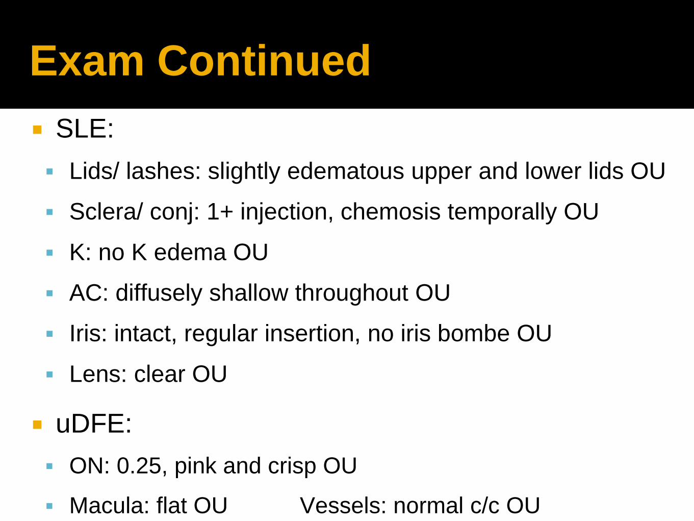
Exam Continued
SLE:
Lids/ lashes: slightly edematous upper and lower lids OU
Sclera/ conj: 1+ injection, chemosis temporally OU
K: no K edema OU
AC: diffusely shallow throughout OU
Iris: intact, regular insertion, no iris bombe OU
Lens: clear OU
uDFE:
ON: 0.25, pink and crisp OU
Macula: flat OU Vessels: normal c/c OU

Normal Ant Segment OCT
Scleral spur
Reflex saturation
beam

Ant Segment OCT: OD
OS
Iridocorneal
apposition
Anterior lens vault Anterior iris
convexity

More History and Exam… in ER
POH: none
Medications: Topiramate 25mg QHS x 2 weeks,
cyproheptadine 1 tsp by mouth QHS
VA: LP at 20’, CF at 14”, 20/20 at 3” OU

DDx of Childhood Narrow
Angles
Congestion
Nanophthalmos
Ciliochoroidal effusion
Inflammation: scleritis, uveitis, JIA
Post-surgical (Sturge Weber,
Klippel Trenaunay Weber)
Tumor
Retinoblastoma
Medulloepithelioma
Exudative RD (Coats’ Disease)
Persistent fetal vasculature
Retinopathy of prematurity
Posterior ‘Pushing’ mechanism:
Anterior rotation of ciliary body:
Pressure from posterior segment:
Contraction of retrolental tissue:
Corneal anomalies: microcornea, cornea
plana/ sclerocornea, megalocornea
Axenfeld-Rieger syndrome, Peters
anomaly, iridoschisis
Aniridia
Ectopia lentis (trauma, Marfan’s
homocystinuria, Ehler’s Danlos, Weill-
Marchesani)
Congenital iris ectropion syndrome
▪ Aphakia
▪ Microspherophakia
Neovascular (tuberous sclerosis)
Peripheral Anterior Synechiae
Without pupillary block
Anterior ‘Pulling’ Mechanism:
With Pupillary block:
Anterior segment dysgenesis:
Drug-induced

Acute angle closure from
Topiramate
First case reported in July 2001 by Banta et al. (1)
Largest case series 2004 by Fraunfelder, et al. (2): n=115
83 cases of bilateral, 3 cases of unilateral angle closure
Ages: 3-70, mean 34
Doses: <50mg (47%), 50-75mg (33%), 100mg (13%,
>100mg (7%)
Time of onset:
5 cases within hours when dose doubled
1-49 days, mean 7 days; 85% occurred within first 2
weeks
1. Banta, et al. Presumed topiramate-induced bilateral acute angle-closure glaucoma.Am J Ophthalmol, 132 (2001),
pp. 112-114
2. Fraunfedler, et al. Topiramate-assoicated acute, bilateral, secondary angle-closure glaucoma. Ophth. 2004; p. 109-
111.

Mechanism
Underlying mechanism- ciliochoroidal effusion--> ciliary body
edema-->relaxation of zonular fibers -->lens thickening --> anterior
displacement of lens -iris complex--> secondary ACG and high
myopia
Cilio-choroidal effusion caused by sulfonamides is an idiosyncratic
response in uveal tissue
Dose independent
Hapten hypothesis: reactive drug metabolites bind to proteins,
forming altered proteins which are recognized as foreign
substances and incite immune reactions
Myopia 87% related to lens thickening and 13% related to anterior
lens displacement, from A-scan measurements
Hook et al. Transient myopia induced by sulfonamides. Am J Ophthalmol. 1986; 101; 495-496.
Senthil S, et al. bilateral simultaneous acute angle closure caused by sulphonamide derivatives: a case series. Indian J Ophthalmol. 2010; 58(3):248-52.
Fraunfedler, et al. Topiramate-assoicated acute, bilateral, secondary angle-closure glaucoma. Ophth. 2004; p. 109-111.

Other Sulfonamides
Other sulfonamides have been reported to cause
similar clinical syndrome:
acetazolamide
indapamide (thiazide diuretic) (1) (2)
sulfasalazine (3)
hydrochlorothiazide (3)
Risk of adverse reaction to sulfonamides is 3% (4)
1. Senthil S, et al. bilateral simultaneous acute angle closure caused by sulphonamide derivatives: a case series. Indian J Ophthalmol. 2010;
58(3):248-52.
2. Blain P, Pβques M, Massin P, Erginay A, Santiago P, Gaudric A. Acute Transient Myopia Induced by Indapamide. Am J Ophthalmol
2000;129:538-4
3. Lee GC, et al. Bilateral angle closure glaucoma induced by sulphonamide-derived medications. Clin Experiment Ophthalmol. 2007;
35(1):55-8.
4. Panday VA, et al. Review of sulfonamide induced acute myopia and acute bilateral angle-closure glaucoma. Compr
Ophthalmol Update. 2007; 8(5): 271-6

Clinical Findings from
Topiramate
Macular folds (from choroidal effusion)
1. Kumar, M, et al. Macular folds: an unusual association in topiramate toxicity. Clin Exp Optom. 2012 95(4):449-52.

Clinical Findings from
Topiramate
Choroidal detachment
1. Kumar, M, et al. Macular folds: an unusual association in topiramate toxicity. Clin Exp Optom. 2012 95(4):449-52.

Clinical Findings from
Topiramate
Ciliochoroidal detachment
1. Kumar, M, et al. Macular folds: an unusual association in topiramate toxicity. Clin Exp Optom. 2012 95(4):449-52.

Treatment
Immediate d/c of topiramate
Aqueous suppressants: PO/ IV Acetazolamide, +/- Mannitol
Topical ocular hypotensives
Topical cycloplegics - retracts ciliary processes and deepen AC
Angle closure usually resolves within 24-48 hrs with medical treatment
Myopia resolves 1-2 weeks of discontinuing the topiramate
Peripheral iridectomy and miotics are not indicated (pupillary block not an
underlying mechanism of this syndrome)
If refractory, PO/ IV steroids(1), Argon laser peripheral iridoplasty (2),
surgical intervention (choroidal drainage (3), vitrectomy, CE/ IOL, glaucoma
surgeries)
1. Rhee DJ, et al. Rapid resolution of topiramate-induced angle-closure glaucoma with methylprednisolone and mannitol
2. Zalta AH, et al. Peripheral iridoplasty efficacy in refractory topiramate-associated bilateral acute angle-closure glaucoma. Ophthalmol. 2008; 126(11):1603-5
3. Parikh, R, et al. J Glaucoma. 2007. 16(8):691-3.

Few case reports in Children
Topiramate is approved over age >2
Included in series but no information provided:
Fraunfedler et al. : age 3 (1)
Thambi, et al.: age 5(2)
2 Case reports:
8 y/o M - c/o “blind” both eyes, Mrx: -6.00 OU, IOP 19 OU. Tx: d/c Topiramate, vision
normalized within 48 hrs (3)
5y/o F - HA, N, fatigue; 10 days, IOPs 50, 46, conj hyperemia, microcystic K edema,
shallow ACs without iris bombe or IK touch; used pilocarpine, timolol, Trusopt, switched
to cyclopentolate and pred forte after Bscan showed 360 degrees of ciliochroidal
effusion; Mrx: -5.50, -6.00 correlated to 9.6, 7.5 diopter shift (4)
1. Fraunfedler, et al. Topiramate-assoicated acute, bilateral, secondary angle-closure glaucoma. Ophth. 2004; p. 109-111.
2. Thabmi, L, et al. Topiramate- Associated Secondary Angle-Closure Glaucoma: A Case Series. Arch Ophthalmol. 2002; 120():1108.
3. Hussein MAW,et al. Acute transient myopia in a child. Medscape Ophthalmology (online) 2002; 3(2)
4. Lin J, et al. Bilateral angle closure glaucoma in a child receiving oral topiramate. J of AAPOS 2003;7:66-8.

Treatment Course in clinic
Date OD OS IOP OD OS
5/22 20/200 20/150 23 21
20/60 20/40+3
Cont PO Diamox, Cosopt, add Cyclogel TID
5/24 20/25- 20/40+3 16 8
D/C Diamox, cont Cosopt, Cyclogel
5/29 20/25+3, 20/25 12 11
20/20 20/20
D/C Cosopt, Cyclogel
7/24 20/20 20/20 13 12

Imaging 7/24/13
26
Take away message
Primary narrow angle glaucoma is rare under 40 years of
age
Consider secondary angle closure glaucoma in pediatric
patients
Bilateral blurred vision is frequently the presenting
symptom of angle closure from Topiramate toxicity
85% of cases of IOP elevation occurs within two weeks of
use

27
Thank you
Dr. Joos
Dr. Benegas
Dr. Kuchtey