Embed Size (px)
Citation preview
CH02 07/12/2012 13:56:15 Page 5
2Yeast Cell Architecture and Functions2.1General Morphology
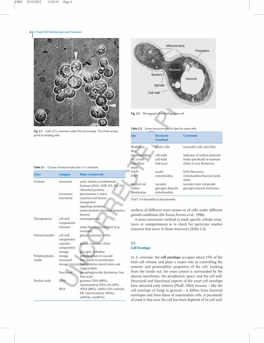
Cell structure and appearance. Yeast cells exhibit great diver-sity with respect to cell size, shape, and color. Even individualcells from a pure strain of a single species can display mor-phological heterogeneity. Additionally, profound alterationsin individual cell morphology will be induced by changingthe physical or chemical conditions at growth. Yeast cell sizevaries widely – some yeasts may be only 2–3mm in length,while other species may reach lengths of 20–50mm. Cellwidth is less variable at about 1–10mm. Under a microscope,Saccharomyces cerevisiae cells appear as ovoid or ellipsoidalstructures, surrounded by a rather thick cell wall (Figure 2.1).Mean values for the large diameter range between 5 and10mm, and for the small diameter between 1 and 7mm.Cell size in brewing strains is usually bigger than that inlaboratory strains. Mean cell size of S. cerevisiae alsoincreases with age.
With regard to cell shape, many yeast species are ellipsoi-dal or ovoid. Some, like the Schizosaccharomyces, are cylindri-cal with hemispherical ends. Candida albicans and Yarrowialipolytica, for example, are mostly filamentous (with pseudo-hyphae and septate hyphae). There are also spherical yeasts(like Debaryomyces species) or elongated forms (with manyyeasts depending on growth conditions).
In principle, the status of S. cerevisiae as a eukaryotic cell isreflected by the fact that similar macromolecular constitu-ents are assembled into the structural components of the cell(Table 2.1). There are, however, some compounds that do notoccur in mammalian cells or in cells of other higher eukar-yotes, such as those building the rigid cell wall or storagecompounds in yeast.
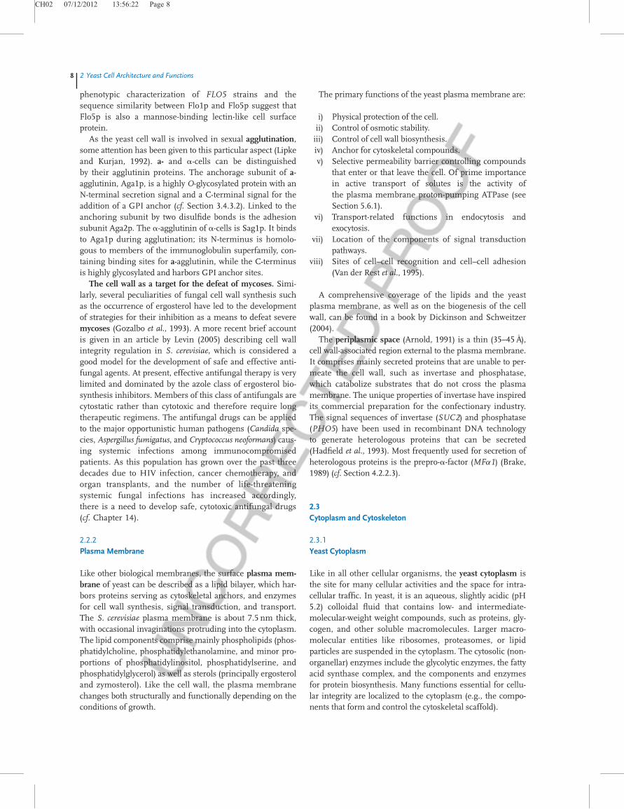
For a better understanding of what I will discuss in thefollowing sections, Figure 2.2 presents a micrograph of adividing yeast cell, indicating some of its major componentsand organelles. We will deal with the yeast envelope, the cyto-plasm, and the cell skeleton, and briefly touch upon thenucleus. The major genetic material distributed throughoutthe 16 chromosomes residing within the nucleus and othergenetic elements, such as the nucleic acids, the retrotranspo-sons, and some extrachromosomal elements, are considered
later in Chapter 5. Section 2.5 presents an overview of otheryeast cellular structures.
Preparations to view cells. Unstained yeast cells can onlybe visualized poorly by light microscopy. At 1000-fold magni-fication, it may be possible to see the yeast vacuole and cyto-solic inclusion bodies. By using phase-contrast microscopy,together with appropriate staining techniques, several cellu-lar structures become distinguishable. Fluorochromic dyes(cf. Table 2.2) can be used with fluorescence microscopy tohighlight features within the cells as well as on the cell sur-face (Pringle et al., 1991).
The range of cellular features visualized is greatlyincreased, when monospecific antibodies raised againststructural proteins are coupled to fluorescent dyes, such asfluorescein isothiocyanate (FITC) or Rhodamine B.
Flow cytometry has several applications in yeast studies(Davey and Kell, 1996). For example, fluorescence-activatedcell sorting (FACS) can monitor yeast cell cycle progression,when cell walls are labeled with concanavalin A conjugatedto FITC and cell protein with tetramethylrhodamine isothio-cyanate (TRITC). These tags enable us to collect quantitativeinformation on the growth properties of individual yeastcells as they progress through their cell cycle.
A very convenient tool to localize and even to follow themovement of particular proteins within yeast cells is the useof the Green Fluorescent Protein (GFP) from the jellyfish(Aequorea victoria) as a reporter molecule (Prasher et al.,1992), as well as several derivatives of GFP with fluorescencespectra shifted to other wavelengths (Heim et al., 1994;Heim, Cubitt, and Tsien, 1995). Fusions of genes of interestwith the fluorescent protein gene (N- or C-terminal) alsoallow us to follow the expression and destiny of the fusionproteins followed by fluorescence microscopy (Niedenthalet al., 1996; Wach et al., 1997; Hoepfner et al., 2000; see alsoChapter 4).
Organelle ultrastructure and macromolecular architecturecan only be obtained with the aid of electron microscopy,which in scanning procedures is useful for studying celltopology, while ultrathin sections are essential in transmis-sion electron microscopy to visualize intracellular fine struc-ture (Streiblova, 1988). Atomic force microscopy can beapplied to uncoated, unfixed cells for imaging the cell
j5
Yeast: Molecular and Cell Biology, Second Edition. Edited by Horst Feldmann.# 2012 Wiley-VCH Verlag GmbH & Co. KGaA. Published 2012 by Wiley-VCH Verlag GmbH & Co. KGaA.
CH02 07/12/2012 13:56:19 Page 6
surfaces of different yeast strains or of cells under differentgrowth conditions (De Souza Pereira et al., 1996).
A most convenient method to mark specific cellular struc-tures or compartments is to check for particular markerenzymes that occur in those structures (Table 2.3).
2.2Cell Envelope
In S. cerevisiae, the cell envelope occupies about 15% of thetotal cell volume and plays a major role in controlling theosmotic and permeability properties of the cell. Lookingfrom the inside out, the yeast cytosol is surrounded by theplasma membrane, the periplasmic space, and the cell wall.Structural and functional aspects of the yeast cell envelopehave attracted early interest (Phaff, 1963) because – like thecell envelope of fungi in general – it differs from bacterialenvelopes and from those of mammalian cells. A peculiarityof yeast is that once the cell has been depleted of its cell wall,
Table 2.1 Classes of macromolecules in S. cerevisiae.
Class Category Major compounds
Proteins structural actin, tubulin (cytoskeleton)histones (H2A, H2B, H3, H4, H1)ribosomal proteins
hormones pheromones a and afunctional enzymes and factors
transporterssignaling receptorsmotor proteins (myosins, kinesins,dynein)
Glycoproteins cell wallcomponents
mannoproteins
enzymes many functional enzymes (e.g.,invertase)
Polysaccharides cell wallcomponents
glucan, mannan, chitin
capsularcomponents
glucan, mannan, chitin
storage glycogen, trehalosePolyphosphates storage polyphosphate in vacuoleLipids structural free sterols in membranes
storage lipid particles (sterol esters andtriglycerides)
functional phosphoglyceride derivatives, freefatty acids
Nucleic acids DNA genomic DNA (80%),mitochondrial DNA (10–20%)
RNA rRNA (80%), mRNA (5% cytosolic,ER, mitochondria), tRNAs,snRNAs, snoRNAs
Fig. 2.1 Cells of S. cerevisiae under the microscope. The white arrows
point to dividing cells.
Fig. 2.2 Micrograph of a dividing yeast cell.
Table 2.2 Some structure-specific dyes for yeast cells.
Dye Structuresvisualized
Comments
Methyleneblue
whole cells nonviable cells stain blue
Aminoacridine cell walls indicator of surface potentialF-C ConA cell walls binds specifically to mannanCalcofluorwhite
bud scars chitin in scar fluoresces
DAPI nuclei DNA fluorescesDAPI mitochondria mitochondria fluoresce pink-
whiteNeutral red vacuoles vacuoles stain red-purpleIodine glycogen deposits glycogen stained red-brownRhodamine mitochondria
DAPI, 4,6-diamidino-2-phenylindole.
6j2 Yeast Cell Architecture and Functions

CH02 07/12/2012 13:56:22 Page 7
protoplasts are generated that are able to completely regener-ate the wall (Necas, 1971).
2.2.1Cell Wall
Yeast cell wall. The outer shell is a rigid structure about 100–200 nm thick and constituting about 25% of the total drymass of the cell (Figure 2.3). The cell wall is composed ofonly four classes of macromolecules: highly glycosylated gly-coproteins (“mannoproteins”), two types of b-glucans, andchitin. The composition of the cell wall is subject to consider-able variation according to growth conditions, and the bio-synthesis of the single compounds is highly controlled both
in space and in time. The literature that has accumulated onthese issues has grown so voluminous that reference is givenhere to only a few review articles (Klis, 1994; Lipke andOvalle, 1998; Cabib et al., 2001). Details of cell wall synthesisduring yeast growth and budding, as well as septum forma-tion (Cid et al., 1995; Cabib et al., 1997; Cabib et al., 2001;Smits, van denEnde, and Klis, 2001), are considered below.
By treatment with lytic enzymes in the presence ofosmotic stabilizers, the yeast cell wall can be removed with-out harming viability or other cellular functions. These“naked” cells are called spheroplasts. The cell wall will regen-erate and this process has been used to study aspects of cellwall biosynthesis. Spheroplasts are amenable to intergenericand intrageneric cell fusions; such hybrids are valuableinstruments in genetic studies and possess a valuable bio-technological potential. A cell wall protein that contains aputative glycosylphosphatidylinositol (GPI)-attachment site,Pst1p, is secreted by regenerating protoplasts. It is upregu-lated by activation of the cell integrity pathway, as mediatedby Rlm1p, as well as upregulated by cell wall damage via dis-ruption of the FKS1 gene, representing the catalytic subunitof glucan synthase (cf. Chapter 3).
Yeast cell aggregation. A phenomenon of particular impor-tance in brewing is flocculation. It is based on asexual cellu-lar aggregation when cells adhere, reversibly, to one another,which leads to the formation of macroscopic flocs sediment-ing out of suspension. Traditionally, brewing yeast strains aredistinguished as highly flocculent bottom yeasts (used forlager or Pilsner fermentations) or weakly flocculent topyeasts (used for ale fermentations or, in Germany, to prepare“top-fermented” beers). Although flocculation is far frombeing completely understood, it appears that the phenome-non is due to specific cell wall lectins in yeast (so-calledflocculins) – surface glycoproteins capable of directly bindingmannoproteins of adjacent cells. Yeast flocculation is geneti-cally determined by the presence of different FLO genes.One such protein is Flo1p, a lectin-like cell-surface proteinthat aggregates cells into “flocs” by binding to mannosesugar chains on the surfaces of other cells. Both the
Table 2.3 Marker enzymes for isolated yeast organelles.
Organelle Compartments Marker enzyme
Cell wall periplasm invertasesecretory pathway acid phosphatase
Plasmamembrane
vanadate-sensitive ATPase
Cytosol glucose-6-phosphatedehydrogenase
Nucleus nucleoplasm RNA polymerasenuclear envelope transmission electron
microscopyER light microsomal
fractionNADPH: cytochrome coxidoreductase
Vacuole membrane a-mannosidasesap protease A and B
Golgiapparatus
b-glucan synthase,mannosyltransferase
Mitochondrion matrix aconitase, fumaraseintermembranespace
cytochrome c peroxidase
inner membrane cytochrome c oxidaseouter membrane kynurenine hydroxylase
Peroxisome catalase, isocitrate lyase,flavin oxidase
--S-S-- --S-S----S-S--
Cytosol
Three membrane-bound synthetases:Csh1 Repair enzymeCsh2 Involved in septum formationCsh3 (Cds2) Cell wall maturation and bud-ring formation
ß-(1,3)-glucanß-(1,6)-glucan
Entrapped mannoproteins
Glucomannoproteins
Chitin ß-(1,4)-poly-N-acetylglucosamine
Plasma membrane
Fig. 2.3 Schematic representation of the yeast cell
wall.
2.2 Cell Envelopej7
CH02 07/12/2012 13:56:22 Page 8
phenotypic characterization of FLO5 strains and thesequence similarity between Flo1p and Flo5p suggest thatFlo5p is also a mannose-binding lectin-like cell surfaceprotein.
As the yeast cell wall is involved in sexual agglutination,some attention has been given to this particular aspect (Lipkeand Kurjan, 1992). a- and a-cells can be distinguishedby their agglutinin proteins. The anchorage subunit of a-agglutinin, Aga1p, is a highly O-glycosylated protein with anN-terminal secretion signal and a C-terminal signal for theaddition of a GPI anchor (cf. Section 3.4.3.2). Linked to theanchoring subunit by two disulfide bonds is the adhesionsubunit Aga2p. The a-agglutinin of a-cells is Sag1p. It bindsto Aga1p during agglutination; its N-terminus is homolo-gous to members of the immunoglobulin superfamily, con-taining binding sites for a-agglutinin, while the C-terminusis highly glycosylated and harbors GPI anchor sites.
The cell wall as a target for the defeat of mycoses. Simi-larly, several peculiarities of fungal cell wall synthesis suchas the occurrence of ergosterol have led to the developmentof strategies for their inhibition as a means to defeat severemycoses (Gozalbo et al., 1993). A more recent brief accountis given in an article by Levin (2005) describing cell wallintegrity regulation in S. cerevisiae, which is considered agood model for the development of safe and effective anti-fungal agents. At present, effective antifungal therapy is verylimited and dominated by the azole class of ergosterol bio-synthesis inhibitors. Members of this class of antifungals arecytostatic rather than cytotoxic and therefore require longtherapeutic regimens. The antifungal drugs can be appliedto the major opportunistic human pathogens (Candida spe-cies, Aspergillus fumigatus, and Cryptococcus neoformans) caus-ing systemic infections among immunocompromisedpatients. As this population has grown over the past threedecades due to HIV infection, cancer chemotherapy, andorgan transplants, and the number of life-threateningsystemic fungal infections has increased accordingly,there is a need to develop safe, cytotoxic antifungal drugs(cf. Chapter 14).
2.2.2Plasma Membrane
Like other biological membranes, the surface plasma mem-brane of yeast can be described as a lipid bilayer, which har-bors proteins serving as cytoskeletal anchors, and enzymesfor cell wall synthesis, signal transduction, and transport.The S. cerevisiae plasma membrane is about 7.5 nm thick,with occasional invaginations protruding into the cytoplasm.The lipid components comprise mainly phospholipids (phos-phatidylcholine, phosphatidylethanolamine, and minor pro-portions of phosphatidylinositol, phosphatidylserine, andphosphatidylglycerol) as well as sterols (principally ergosteroland zymosterol). Like the cell wall, the plasma membranechanges both structurally and functionally depending on theconditions of growth.
The primary functions of the yeast plasma membrane are:
i) Physical protection of the cell.ii) Control of osmotic stability.iii) Control of cell wall biosynthesis.iv) Anchor for cytoskeletal compounds.v) Selective permeability barrier controlling compounds
that enter or that leave the cell. Of prime importancein active transport of solutes is the activity ofthe plasma membrane proton-pumping ATPase (seeSection 5.6.1).
vi) Transport-related functions in endocytosis andexocytosis.
vii) Location of the components of signal transductionpathways.
viii) Sites of cell–cell recognition and cell–cell adhesion(Van der Rest et al., 1995).
A comprehensive coverage of the lipids and the yeastplasma membrane, as well as on the biogenesis of the cellwall, can be found in a book by Dickinson and Schweitzer(2004).
The periplasmic space (Arnold, 1991) is a thin (35–45A�),
cell wall-associated region external to the plasma membrane.It comprises mainly secreted proteins that are unable to per-meate the cell wall, such as invertase and phosphatase,which catabolize substrates that do not cross the plasmamembrane. The unique properties of invertase have inspiredits commercial preparation for the confectionary industry.The signal sequences of invertase (SUC2) and phosphatase(PHO5) have been used in recombinant DNA technologyto generate heterologous proteins that can be secreted(Hadfield et al., 1993). Most frequently used for secretion ofheterologous proteins is the prepro-a-factor (MFa1) (Brake,1989) (cf. Section 4.2.2.3).
2.3Cytoplasm and Cytoskeleton
2.3.1Yeast Cytoplasm
Like in all other cellular organisms, the yeast cytoplasm isthe site for many cellular activities and the space for intra-cellular traffic. In yeast, it is an aqueous, slightly acidic (pH5.2) colloidal fluid that contains low- and intermediate-molecular-weight weight compounds, such as proteins, gly-cogen, and other soluble macromolecules. Larger macro-molecular entities like ribosomes, proteasomes, or lipidparticles are suspended in the cytoplasm. The cytosolic (non-organellar) enzymes include the glycolytic enzymes, the fattyacid synthase complex, and the components and enzymesfor protein biosynthesis. Many functions essential for cellu-lar integrity are localized to the cytoplasm (e.g., the compo-nents that form and control the cytoskeletal scaffold).
8j2 Yeast Cell Architecture and Functions
CH02 07/12/2012 13:56:22 Page 9
2.3.2Yeast Cytoskeleton
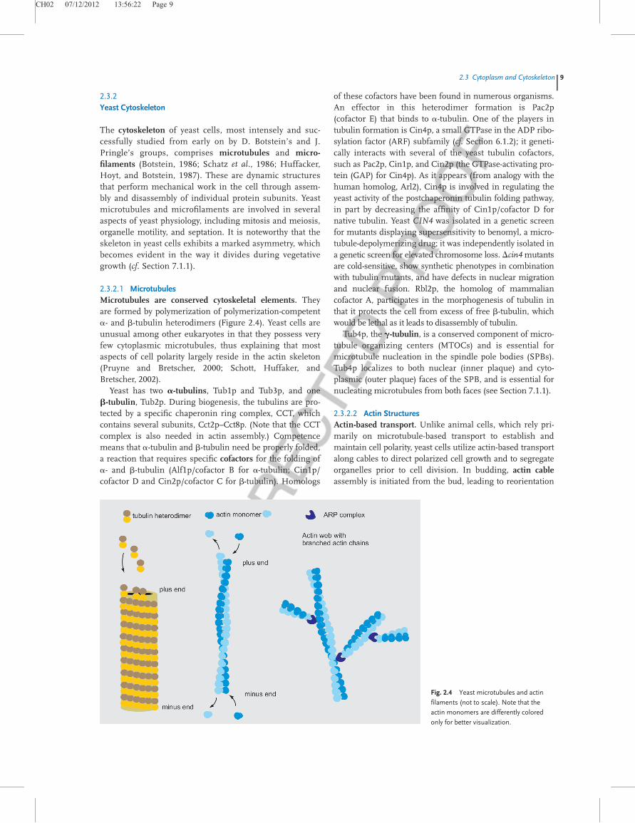
The cytoskeleton of yeast cells, most intensely and suc-cessfully studied from early on by D. Botstein’s and J.Pringle’s groups, comprises microtubules and micro-filaments (Botstein, 1986; Schatz et al., 1986; Huffacker,Hoyt, and Botstein, 1987). These are dynamic structuresthat perform mechanical work in the cell through assem-bly and disassembly of individual protein subunits. Yeastmicrotubules and microfilaments are involved in severalaspects of yeast physiology, including mitosis and meiosis,organelle motility, and septation. It is noteworthy that theskeleton in yeast cells exhibits a marked asymmetry, whichbecomes evident in the way it divides during vegetativegrowth (cf. Section 7.1.1).
2.3.2.1 MicrotubulesMicrotubules are conserved cytoskeletal elements. Theyare formed by polymerization of polymerization-competenta- and b-tubulin heterodimers (Figure 2.4). Yeast cells areunusual among other eukaryotes in that they possess veryfew cytoplasmic microtubules, thus explaining that mostaspects of cell polarity largely reside in the actin skeleton(Pruyne and Bretscher, 2000; Schott, Huffaker, andBretscher, 2002).
Yeast has two a-tubulins, Tub1p and Tub3p, and oneb-tubulin, Tub2p. During biogenesis, the tubulins are pro-tected by a specific chaperonin ring complex, CCT, whichcontains several subunits, Cct2p–Cct8p. (Note that the CCTcomplex is also needed in actin assembly.) Competencemeans that a-tubulin and b-tubulin need be properly folded,a reaction that requires specific cofactors for the folding ofa- and b-tubulin (Alf1p/cofactor B for a-tubulin; Cin1p/cofactor D and Cin2p/cofactor C for b-tubulin). Homologs
of these cofactors have been found in numerous organisms.An effector in this heterodimer formation is Pac2p(cofactor E) that binds to a-tubulin. One of the players intubulin formation is Cin4p, a small GTPase in the ADP ribo-sylation factor (ARF) subfamily (cf. Section 6.1.2); it geneti-cally interacts with several of the yeast tubulin cofactors,such as Pac2p, Cin1p, and Cin2p (the GTPase-activating pro-tein (GAP) for Cin4p). As it appears (from analogy with thehuman homolog, Arl2), Cin4p is involved in regulating theyeast activity of the postchaperonin tubulin folding pathway,in part by decreasing the affinity of Cin1p/cofactor D fornative tubulin. Yeast CIN4 was isolated in a genetic screenfor mutants displaying supersensitivity to benomyl, a micro-tubule-depolymerizing drug; it was independently isolated ina genetic screen for elevated chromosome loss. Dcin4mutantsare cold-sensitive, show synthetic phenotypes in combinationwith tubulin mutants, and have defects in nuclear migrationand nuclear fusion. Rbl2p, the homolog of mammaliancofactor A, participates in the morphogenesis of tubulin inthat it protects the cell from excess of free b-tubulin, whichwould be lethal as it leads to disassembly of tubulin.
Tub4p, the g-tubulin, is a conserved component of micro-tubule organizing centers (MTOCs) and is essential formicrotubule nucleation in the spindle pole bodies (SPBs).Tub4p localizes to both nuclear (inner plaque) and cyto-plasmic (outer plaque) faces of the SPB, and is essential fornucleating microtubules from both faces (see Section 7.1.1).
2.3.2.2 Actin StructuresActin-based transport. Unlike animal cells, which rely pri-marily on microtubule-based transport to establish andmaintain cell polarity, yeast cells utilize actin-based transportalong cables to direct polarized cell growth and to segregateorganelles prior to cell division. In budding, actin cableassembly is initiated from the bud, leading to reorientation
Fig. 2.4 Yeast microtubules and actin
filaments (not to scale). Note that the
actin monomers are differently colored
only for better visualization.
2.3 Cytoplasm and Cytoskeletonj9
CH02 07/12/2012 13:56:23 Page 10
of actin cables, and thus targeting of growth and secretion tothe future bud tip (cf. Section 7.1). Polarized growth towardsthe bud tip (or cap) continues through a medium-buddedstage, and depends on actin cables emanating from the budtip and neck. These cables serve as polarized tracks for typeV myosin-dependent delivery of cargos needed to build thedaughter cell.
Types of actin filaments. Actin is an ATP-binding proteinthat exists both in monomeric (G-actin) and filamentous (F-actin) forms. Actin is encoded in yeast by the single geneACT1 (Ng and Abelson, 1980). Actin filaments areassembled by the reversible polymerization of monomersand have an intrinsic polarity; the fast-growing end is calledthe barbed end and the slow-growing end is called thepointed end (Figure 2.4). Yeast cells contain three types offilamentous actin structures: (i) actin cables, (ii) an actin-myosin contractile ring (Bi et al., 1998), and (iii) actin corticalpatches, all of which are subjected to extensivereorganization throughout the cell cycle. Actin cables serveas tracks for polarized secretion, organelle and mRNA trans-port, and mitotic spindle alignment. The actin–myosin con-tractile ring forms transiently at the mother–daughter neckand is important for cytokinesis. Cortical patches arebranched actin filaments involved in endocytosis and mem-brane growth and polarity. Genetic screens and biochemicalpurifications have been fruitful in identifying numerous fac-tors that regulate actin cytoskeleton dynamics, organization,and function (review: Moseley and Goode, 2006).
Assembly of actin filaments. The S. cerevisiae genome en-codes two genes, BNI1 and BNR1, that are members of theformin family assembling linear actin cables in the bud andbud neck, respectively. Formins constitute a well-conservedfamily of proteins that promote the assembly of actin fila-ments, which are necessary in remodeling of the actin cyto-skeleton during such processes as budding, mating,cytokinesis, or endocytosis (and in higher cells, cell adhesionand migration). The formin proteins are characterized by thepresence of two highly conserved FH (formin homology)domains: the FH1 domain, containing polyproline motifsthat mediate binding to profilin (actin- and phosphatidylino-sitol-4,5-bisphosphate (PI(4,5)P2)-binding protein, Pfy1p),which in turn binds actin monomers, and the FH2 domain,which nucleates actin assembly. The FH2 domains of Bni1pand Bnr1p are distinct from those of the metazoan groups,containing a yeast-specific insert that is not found in otherorganisms. In addition to FH1 and FH2 domains, forminscontain a regulatory Rho-binding domain (RBD) and a Dia-autoregulatory domain (DAD).
A model for formin-mediated actin assembly has sug-gested the following sequence of events. Activated Rho pro-tein binds to the formin RBD domain and releases theformin from a conformation in which it is autoinhibited(due to an interaction between its N- and C-termini) to adopta conformation that exposes the FH1 and FH2 domains. TheFH1 domain then interacts with profilin-bound actin mono-mers, handing them over to the FH2 domain, a dimeric
structure that may interact with two actin monomers to stabi-lize a dimeric actin form, prior to polymerization, wherebyactin cables are formed. The FH2 domain remains associ-ated with the growing end of the filament to protect it frominteraction with capping proteins (a FH2 function termed“processive capping”).
Consistent with this model, Bni1p has been identified as adownstream target of Rho1p, which regulates reorganizationof the actin cytoskeleton, and hence the process of bud for-mation (cf. Section 7.1.1). Additionally, Bni1p activation isregulated by the small GTPases Rho3p and Rho4p, whichaffect the inhibitory interaction between the RBD and theDAD domains in the formin, while the Rho protein Cdc42pis needed for proper cable assembly during initiation of budgrowth. Bni1p autoinhibition (as mentioned before) can alsobe aborted by phosphorylation of Bni1p affected by Prk1pkinase. Support for the model also comes from crystal struc-ture studies of the Bni1p FH2 domain complexed with actin.
Actin filament assembly. Long actin filament bundles areformed by Crn1p (coronin) (Rybakin and Clemen, 2005),which binds actin filaments (F-actin) and cross-links them.Crn1p also regulates the actin filament nucleation and theformation of branched actin filaments as found in corticalpatches. Crn1p is composed of five N-terminal WD repeats,forming a b-propeller structure, a microtubule bindingdomain, and a C-terminal a-helical coiled-coil structure,whereby the b-propeller and coiled-coil domains arerequired for recruitment of Crn1p to cortical patches.
The highly conserved actin nucleation center required forthe motility and integrity of actin patches, involved in endo-cytosis and membrane growth, is the Arp2/3 complex. Inyeast, the complex consists of seven proteins, two of which(Arp2p and Arp3p) are actin-related, while five components(Arc15p, Arc18p, Arc19p, Arc35p, and Arc40p) are non-actin-related proteins (Winter et al., 1997; Evangelista et al.,2002). The Arp2/3 complex nucleates the formation ofbranched actin filaments by binding to the side of an existing(mother) filament and nucleating the formation of a new(daughter) actin filament at a 708 angle (Figure 2.4). Arp2pand Arp3p serve as the first two subunits of the daughter fila-ment, likely mimicking actin monomers due to their struc-tural similarity to actin. However, the Arp2/3 complex doesnot play a role in the formation of actin cables (unbranchedactin structures). To achieve optimal actin nucleation activity,the Arp2/3 complex is assisted by an assembly protein, suchas Las17p (also Bee1p, of the SCAR/WASP family), myosin I,Abp1p (Olazabal and Machesky, 2001), or Pan1p.
Las17p/Bee1p as an activator of the Arp2/3 protein com-plex is the only S. cerevisiae homolog of the human Wiskott–Aldrich syndrome protein (WASP), which itself is a memberof the larger WASP/SCAR/WAVE protein family. Las17p wasidentified biochemically as an essential nucleation factor inthe reconstitution of cortical actin patches. Las17p localizeswith the Arp2/3 complex to actin patches; disruption ofLAS17 leads to the loss of actin patches and a block in endo-cytosis. In the physical interaction between Las17p and the
10j2 Yeast Cell Architecture and Functions
CH02 07/12/2012 13:56:24 Page 11
Arp2/3 complex, the C-terminal WA (WH2 (WASP homol-ogy 2) and A (acidic)) domain of Las17p are required as arethe two subunits of the Arp2/3 complex, Arc15p andArc19p. The WA domain is sufficient for Arp2/3 complexbinding and activation; it shares sequence similarity with anacidic domain in myosin type I (Myo3p and Myo5p in S. cer-evisiae), which also interacts with the Arp2/3 complex.Genetic and biochemical studies have identified numerousproteins that physically interact with Las17p. The WH1domain of Las17p binds strongly to verprolin (Vrp1p/End5p(Thanabalu and Munn, 2001)), the yeast homolog of humanWIP (WASP-interacting protein), which is involved inLas17p localization. The proline-rich region of Las17p bindsto SH3 domain-containing proteins, including Sla1p (anactin patch protein with a role in endocytosis) and manyothers that may regulate the activity of Las17p.
Two other proteins involved in formation and stabilizationof actin bundles in cables and patches are Sac6p (fimbrin)and Scp1p (calponin/transgelin), which work together. Thestabilization of actin filaments in patches also strictlydepends on capping of the “barbed” ends by small cappingproteins, Cap1p and Cap2p.
Actin filament disassembly. Debranching of the actin fila-ments in cortical patches by the Arp2/3 complex is inducedby Gmf2p/Aim7p, which also inhibits further actin nuclea-tion (Gandhi et al., 2010). The protein has similarity to yeastCof1p (cofilin) and to the human glia maturation factor(GMF). Cofilin, Cof1p, promotes actin filamentdepolarization in a pH-dependent manner. It binds bothactin monomers and filaments; its main task is to sever fila-ments (Moon et al., 1993; Theriot, 1997). Cofilin is regulated
by phosphorylation at Ser4; homologs are ubiquitous andessential in eukaryotes. Aip1p promotes filament dis-assembly by enhancing cofilin severing and protecting sev-ered filaments by capping.
Scd5p is an essential protein that colocalizes with corticalactin and as an adapter protein functionally links corticalactin organization with endocytosis. Scd5p and the clathrinheavy and light chains (Chc1p and Clc1p, respectively) physi-cally associate with Sla2p (Wesp et al., 1997), a trans-membrane actin-binding protein involved in membranecytoskeleton assembly and cell polarization, which is also ahomolog of the mammalian huntingtin-interacting proteinHIP1 and the related HIP1R. Both Scd5p and clathrin arerequired for Sla2p localization at the cell cortex. Scd5pactivity appears to be regulated by phosphorylation/dephosphorylation. Phosphorylation of Scd5p by proteinkinase Prk1p results in its negative regulation, whereasdephosphorylation by the Glc7p type 1 protein phosphataserelieves this inhibition. Mutations in GLC7 that abolishGlc7p interactions with Scd5p result in defects in endocyto-sis and actin organization. Loss of function scd5mutants suf-fer from defects in receptor-mediated endocytosis andnormal actin organization. They exhibit larger and depolar-ized cortical actin patches and a prevalence of G-actin bars.
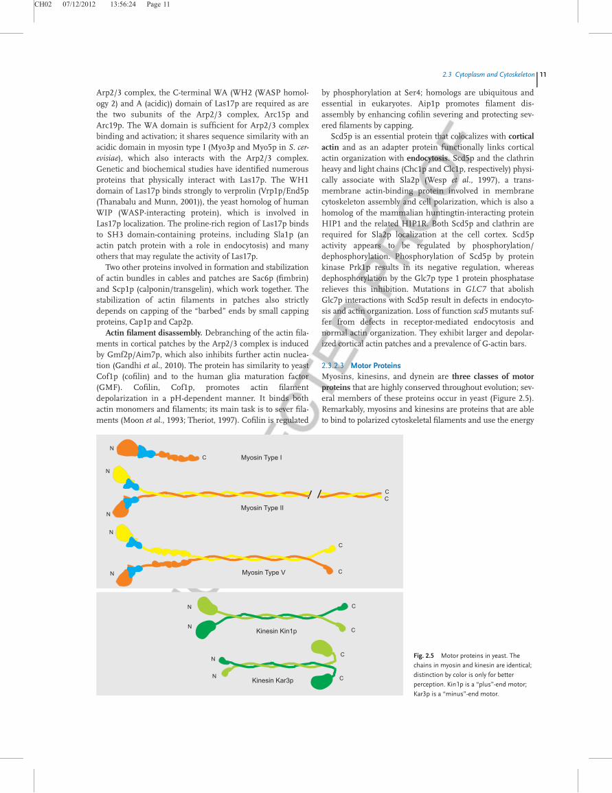
2.3.2.3 Motor ProteinsMyosins, kinesins, and dynein are three classes of motorproteins that are highly conserved throughout evolution; sev-eral members of these proteins occur in yeast (Figure 2.5).Remarkably, myosins and kinesins are proteins that are ableto bind to polarized cytoskeletal filaments and use the energy
Myosin Type II
Myosin Type V
Kinesin Kin1p
Kinesin Kar3p
Myosin Type IN
N
N
N
N
N
N
N
N
C
C
C
C
C
C
C
CC
Fig. 2.5 Motor proteins in yeast. The
chains in myosin and kinesin are identical;
distinction by color is only for better
perception. Kin1p is a “plus”-end motor;
Kar3p is a “minus”-end motor.
2.3 Cytoplasm and Cytoskeletonj11
CH02 07/12/2012 13:56:25 Page 12
derived from repeated cycles of ATP hydrolysis to movealong them. By unidirectional movement, these moleculescan carry cargo from one point to a distant location withinthe cell; other motor proteins may cause filaments to slideagainst each other, so that the generated force drives pro-cesses like nuclear migration and cell division (Hoyt,Hyman, and B€ahler, 1997; Moore and Cooper, 2010).
2.3.2.3.1 Myosins Myosins are rod-like, extended struc-tures (around 2 nm wide and greater than 150 nm long) nor-mally consisting of two heavy and four light chains, wherebythe heavy chains wrap around each other to form a coiled-coil of two a-helices (called the tail), while the light chainsare part of motor domains at the N-terminus (called thehead); between the head and tail are so-called IQ domains.Of the 14 different types of myosins in the myosin super-family, S. cerevisiae has members of type I, type II, andtype V (Brown, 1997). Type I members are characterizedby the occurrence of only one head per molecule, whereastype II members carry two heads, and type V members havetwo extended head regions.
Type II myosins. The only type II myosin in yeast isMyo1p; it fulfills a specialized function as part of the ring-shaped actomyosin complex that (early in the cell cycle)localizes to the presumptive bud site and remains at themother–bud neck until cytokinesis is completed (VerPlankand Li, 2005). Formation, but not maintenance, of this con-tractile ring requires the intact septin collar at the bud neck(cf. Section 7.2). Late in anaphase, F-actin Act1p and theIQGAP-related protein, Iqg1p (Epp and Chant, 1997), alsoaccumulate in the neck ring, whereby incorporation ofF-actin depends on Myo1p, and Iqg1p determines the local-ization of axial markers Bud4p and Cdc12p. At the end ofanaphase, the actinomyosin ring begins to contract. Myo1pis regulated by two light chains, an essential light chain(ELC), Mlc1p, and a regulatory light chain (RLC), Mlc2p,which displays significant sequence homology to calmodulinor myosin light chain related proteins. Like other lightchains, Mlc2p contains an EF hand and a phosphorylatableserine residue, both close to the N-terminus. Mlc1p interactswith one of the two motifs (IQ1), which, however, does notplay a major role in regulating Myo1p; instead, this interac-tion regulates actin ring formation and targeted secretionthrough further interactions with Myo1p, Iqg1p, and Myo2p.Mlc2p interacts with the IQ2 motif and most likely plays arole in the disassembly of the Myo1p ring. The human coun-terpart to Myo1p, MYH11, may give rise to leukemia orfamilial aortic aneurysm.
Type V myosin subfamily. Myo2p and Myo4p belong tothe type V myosin subfamily. Myo2p promotes polarizedgrowth by orienting the mitotic spindle and by taking overthe vectorial transport of organelles along actin cables to sitessuch as the growing bud during vegetative growth, the budneck during cytokinesis, and the shmoo tip during mating.Even organelles, including secretory vesicles, vacuoles, per-oxisomes, and late Golgi elements, are transported into the
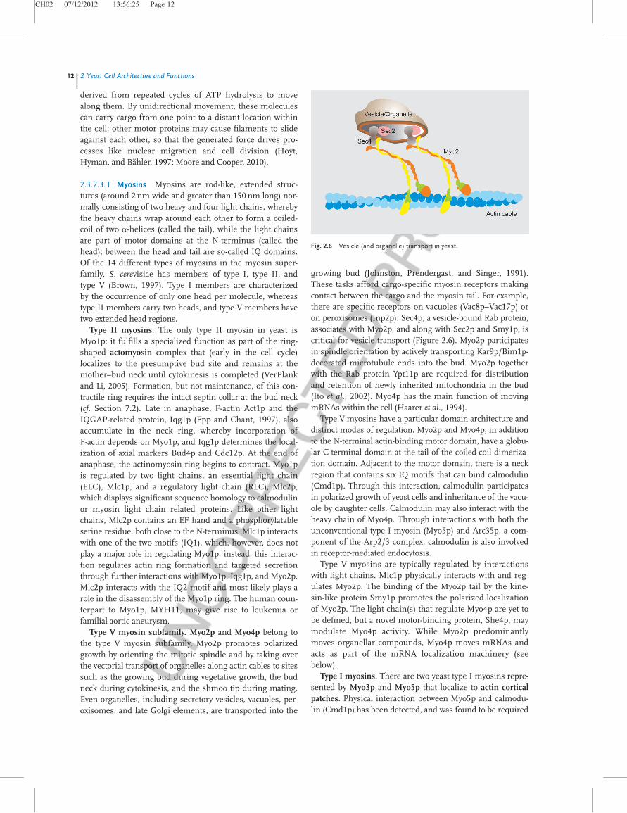
growing bud (Johnston, Prendergast, and Singer, 1991).These tasks afford cargo-specific myosin receptors makingcontact between the cargo and the myosin tail. For example,there are specific receptors on vacuoles (Vac8p–Vac17p) oron peroxisomes (Inp2p). Sec4p, a vesicle-bound Rab protein,associates with Myo2p, and along with Sec2p and Smy1p, iscritical for vesicle transport (Figure 2.6). Myo2p participatesin spindle orientation by actively transporting Kar9p/Bim1p-decorated microtubule ends into the bud. Myo2p togetherwith the Rab protein Ypt11p are required for distributionand retention of newly inherited mitochondria in the bud(Ito et al., 2002). Myo4p has the main function of movingmRNAs within the cell (Haarer et al., 1994).
Type V myosins have a particular domain architecture anddistinct modes of regulation. Myo2p and Myo4p, in additionto the N-terminal actin-binding motor domain, have a globu-lar C-terminal domain at the tail of the coiled-coil dimeriza-tion domain. Adjacent to the motor domain, there is a neckregion that contains six IQ motifs that can bind calmodulin(Cmd1p). Through this interaction, calmodulin participatesin polarized growth of yeast cells and inheritance of the vacu-ole by daughter cells. Calmodulin may also interact with theheavy chain of Myo4p. Through interactions with both theunconventional type I myosin (Myo5p) and Arc35p, a com-ponent of the Arp2/3 complex, calmodulin is also involvedin receptor-mediated endocytosis.
Type V myosins are typically regulated by interactionswith light chains. Mlc1p physically interacts with and reg-ulates Myo2p. The binding of the Myo2p tail by the kine-sin-like protein Smy1p promotes the polarized localizationof Myo2p. The light chain(s) that regulate Myo4p are yet tobe defined, but a novel motor-binding protein, She4p, maymodulate Myo4p activity. While Myo2p predominantlymoves organellar compounds, Myo4p moves mRNAs andacts as part of the mRNA localization machinery (seebelow).
Type I myosins. There are two yeast type I myosins repre-sented by Myo3p and Myo5p that localize to actin corticalpatches. Physical interaction between Myo5p and calmodu-lin (Cmd1p) has been detected, and was found to be required
Fig. 2.6 Vesicle (and organelle) transport in yeast.
12j2 Yeast Cell Architecture and Functions
CH02 07/12/2012 13:56:26 Page 13
for endocytosis. Myo5p also interacts physically with verpro-lin (Vrp1p), a proline-rich protein. Deletion of the geneVRP1 causes delocalization of Myo5p-containing patches.
Tropomyosin. In addition to the myosins, yeast harborstwo isoforms of tropomyosin. Tmp1p is the major isoformthat binds to and stabilizes actin cables and filaments, whichdirect polarized cell growth and the distribution of severalorganelles. The protein is acetylated by the NatB complex;the acetylated form will bind actin more efficiently. Tmp2p,the minor isoform, largely has functions overlapping withthose of Tmp1p.
2.3.2.3.2 Kinesins Both kinesins and kinesin-related pro-teins are motor proteins remarkably similar to type V myo-sins. They generally function in mitotic spindle assemblyand organization (see also Section 7.2.2.2), although eachone takes over specialized functions. Cin8p, a kinesin motorprotein, has an additional role in chromosome segregation.Functionally redundant with Cin8p is the kinesin-relatedmotor protein Kip1(Cin9p), which, however, has an addi-tional role in partitioning the 2 mm plasmid. The kinesin-related motor protein Kip2p stabilizes microtubules by tar-geting Bik1p, a microtubule-associated protein and compo-nent of the interface between microtubules and kinetochore(Berlin, Styles, and Fink, 1990; Moore and Cooper, 2010), tothe plus end; Kip2p levels are controlled during the cell cycle.Kip3p is a further kinesin-related protein involved in spindlepositioning. Cik1p is a kinesin-associated protein that stablyand specifically targets the karyogamy protein Kar3p, aminus-end-directed microtubule motor that functions inmitosis and meiosis, localizes to the SPB, and is required fornuclear fusion during mating. Smy1p, a protein whose N-ter-minal domain is related to the motor domain of kinesins andthat interacts with Myo2p, has already been mentioned; itmay be required for exocytosis.
2.3.2.3.3 Dynein Cytoplasmic dynein, Dyn1p (Pac6p), isthe largest motor protein in yeast and a “minus”-endmotor of microtubules. Dyn1p is active in the movementof the mitotic spindle that must move into the narrowneck between the mother cell and the bud in order to seg-regate duplicated chromosomes accurately. The processbegins with the dynactin complex, directing spindle orien-tation and nuclear migration. This complex is composedof the actin-related protein Arp1p, together with Jnm1p(Pac3p) and Nip100p (Pac13p).
The movement of the spindle occurs in two main steps aspart of nuclear migration into the neck region. (i) Thenucleus moves to a position adjacent to the neck, a processinvolving cytoplasmic microtubules, the motor proteinKip3p, and Kar9p, a karyogamy protein required for correctpositioning of the mitotic spindle and for orienting cyto-plasmic microtubules; Kar9p localizes to the shmoo tip inmating cells and to the tip of the growing bud. (ii) Themitotic spindle is moved into the neck, which requires cyto-plasmic microtubules from the SPB sliding along the bud
cortex, and pulling the nucleus and the elongating spindle.Sliding depends on the heavy chain of cytoplasmic dynein(Dyn1p), the dynactin complex, and the regulators Num1p(Pac12p) and Ndl1p. In the second step, Pac1p functions inaiding the recruitment of dynein to the “plus” ends of micro-tubules. In this function, Pac1p is regulated by Ndl1p, ahomolog of nuclear distribution factor NudE that interactswith Pac1p (Li, Lee, and Cooper, 2005). Cortical Num1pbrings together the dynein intermediate chain Pac11p andthe cytoplasmic microtubules (Farkasovsky and Kuntzel,2001). Finally, Bim1p, a microtubule-binding protein, alsoknown as Yeb1p (EB1, microtubule plus-end binding)together with Kar9p serves as the cortical microtubule cap-ture site. In case the spindle is oriented abnormally, Bim1pwill delay the exit from mitosis (Schwartz, Richards, andBotstein, 1997; Miller, Cheng, and Rose, 2000; Moore,Stuchell-Brereton, and Cooper, 2009).
2.3.2.4 Other Cytoskeletal Factors
2.3.2.4.1 Proteins Interacting with the Cytoskeleton Otherproteins that have been implicated in actin cytoskeletonreorganization and establishment of cell polarity are the pro-teins Boi1p and its functionally redundant homolog Boi2p.Both Boi1p and Boi2p contain SH3, pleckstrin homology(PH), and proline-rich domains. Several structure–functionand genetic analysis experiments have tried to determinewhich domains are important for interactions with other pro-teins involved in the above processes. These studies showedthat the Boi proteins interact physically and/or geneticallywith Bem1p, another SH3 domain protein, as well as threeRho-type GTPases – Cdc42p, Rho3p and the Rho3-relatedRho4p (cf. Section 7.1.1).
Stt4p, the phosphatidylinositol-4-kinase involved in sphin-golipid biosynthesis and in regulation of the intracellulartransport of aminophospholipid phosphatidylserine fromthe endoplasmic reticulum (ER) to the Golgi, is required foractin cytoskeleton organization as well. Stt4p binds to theplasma membrane via the protein Sfk1p, thus promotingcell wall synthesis, actin cytoskeleton organization, and theRho1/Pkc1-mediated mitogen-activated protein (MAP)kinase cascade (cf. Section 10.2). STT4 is an essential gene insome backgrounds, but not in others. Dstt4 mutants lackmost of the phosphatidylinositol-4-kinase activity that isdetected in the wild-type and are arrested in the G2/M phaseof the cell cycle. Inactivation of Stt4p results in mislocaliza-tion of the Rho-GTPase guanine nucleotide exchange factor(GEF) Rom2p and also in the rapid attenuation of translationinitiation.
2.3.2.4.2 Transport of Organellar Components Of impor-tance for the proper transfer of organellar components to thebud or, on the contrary, to restrict certain compounds to beaccumulated in the bud is a specific mRNA localizationmachinery that becomes active during budding. In particular,mating-type switching should occur only in mother cells,
2.3 Cytoplasm and Cytoskeletonj13
CH02 07/12/2012 13:56:26 Page 14
meaning that HO transcription in daughter cells has to be pre-vented (cf. Chapter 7). This effect is brought about byAsh1p, a protein specifically localized to daughter nucleilate in the cell cycle, where it is poised to inhibit HO tran-scription in the following G1 phase. This asymmetriclocalization is achieved by the delivery of ASH1 mRNA todaughter cells by the products of the SHE genes. She2pand Loc1p bind to ASH1 mRNA in mother and daughternuclei, and mediate export to the cytoplasm. She3p thenassociates with the ribonucleoprotein particle (mRNP) andacts as an adapter for its association with myosin Myo4p(also called She1p). Myo4p transports the mRNP complexalong actin cables to the bud tip. During telophase, ASH1mRNA becomes anchored to the bud tip by Bni1p and/orHek2p and/or Bud6p. Translation of ASH1 mRNA isdelayed as long as the message is in transit.
2.4Yeast Nucleus
2.4.1Overview
The nuclear structure in yeasts is a nearly round organelle ofabout 1.5mm diameter located in the center of the cell orslightly excentrically. The nucleoplasm is surrounded by adouble membrane bilayer (inner and outer nuclear mem-brane), thus separating the nucleoplasm from the cytoplasm.Nuclear pore complexes (NPCs) of about 50–100 nm indiameter form the natural channels for exchange of compo-nents between the nucleus and cytosol, whereby export andimport pathways can be distinguished (see Section 8.2). Theouter nuclear membrane is largely contiguous with themembrane of the ER. Unlike most eukaryotic cells, the yeastnuclear membrane is not resolved during mitosis, while isbreaks down in higher eukaryotic cells. This latter fact neces-sitates the resynthesis of the complete nuclear structure,including nuclear pores, for example, in animal cells. None-theless, biogenesis of nuclear pores has also been studied inyeast, as de novo synthesis has to occur also in this organism(D’Angelo and Hetzer, 2008).
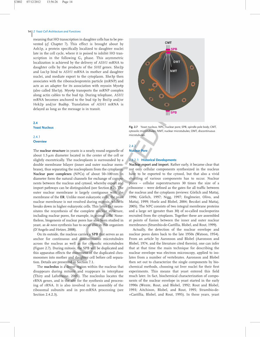
On its outside, the nucleus carries a SPB that serves as ananchor for continuous and discontinuous microtubulesacross the nucleus as well as for cytosolic microtubules(Figure 2.7). During mitosis, the SPB will be duplicated andthis apparatus effects the movement of the duplicated chro-mosomes into mother and daughter cell before cell separa-tion. Details are presented in Section 7.1.
The nucleolus is a dense region within the nucleus thatdisappears during mitosis and reappears in interphase(Thiry and Lafontaine, 2005). The nucleolus locates therRNA genes, and is the site for the synthesis and process-ing of rRNA. It is also involved in the assembly of theribosomal subunits and in pre-mRNA processing (seeSection 2.4.2.3).
2.4.2Nuclear Pore
2.4.2.1 Historical DevelopmentsNuclear export and import. Rather early, it became clear thatnot only cellular components synthesized in the nucleushave to be exported to the cytosol, but that also a vividshuttling of various components has to occur. Nuclearpores – cellular superstructures 30 times the size of aribosome – were defined as the gates for all traffic betweenthe nucleus and the cytoplasm (reviews: G€orlich and Mattaj,1996; G€orlich, 1997; Nigg, 1997; Englmeier, Olivo, andMattaj, 1999; Hoelz and Blobel, 2004; Becskei and Mattaj,2005). The NPC consists of two integral membrane proteinsand a large set (greater than 30) of so-called nucleoporinsrecruited from the cytoplasm. Together these are assembledat points of fusion between the inner and outer nuclearmembranes (Strambio-de-Castillia, Blobel, and Rout, 1999).
Actually, the detection of the nuclear envelope andnuclear pores dates back to the late 1950s (Watson, 1954).From an article by Aaronson and Blobel (Aaronson andBlobel, 1974, and the literature cited therein), one can inferthat at that time the main technique for describing thenuclear envelope was electron microscopy, applied to iso-lates from a number of vertebrates. Aaronson and Blobelthen set out to characterize the single components by bio-chemical methods, choosing rat liver nuclei for their firstexperiments. This means that yeast entered this fieldmuch later. In fact, biochemical characterization of compo-nents of the nuclear envelope in yeast started in the early1990s (Wente, Rout, and Blobel, 1992; Rout and Blobel,1993; Aitchison, Blobel, and Rout, 1995; Strambio-de-+Castillia, Blobel, and Rout, 1995). In these years, yeast
Fig. 2.7 Yeast nucleus. NPC, nuclear pore; SPB, spindle pole body; CMT,
cytosolic microtubules; NMT, nuclear microtubules; DMT, discontinuous
microtubules.
14j2 Yeast Cell Architecture and Functions
CH02 07/12/2012 13:56:27 Page 15
factors implicated in nuclear import and export were alsocharacterized (Corbett et al., 1995; Koepp and Silver, 1996).By the end of the 1990s, a rather comprehensive descrip-tion of the components of the yeast nuclear pore (which issomewhat simpler than that of metazoans) was available(Rout et al., 2000). Likewise, the majority of the yeast andvertebrate components, as well as interesting aspects ofnuclear traffic, were described (Ryan and Wente, 2000;Vasu and Forbes, 2001).
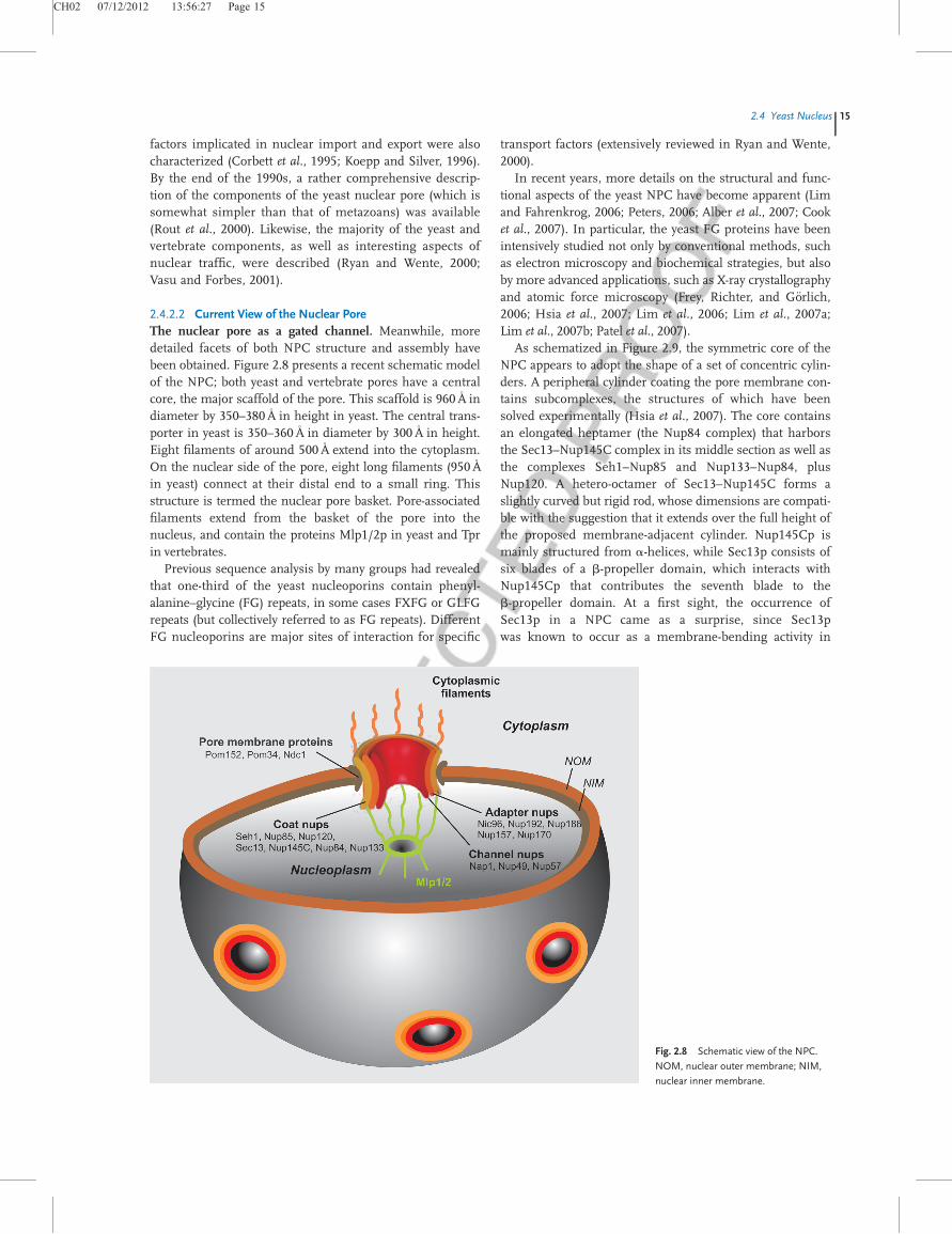
2.4.2.2 Current View of the Nuclear PoreThe nuclear pore as a gated channel. Meanwhile, moredetailed facets of both NPC structure and assembly havebeen obtained. Figure 2.8 presents a recent schematic modelof the NPC; both yeast and vertebrate pores have a centralcore, the major scaffold of the pore. This scaffold is 960A
�in
diameter by 350–380A�in height in yeast. The central trans-
porter in yeast is 350–360A�in diameter by 300A
�in height.
Eight filaments of around 500A�extend into the cytoplasm.
On the nuclear side of the pore, eight long filaments (950A�
in yeast) connect at their distal end to a small ring. Thisstructure is termed the nuclear pore basket. Pore-associatedfilaments extend from the basket of the pore into thenucleus, and contain the proteins Mlp1/2p in yeast and Tprin vertebrates.
Previous sequence analysis by many groups had revealedthat one-third of the yeast nucleoporins contain phenyl-alanine–glycine (FG) repeats, in some cases FXFG or GLFGrepeats (but collectively referred to as FG repeats). DifferentFG nucleoporins are major sites of interaction for specific
transport factors (extensively reviewed in Ryan and Wente,2000).
In recent years, more details on the structural and func-tional aspects of the yeast NPC have become apparent (Limand Fahrenkrog, 2006; Peters, 2006; Alber et al., 2007; Cooket al., 2007). In particular, the yeast FG proteins have beenintensively studied not only by conventional methods, suchas electron microscopy and biochemical strategies, but alsoby more advanced applications, such as X-ray crystallographyand atomic force microscopy (Frey, Richter, and G€orlich,2006; Hsia et al., 2007; Lim et al., 2006; Lim et al., 2007a;Lim et al., 2007b; Patel et al., 2007).
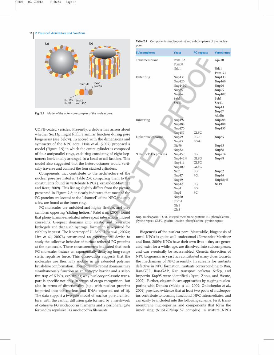
As schematized in Figure 2.9, the symmetric core of theNPC appears to adopt the shape of a set of concentric cylin-ders. A peripheral cylinder coating the pore membrane con-tains subcomplexes, the structures of which have beensolved experimentally (Hsia et al., 2007). The core containsan elongated heptamer (the Nup84 complex) that harborsthe Sec13–Nup145C complex in its middle section as well asthe complexes Seh1–Nup85 and Nup133–Nup84, plusNup120. A hetero-octamer of Sec13–Nup145C forms aslightly curved but rigid rod, whose dimensions are compati-ble with the suggestion that it extends over the full height ofthe proposed membrane-adjacent cylinder. Nup145Cp ismainly structured from a-helices, while Sec13p consists ofsix blades of a b-propeller domain, which interacts withNup145Cp that contributes the seventh blade to theb-propeller domain. At a first sight, the occurrence ofSec13p in a NPC came as a surprise, since Sec13pwas known to occur as a membrane-bending activity in
Fig. 2.8 Schematic view of the NPC.
NOM, nuclear outer membrane; NIM,
nuclear inner membrane.
2.4 Yeast Nucleusj15
CH02 07/12/2012 13:56:33 Page 16
COPII-coated vesicles. Presently, a debate has arisen aboutwhether Sec13p might fulfill a similar function during porebiogenesis (see below). In accord with the dimensions andsymmetry of the NPC core, Hsia et al. (2007) proposed amodel (Figure 2.9) in which the entire cylinder is composedof four antiparallel rings, each ring consisting of eight hep-tamers horizontally arranged in a head-to-tail fashion. Thismodel also suggested that the hetero-octamer would verti-cally traverse and connect the four stacked cylinders.
Components that contribute to the architecture of thenuclear pore are listed in Table 2.4, comparing them to theconstituents found in vertebrate NPCs (Fernandez-Martinezand Rout, 2009). This listing slightly differs from the picturepresented in Figure 2.8; it clearly indicates that most of theFG proteins are located to the “channel” of the NPC and onlya few are found at the inner ring.
FG molecules are unfolded and highly flexible, and theycan form opposing “sliding helices.” Patel et al. (2007) foundthat phenylalanine-mediated inter-repeat interactions indeedcross-link G-repeat domains into elastic and reversiblehydrogels and that such hydrogel formation is required forviability in yeast. The laboratory of U. Aebi (Lim et al., 2007a;Lim et al., 2007b) constructed an experimental device tostudy the collective behavior of surface-tethered FG proteinsat the nanoscale. These measurements indicated that suchFG molecules induce an exponentially decaying long-rangesteric repulsive force. This observation suggests that themolecules are thermally mobile in an extended polymerbrush-like conformation. Therefore, FG-repeat domains maysimultaneously function as an entropic barrier and a selec-tive trap of NPCs, explaining why nucleocytoplasmic trans-port is specific not only in terms of cargo recognition, butalso in terms of directionality (e.g., with nuclear proteinsimported into the nucleus and RNAs exported out of it).The data support a two-gate model of nuclear pore architec-ture, with the central diffusion gate formed by a meshworkof cohesive FG nucleoporin filaments and a peripheral gateformed by repulsive FG nucleoporin filaments.
Biogenesis of the nuclear pore. Meanwhile, biogenesis ofnovel NPCs is quite well understood (Fernandez-Martinezand Rout, 2009): NPCs have their own lives – they are gener-ated, exist for a while, age, are dissolved into subcomplexes,and can eventually be reassembled. Genetic dissection ofNPC biogenesis in yeast has contributed many clues towardsthe mechanism of NPC assembly. In screens for mutantsdefective in NPC formation, mutants corresponding to Ran,Ran-GEF, Ran-GAP, Ran transport cofactor Ntf2p, andimportin Kap95 were identified (Ryan, Zhou, and Wente,2007). Further, elegant in vivo approaches by tagging nucleo-porins with Dendra (Makio et al., 2009; Onischenko et al.,2009) provided evidence that at least two pools of nucleopor-ins contribute to forming functional NPC intermediates, andcan easily be included into the following scheme. First, trans-membrane nucleoporins and components that form theinner ring (Nup170/Nup157 complex) in mature NPCs
Fig. 2.9 Model of the outer core complex of the nuclear pore.
Table 2.4 Components (nucleoporins) and subcomplexes of the nuclearpore.
Subcomplexes Yeast FG repeats Vertebrates
Transmembrane Pom152 Gp210Pom34Ndc1 Ndc1
Pom121Outer ring Nup133 Nup133
Nup120 Nup160Nup145C Nup96Nup85 Nup75Nup84 Nup107Seh1 Seh1Sec13 Sec13
Nup43Nup37Aladin
Inner ring Nup192 Nup205Nup188 Nup188Nup170 Nup155Nup157 GLFG
Linker nucleoporins Nup59 FG-6 Nup35Nup53 FG-4Nic96 Nup93Nup82 Nup88
“Channel” FG proteins Nup159 FG Nup214Nup145N GLFG Nup98Nup116 GLFGNup100 GLFGNsp1 FG Nup62Nup57 FG Nup54Nup49 Nup58/45Nup42 FG NLP1Nup1 FGNup2 FGNup60Cdc31Gle1Gle2
Nup, nucleoporin; POM, integral membrane protein; FG, phenylalanine–glycine repeat, GLFG, glycine–leucine–phenylalanine–glycine repeat.
16j2 Yeast Cell Architecture and Functions
CH02 07/12/2012 13:56:36 Page 17
congregate on both sides of the nuclear envelope (D’Angeloet al., 2006), probably starting the process of bending theouter and inner membranes. Perhaps to accomplish this,Nup170p homologs make use of a membrane-bindingamphipathic a-helix; this complex fuses to form a prepore.In a second step, the outer ring Nup84 complex builds up ascaffold to coat the whole pore membrane; nucleoporinsNup53p and Nup59p might directly interact with theNup170p complex to stabilize this prepore structure. Thescaffold finally recruits the residual linker nucleoporins andthe FG nucleoporins to complete a mature NPC.
It appears that other candidates for assembly factors ofnovel NPCs include the ER protein Apq12p (Scarcelli,Hodge, and Cole, 2007), and members of the reticulons(RTNs) and Yop1p protein families (Dawson et al., 2009).RTNs and Yop1p (DP1 in vertebrates) proteins are of particu-lar interest, as they can bend membranes and also have func-tions in tubular ER maintenance.
2.4.2.3 Yeast NucleolusAs in all other eukaryotes, the nucleolus in yeast is a separatecompartment within the nucleus, forming a crescent-shapedregion abutting the nuclear envelope (Shaw and Doonan,2005). This differs from nucleoli in higher organisms, wherethey appear as more or less spherical bodies. In all cases, thenucleolus is the specialized subnuclear compartment forribosome synthesis, centered around the nucleolar organiz-ing regions (NORs) – landmarks within the genome thatencode the repeated rRNA genes (Boisvert et al., 2007).
The genes for the rRNAs attached in tandem copies, aretranscribed by RNA polymerase I (cf. Chapter 9) with theexception of the 5S RNA gene. The rRNA precursor mole-cules are processed in the nucleolus by specific trimmingenzymes and modified at roughly greater than 200 nucleo-tide positions – either by the action of specific methylases orpseudouridine synthases. Likewise, a large number ofassembly steps of the rRNAs with ribosomal proteins occurin this compartment (cf. Chapter 5). Accordingly, a plethoraof proteins must be involved in these procedures. Proteomeanalyses in human nucleoli have identified more than 700proteins acting in this compartment. However, some ofthese components (such as the small nucleolar RNAs (snoR-NAs)) seem to be involved in processes other than ribosomebiogenesis (e.g., in mRNA splicing).
During mitosis the nuclear envelope, NPCs, and nucleo-lus must also be segregated. Yeast cells achieve this in a“closed” form of mitosis (i.e., in yeast these nuclear struc-tures remain intact), while in higher organisms mitosisoccurs in more or less “open” forms in which these nuclearstructures are disassembled (DeSouza and Osmani, 2009).Although not all problems have been solved about howchromosome segregation is achieved (cf. Chapter 7), it hasbeen established that breakdown and separation of thenucleolus in yeast occurs late in mitosis; it persists as anintact region until anaphase. A peculiarity of rDNA-contain-ing chromosomes is their direct association with condensin
and thus high compaction of rDNA chromatin in the nucle-olus. This condensation is promoted by Cdc14p in theFEAR pathway of mitotic exit, but independent from theMEN pathway (Freeman, Aragon-Alcaide, and Strunnikov,2000) (cf. Chapter 7).
2.4.3Yeast Chromosomes
The nucleoplasm harbors the nuclear chromosomes packedinto chromatin structure. In contrast to higher eukaryoticcells, yeast nucleosomes occupy a length of around 145 bp ofDNA. While the genome sizes of (Hemiascomycetous)yeasts are relatively constant and generally range from 10 to15 Mb, the number and sizes of the single chromosomesvary between species (Table 2.5).
Yeast genomes have been analyzed by karyotyping – theseparation and size determination of the single chromo-somes by pulsed-field gel electrophoresis (PFGE; Figure2.10) (Carle and Olson, 1985).
Genetic elements of the nuclear chromosomes and theextrachromosomal genetic elements are considered in detailin Chapter 5.
2.5Organellar Compartments
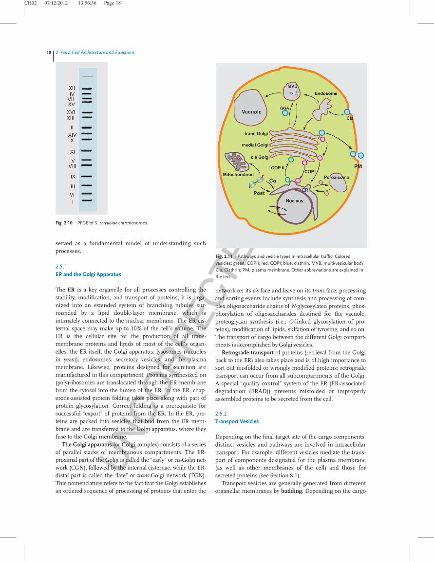
Various compartments surrounded by individual mem-branes are located within the yeast cytoplasm, which playkey roles in the manufacturing and trafficking of proteins(Figure 2.11). Transport of proteins between cellular com-partments is bound to different forms of transport vesi-cles and is found in all eukaryotic cells, but yeast has
Table 2.5 Genome sizes of some yeasts.
Species Ploidy Chromosomenumber
Genomesize (Mb)
Saccharomyces cerevisiae n 16 12.1Saccharomycesparadoxus
2n 16 12.2
Saccharomyces bayanus 2n 16 10.2Saccharomyces exiguus 2n 14–16Saccharomyces servazii 2n 9–13Candida glabrata n 13 14.7Saccharomyces castellii 9 11.4Kluyveromyces waltii 8 10.7Kluyveromycesmarxianus
10
Saccharomyces kluyveri 2n 8 11.3Eremothecium gossypii n 7 8.7Kluyveromyces lactis n 6 10.7Debaryomyces hansenii n 7 12.2Yarrowia lipolytica n 6 20.5
See also Chapters NaN, 16, and Appendix B.
2.5 Organellar Compartmentsj17
CH02 07/12/2012 13:56:36 Page 18
served as a fundamental model of understanding suchprocesses.
2.5.1ER and the Golgi Apparatus
The ER is a key organelle for all processes controlling thestability, modification, and transport of proteins; it is orga-nized into an extended system of branching tubules sur-rounded by a lipid double-layer membrane, which isintimately connected to the nuclear membrane. The ER cis-ternal space may make up to 10% of the cell’s volume. TheER is the cellular site for the production of all trans-membrane proteins and lipids of most of the cell’s organ-elles: the ER itself, the Golgi apparatus, lysosomes (vacuolesin yeast), endosomes, secretory vesicles, and the plasmamembrane. Likewise, proteins designed for secretion aremanufactured in this compartment. Proteins synthesized on(poly)ribosomes are translocated through the ER membranefrom the cytosol into the lumen of the ER. In the ER, chap-erone-assisted protein folding takes place along with part ofprotein glycosylation. Correct folding is a prerequisite forsuccessful “export” of proteins from the ER. In the ER, pro-teins are packed into vesicles that bud from the ER mem-brane and are transferred to the Golgi apparatus, where theyfuse to the Golgi membrane.
The Golgi apparatus (or Golgi complex) consists of a seriesof parallel stacks of membranous compartments. The ER-proximal part of the Golgi is called the “early” or cis-Golgi net-work (CGN), followed by the internal cisternae, while the ER-distal part is called the “late” or trans-Golgi network (TGN).This nomenclature refers to the fact that the Golgi establishesan ordered sequence of processing of proteins that enter the
network on its cis face and leave on its trans face; processingand sorting events include synthesis and processing of com-plex oligosaccharide chains of N-glycosylated proteins, phos-phorylation of oligosaccharides destined for the vacuole,proteoglycan synthesis (i.e., O-linked glycosylation of pro-teins), modification of lipids, sulfation of tyrosine, and so on.The transport of cargo between the different Golgi compart-ments is accomplished by Golgi vesicles.
Retrograde transport of proteins (retrieval from the Golgiback to the ER) also takes place and is of high importance tosort out misfolded or wrongly modified proteins; retrogradetransport can occur from all subcompartments of the Golgi.A special “quality control” system of the ER (ER-associateddegradation (ERAD)) prevents misfolded or improperlyassembled proteins to be secreted from the cell.
2.5.2Transport Vesicles
Depending on the final target site of the cargo components,distinct vesicles and pathways are involved in intracellulartransport. For example, different vesicles mediate the trans-port of components designated for the plasma membrane(as well as other membranes of the cell) and those forsecreted proteins (see Section 8.1).
Transport vesicles are generally generated from differentorganellar membranes by budding. Depending on the cargo
Fig. 2.10 PFGE of S. cerevisiae chromosomes.
Fig. 2.11 Pathways and vesicle types in intracellular traffic. Colored
vesicles: green, COPII; red, COPI; blue, clathrin. MVB, multi-vesicular body;
Cla, Clathrin; PM, plasma membrane. Other abbreviations are explained in
the text.
18j2 Yeast Cell Architecture and Functions
CH02 07/12/2012 13:56:40 Page 19
and its destination, the vesicles – in addition to their lipidbilayer envelope, are endowed with a characteristic coat. Inyeast, three types of coated vesicles can be distinguished(Table 2.6).
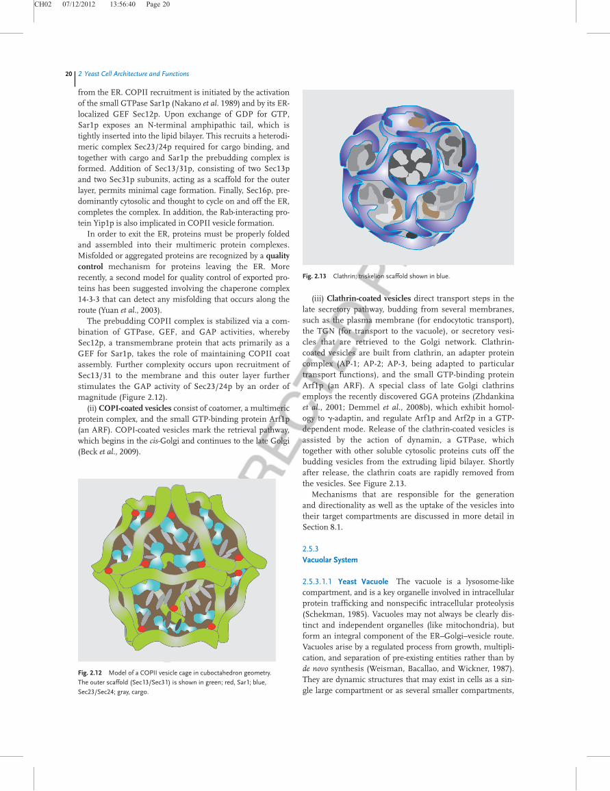
(i) COPII-coated vesicles are employed for the anterograde(forward) transport of cargo molecules from the ER to theGolgi, a function that is meanwhile well understood also in amechanistic sense; the yeast system has contributed integralinformation (Hughes and Stephens, 2008). COPII-coatedvesicles are assembled at the ER membrane from three
components: the small GTP-binding protein (Sar1p), theSec23/24p complex, and the Sec13/31p complex; these aresufficient to build a vesicle in vitro. Packaging of the types oftransported molecules is not random, but a selective process(Bickford, Mossessova, and Goldberg, 2004; Lee and Miller,2007; Sato and Nakano, 2007; Fromme, Orci, and Schekman,2008). Each outward movement has to be counterbalancedby a retrieval step whereby membrane and selected proteinsare returned to their original compartment of origin; compo-nents of the complex may undergo several rounds of export
Table 2.6 Components of coated vesicles in yeast.
Vesicle Subcomponent Geneproduct
Description
COPII Sec12 GEF; ER membrane protein; Sar1p activatorSar1 small GTP-binding protein; Sar1p-GTP recruits Sec23–Sec24 complexSec23 GAP for Sar1pSec24 involved in cargo selection and formation of prebudding complexSfb2Sfb3Sec13 forming outer layer (scaffold) of COPII coat; Sec13p has membrane bending activitySec31Sec16 stabilizes prebudding vesicles
COPI coatomer a Cop1 COPI vesicle coatomer complexcoatomer b Sec26 involved in ER–Golgi protein trafficking and maintenance of normal ER morphologycoatomer b0 Sec27 involved in ER–Golgi and Golgi–ER transport; contains WD40 domains that mediate cargo selective
interactionscoatomer g Sec21 involved in ER–Golgi transport of selective cargocoatomer d Ret2 involved in retrograde transport between Golgi and ERcoatomer e Sec28 regulates retrograde Golgi–ER protein traffic; stabilizes Cop1p and the coatomer complexcoatomer z Ret3 involved in retrograde transport between Golgi and ER
Arf1 small GTP-binding proteinDsl1 peripheral membrane protein needed for Golgi–ER retrograde traffic; forms a complex with Sec39p
and Tip20p that interacts with ER SNAREs Sec20p and Use1p; component of the ER target site thatinteracts with coatomer
Clathrin triskelion Chc1 clathrin heavy chain, triskelion structural componentClc1 clathrin light chain, triskelion structural component; regulates clathrin function
AP-1 Apl2 b-adaptin, large subunit of the clathrin-associated protein (AP-1) complex; binds clathrinApl4 g-adaptin, large subunit of the clathrin-associated protein (AP-1) complex; binds clathrinAps1 small subunit of the clathrin-associated adapter complex AP-1Laa1 AP-1 accessory protein; colocalizes with clathrin to the late Golgi apparatus; involved in TGN–
endosome transport; physically interacts with AP-1Apm1 Mu1-like medium subunit of the clathrin-associated protein complex (AP-1); binds clathrin
AP-2 Apl1 b-adaptin, large subunit of the clathrin-associated protein (AP-2) complex; binds clathrinApl3 a-adaptin, large subunit of the clathrin associated protein complex (AP-2)Aps2 small subunit of the clathrin-associated adapter complex AP-2; involved in protein sorting at the
plasma membraneApm4 Mu2-like subunit of the clathrin associated protein complex (AP-2)
AP-3 Apl5 d-adaptin-like subunit of the clathrin associated protein complex (AP-3); functions in transport ofalkaline phosphatase to the vacuole
Aps3 small subunit of the clathrin-associated adapter complex AP-3, involved in vacuolar protein sortingApm3p Mu3-like subunit of the clathrin-associated protein complex (AP-3); functions in transport of alkaline
phosphatase to the vacuoleApm2 protein of unknown function, homologous to the medium chain of mammalian clathrin-associated
protein complexGga1 Golgi-localized protein with homology to g-adaptin, regulates Arf1p and Arf2p in a GTP-dependent
manner to facilitate traffic through the late GolgiGga2 protein that regulates Arf1p and Arf2p in a GTP-dependent manner to facilitate traffic through the
late Golgi; binds InsP(4), which plays a role in TGN localizationSwa2 clathrin-binding protein required for uncoating of clathrin-coated vesicles
2.5 Organellar Compartmentsj19
CH02 07/12/2012 13:56:40 Page 20
from the ER. COPII recruitment is initiated by the activationof the small GTPase Sar1p (Nakano et al. 1989) and by its ER-localized GEF Sec12p. Upon exchange of GDP for GTP,Sar1p exposes an N-terminal amphipathic tail, which istightly inserted into the lipid bilayer. This recruits a heterodi-meric complex Sec23/24p required for cargo binding, andtogether with cargo and Sar1p the prebudding complex isformed. Addition of Sec13/31p, consisting of two Sec13pand two Sec31p subunits, acting as a scaffold for the outerlayer, permits minimal cage formation. Finally, Sec16p, pre-dominantly cytosolic and thought to cycle on and off the ER,completes the complex. In addition, the Rab-interacting pro-tein Yip1p is also implicated in COPII vesicle formation.
In order to exit the ER, proteins must be properly foldedand assembled into their multimeric protein complexes.Misfolded or aggregated proteins are recognized by a qualitycontrol mechanism for proteins leaving the ER. Morerecently, a second model for quality control of exported pro-teins has been suggested involving the chaperone complex14-3-3 that can detect any misfolding that occurs along theroute (Yuan et al., 2003).
The prebudding COPII complex is stabilized via a com-bination of GTPase, GEF, and GAP activities, wherebySec12p, a transmembrane protein that acts primarily as aGEF for Sar1p, takes the role of maintaining COPII coatassembly. Further complexity occurs upon recruitment ofSec13/31 to the membrane and this outer layer furtherstimulates the GAP activity of Sec23/24p by an order ofmagnitude (Figure 2.12).
(ii) COPI-coated vesicles consist of coatomer, a multimericprotein complex, and the small GTP-binding protein Arf1p(an ARF). COPI-coated vesicles mark the retrieval pathway,which begins in the cis-Golgi and continues to the late Golgi(Beck et al., 2009).
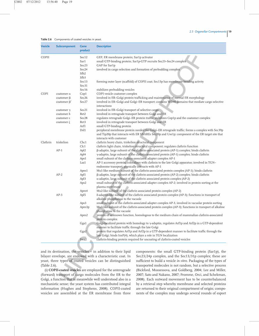
(iii) Clathrin-coated vesicles direct transport steps in thelate secretory pathway, budding from several membranes,such as the plasma membrane (for endocytotic transport),the TGN (for transport to the vacuole), or secretory vesi-cles that are retrieved to the Golgi network. Clathrin-coated vesicles are built from clathrin, an adapter proteincomplex (AP-1; AP-2; AP-3, being adapted to particulartransport functions), and the small GTP-binding proteinArf1p (an ARF). A special class of late Golgi clathrinsemploys the recently discovered GGA proteins (Zhdankinaet al., 2001; Demmel et al., 2008b), which exhibit homol-ogy to g-adaptin, and regulate Arf1p and Arf2p in a GTP-dependent mode. Release of the clathrin-coated vesicles isassisted by the action of dynamin, a GTPase, whichtogether with other soluble cytosolic proteins cuts off thebudding vesicles from the extruding lipid bilayer. Shortlyafter release, the clathrin coats are rapidly removed fromthe vesicles. See Figure 2.13.
Mechanisms that are responsible for the generationand directionality as well as the uptake of the vesicles intotheir target compartments are discussed in more detail inSection 8.1.
2.5.3Vacuolar System
2.5.3.1.1 Yeast Vacuole The vacuole is a lysosome-likecompartment, and is a key organelle involved in intracellularprotein trafficking and nonspecific intracellular proteolysis(Schekman, 1985). Vacuoles may not always be clearly dis-tinct and independent organelles (like mitochondria), butform an integral component of the ER–Golgi–vesicle route.Vacuoles arise by a regulated process from growth, multipli-cation, and separation of pre-existing entities rather than byde novo synthesis (Weisman, Bacallao, and Wickner, 1987).They are dynamic structures that may exist in cells as a sin-gle large compartment or as several smaller compartments,
Fig. 2.12 Model of a COPII vesicle cage in cuboctahedron geometry.
The outer scaffold (Sec13/Sec31) is shown in green; red, Sar1; blue,
Sec23/Sec24; gray, cargo.
Fig. 2.13 Clathrin; triskelion scaffold shown in blue.
20j2 Yeast Cell Architecture and Functions
CH02 07/12/2012 13:56:42 Page 21
called “prevacuolar compartments” (PVCs), “prevacuolarendosomes” (PVEs), or “late endosomes” (LEs). They arebound by a single membrane, which has a phospholipid,unsaturated fatty acid, and sterol content different from theplasma membrane. Phosphatidylinositol phosphates (e.g.,phosphatidylinositol-4-phosphate (PI(4)P)) (Audhya, Foti,and Emr, 2000) are essential for the maintenance of vacuolarmorphology. In yeast, the vacuole(s) usually occupy up to30% of the total cellular volume.
The vacuole is a “drain.” The vacuole is the compartmentthat receives proteins from different routes: (i) proteinssorted away from the secretory pathway at the Golgi appara-tus, (ii) proteins derived from the plasma membrane, (iii)proteins imported by endocytic traffic, and (iv) productsfrom autophagy, which represents a “destructive” pathway toliberate the cell from old organelles or organellar remnants(cf. Section 8.1.3.5.1).
In the first stages of endocytosis, plasma membraneinvaginations are formed that pinch off to generate vesiclesthat finally deliver their load to the endosomes. In mostcases, the endocytosed proteins are directed – via severalforms of multivesicular bodies (MVBs) – to the vacuole fordegradation. However, recently it became clear that alsoretrieval pathways (to the Golgi) for endocytosed proteinsdo exist in yeast. Details of these processes are presentedin Section 8.1.
2.5.3.1.2 Vacuolar Degradation The degradative processesare catalyzed by the activities of the more than 40 differentintravacuolar hydrolases: endopeptidases, aminopeptidases,and carboxypeptidases (Achstetter et al., 1984; Jones, 1984;Jones, 1991; Vida et al., 1991; Knop et al., 1993), and nucle-ases, glycosidases, lipases, phospholipases, and phospha-tases. Delivery of these enzymes to the vacuole is mediatedby a portion of the secretory pathway (Rothman et al., 1989;Fratti et al., 2004) and there is a selective uptake of substratesto be degraded (Chiang and Schekman, 1991; Chiang,Schekman, and Hamamoto, 1996). Apart from their role indegradative processes, vacuoles are involved in several otherphysiological functions, such as being storage compartmentsfor basic amino acids, polyphosphates, and certain metal cat-ions (Kþ, Mg2þ, and Ca2þ). They also participate in osmo-regulation and the homeostatic regulation of cytosolic ionconcentration and pH. pH is controlled by the vacuolarplasma membrane ATPase (see Section 8.3); while the cyto-solic pH is about 7.2, the vacuolar pH is adapted to 5.0 – theoptimum for the hydrolytic enzymes.
2.5.4Endocytosis and Exocytosis
Endocytosis has to fulfill two tasks: (i) internalize anddegrade components that might be hazardous to the cell,and (ii) recycle membrane components for repeated use(retrieval of receptors) or downregulate the activity of par-ticular membrane receptors, both of which are of major
importance to keep cellular integrity. In many cases,selected extracellular macromolecules are endocytosed bybinding to specific membrane receptors. One example inyeast is the receptor protein for the a- or a-matingpheromones.
Two methods are employed in preparing the cargo to beimported; further details are discussed in Section 8.1.3.6.
Exocytosed material is packaged into clathrin-coated vesi-cles in the late Golgi network. There exists a constitutivesecretory pathway for proteoglycans and glycoproteins thatwill form constituents of the plasma membrane. Regulatedpathways are designed for the export of transmembrane pro-teins, such as receptors or transporters.
One prominent example of exocytosed material are lipidrafts, which form in the membrane of the trans-Golgi by self-aggregation into microaggregates and thus can transport par-ticular combinations of membrane constituents to the cellsurface. Lipid rafts may comprise proteins with extendedtransmembrane domains, glycolipids, and GPI-anchoredproteins (cf. Section 3.4.3.2).
2.5.5Mitochondria
For a long time, yeast mitochondria have been employed bymany researchers as the model system in which mitochon-drial structure, function, and biogenesis have been studied.Yeast mitochondria not only resemble these organellesfound in higher eukaryotes, but are of outstanding impor-tance for the understanding of fermentation processes. Yeastmitochondria are easy to isolate as respiratory-competentorganelles and the genetics of yeast mitochondria has beenstudied in great detail.
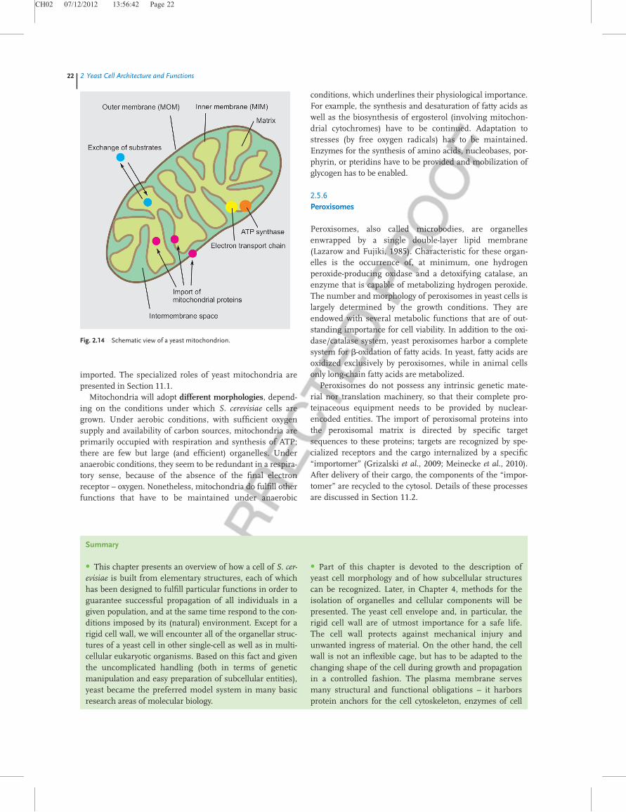
2.5.5.1 Mitochondrial StructureYeast mitochondria, like their mammalian counterparts, aresurrounded by two types of lipid bilayers, an outer mem-brane (MOM) and an inner membrane (MIM), the two ofwhich embody an intermembrane space (IMS). The inner ofthe mitochondrion is called the “mitochondrial matrix.” Theouter membrane is sort of a shelter that also containsenzymes involved in lipid metabolism. The inner membranecontains (i) cytochromes for the respiratory chain, (ii) theATP synthase coupled to the respiratory chain, and (iii) anumber of transport proteins for the exchange of low-molec-ular-weight components. The matrix is the site for the citricacid cycle (tricarboxylic acid (TCA) cycle) and contains themitochondrial DNA, together with the protein synthesizingmachinery including mitochondrial ribosomes. One of themost important features of the setup comprising all compart-ments of the mitochondria are the systems for the internal-ization and processing of proteins that are manufactured oncytosolic ribosomes and imported into the mitochondria(Figure 2.14). Only a few proteins are synthesized by the useof the mitochondrial machinery, whereas the vast majority ofthe mitochondrial proteins (greater than 800) have to be
2.5 Organellar Compartmentsj21
CH02 07/12/2012 13:56:42 Page 22
imported. The specialized roles of yeast mitochondria arepresented in Section 11.1.
Mitochondria will adopt different morphologies, depend-ing on the conditions under which S. cerevisiae cells aregrown. Under aerobic conditions, with sufficient oxygensupply and availability of carbon sources, mitochondria areprimarily occupied with respiration and synthesis of ATP;there are few but large (and efficient) organelles. Underanaerobic conditions, they seem to be redundant in a respira-tory sense, because of the absence of the final electronreceptor – oxygen. Nonetheless, mitochondria do fulfill otherfunctions that have to be maintained under anaerobic
conditions, which underlines their physiological importance.For example, the synthesis and desaturation of fatty acids aswell as the biosynthesis of ergosterol (involving mitochon-drial cytochromes) have to be continued. Adaptation tostresses (by free oxygen radicals) has to be maintained.Enzymes for the synthesis of amino acids, nucleobases, por-phyrin, or pteridins have to be provided and mobilization ofglycogen has to be enabled.
2.5.6Peroxisomes
Peroxisomes, also called microbodies, are organellesenwrapped by a single double-layer lipid membrane(Lazarow and Fujiki, 1985). Characteristic for these organ-elles is the occurrence of, at minimum, one hydrogenperoxide-producing oxidase and a detoxifying catalase, anenzyme that is capable of metabolizing hydrogen peroxide.The number and morphology of peroxisomes in yeast cells islargely determined by the growth conditions. They areendowed with several metabolic functions that are of out-standing importance for cell viability. In addition to the oxi-dase/catalase system, yeast peroxisomes harbor a completesystem for b-oxidation of fatty acids. In yeast, fatty acids areoxidized exclusively by peroxisomes, while in animal cellsonly long-chain fatty acids are metabolized.
Peroxisomes do not possess any intrinsic genetic mate-rial nor translation machinery, so that their complete pro-teinaceous equipment needs to be provided by nuclear-encoded entities. The import of peroxisomal proteins intothe peroxisomal matrix is directed by specific targetsequences to these proteins; targets are recognized by spe-cialized receptors and the cargo internalized by a specific“importomer” (Grizalski et al., 2009; Meinecke et al., 2010).After delivery of their cargo, the components of the “impor-tomer” are recycled to the cytosol. Details of these processesare discussed in Section 11.2.
Summary
� This chapter presents an overview of how a cell of S. cer-evisiae is built from elementary structures, each of whichhas been designed to fulfill particular functions in order toguarantee successful propagation of all individuals in agiven population, and at the same time respond to the con-ditions imposed by its (natural) environment. Except for arigid cell wall, we will encounter all of the organellar struc-tures of a yeast cell in other single-cell as well as in multi-cellular eukaryotic organisms. Based on this fact and giventhe uncomplicated handling (both in terms of geneticmanipulation and easy preparation of subcellular entities),yeast became the preferred model system in many basicresearch areas of molecular biology.
� Part of this chapter is devoted to the description ofyeast cell morphology and of how subcellular structurescan be recognized. Later, in Chapter 4, methods for theisolation of organelles and cellular components will bepresented. The yeast cell envelope and, in particular, therigid cell wall are of utmost importance for a safe life.The cell wall protects against mechanical injury andunwanted ingress of material. On the other hand, the cellwall is not an inflexible cage, but has to be adapted to thechanging shape of the cell during growth and propagationin a controlled fashion. The plasma membrane servesmany structural and functional obligations – it harborsprotein anchors for the cell cytoskeleton, enzymes of cell
Fig. 2.14 Schematic view of a yeast mitochondrion.
22j2 Yeast Cell Architecture and Functions
CH02 07/12/2012 13:56:44 Page 23
wall synthesis, receptors for communication with theenvironment, including osmotic control, and selectivetransporters for compounds that enter or that leave thecell. We describe in some detail the components (i.e.,microtubules and microfilaments) that build up adynamic cytoskeletal scaffold. The cytoskeleton is mainlydesigned for two functions in yeast physiology: (i) trans-port of cargo (from simple molecules through complexstructures to whole organelles) across the cell cytoplasm,and (ii) participation in mitosis and meiosis, determiningcell polarity during budding or mating as well as septationbefore cell separation. Motor proteins such as myosins,kinesins, or dynein provide the energy necessary formotility.
� The cell nucleus is the central organelle that handles theduplication of the chromosomes during cell division andthe expression of the majority of genetic information.Thereby, the nuclear pores act as instruments controllingthe exchange of macromolecules between the nucleus andthe cytosol, or sequestration of particular types of factorswithin the nucleus. These features are paralleled in yeastand mammals, as intense work in both systems has dem-onstrated. Here, we rather concentrate on morphological
aspects and will discuss some functional aspects later inChapter 8.
� Cellular organelles taking care of protein processing,their cellular distribution, and programmed degradationare the ER, connected to the nuclear membrane, the Golgiapparatus, and the yeast vacuole (which is similar to lyso-somes), including the various vesicular bodies that mediateinterorganellar transport. Most of the proteins are fabri-cated by cytoplasmic ribosomes and imported into the ER,which then controls their correct folding. Handed over tothe Golgi, the proteins are subjected to various modifica-tions, thus endowing each of them with attributes that facil-itate their targeting to the final destination within the cellor label them for exocytosis. Proteins imported into the cellor retrieved from its surface are channeled into this systemas well. The intracellular trafficking was one of the earliestissues studied in yeast and opened up ways to look intosimilar aspects in higher organisms.
� Finally, this chapter briefly outlines the structural andfunctional characteristics of the mitochondria and the per-oxisomes, both of which are organelles also indispensablein higher eukaryotes.
Further Reading
Beck, R., Rawet, M., Wieland, F.T., and Cassel, D. (2009) TheCOPI system: molecular mechanisms and function. FEBSLetters, 583, 2709.
Bickford, L.C., Mossessova, E., and Goldberg, J. (2004) A struc-tural view of the COPII vesicle coat. Current Opinion inStructural Biology, 14, 147–153 (review).
Boisvert, F.M., van Koningsbruggen, S., Navascu�es, J., andLamond, A.I. (2007) The multifunctional nucleolus. NatureReviews Molecular Cell Biology, 8, 574–585.
Bowers, K. and Stevens, T.H. (2005) Protein transportfrom the late Golgi to the vacuole in the yeast Saccharomycescerevisiae. Biochemica et Biophysica Acta, 1744, 438–454(review).
Bryant, N.J. and Stevens, T.H. (1998) Vacuole biogenesis inSaccharomyces cerevisiae: protein transport pathways to theyeast vacuole. Microbiology and Molecular Biology Reviews,62, 230–47.
D’Angelo, M.A. and Hetzer, M.W. (2008) Structure, dynamicsand function of nuclear pore complexes. Trends in Cell Biol-ogy, 18, 456–466.
Fernandez-Martinez, J. and Rout, M.P. (2009) Nuclear porecomplex biogenesis. Current Opinion in Cell Biology, 21,603–612.
Fromme, J.C., Orci, L., and Schekman, R. (2008) Coordinationof COPII vesicle trafficking by Sec23. Trends in Cell Biology,18, 330–6.
Girzalski, W., Platta, H.W., and Erdmann, R. (2009) Proteintransport across the peroxisomal membrane. BiologicalChemistry, 390, 745–751.
Glick, B.S. (2000) Organization of the Golgi apparatus. CurrentOpinion in Cell Biology, 12, 450–456.
Hoyt, M.A., Hyman, A.A., and B€ahler, M. (1997) Motor pro-teins of the eukaryotic cytoskeleton. Proceedings of theNational Academy of Sciences of the United States of America,94, 12747–12748.
Joglekar, A.P., Bloom, K.S., and Salmon, E.D. (2010) Mecha-nisms of force generation by end-on kinetochore-micro-tubule attachments. Current Opinion in Cell Biology, 22, 57.
Kirchhausen, T. (2000) Three ways to make a vesicle. NatureReviews Molecular Cell Biology, 1, 187–198.
Lee, M.C.S. and Miller, E.A. (2007) Molecular mechanisms ofCOPII vesicle formation. Seminars in Cell and DevelopmentalBiology, 18, 424–434.
Levin, D.E. (2005) Cell wall integrity signaling in Saccharomy-ces cerevisiae. Microbiology and Molecular Biology Reviews, 69,262–291.
Liu, J., Sun, Y., Oster, G.F., and Drubin, D.G. (2010) Mechano-chemical crosstalk during endocytic vesicle formation. Cur-rent Opinion in Cell Biology, 22, 36.
Meinecke, M., Cismowski, C., Schliebs, W. et al. (2010) Theperoxisomal importomer constitutes a large and highlydynamic pore. Nature Cell Biology, 12, 273–277.
Further Readingj23
CH02 07/12/2012 13:56:44 Page 24
Moore, J.K., Stuchell-Brereton, M.D., and Cooper, J.A. (2009)Function of dynein in budding yeast: mitotic spindle posi-tioning in a polarized cell. Cell Motility and the Cytoskeleton,66, 546–555.
Moseley, J.B. and Goode, B.L. (2006) The yeast actin cyto-skeleton: from cellular function to biochemical mechanism.Microbiology and Molecular Biology Reviews, 70, 605–645.
Neupert, W. and Herrmann, J.M. (2007) Translocation of pro-teins into mitochondria. Annual Review of Biochemistry, 76,723–749.
Sato, K. and Nakano, A. (2007) Mechanisms of COPIIvesicle formation and protein sorting. FEBS Letters, 581,2076–2082.
Schott, D., Huffaker, T., and Bretscher, A. (2002) Microfila-ments and microtubules: the news from yeast. CurrentOpinion in Microbiology, 5, 564–574.
Tolliday, N., Bouquin, N., and Li, R. (2001) Assembly and regu-lation of the cytokinetic apparatus in budding yeast. CurrentOpinion in Microbiology, 4, 690–695.
Tsien, R.Y. (2008) Constructing and exploiting the fluorescent pro-tein paintbox. Nobel Lecture, December 8, 2008.
Van der Rest, M.E., Kamminga, A.H., Nakano, A., Anraku, Y.,Pollman, B., and Konings, W.N. (1995) The plasma mem-brane of Saccharomyces cerevisiae: structure, function, andbiogenesis. Microbiology and Molecular Biology Reviews, 59,304–322.
24j2 Yeast Cell Architecture and Functions