Embed Size (px)
Citation preview
Vuong et al. Biotechnology for Biofuels 2013, 6:148http://www.biotechnologyforbiofuels.com/content/6/1/148
RESEARCH Open Access
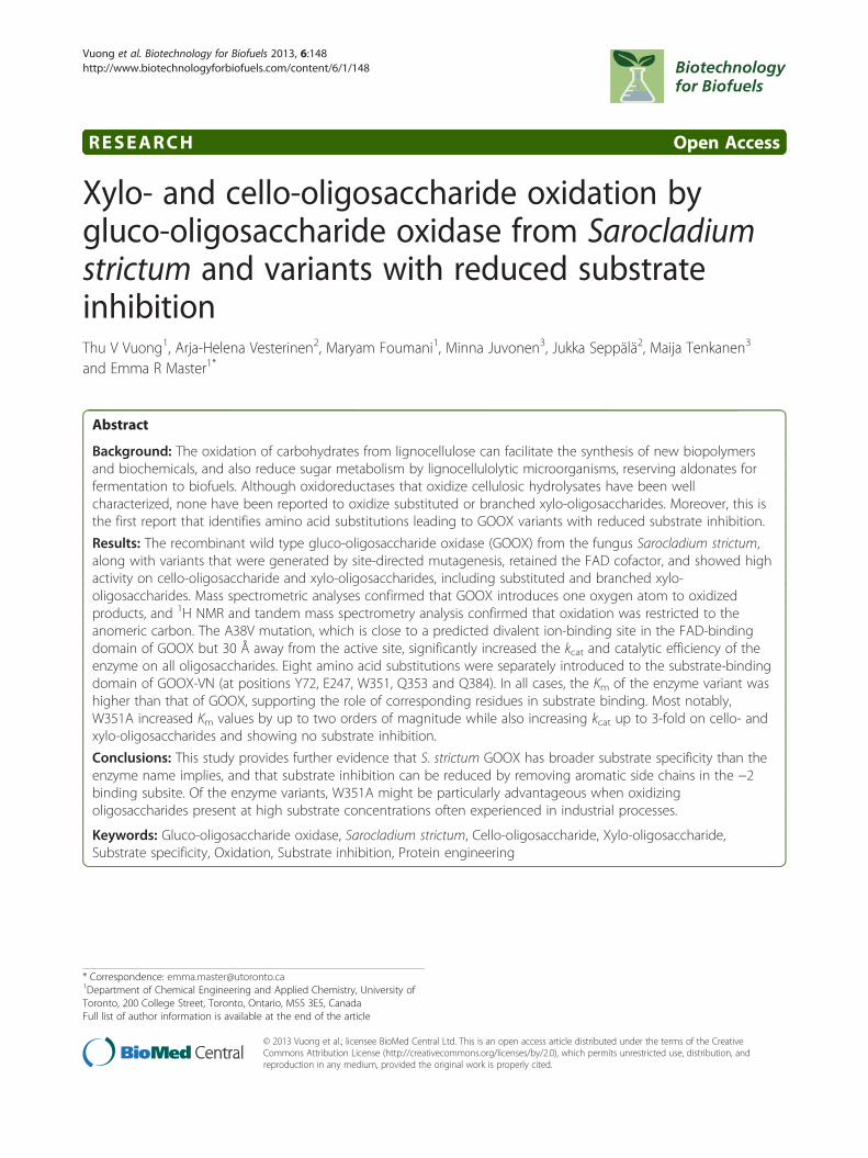
Xylo- and cello-oligosaccharide oxidation bygluco-oligosaccharide oxidase from Sarocladiumstrictum and variants with reduced substrateinhibitionThu V Vuong1, Arja-Helena Vesterinen2, Maryam Foumani1, Minna Juvonen3, Jukka Seppälä2, Maija Tenkanen3
and Emma R Master1*
Abstract
Background: The oxidation of carbohydrates from lignocellulose can facilitate the synthesis of new biopolymersand biochemicals, and also reduce sugar metabolism by lignocellulolytic microorganisms, reserving aldonates forfermentation to biofuels. Although oxidoreductases that oxidize cellulosic hydrolysates have been wellcharacterized, none have been reported to oxidize substituted or branched xylo-oligosaccharides. Moreover, this isthe first report that identifies amino acid substitutions leading to GOOX variants with reduced substrate inhibition.
Results: The recombinant wild type gluco-oligosaccharide oxidase (GOOX) from the fungus Sarocladium strictum,along with variants that were generated by site-directed mutagenesis, retained the FAD cofactor, and showed highactivity on cello-oligosaccharide and xylo-oligosaccharides, including substituted and branched xylo-oligosaccharides. Mass spectrometric analyses confirmed that GOOX introduces one oxygen atom to oxidizedproducts, and 1H NMR and tandem mass spectrometry analysis confirmed that oxidation was restricted to theanomeric carbon. The A38V mutation, which is close to a predicted divalent ion-binding site in the FAD-bindingdomain of GOOX but 30 Å away from the active site, significantly increased the kcat and catalytic efficiency of theenzyme on all oligosaccharides. Eight amino acid substitutions were separately introduced to the substrate-bindingdomain of GOOX-VN (at positions Y72, E247, W351, Q353 and Q384). In all cases, the Km of the enzyme variant washigher than that of GOOX, supporting the role of corresponding residues in substrate binding. Most notably,W351A increased Km values by up to two orders of magnitude while also increasing kcat up to 3-fold on cello- andxylo-oligosaccharides and showing no substrate inhibition.
Conclusions: This study provides further evidence that S. strictum GOOX has broader substrate specificity than theenzyme name implies, and that substrate inhibition can be reduced by removing aromatic side chains in the −2binding subsite. Of the enzyme variants, W351A might be particularly advantageous when oxidizingoligosaccharides present at high substrate concentrations often experienced in industrial processes.
Keywords: Gluco-oligosaccharide oxidase, Sarocladium strictum, Cello-oligosaccharide, Xylo-oligosaccharide,Substrate specificity, Oxidation, Substrate inhibition, Protein engineering
* Correspondence: [email protected] of Chemical Engineering and Applied Chemistry, University ofToronto, 200 College Street, Toronto, Ontario, M5S 3E5, CanadaFull list of author information is available at the end of the article
© 2013 Vuong et al.; licensee BioMed Central Ltd. This is an open access article distributed under the terms of the CreativeCommons Attribution License (http://creativecommons.org/licenses/by/2.0), which permits unrestricted use, distribution, andreproduction in any medium, provided the original work is properly cited.
Vuong et al. Biotechnology for Biofuels 2013, 6:148 Page 2 of 14http://www.biotechnologyforbiofuels.com/content/6/1/148
BackgroundRecently, a new classification of carbohydrate active en-zymes termed auxiliary activities (or AA), was introducedto the carbohydrate-active enzyme database (CAZy; http://www.cazy.org) [1]. Many of the enzymes classified into AAfamilies are carbohydrate oxidases. Well-known examplesinclude cellobiose dehydrogenase (CDH, EC 1.1.99.18,AA3_1) [2], glucose 1-oxidase (EC 1.1.3.4, AA3_2) [3], py-ranose 2-oxidase (EC 1.1.3.10, AA3_4) [4], and galactose 6-oxidase (EC 1.1.3.9, AA5_2) [5]. Comparatively few publica-tions describe the activity of gluco-oligosaccharide oxidases(GOOX, EC 1.1.3.-), which are classified as family AA7 en-zymes, and exhibit high catalytic activity on oligomeric sub-strates [6,7].Early reports of GOOX-T1 from the fungus Sarocladium
strictum T1 (previously known as Acremonium strictum T1[8]) confirmed oxidation of the hydroxyl group attached tothe anomeric carbon of maltose [6]; other analyses revealedeven higher activities on cello-oligosaccharides, particularlycellotriose [9,10]. Like other flavin carbohydrate oxidasesthat target the hydroxyl group of the anomeric carbon,GOOX-T1 is thought to mediate oxidoreductase activitythrough two half-reactions: 1) oxidation of the reducingsugar to the corresponding lactone, and 2) reduction ofmolecular oxygen to hydrogen peroxide [11]. Subsequenthydrolysis of the lactone product to the corresponding car-boxylic acid may then occur. While the biological functionof GOOX is uncertain, hydrogen peroxide generatedthrough carbohydrate oxidation could be used by ligninperoxidases and manganese peroxidase in lignin degrad-ation. From an applied perspective, gluco-oligosaccharideoxidases could provide an alternative to CDHs used in am-perometric enzyme biosensors for real-time measurementof cellulase activity on insoluble cellulose [12]. More recentapplications of CDH also demonstrate the benefit of carbo-hydrate oxidation to reduce sugar consumption bylignocellulolytic fungi, thereby maximizing ethanol yieldsfrom fermenting microorganisms [13].The crystal structure of GOOX-T1 reveals a monomeric
glycoprotein with a flavin adenine dinucleotide (FAD)-bind-ing domain coordinated by a bi-covalent linkage to H70(8α-N1-histidyl) and C130 (6-S-cysteinyl); GOOX-T1 isalso characterized by having a comparatively opensubstrate-binding site [14]. Site-directed mutagenesis con-firmed the requirement of bi-covalent coordination of FADfor enzyme activity; this unique coordination is also corre-lated to the relatively high redox potential of GOOX-T1[14,15]. In our recent study, GOOX-VN from S. strictumstrain CBS 346.70 was recombinantly expressed and bio-chemically characterized using a range of sugars and oligo-saccharides, including cello-oligosaccharides and xylo-oligosaccharides with up to 3 sugar units [7]. Fifteen aminoacid differences distinguish GOOX-VN and GOOX-T1: 13are intrinsic differences in the wild-type gene sequences
while 2 (A38V and S388N) arose from random mutationsduring the construction of the GOOX-VN expression sys-tem [7] (Additional file 1: Figure S1). GOOX-VN was foundto oxidize xylose as well as xylobiose and xylotriose [7].Given the high sequence identity between GOOX-VN andGOOX-T1 (97%), and since none of the amino acid substi-tutions between GOOX-VN and GOOX-T1 are predictedto directly participate in substrate binding, it is likely thatGOOX-T1 also oxidizes xylo-oligosaccharides even thoughxylo-oligosaccharide oxidation by GOOX-T1 has not beenreported [7,10]. Notably, resulting enzymatically oxidizedoligosaccharides could be used as carbohydrate standardsthat replaces the comparatively arduous chemical syn-thesis approach [16], facilitating the characterization ofcarbohydrate-oxidizing enzymes whose activity can not beeasily measured by colorimetric assays.To investigate the role of selected amino acids on sub-
strate preference, three amino acids in the GOOX-VNsubstrate binding site were previously substituted to cor-responding residues in chito-oligosaccharide oxidase(ChitO) from Fusarium graminearum [15] or carbohy-drate oxidase from Microdochium nivale [17], whichshow 45% and 42% sequence identity to GOOX-VN, re-spectively [7]. Of these, Y300A nearly doubled kcat valuesfor oligosaccharides while also increasing correspondingKm values [7]. The current study describes a more com-prehensive assessment of substrate preference and ca-talysis by GOOX-VN by 1) constructing eight additionalamino acid substitutions within the substrate bindingsite of this enzyme, 2) generating V38A and N388S sub-stitutions that convert GOOX-VN to the wild-typeGOOX sequence, and 3) using several oligosaccharides,including branched xylo-oligosaccharides (Figure 1) tocharacterize the catalytic efficiency, substrate selectivityand substrate inhibition of GOOX-VN enzyme variants.These analyses confirmed comparable kinetic efficiencieson cello-oligosaccharides and xylo-oligosaccharides,suggesting that gluco-oligosaccharide oxidases character-ized to date have broader substrate specificity than theenzyme name implies. This study also identified enzymevariants with high catalytic activity but lower substrateinhibition, which could improve oligosaccharide oxida-tion at high substrate concentrations often experiencedin industrial bioprocesses.
Results and discussionProtein expression and biophysical characterizationRecombinantly expressed GOOX-VN and enzyme vari-ants were purified to more than 95% homogeneity by af-finity chromatography (Figure 2A, Additional file 2:Figure S2). Amino acid substitutions did not affect pro-tein yields, and in general, between 5 and 10 mg/L ofpurified protein were recovered. The observed mass ofall enzymes was approximately 70 kDa (Additional file 2:
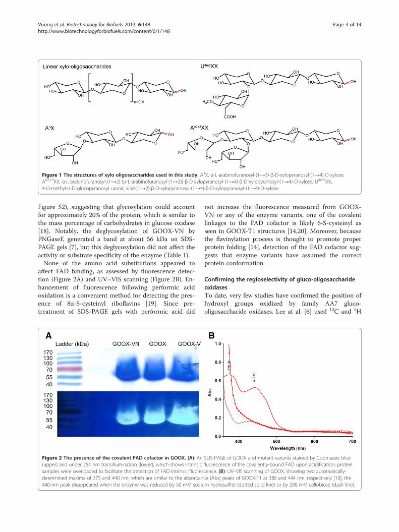
Figure 1 The structures of xylo-oligosaccharides used in this study. A3X, α-L-arabinofuranosyl-(1→3)-β-D-xylopyranosyl-(1→4)-D-xylose;Ad2+3XX, α-L-arabinofuranosyl-(1→2)-[α-L-arabinofuranosyl-(1→3)]-β-D-xylopyranosyl-(1→4)-β-D-xylopyranosyl-(1→4)-D-xylose; U4m2XX,4-O-methyl-α-D-glucopyranosyl uronic acid-(1→2)-β-D-xylopyranosyl-(1→4)-β-D-xylopyranosyl-(1→4)-D-xylose.
Vuong et al. Biotechnology for Biofuels 2013, 6:148 Page 3 of 14http://www.biotechnologyforbiofuels.com/content/6/1/148
Figure S2), suggesting that glycosylation could accountfor approximately 20% of the protein, which is similar tothe mass percentage of carbohydrates in glucose oxidase[18]. Notably, the deglycosylation of GOOX-VN byPNGaseF, generated a band at about 56 kDa on SDS-PAGE gels [7], but this deglycosylation did not affect theactivity or substrate specificity of the enzyme (Table 1).None of the amino acid substitutions appeared to
affect FAD binding, as assessed by fluorescence detec-tion (Figure 2A) and UV–VIS scanning (Figure 2B). En-hancement of fluorescence following performic acidoxidation is a convenient method for detecting the pres-ence of 8α-S-cysteinyl riboflavins [19]. Since pre-treatment of SDS-PAGE gels with performic acid did
Figure 2 The presence of the covalent FAD cofactor in GOOX. (A): An(upper) and under 254 nm transillumination (lower), which shows intrinsicsamples were overloaded to facilitate the detection of FAD intrinsic fluoresdetermined maxima of 375 and 440 nm, which are similar to the absorban440-nm peak disappeared when the enzyme was reduced by 50 mM sodiu
not increase the fluorescence measured from GOOX-VN or any of the enzyme variants, one of the covalentlinkages to the FAD cofactor is likely 6-S-cysteinyl asseen in GOOX-T1 structures [14,20]. Moreover, becausethe flavinylation process is thought to promote properprotein folding [14], detection of the FAD cofactor sug-gests that enzyme variants have assumed the correctprotein conformation.
Confirming the regioselectivity of gluco-oligosaccharideoxidasesTo date, very few studies have confirmed the position ofhydroxyl groups oxidized by family AA7 gluco-oligosaccharide oxidases. Lee at al. [6] used 13C and 1H
SDS-PAGE of GOOX and mutant variants stained by Coomassie bluefluorescence of the covalently-bound FAD upon acidification; proteincence. (B): UV–VIS scanning of GOOX, showing two automatically-ce (Abs) peaks of GOOX-T1 at 380 and 444 nm, respectively [10]; them hydrosulfite (dotted solid line) or by 200 mM cellobiose (dash line).
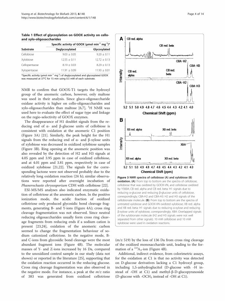
Figure 3 NMR spectra of cellobiose (A) and xylobiose (B)oxidation. (A): From top to bottom are the spectra of cellobiose,cellobiose that was oxidized by GOOX-VN, and cellobiose oxidizedby Y300A; CB red. alpha and CB red. beta: H1 signals due toreducing α-glucose and reducing β-glucose units of cellobiose,correspondingly; CBA-H2 and CBA-H3: H2 and H3 signals of thecellobionate molecule. (B): From top to bottom are the spectra ofuntreated xylobiose and GOOX-VN oxidized xylobiose; XB red. alphaand XB red. beta: H1 signals due to reducing α-xylose and reducingβ-xylose units of xylobiose, correspondingly; XBA: Overlapped signalsof the xylobionate molecule (H2 and H3 signals were not wellseparated from other signals). 10 mM cellobiose and 10 mMxylobiose were used in oxidation reactions.
Table 1 Effect of glycosylation on GOOX activity on cello-and xylo-oligosaccharides
Specific activity of GOOX (μmol min-1 mg-1)*
Substrate Deglycosylated Glycosylated
Cellobiose 9.03 ± 0.05 9.20 ± 0.11
Xylobiose 12.55 ± 0.11 12.72 ± 0.13
Cellopentaose 8.19 ± 0.03 8.29 ± 0.13
Xylopentaose 11.91 ± 0.09 11.93 ± 0.01
*Specific activity (μmol min-1 mg-1) of deglycosylated and glycosylated GOOXwas measured at 37°C for 15 min using 0.5 mM of each substrate.
Vuong et al. Biotechnology for Biofuels 2013, 6:148 Page 4 of 14http://www.biotechnologyforbiofuels.com/content/6/1/148
NMR to confirm that GOOX-T1 targets the hydroxylgroup of the anomeric carbon, however, only maltosewas used in their analysis. Since gluco-oligosaccharideoxidase activity is higher on cello-oligosaccharides andxylo-oligosaccharides than maltose [6,7], 1H NMR wasused here to evaluate the effect of sugar type and linkageon the regio-selectivity of GOOX enzymes.The disappearance of H1 doublet signals from the re-
ducing end of α- and β-glucose units of cellobiose isconsistent with oxidation at the anomeric C1 position(Figure 3A) [21]. Similarly, the peak height for the H1signals from the reducing end of α- and β-xylose unitsof xylobiose was decreased in oxidized xylobiose samples(Figure 3B). Ring opening at the anomeric position wasalso revealed by the detection of H2 and H3 signals at4.05 ppm and 3.95 ppm in case of oxidized cellobiose,and at 4.01 ppm and 3.81 ppm, respectively in case ofoxidized xylobiose [21,22]. The signals for the corre-sponding lactone were not observed probably due to therelatively long oxidation reaction (24 h); similar observa-tions were reported after overnight incubation ofPhanerochaete chrysosporium CDH with cellobiose [22].ESI-MS/MS analyses also indicated enzymatic oxida-
tion of cellotriose at the anomeric carbon. In the positiveionization mode, the acidic fraction of oxidizedcellotriose only produced glycosidic bond cleavage frag-ments, generating B- and Y-ions (Figure 4A); cross ringcleavage fragmentation was not observed. Since neutralreducing oligosaccharides usually form cross ring cleav-age fragments from reducing ends if a sodium cation ispresent [23,24], oxidation of the anomeric carbonseemed to change the fragmentation behaviour of so-dium cationized cellotriose. In the negative mode, B-and C-ions from glycosidic bond cleavage were the mostabundant fragment ions (Figure 4B). The molecularmasses of Y- and Z-ions increased by 16 Da, comparedto the unoxidized control sample in our study (data notshown) or reported in the literature [25], supporting thatthe oxidation reaction occurred in the reducing glucose.Cross ring cleavage fragmentation was also observed inthe negative mode. For instance, a peak at the m/z ratioof 383 was generated from oxidized cellotriose
(m/z 519) by the loss of 136 Da from cross ring cleavageof the oxidized monosaccharide unit, leading to the for-mation of a 2,4A3-ion (Figure 4B).Additional, indirect evidence, from colorimetric assays,
for the oxidation at C1 is that no activity was detectedon D-glucose derivatives lacking a C1 hydroxyl group,including 1,5-anhydroglucitol (D-glucose with -H in-stead of -OH at C1) and methyl-β-D-glucopyranoside(D-glucose with -OCH3 instead of -OH at C1).
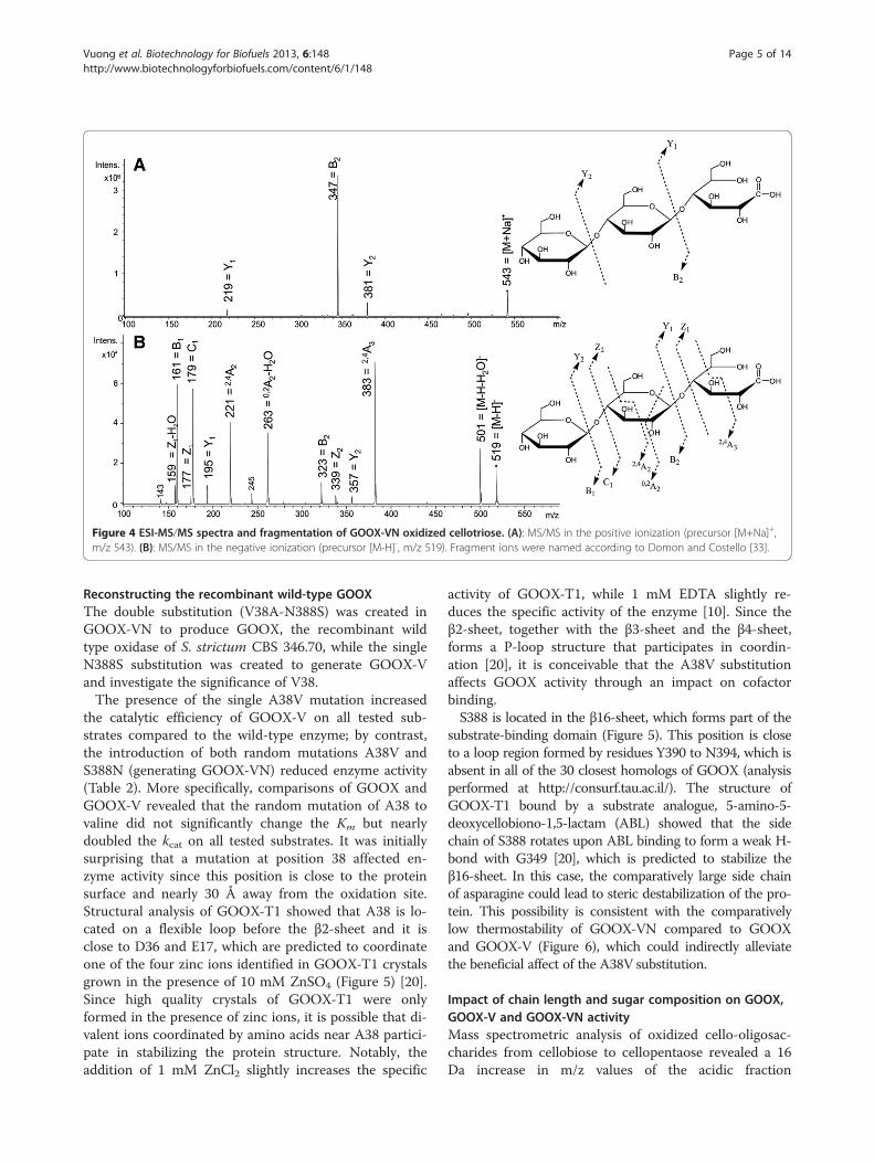
Figure 4 ESI-MS/MS spectra and fragmentation of GOOX-VN oxidized cellotriose. (A): MS/MS in the positive ionization (precursor [M+Na]+,m/z 543). (B): MS/MS in the negative ionization (precursor [M-H]-, m/z 519). Fragment ions were named according to Domon and Costello [33].
Vuong et al. Biotechnology for Biofuels 2013, 6:148 Page 5 of 14http://www.biotechnologyforbiofuels.com/content/6/1/148
Reconstructing the recombinant wild-type GOOXThe double substitution (V38A-N388S) was created inGOOX-VN to produce GOOX, the recombinant wildtype oxidase of S. strictum CBS 346.70, while the singleN388S substitution was created to generate GOOX-Vand investigate the significance of V38.The presence of the single A38V mutation increased
the catalytic efficiency of GOOX-V on all tested sub-strates compared to the wild-type enzyme; by contrast,the introduction of both random mutations A38V andS388N (generating GOOX-VN) reduced enzyme activity(Table 2). More specifically, comparisons of GOOX andGOOX-V revealed that the random mutation of A38 tovaline did not significantly change the Km but nearlydoubled the kcat on all tested substrates. It was initiallysurprising that a mutation at position 38 affected en-zyme activity since this position is close to the proteinsurface and nearly 30 Å away from the oxidation site.Structural analysis of GOOX-T1 showed that A38 is lo-cated on a flexible loop before the β2-sheet and it isclose to D36 and E17, which are predicted to coordinateone of the four zinc ions identified in GOOX-T1 crystalsgrown in the presence of 10 mM ZnSO4 (Figure 5) [20].Since high quality crystals of GOOX-T1 were onlyformed in the presence of zinc ions, it is possible that di-valent ions coordinated by amino acids near A38 partici-pate in stabilizing the protein structure. Notably, theaddition of 1 mM ZnCl2 slightly increases the specific
activity of GOOX-T1, while 1 mM EDTA slightly re-duces the specific activity of the enzyme [10]. Since theβ2-sheet, together with the β3-sheet and the β4-sheet,forms a P-loop structure that participates in coordin-ation [20], it is conceivable that the A38V substitutionaffects GOOX activity through an impact on cofactorbinding.S388 is located in the β16-sheet, which forms part of the
substrate-binding domain (Figure 5). This position is closeto a loop region formed by residues Y390 to N394, which isabsent in all of the 30 closest homologs of GOOX (analysisperformed at http://consurf.tau.ac.il/). The structure ofGOOX-T1 bound by a substrate analogue, 5-amino-5-deoxycellobiono-1,5-lactam (ABL) showed that the sidechain of S388 rotates upon ABL binding to form a weak H-bond with G349 [20], which is predicted to stabilize theβ16-sheet. In this case, the comparatively large side chainof asparagine could lead to steric destabilization of the pro-tein. This possibility is consistent with the comparativelylow thermostability of GOOX-VN compared to GOOXand GOOX-V (Figure 6), which could indirectly alleviatethe beneficial affect of the A38V substitution.
Impact of chain length and sugar composition on GOOX,GOOX-V and GOOX-VN activityMass spectrometric analysis of oxidized cello-oligosac-charides from cellobiose to cellopentaose revealed a 16Da increase in m/z values of the acidic fraction
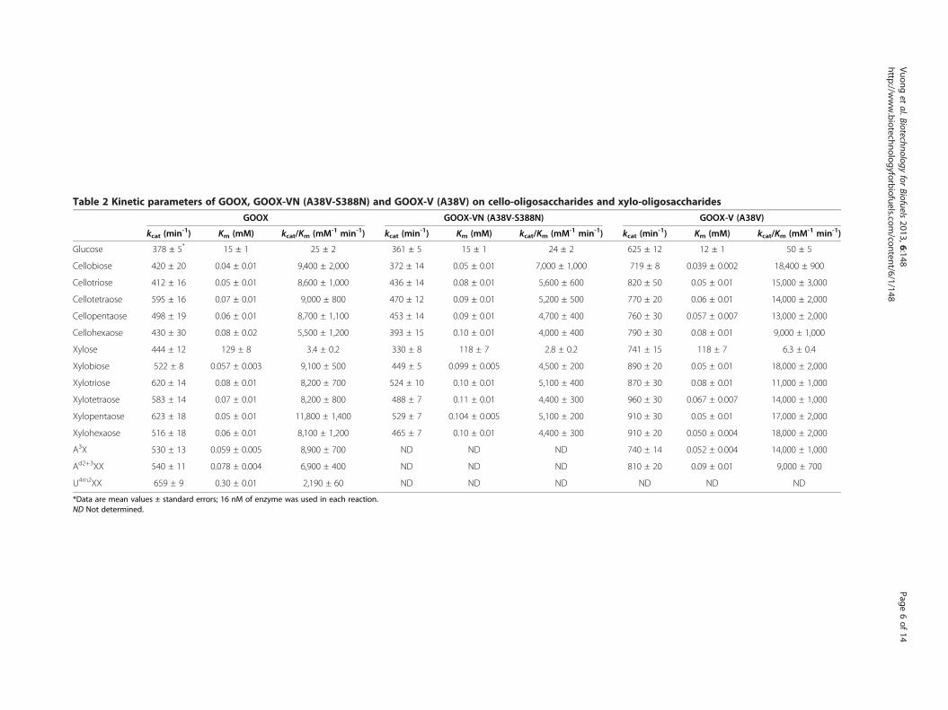
Table 2 Kinetic parameters of GOOX, GOOX-VN (A38V-S388N) and GOOX-V (A38V) on cello-oligosaccharides and xylo-oligosaccharides
GOOX GOOX-VN (A38V-S388N) GOOX-V (A38V)
kcat (min-1) Km (mM) kcat/Km (mM-1 min-1) kcat (min-1) Km (mM) kcat/Km (mM-1 min-1) kcat (min-1) Km (mM) kcat/Km (mM-1 min-1)
Glucose 378 ± 5* 15 ± 1 25 ± 2 361 ± 5 15 ± 1 24 ± 2 625 ± 12 12 ± 1 50 ± 5
Cellobiose 420 ± 20 0.04 ± 0.01 9,400 ± 2,000 372 ± 14 0.05 ± 0.01 7,000 ± 1,000 719 ± 8 0.039 ± 0.002 18,400 ± 900
Cellotriose 412 ± 16 0.05 ± 0.01 8,600 ± 1,000 436 ± 14 0.08 ± 0.01 5,600 ± 600 820 ± 50 0.05 ± 0.01 15,000 ± 3,000
Cellotetraose 595 ± 16 0.07 ± 0.01 9,000 ± 800 470 ± 12 0.09 ± 0.01 5,200 ± 500 770 ± 20 0.06 ± 0.01 14,000 ± 2,000
Cellopentaose 498 ± 19 0.06 ± 0.01 8,700 ± 1,100 453 ± 14 0.09 ± 0.01 4,700 ± 400 760 ± 30 0.057 ± 0.007 13,000 ± 2,000
Cellohexaose 430 ± 30 0.08 ± 0.02 5,500 ± 1,200 393 ± 15 0.10 ± 0.01 4,000 ± 400 790 ± 30 0.08 ± 0.01 9,000 ± 1,000
Xylose 444 ± 12 129 ± 8 3.4 ± 0.2 330 ± 8 118 ± 7 2.8 ± 0.2 741 ± 15 118 ± 7 6.3 ± 0.4
Xylobiose 522 ± 8 0.057 ± 0.003 9,100 ± 500 449 ± 5 0.099 ± 0.005 4,500 ± 200 890 ± 20 0.05 ± 0.01 18,000 ± 2,000
Xylotriose 620 ± 14 0.08 ± 0.01 8,200 ± 700 524 ± 10 0.10 ± 0.01 5,100 ± 400 870 ± 30 0.08 ± 0.01 11,000 ± 1,000
Xylotetraose 583 ± 14 0.07 ± 0.01 8,200 ± 800 488 ± 7 0.11 ± 0.01 4,400 ± 300 960 ± 30 0.067 ± 0.007 14,000 ± 1,000
Xylopentaose 623 ± 18 0.05 ± 0.01 11,800 ± 1,400 529 ± 7 0.104 ± 0.005 5,100 ± 200 910 ± 30 0.05 ± 0.01 17,000 ± 2,000
Xylohexaose 516 ± 18 0.06 ± 0.01 8,100 ± 1,200 465 ± 7 0.10 ± 0.01 4,400 ± 300 910 ± 20 0.050 ± 0.004 18,000 ± 2,000
A3X 530 ± 13 0.059 ± 0.005 8,900 ± 700 ND ND ND 740 ± 14 0.052 ± 0.004 14,000 ± 1,000
Ad2+3XX 540 ± 11 0.078 ± 0.004 6,900 ± 400 ND ND ND 810 ± 20 0.09 ± 0.01 9,000 ± 700
U4m2XX 659 ± 9 0.30 ± 0.01 2,190 ± 60 ND ND ND ND ND ND
*Data are mean values ± standard errors; 16 nM of enzyme was used in each reaction.ND Not determined.
Vuonget
al.Biotechnologyfor
Biofuels2013,6:148
Page6of
14http://w
ww.biotechnologyforbiofuels.com
/content/6/1/148
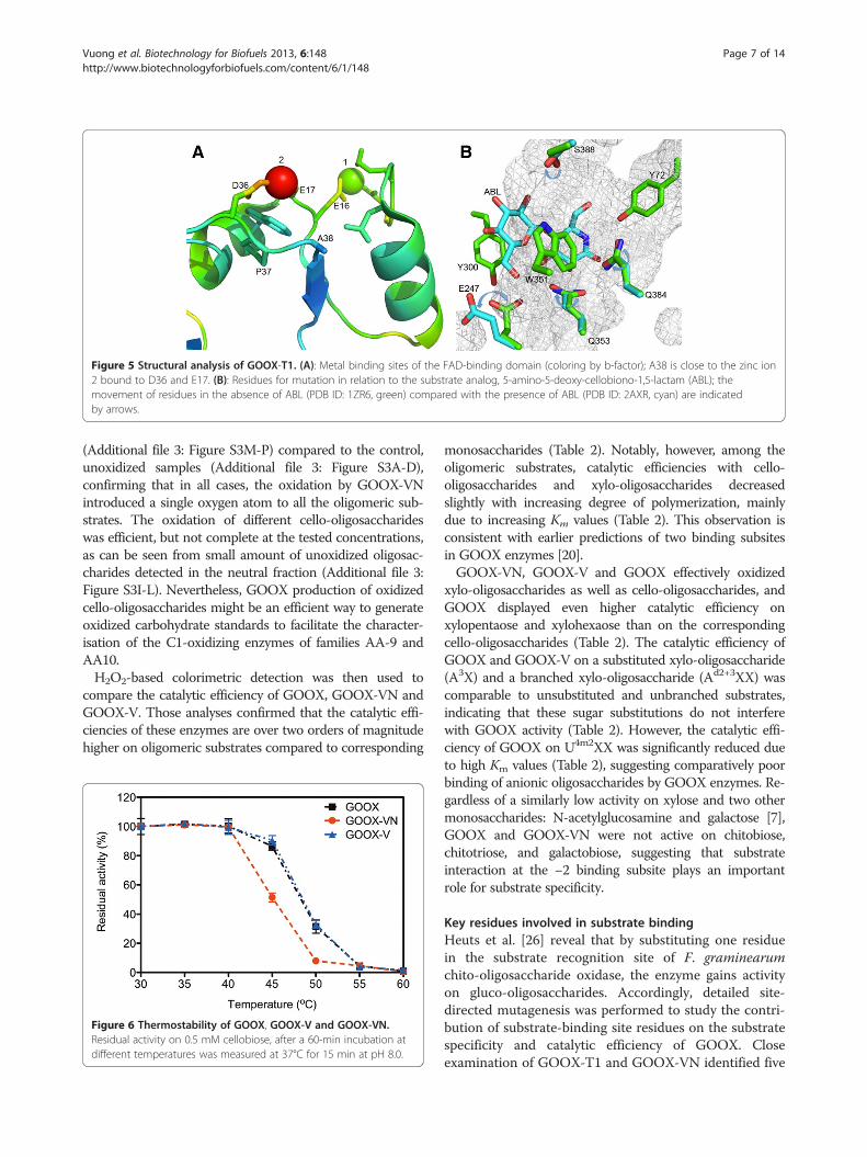
Figure 5 Structural analysis of GOOX-T1. (A): Metal binding sites of the FAD-binding domain (coloring by b-factor); A38 is close to the zinc ion2 bound to D36 and E17. (B): Residues for mutation in relation to the substrate analog, 5-amino-5-deoxy-cellobiono-1,5-lactam (ABL); themovement of residues in the absence of ABL (PDB ID: 1ZR6, green) compared with the presence of ABL (PDB ID: 2AXR, cyan) are indicatedby arrows.
Vuong et al. Biotechnology for Biofuels 2013, 6:148 Page 7 of 14http://www.biotechnologyforbiofuels.com/content/6/1/148
(Additional file 3: Figure S3M-P) compared to the control,unoxidized samples (Additional file 3: Figure S3A-D),confirming that in all cases, the oxidation by GOOX-VNintroduced a single oxygen atom to all the oligomeric sub-strates. The oxidation of different cello-oligosaccharideswas efficient, but not complete at the tested concentrations,as can be seen from small amount of unoxidized oligosac-charides detected in the neutral fraction (Additional file 3:Figure S3I-L). Nevertheless, GOOX production of oxidizedcello-oligosaccharides might be an efficient way to generateoxidized carbohydrate standards to facilitate the character-isation of the C1-oxidizing enzymes of families AA-9 andAA10.H2O2-based colorimetric detection was then used to
compare the catalytic efficiency of GOOX, GOOX-VN andGOOX-V. Those analyses confirmed that the catalytic effi-ciencies of these enzymes are over two orders of magnitudehigher on oligomeric substrates compared to corresponding
Figure 6 Thermostability of GOOX, GOOX-V and GOOX-VN.Residual activity on 0.5 mM cellobiose, after a 60-min incubation atdifferent temperatures was measured at 37°C for 15 min at pH 8.0.
monosaccharides (Table 2). Notably, however, among theoligomeric substrates, catalytic efficiencies with cello-oligosaccharides and xylo-oligosaccharides decreasedslightly with increasing degree of polymerization, mainlydue to increasing Km values (Table 2). This observation isconsistent with earlier predictions of two binding subsitesin GOOX enzymes [20].GOOX-VN, GOOX-V and GOOX effectively oxidized
xylo-oligosaccharides as well as cello-oligosaccharides, andGOOX displayed even higher catalytic efficiency onxylopentaose and xylohexaose than on the correspondingcello-oligosaccharides (Table 2). The catalytic efficiency ofGOOX and GOOX-V on a substituted xylo-oligosaccharide(A3X) and a branched xylo-oligosaccharide (Ad2+3XX) wascomparable to unsubstituted and unbranched substrates,indicating that these sugar substitutions do not interferewith GOOX activity (Table 2). However, the catalytic effi-ciency of GOOX on U4m2XX was significantly reduced dueto high Km values (Table 2), suggesting comparatively poorbinding of anionic oligosaccharides by GOOX enzymes. Re-gardless of a similarly low activity on xylose and two othermonosaccharides: N-acetylglucosamine and galactose [7],GOOX and GOOX-VN were not active on chitobiose,chitotriose, and galactobiose, suggesting that substrateinteraction at the −2 binding subsite plays an importantrole for substrate specificity.
Key residues involved in substrate bindingHeuts et al. [26] reveal that by substituting one residuein the substrate recognition site of F. graminearumchito-oligosaccharide oxidase, the enzyme gains activityon gluco-oligosaccharides. Accordingly, detailed site-directed mutagenesis was performed to study the contri-bution of substrate-binding site residues on the substratespecificity and catalytic efficiency of GOOX. Closeexamination of GOOX-T1 and GOOX-VN identified five
Vuong et al. Biotechnology for Biofuels 2013, 6:148 Page 8 of 14http://www.biotechnologyforbiofuels.com/content/6/1/148
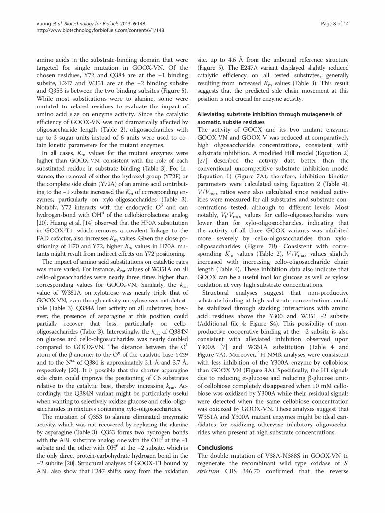
amino acids in the substrate-binding domain that weretargeted for single mutation in GOOX-VN. Of thechosen residues, Y72 and Q384 are at the −1 bindingsubsite, E247 and W351 are at the −2 binding subsiteand Q353 is between the two binding subsites (Figure 5).While most substitutions were to alanine, some weremutated to related residues to evaluate the impact ofamino acid size on enzyme activity. Since the catalyticefficiency of GOOX-VN was not dramatically affected byoligosaccharide length (Table 2), oligosaccharides withup to 3 sugar units instead of 6 units were used to ob-tain kinetic parameters for the mutant enzymes.In all cases, Km values for the mutant enzymes were
higher than GOOX-VN, consistent with the role of eachsubstituted residue in substrate binding (Table 3). For in-stance, the removal of either the hydroxyl group (Y72F) orthe complete side chain (Y72A) of an amino acid contribut-ing to the −1 subsite increased the Km of corresponding en-zymes, particularly on xylo-oligosaccharides (Table 3).Notably, Y72 interacts with the endocyclic O5 and canhydrogen-bond with OH6 of the cellobionolactone analog[20]. Huang et al. [14] observed that the H70A substitutionin GOOX-T1, which removes a covalent linkage to theFAD cofactor, also increases Km values. Given the close po-sitioning of H70 and Y72, higher Km values in H70A mu-tants might result from indirect effects on Y72 positioning.The impact of amino acid substitutions on catalytic rates
was more varied. For instance, kcat values of W351A on allcello-oligosaccharides were nearly three times higher thancorresponding values for GOOX-VN. Similarly, the kcatvalue of W351A on xylotriose was nearly triple that ofGOOX-VN, even though activity on xylose was not detect-able (Table 3). Q384A lost activity on all substrates; how-ever, the presence of asparagine at this position couldpartially recover that loss, particularly on cello-oligosaccharides (Table 3). Interestingly, the kcat of Q384Non glucose and cello-oligosaccharides was nearly doubledcompared to GOOX-VN. The distance between the O1
atom of the β anomer to the Oη of the catalytic base Y429and to the Nε2 of Q384 is approximately 3.1 Å and 3.7 Å,respectively [20]. It is possible that the shorter asparagineside chain could improve the positioning of C6 substratesrelative to the catalytic base, thereby increasing kcat. Ac-cordingly, the Q384N variant might be particularly usefulwhen wanting to selectively oxidize glucose and cello-oligo-saccharides in mixtures containing xylo-oligosaccharides.The mutation of Q353 to alanine eliminated enzymatic
activity, which was not recovered by replacing the alanineby asparagine (Table 3). Q353 forms two hydrogen bondswith the ABL substrate analog: one with the OH3 at the −1subsite and the other with OH6 at the −2 subsite, which isthe only direct protein-carbohydrate hydrogen bond in the−2 subsite [20]. Structural analyses of GOOX-T1 bound byABL also show that E247 shifts away from the oxidation
site, up to 4.6 Å from the unbound reference structure(Figure 5). The E247A variant displayed slightly reducedcatalytic efficiency on all tested substrates, generallyresulting from increased Km values (Table 3). This resultsuggests that the predicted side chain movement at thisposition is not crucial for enzyme activity.
Alleviating substrate inhibition through mutagenesis ofaromatic, subsite residuesThe activity of GOOX and its two mutant enzymesGOOX-VN and GOOX-V was reduced at comparativelyhigh oligosaccharide concentrations, consistent withsubstrate inhibition. A modified Hill model (Equation 2)[27] described the activity data better than theconventional uncompetitive substrate inhibition model(Equation 1) (Figure 7A); therefore, inhibition kineticsparameters were calculated using Equation 2 (Table 4).Vi/Vmax ratios were also calculated since residual activ-ities were measured for all substrates and substrate con-centrations tested, although to different levels. Mostnotably, Vi/Vmax values for cello-oligosaccharides werelower than for xylo-oligosaccharides, indicating thatthe activity of all three GOOX variants was inhibitedmore severely by cello-oligosaccharides than xylo-oligosaccharides (Figure 7B). Consistent with corre-sponding Km values (Table 2), Vi/Vmax values slightlyincreased with increasing cello-oligosaccharide chainlength (Table 4). These inhibition data also indicate thatGOOX can be a useful tool for glucose as well as xyloseoxidation at very high substrate concentrations.Structural analyses suggest that non-productive
substrate binding at high substrate concentrations couldbe stabilized through stacking interactions with aminoacid residues above the Y300 and W351 -2 subsite(Additional file 4: Figure S4). This possibility of non-productive cooperative binding at the −2 subsite is alsoconsistent with alleviated inhibition observed uponY300A [7] and W351A substitution (Table 4 andFigure 7A). Moreover, 1H NMR analyses were consistentwith less inhibition of the Y300A enzyme by cellobiosethan GOOX-VN (Figure 3A). Specifically, the H1 signalsdue to reducing α-glucose and reducing β-glucose unitsof cellobiose completely disappeared when 10 mM cello-biose was oxidized by Y300A while their residual signalswere detected when the same cellobiose concentrationwas oxidized by GOOX-VN. These analyses suggest thatW351A and Y300A mutant enzymes might be ideal can-didates for oxidizing otherwise inhibitory oligosaccha-rides when present at high substrate concentrations.
ConclusionsThe double mutation of V38A-N388S in GOOX-VN toregenerate the recombinant wild type oxidase of S.strictum CBS 346.70 confirmed that the reverse
Table 3 Kinetics of GOOX-VN mutant enzymes on cello- and xylo-oligosaccharides
Substrate Parameter GOOX-VN Y72F Y72A E247A W351A Q353N Q353A Q384N Q384A
Glucose kcat (min-1) 361 ± 5 - - 472 ± 6 (1,280 ± 70)* - - (713 ± 14) -
Km (mM) 15 ± 1 - - 62 ± 2 (890 ± 60) - - (325 ± 10) -
kcat/Km (mM-1 min-1) 24 ± 2 - - 7.7 ± 0.3 (1.4 ± 0.1) - - (2.2 ± 0.1) -
Cellobiose kcat (min-1) 372 ± 14 497 ± 7 644 ± 8 480 ± 14 1,040 ± 30 117 ± 2 16 ± 1 916 ± 13 210 ± 4
Km (mM) 0.05 ± 0.01 1.3 ± 0.1 2.5 ± 0.1 0.17 ± 0.02 13 ± 1 11.3 ± 0.4 1.1 ± 0.2 0.63 ± 0.03 8.8 ± 0.3
kcat/Km (mM-1 min-1) 7,000 ± 1,000 370 ± 20 250 ± 12 2,800 ± 300 79 ± 5 10.3 ± 0.4 15 ± 3 1,450 ± 80 24 ± 1
Cellotriose kcat (min-1) 436 ± 14 533 ± 5 695 ± 6 465 ± 17 1,057 ± 15 124 ± 2 20.0 ± 0.3 1,032 ± 12 245 ± 3
Km (mM) 0.08 ± 0.01 0.62 ± 0.02 1.17 ± 0.03 0.11 ± 0.01 6.6 ± 0.2 6.2 ± 0.2 0.57 ± 0.03 0.20 ± 0.01 4.6 ± 0.1
kcat/Km (mM-1 min-1) 5,600 ± 600 860 ± 30 596 ± 15 4,300 ± 500 160 ± 5 20 ± 1 35 ± 2 5,000 ± 200 54 ± 2
Xylose kcat (min-1) 330 ± 8 - - 189 ± 5 - - - - -
Km (mM) 118 ± 7 - - 167 ± 11 - - - - -
kcat/Km (mM-1 min-1) 2.8 ± 0.2 - - 1.1 ± 0.1 - - - - -
Xylobiose kcat (min-1) 449 ± 5 (446 ± 9) 764 ± 17 536 ± 6 (670 ± 50) - - (332 ± 11) (115 ± 7)
Km (mM) 0.099 ± 0.005 (10.3 ± 0.3) 7.0 ± 0.3 0.43 ± 0.02 (48 ± 4) - - (11 ± 1) (23 ± 2)
kcat/Km (mM-1 min-1) 4,500 ± 200 (43 ± 2) 109 ± 5 1,250 ± 60 (14 ± 2) - - (30 ± 2) (5.0 ± 0.5)
Xylotriose kcat (min-1) 524 ± 10 (590 ± 30) 920 ± 40 620 ± 20 (1,500 ± 300) - - (260 ± 30) (270 ± 30)
Km (mM) 0.10 ± 0.01 (11 ± 1) 6.1 ± 0.5 0.34 ± 0.04 (100 ± 20) - - (10 ± 2) (41 ± 6)
kcat/Km (mM-1 min-1) 5,100 ± 400 (54 ± 6) 150 ± 14 1,800 ± 200 (15 ± 5) - - (27 ± 5) (7 ± 1)*Estimated data are in parenthesis since Km values exceeded the tested substrate concentrations.-Activity was not detected.
Vuonget
al.Biotechnologyfor
Biofuels2013,6:148
Page9of
14http://w
ww.biotechnologyforbiofuels.com
/content/6/1/148
Figure 7 Substrate inhibition. (A): Substrate inhibition models ofGOOX, GOOX-V and GOOX-VN; dashed lines indicate substrateinhibition curves fitted with eq. 1 (uncompetitive substrate inhibitionmodel); solid lines indicate inhibition curves fitted with eq. 2 (amodified Hill equation [27]). (B): Inhibition of xylo-oligosaccharides(dashed lines) and cello-oligosaccharides (solid lines) on GOOXactivity; curves fitted using eq. 2; X2 to X6 - xylobiose toxylohexaose, correspondingly and G2 to G6 - cellobiose tocellohexaose, correspondingly.
Vuong et al. Biotechnology for Biofuels 2013, 6:148 Page 10 of 14http://www.biotechnologyforbiofuels.com/content/6/1/148
mutations do not explain the difference in substratepreference between GOOX-VN and GOOX-T1. Thecurrent analysis also further highlights that GOOX en-zymes characterized to date are not specific to glucose-based substrates, and instead show broad substratespecificity on a number of oligosaccharides includingcello- and xylo-oligosaccharides, as well as substitutedand branched xylo-oligosaccharides. The substratepromiscuity of GOOX, along with variants with highercatalytic activity and lower substrate inhibition, broadensits applications in biomass processing at high polysac-charide and oligosaccharide concentrations.
MethodsMaterialsSarocladium strictum type strain CBS 346.70 was obtainedfrom the American Type Culture Collection (ATCC)No.34717. Glucose, xylose, and cellobiose were purchased
from Sigma (St. Louis, USA), while other cello-oligosaccharides as well as xylo-oligosaccharides, chito-oligosaccharides and galactobiose were purchased fromMegazyme (Megazyme International, Ireland). Substitutedxylo-oligosaccharides including α-L-arabinofuranosyl-(1→3)-β-D-xylopyranosyl-(1→4)-D-xylose (A3X), α-L-arabinofuranosyl-(1→2)-[α-L-arabinofuranosyl-(1→3)]-β-D-xylopyranosyl-(1→4)-β-D-xylopyranosyl-(1→4)-D-xylose(Ad2+3XX) and 4-O-methyl-α-D-glucopyranosyl uronicacid-(1→2)-β-D-xylopyranosyl- (1→4)-β-D-xylopyranosyl-(1→4)-D-xylose (U4m2XX) were prepared as previouslydescribed [28-30].
Site-directed mutagenesisThe QuikChange kit (Agilent Technologies, USA) andten primer pairs (Additional file 5: Table S1) were usedto separately introduce ten amino acid substitutions toGOOX-VN. The GOOX-VN gene from S. strictum CBS346.70 that was previously cloned into the pPICZαA ex-pression vector [7] was used as the template for site-directed mutagenesis. Expression plasmids containingthe mutated gene were sequenced at the Centre for Ap-plied Genomics (TCAG, the Hospital for Sick Children).
Recombinant protein expressionMutated plasmids were transformed into Pichia pastorisstrains according to the manufacturer’s instructions (LifeTechnologies, USA). The transformants were screenedfor protein expression by immuno-colony blot as previ-ously described [7] as well as using an overlay activityassay. Briefly, approximately 10 mL of the overlay mix-ture (0.3% agarose, 2% cellobiose, 2 mM phenol, 0.4 mM4-aminoantipyrine (4-AA) and 15 U/mL horseradishperoxidase in 50 mM Tris–HCl pH 8.0) were appliedover P. pastoris colonies that had been induced for 3days by daily addition of 0.5% methanol. Following 30min to 60 min of incubation at 37°C, transformants thatexpressed active forms of the recombinant enzyme wereidentified by the formation of a pink halo around thecolony. Positive transformants were grown at 30°C and250 rpm for 5 days, and 0.5% methanol was added every24 h to induce recombinant protein expression.Culture supernatants were collected by centrifugation
at 5,000 g for 10 min, and then concentrated to approxi-mately 15 mL using Jumbosep™ centrifugal concentratorunits (Pall Corporation, USA) before being passedthrough a HiTrap™ desalting column (GE Healthcare,UK) using a BioLogic Duoflow FPLC system (Bio-RadLaboratories, USA). The protein fractions were loadedonto a GE HisTrap™ column (GE Healthcare, UK),washed with the washing buffer (50 mM NaH2PO4, 300mM NaCl, 20 mM imidazole, pH 8.0) and then elutedusing the elution buffer, which is the same as the wash-ing buffer but with 250 mM imidazole. Eluted fractions
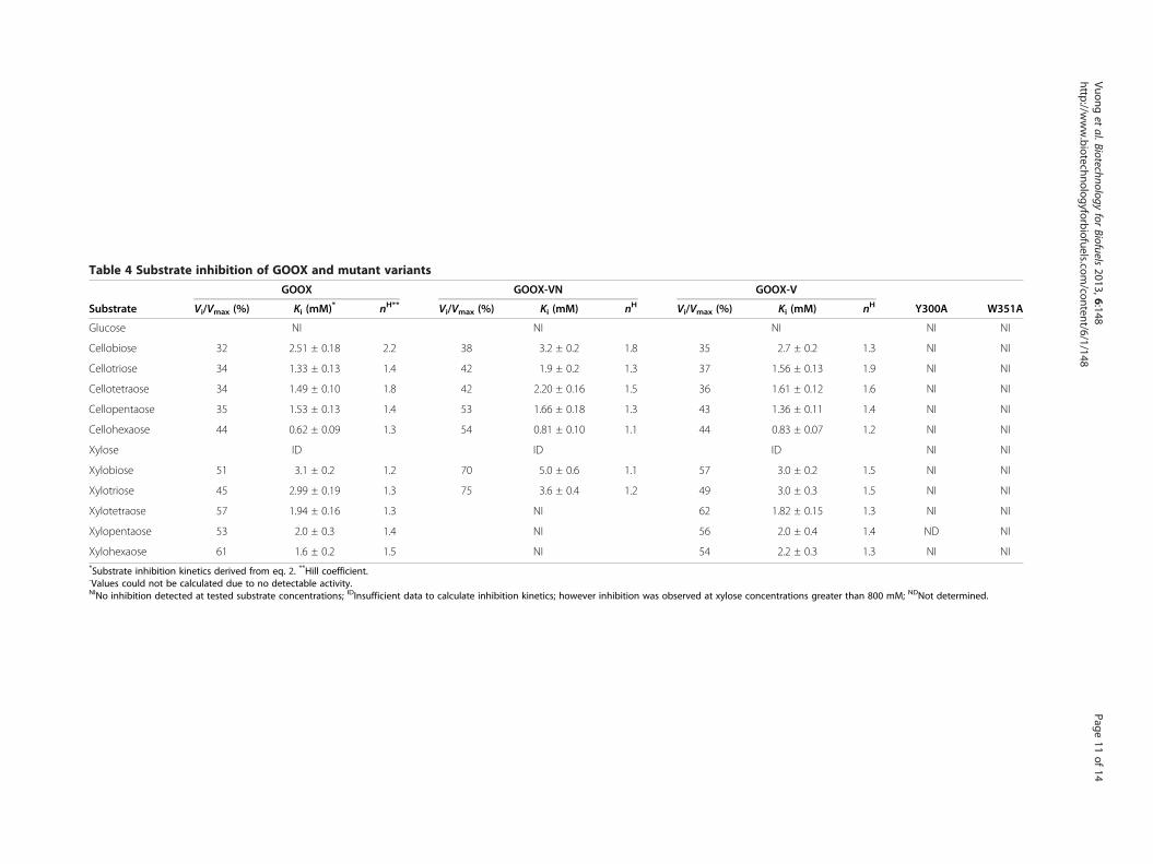
Table 4 Substrate inhibition of GOOX and mutant variants
Substrate
GOOX GOOX-VN GOOX-V
Y300A W351AVi/Vmax (%) Ki (mM)* nH** Vi/Vmax (%) Ki (mM) nH Vi/Vmax (%) Ki (mM) nH
Glucose NI NI NI NI NI
Cellobiose 32 2.51 ± 0.18 2.2 38 3.2 ± 0.2 1.8 35 2.7 ± 0.2 1.3 NI NI
Cellotriose 34 1.33 ± 0.13 1.4 42 1.9 ± 0.2 1.3 37 1.56 ± 0.13 1.9 NI NI
Cellotetraose 34 1.49 ± 0.10 1.8 42 2.20 ± 0.16 1.5 36 1.61 ± 0.12 1.6 NI NI
Cellopentaose 35 1.53 ± 0.13 1.4 53 1.66 ± 0.18 1.3 43 1.36 ± 0.11 1.4 NI NI
Cellohexaose 44 0.62 ± 0.09 1.3 54 0.81 ± 0.10 1.1 44 0.83 ± 0.07 1.2 NI NI
Xylose ID ID ID NI NI
Xylobiose 51 3.1 ± 0.2 1.2 70 5.0 ± 0.6 1.1 57 3.0 ± 0.2 1.5 NI NI
Xylotriose 45 2.99 ± 0.19 1.3 75 3.6 ± 0.4 1.2 49 3.0 ± 0.3 1.5 NI NI
Xylotetraose 57 1.94 ± 0.16 1.3 NI 62 1.82 ± 0.15 1.3 NI NI
Xylopentaose 53 2.0 ± 0.3 1.4 NI 56 2.0 ± 0.4 1.4 ND NI
Xylohexaose 61 1.6 ± 0.2 1.5 NI 54 2.2 ± 0.3 1.3 NI NI*Substrate inhibition kinetics derived from eq. 2. **Hill coefficient.-Values could not be calculated due to no detectable activity.NINo inhibition detected at tested substrate concentrations; IDInsufficient data to calculate inhibition kinetics; however inhibition was observed at xylose concentrations greater than 800 mM; NDNot determined.
Vuonget
al.Biotechnologyfor
Biofuels2013,6:148
Page11
of14
http://www.biotechnologyforbiofuels.com
/content/6/1/148
Vuong et al. Biotechnology for Biofuels 2013, 6:148 Page 12 of 14http://www.biotechnologyforbiofuels.com/content/6/1/148
were replaced by 50 mM Tris–HCl (pH 8.0) usingVivaspin 20 concentration units (Sartorius, Germany).
Confirmation of protein purity and identityProtein concentrations were measured using the Brad-ford method (Bio-Rad Laboratories, USA) and con-firmed using SDS-PAGE densitometry, where the banddensity of GOOX and the BSA reference protein weredetermined using ImageJ (http://rsbweb.nih.gov/ij/). Re-tention of the FAD cofactor in mutant enzymes wasassessed by verifying sample absorbance at 350–700 nmusing a Varian Cary 50 Bio UV–VIS spectrophotometer(Agilent Technologies, USA). The presence of the FADcofactor in intact protein samples was further confirmedby running the enzyme samples using SDS-PAGE andincubating the gel in 10% acetic acid for 10 min beforevisualization of fluorescent bands under a hand-heldMineralight® UV lamp (UVP, USA). A second, identicalSDS-PAGE gel was treated with performic acid beforethe acetic acid treatment to check for increase in fluor-escence intensity [19] (note: extra caution is requiredwhen handling performic acid).To confirm the introduction of single amino acid sub-
stitutions, protein samples were exchanged to MilliQwater using 10 kDa Amicon filter units (EMD Millipore,USA), and then 2000 pmol of each protein wereprocessed using a Waters Pico-Tag System to evaluatetotal amino acid composition (Advanced Protein Tech-nology Centre, Hospital for Sick Children, Toronto,Canada). Protein samples were also digested with modi-fied sequencing-grade trypsin (Promega, USA) andpeptide sequences were obtained by tandem mass spec-trometry using an LTQ-XL™ mass spectrometer (ThermoFisher Scientific, USA).
Enzymatic kinetics and thermostabilityA 96-well chromogenic assay was used to measurehydrogen peroxide production [7,10]. Briefly, the pro-duction of H2O2 was coupled to the oxidation of 4-AAby horseradish peroxidase and measured continuously at500 nm and 37°C for 15 min. To determine specific ac-tivity, 16 nM of enzyme was assayed with 0.5 mM oligo-saccharides. Kinetic parameters were determined byusing 16 nM of enzyme and a range of substrate concen-trations: 0.05 - 300 mM glucose, 0.05 - 1200 mM xylose,0.05 - 20 mM cellobiose, 0.01 - 10 mM cellotriose,xylobiose, and xylotriose, 0.01 - 4 mM of longer cello-and xylo-oligosaccharides, 0.01 - 1 mM A3X andAd2+3XX, and 0.04 - 0.4 mM U4m2XX. At least eightsubstrate concentrations in triplicates were assayed foreach substrate, and then kinetic parameters were calcu-lated using the Michaelis–Menten equation of GraphPadPrism5 software (GraphPad Software, USA). Substrateinhibition kinetics were calculated using a conventional
substrate inhibition equation (Equation 1) and a modi-fied Hill equation (Equation 2) [27]:
v ¼ Vmax � S½ �Km þ S½ � þ S½ �2
Ki
ð1Þ
v ¼Vmax þ V i
S½ �2K2
i
� �
1þ KnHs
S½ �nH þ S½ �2K2
i
ð2Þ
Where,Vi is the reaction velocity in the presence of in-hibition and nH is the Hill coefficient.To determine the temperature stabilities of enzyme
variants, 16 nM of each enzyme was incubated for 1 h attemperatures between 30 and 60°C, and residual activ-ities were measured using the conventional 4-AAchromogenic assay and 0.5 mM cellobiose.
Mass spectrometric analysis of oxidized productsReaction mixtures containing 1 mM of cello-oligosac-charides, from cellobiose to cellohexaose, and 160 nMGOOX-VN or GOOX-Y300A, in 50 mM Tris HCl (pH8.0) were incubated overnight at 37°C. To characterizeoxidized products, 100 μL of each reaction mixture werediluted in 900 μL of MilliQ-water, and diluted sampleswere purified and fractionated to neutral and acidic oli-gosaccharides using a Hypersep porous graphitized car-bon column (Thermo Scientific, MA, USA), followingthe protocols of Packer et al. [31] and Chong et al. [32]with modifications. Neutral sugars were eluted using40% acetonitrile, and mixture of 50% acetonitrile and0.05% TFA were used to elute acidic sugars. Collectedfractions were dried with nitrogen gas for 20 min andthen freeze-dried overnight.Mass spectrometric analyses were performed using an
Agilent XCT Plus model ion trap mass spectrometer(Agilent Technologies, Waldbronn, Germany) equippedwith an electrospray source. For ESI-MS and ESI-MS/MS analyses, freeze dried samples were dissolved in20 μL of MilliQ-water, and 6 μL of each sample was di-luted in 100 μL of methanol–water-formic acid solvent(50:49:1 (v:v:v)). Sample solutions were introduced intothe ES source at a flow rate of 5 μL/min via a syringepump. The drying gas temperature was set to 325°C;drying gas flow was 4 L/min; the nebulizer pressure was15 psi, and the ES capillary voltage was set to 3164 V.Ions were collected in the m/z range of 50 to 1000. ESI-MS/MS analyses were performed in both positive andnegative ionization modes. Fragmentation amplitude wasset to 0.60 V in the positive mode and 0.80 V in thenegative mode, and the precursor ion isolation widthwas set to 1.0 m/z and 1.5 m/z, respectively.
Vuong et al. Biotechnology for Biofuels 2013, 6:148 Page 13 of 14http://www.biotechnologyforbiofuels.com/content/6/1/148
NMR analysis of oxidized productsReaction mixtures containing 10 mM cellobiose or 10mM xylobiose, and 160 nM GOOX-VN or GOOX-Y300A, in 50 mM Tris HCl (pH 8.0) were incubatedovernight at 37°C. Oxidized products were analyzed byproton nuclear magnetic resonance (1H NMR) usinga Bruker 400 MHz NMR Spectrometer (BrukerUltrashield 400 Plus, USA). Samples were measured dir-ectly in the reaction solvent with water suppressionusing 10% deuterium oxide as a co-solvent for deuter-ium lock. The peaks were identified using the estimationprogram of ChemBioDrawUltra 12.0 (CambridgeSoft).
Additional files
Additional file 1: Figure S1. Protein sequence alignment of GOOX-T1,GOOX and GOOX-VN. The protein sequence of GOOX-T1 from S. strictumT1 was aligned with those of GOOX and GOOX-VN from S. strictum CBS346.70. The positions of amino acid differences are numbered while theamino acid substitutions created in GOOX-VN for the current study areindicated by rectangles. Amino acid substitutions introduced tore-construct GOOX from GOOX-VN are indicated by an asterisk.
Additional file 2: Figure S2. SDS-PAGE analysis of purified proteinpreparations. SDS-PAGE was performed using a 12% polyacrylamide gel,which was then stained with Coomassie Brilliant Blue R-250. PageRuler™Plus prestained protein ladder (Fermentas) was used.
Additional file 3: Figure S3. Positive ion ESI-MS spectra of four cello-oligosaccharide samples before and after oxidation. Samples wereseparated to neutral and acidic fractions prior analysis. G2: Cellobiose; G3:Cellotriose; G4: Cellotetraose; G5: Cellopentaose. (A)-(H): Unoxidizedcello-oligosaccharide samples; (I)-(P): GOOX-VN oxidized cello-oligosaccharide samples; (A)-(D) and (I)-(L): Neutral fractions: (E)-(H) and(M)-(P): Acidic fractions. Na: Sodium, K: Potassium and H: Proton adducts,respectively.
Additional file 4: Figure S4. A potential non-productive bindingsubsite. (Left): A potential binding pocket (yellow eclipse) above Y300and W351, as seen in the GOOX-T1 structure with the presence of ABL(PDB ID: 2AXR). (Right): A 45°-rotated view, the distance between twostacking residues is 8.1 Å.
Additional file 5: Table S1. The sequences of forward oligonucleotideprimers used for site-directed mutagenesis.
Abbreviations4-AA: 4-aminoantipyrine; AA: auxiliary activities; ABL: 5-amino-5-deoxy-cellobiono-1,5-lactam; CDH: cellobiose dehydrogenases; FAD: flavin adeninedinucleotide; GOOX: gluco-oligosaccharide oxidase; A3X: α-L-arabinofuranosyl-(1→3)-β-D-xylopyranosyl-(1→4)-D-xylose; Ad2+3XX: α-L-arabinofuranosyl-(1→2)-[α-L-arabinofuranosyl-(1→3)]-β-D-xylopyranosyl-(1→4)-β-D-xylopyranosyl-(1→4)-D-xylose; U4m2XX: 4-O-methyl-α-D-glucopyranosyl uronic acid-(1→2)-β-D-xylopyranosyl-(1→4)-β-D-xylopyranosyl-(1→4)-D-xylose.
Competing interestsThe authors declare there are no competing interests.
Authors’ contributionsTVV, JS, MT and ERM designed the experiments. TVV, AHV, MF and MJperformed the experiments and analyzed the data. All authors discussed theresults and implications and commented on the manuscript at all stages. Allauthors read and approved the final manuscript.
AcknowledgementsFunding for this research was provided by the Government of Ontario forthe project “Forest FAB: Applied Genomics for Functionalized Fibre and
Biochemicals” (ORF-RE-05-005), grants from the Natural Sciences andEngineering Research Council, and a FiDiPro Fellowship to ERM from theFinnish Funding Agency for Technology and Innovation (Tekes). This studywas also funded by the Academy of Finland for the ENOX project (Number252183) to MT and (Number 252827) to ERM.
Author details1Department of Chemical Engineering and Applied Chemistry, University ofToronto, 200 College Street, Toronto, Ontario, M5S 3E5, Canada. 2Departmentof Biotechnology and Chemical Technology, Aalto University, Kemistintie 1D1, Espoo 02150, Finland. 3Department of Food and Environmental Sciences,University of Helsinki, P.O. Box 27, Helsinki 00014, Finland.
Received: 7 June 2013 Accepted: 4 October 2013Published: 12 October 2013
References1. Levasseur A, Drula E, Lombard V, Coutinho PM, Henrissat B: Expansion of
the enzymatic repertoire of the CAZy database to integrate auxiliaryredox enzymes. Biotechnol Biofuels 2013, 6:41.
2. Henriksson G, Johansson G, Pettersson G: A critical review of cellobiosedehydrogenases. J Biotechnol 2000, 78:93–113.
3. Bankar SB, Bule MV, Singhal RS, Ananthanarayan L: Glucose oxidase-anoverview. Biotechnol Adv 2009, 27:489–501.
4. Prongjit M, Sucharitakul J, Wongnate T, Haltrich D, Chaiyen P: Kineticmechanism of pyranose 2-oxidase from Trametes multicolor.Biochemistry 2009, 48:4170–4180.
5. Whittaker JW: Free radical catalysis by galactose oxidase. Chem Rev 2003,103:2347–2363.
6. Lee MH, Lai WL, Lin SF, Hsu CS, Liaw SH, Tsai YC: Structuralcharacterization of glucooligosaccharide oxidase from Acremoniumstrictum. Appl Environ Microbiol 2005, 71:8881–8887.
7. Foumani M, Vuong TV, Master ER: Altered substrate specificity of thegluco-oligosaccharide oxidase from Acremonium strictum.Biotechnol Bioeng 2011, 108:2261–2269.
8. Summerbell RC, Gueidan C, Schroers HJ, de Hoog GS, Starink M, Rosete YA,Guarro J, Scott JA: Acremonium phylogenetic overview and revision ofGliomastix, Sarocladium, and Trichothecium. Stud Mycol 2011, 68:139–162.
9. Fan Z, Oguntimein GB, Reilly PJ: Characterization of kinetics andthermostability of Acremonium strictum glucooligosaccharide oxidase.Biotechnol Bioeng 2000, 68:231–237.
10. Lin S-F, Yang T-Y, Inukai T, Yamasaki M, Tsai Y-C: Purification andcharacterization of a novel glucooligosaccharide oxidase fromAcremonium strictum T1. Biochim Biophys Acta 1991, 1118:41–47.
11. van Hellemond EW, Leferink NG, Heuts DP, Fraaije MW, van Berkel WJ:Occurrence and biocatalytic potential of carbohydrate oxidases.Adv Appl Microbiol 2006, 60:17–54.
12. Cruys-Bagger N, Ren G, Tatsumi H, Baumann MJ, Spodsberg N, AndersenHD, Gorton L, Borch K, Westh P: An amperometric enzyme biosensor forreal-time measurements of cellobiohydrolase activity on insolublecellulose. Biotechnol Bioeng 2012, 109:3199–3204.
13. Fan Z, Wu W, Hildebrand A, Kasuga T, Zhang R, Xiong X: A novelbiochemical route for fuels and chemicals production from cellulosicbiomass. PLoS One 2012, 7:e31693.
14. Huang CH, Winkler A, Chen CL, Lai WL, Tsai YC, Macheroux P, Liaw SH:Functional roles of the 6-S-cysteinyl, 8alpha-N1-histidyl FAD inglucooligosaccharide oxidase from Acremonium strictum. J Biol Chem2008, 283:30990–30996.
15. Heuts DP, Winter RT, Damsma GE, Janssen DB, Fraaije MW: The role ofdouble covalent flavin binding in chito-oligosaccharide oxidase fromFusarium graminearum. Biochem J 2008, 413:175–183.
16. Forsberg Z, Vaaje-Kolstad G, Westereng B, Bunaes AC, Stenstrom Y,MacKenzie A, Sorlie M, Horn SJ, Eijsink VG: Cleavage of cellulose by aCBM33 protein. Protein Sci 2011, 20:1479–1483.
17. Nordkvist M, Nielsen PM, Villadsen J: Oxidation of lactose to lactobionicacid by a Microdochium nivale carbohydrate oxidase: kinetics andoperational stability. Biotechnol Bioeng 2007, 97:694–707.
18. Kalisz HM, Hecht HJ, Schomburg D, Schmid RD: Effects of carbohydratedepletion on the structure, stability and activity of glucose oxidase fromAspergillus niger. Biochim Biophys Acta 1991, 1080:138–142.
Vuong et al. Biotechnology for Biofuels 2013, 6:148 Page 14 of 14http://www.biotechnologyforbiofuels.com/content/6/1/148
19. Scrutton NS: Identification of covalent flavoproteins and analysis of thecovalent link. Methods Mol Biol 1999, 131:181–193.
20. Huang CH, Lai WL, Lee MH, Chen CJ, Vasella A, Tsai YC, Liaw SH: Crystalstructure of glucooligosaccharide oxidase from Acremonium strictum: anovel flavinylation of 6-S-cysteinyl, 8alpha-N1-histidyl FAD. J Biol Chem2005, 280:38831–38838.
21. Nouaille R, Matulova M, Patoprsty V, Delort AM, Forano E: Production ofoligosaccharides and cellobionic acid by Fibrobacter succinogenes S85growing on sugars, cellulose and wheat straw. Appl Microbiol Biotechnol2009, 83:425–433.
22. Higham CW, Gordon-Smith D, Dempsey CE, Wood PM: Direct 1H NMRevidence for conversion of beta-D-cellobiose to cellobionolactone bycellobiose dehydrogenase from Phanerochaete chrysosporium.FEBS Lett 1994, 351:128–132.
23. Asam MR, Glish GL: Tandem mass spectrometry of alkali cationizedpolysaccharides in a quadrupole ion trap. J Am Soc Mass Spectr 1997,8:987–995.
24. Hofmeister GE, Zhou Z, Leary JA: Linkage position determination inlithium-cationized disaccharides: tandem mass spectrometry andsemiempirical calculations. J Am Chem Soc 1991, 113:5964–5970.
25. Pasanen S, Janis J, Vainiotalo P: Cello-, malto- and xylooligosaccharidefragmentation by collision-induced dissociation using QIT and FT-ICRmass spectrometry: a systematic study. Int J Mass Spectrom 2007,263:22–29.
26. Heuts DPHM, Janssen DB, Fraaije MW: Changing the substrate specificityof a chitooligosaccharide oxidase from Fusarium graminearum by model-inspired site-directed mutagenesis. FEBS Lett 2007, 581:4905–4909.
27. LiCata VJ, Allewell NM: Is substrate inhibition a consequence of allosteryin aspartate transcarbamylase? Biophys Chem 1997, 64:225–234.
28. Pastell H, Tuomainen P, Virkki L, Tenkanen M: Step-wise enzymaticpreparation and structural characterization of singly and doublysubstituted arabinoxylo-oligosaccharides with non-reducing endterminal branches. Carbohydr Res 2008, 343:3049–3057.
29. Rantanen H, Virkki L, Tuomainen P, Kabel M, Schols H, Tenkanen M:Preparation of arabinoxylobiose from rye xylan using family 10Aspergillus aculeatus endo-1,4-beta-D-xylanase. Carbohyd Polym 2007,68:350–359.
30. Koutaniemi S, Guillon F, Tranquet O, Bouchet B, Tuomainen P, Virkki L,Petersen HL, Willats WG, Saulnier L, Tenkanen M: Substituent-specificantibody against glucuronoxylan reveals close association of glucuronicacid and acetyl substituents and distinct labeling patterns in treespecies. Planta 2012, 236:739–751.
31. Packer NH, Lawson MA, Jardine DR, Redmond JW: A general approach todesalting oligosaccharides released from glycoproteins. Glycoconj J 1998,15:737–747.
32. Chong SL, Nissila T, Ketola RA, Koutaniemi S, Derba-Maceluch M,Mellerowicz EJ, Tenkanen M, Tuomainen P: Feasibility of usingatmospheric pressure matrix-assisted laser desorption/ionization withion trap mass spectrometry in the analysis of acetylatedxylooligosaccharides derived from hardwoods and Arabidopsis thaliana.Anal Bioanal Chem 2011, 401:2995–3009.
33. Domon B, Costello C: A systematic nomenclature for carbohydratefragmentations in FAB-MS/MS spectra of glycoconjugates.Glycoconj J 1988, 5:397–409.
doi:10.1186/1754-6834-6-148Cite this article as: Vuong et al.: Xylo- and cello-oligosaccharideoxidation by gluco-oligosaccharide oxidase from Sarocladium strictumand variants with reduced substrate inhibition. Biotechnology for Biofuels2013 6:148.
Submit your next manuscript to BioMed Centraland take full advantage of:
• Convenient online submission
• Thorough peer review
• No space constraints or color figure charges
• Immediate publication on acceptance
• Inclusion in PubMed, CAS, Scopus and Google Scholar
• Research which is freely available for redistribution
Submit your manuscript at www.biomedcentral.com/submit