Embed Size (px)
Citation preview

Receptor Binding
X-Ray Crystallography and Free Energy Calculations Reveal theBinding Mechanism of A2A Adenosine Receptor Antagonists**Willem Jespers, Gr8gory Verdon, Jhonny Azuaje, Maria Majellaro, Henrik Ker-nen,Xerardo Garc&a-Mera, Miles Congreve, Francesca Deflorian, Chris de Graaf, Andrei Zhukov,Andrew S. Dor8, Jonathan S. Mason, Johan cqvist, Robert M. Cooke, Eddy Sotelo, andHugo Guti8rrez-de-Ter#n*
Abstract: We present a robust protocol based on iterations offree energy perturbation (FEP) calculations, chemical syn-thesis, biophysical mapping and X-ray crystallography toreveal the binding mode of an antagonist series to the A2A
adenosine receptor (AR). Eight A2AAR binding site mutationsfrom biophysical mapping experiments were initially analyzedwith sidechain FEP simulations, performed on alternatebinding modes. The results distinctively supported one bindingmode, which was subsequently used to design new chromonederivatives. Their affinities for the A2AAR were experimentallydetermined and investigated through a cycle of ligand-FEPcalculations, validating the binding orientation of the differentchemical substituents proposed. Subsequent X-ray crystallog-raphy of the A2AAR with a low and a high affinity chromonederivative confirmed the predicted binding orientation. Thenew molecules and structures here reported were driven by freeenergy calculations, and provide new insights on antagonistbinding to the A2AAR, an emerging target in immuno-oncology.
Introduction
Computational estimation of shifts in binding free energy,associated with ligand modifications or point mutations in thereceptor macromolecule, can provide the missing link be-tween the structure of a protein-ligand complex and a panel ofexperimental binding affinities. Rigorous free energy pertur-bation (FEP) methods have been used for decades tounderstand the structure-affinity relationships (SAR) arounda given chemical scaffold, and recent advances now allow the
use of this technique routinely in ligand design projects.[1] Thesame methodology can be used to analyze this problem froma complementary perspective, that is to estimate the gain orloss in binding free energy from site-directed mutagenesis(SDM) data. The idea of in silico mutagenesis, initiallyintroduced almost three decades ago by Kollman to study thebinding and catalysis of subtilisin,[2] was recently implementedin computational pipelines that pursue a systematic character-ization of the effect on point mutations on for example,ligand-binding or protein stability.[3–5] The combination ofboth ligand and residue FEP simulations can provide a fullenergetic landscape of the molecular interactions governingprotein-ligand binding, which underlies the design of twocomplementary protocols in our lab, namely QligFEP[6] andQresFEP,[3] integrated in the molecular dynamics (MD)software package Q.[7, 8]
One area where this approach is particularly promising isthe design of ligands for G-protein-coupled receptors(GPCRs), a superfamily of seven-transmembrane (7TM)cellular receptors[9] that mediate the therapeutic effects ofabout 30 % of all marketed drugs.[10] There is a large amountof SAR and SDM data available for these receptors, whichcan be combined with the increasing growth of structuralknowledge of many GPCR targets. The first integratedapproach of ligand and residue FEP simulations was pub-lished by Boukharta et al. to characterize antagonist bindingto the Y1 neuropeptide receptor,[11] which we later expandedto other GPCR families, including the related neuropeptidereceptor Y2,
[12] the orphan receptor GPR139[13] and severalmembers of the family of adenosine receptors.[14–16] Among
[*] W. Jespers, Dr. H. Ker-nen, Prof. J. bqvist, Dr. H. Guti8rrez-de-Ter#nDepartment of Cell and Molecular Biology, Uppsala University,BMC, Biomedical CenterBox 596, Uppsala (Sweden)E-mail: [email protected]
Dr. G. Verdon, Dr. M. Congreve, Dr. F. Deflorian, Dr. C. de Graaf,Dr. A. Zhukov, Dr. A. S. Dor8, Dr. J. S. Mason, Dr. R. M. CookeSosei HeptaresSteinmetz Granta Park, Great Abington, Cambridge CB21 6DG (UK)
Dr. J. Azuaje, Dr. M. Majellaro, Prof. X. Garc&a-Mera, Prof. E. SoteloDepartament of Organic Chemistry, Faculty of Farmacy, Universi-dade de Santiago de Compostela (Spain)
Dr. J. Azuaje, Dr. M. Majellaro, Prof. E. SoteloCentro Singular de Investigacikn en Qu&mica Biolkxica y MateriaisMoleculares (CIQUS), Universidade de Santiago de Compostela(Spain)
Dr. H. Ker-nenPresent address: H. Lundbeck A/SOttiliavej 9, 2500 Valby (Denmark)
[**] A previous version of this manuscript has been deposited ona preprint server (https://doi.org/10.26434/chemrxiv.11444877.v1).
Supporting information and the ORCID identification number(s) forthe author(s) of this article can be found under:https://doi.org/10.1002/anie.202003788.
T 2020 The Authors. Published by Wiley-VCH Verlag GmbH & Co.KGaA. This is an open access article under the terms of the CreativeCommons Attribution License, which permits use, distribution andreproduction in any medium, provided the original work is properlycited.
AngewandteChemieResearch Articles
How to cite: Angew. Chem. Int. Ed. 2020, 59, 16536–16543International Edition: doi.org/10.1002/anie.202003788German Edition: doi.org/10.1002/ange.202003788
16536 T 2020 The Authors. Published by Wiley-VCH Verlag GmbH & Co. KGaA, Weinheim Angew. Chem. Int. Ed. 2020, 59, 16536 – 16543

the latter, the adenosine A2A receptor (A2AAR) was one ofthe first GPCRs to be crystallized[17] and today stands out asone of the better characterized GPCRs from a structuralperspective. Several structures of the inactive and activeforms of the receptor have been solved within the lastdecade, and the integration of the available experimental datahas strongly aided ligand design programs for this recep-tor.[18, 19]
Many A2AAR antagonists have been developed targetinga number of pathologies,[20–22] including recent clinical candi-dates in immuno-oncology. A number of these antagonistshave been co-crystallized with the A2AAR, providing uniquestructural information which, in combination with the exten-sive SDM data available,[23] allow envisaging ligand bindingmechanisms and structure-based drug design (SBDD) pro-grams of antagonist molecules.[18] However, suboptimalproperties of traditional scaffolds, such as poor pharmacoki-netics and low selectivity profiles, motivate the search ofnovel chemical entities as A2AAR antagonists,[24] frequentlythrough high throughput screening (HTS) campaigns. In thesecases, it is not common to obtain a crystal structure of thereceptor-ligand complex, which can hamper further hit to leadoptimization. Instead, approximate binding modes are ofteninferred from the experimental data extracted from SAR ofligand series and SDM data,[23] which can be complementedby computational models of the protein-ligand complex.[25]
Biophysical Mapping (BPM), is an integrated approachthat has been used with success in antagonist design programson the A2AAR and other GPCRs.[27–30] Here, the bindingaffinity of a ligand series is evaluated via surface plasmonresonance (SPR) on a panel of mutant receptors, each bearinga single-point mutation within the putative binding site.[27]
The resulting matrix of binding affinity shifts from wild type(WT) affinities combined with SAR data and mapped toa receptor-ligand model provide further insights in thedeterminants of binding of the scaffold. The first applicationof this technique was based around the co-crystallized A2AARantagonist ZM241385 in combination with 8 receptor mutants(see Figure 1A).[22] These mutations involve residues in directcontact with the ligand, such as N2536.55, L853.33, M1775.38,N1815.42 and I662.64 (Ballesteros Weinstein numbering[31] insuperscript), as well as residues not directly in contact withthe ligand, namely S2777.42, Y2717.36 and L167EL2 (see Fig-ure 1). This approach was extended for 1,2,4-triazines asA2AAR antagonists,[32] and the binding mode was laterconfirmed by X-ray crystallography (see Figure 1B).[29] Inthe same HTS campaign, a series of chromones wereidentified as a novel family of A2AAR antagonists,[32] andconsecutively optimized to yield the potent and selectiveChromone 14 (see Figure 2).[30] At that point, the lead-optimization program was successful in improving the affinityof the initial HTS hit, while not focusing on pharmacokineticoptimization (i.e., the most potent compound Chromone 14contains a metabolically unstable ester group). Interestingly,this structure-based optimization was guided by the interpre-tation of the BPM data and a computational model of thecomplex generated by docking. This led to the proposal of twoputative binding modes compatible with the BPM data. Evenif the SAR of the generated series seemed to favor one of
them, the question remained open due to the lack of an X-raystructure with any of these compounds in the originalstudy.[30,32]
Here, we initially examine the SPR data available forthese A2AAR antagonist families through a recently devel-oped in silico mutagenesis tool based on free energyperturbation (FEP) simulations.[11, 14, 15] The results pointedto a unified binding mode of the chromone series, which ishere used as a basis for the design, synthesis and pharmaco-logical evaluation of an extended series of compounds aimedto further explain the underlying SAR of chromones asA2AAR antagonists. Finally, experimental structures oftwo chromone-A2AAR complexes were solved which con-firmed the binding mode hypothesis from the computationalstudies.
Results and Discussion
Free Energy Perturbation Calculations on Existing BPM Data
The BPM data obtained for the three chemotypes ofA2AAR antagonists were collected from reference [27], andrelative binding free energy changes between mutant and WTreceptor were calculated from Ki values (see Table 1,DDGexp
bind). Thereafter, relative binding free energies for eachligand were calculated based on 3D models of the receptor-ligand complexes (DDGcalc
bind, Table 1). In the case ofZM241385, the model was directly extracted from the high-resolution structure with the A2AAR (see Figure 1), and thecurated receptor model (see Supporting Information, Meth-ods) was used throughout this work. The results showexcellent agreement with the experimental data, with a meanabsolute error (MAE) of 0.41 kcalmol@1 and a correlationcoefficient of R2 = 0.94 (Table 1). Next, the same set of BPMmutations was analyzed for triazine 4g. In this case, thestarting configuration of the complex was obtained by
Figure 1. Binding mode and chemical structures of antagonistsZM241385 (A, crystal structure 4EIY[26]) and triazine 4b (B). Theexperimental pose of the triazine (cyan) was superimposed on thesame crystal structure of the receptor shown in panel A (ribbons).Both compounds had been characterized by BPM (residues labelledand depicted in gray sticks). Receptor-ligand hydrogen bonds aredepicted as magenta lines.
AngewandteChemieResearch Articles
16537Angew. Chem. Int. Ed. 2020, 59, 16536 – 16543 T 2020 The Authors. Published by Wiley-VCH Verlag GmbH & Co. KGaA, Weinheim www.angewandte.org

aligning the triazine 4g-A2AAR complex (3UZA) to thecurated model described before, retaining only the coordi-
nates of the ligand (Figure 1B).[33] The calculated freeenergies (DDGcalc
bind, Table 1) were in general good agreementwith the experimental data (MAE = 0.94 kcal mol@1; R2 =
0.66). In contrast to the previous case, two mutations(Met1775.38 and Asn1815.42) show qualitative discrepancy withthe experimental data for this ligand. Subsequent analysis ofthe MD trajectories suggests that this is likely due toa suboptimal representation of the (water mediated) H-bondnetwork upon mutation, which has previously been recog-nized as a challenging factor for this particular scaffold.[34,35]
Once the QresFEP protocol was validated to reproducethe BPM data on experimentally known structures, we movedon to the chromone scaffold, for which there is analogousBPM data, but no crystal structure available. Here, tworeasonable binding modes were generated by moleculardocking of Chromone 14. The two poses, denoted as A andB (see Figure 2), form at least one hydrogen bond withAsn2536.55 (a highly conserved interaction in AR ligandrecognition)[23] and are related by a symmetry axis along thebicyclic core of the chromone scaffold. According to thedocking score function used, both poses were energeticallyequivalent (@9.30 vs. @9.17 for pose A and B respectively)making it difficult at this point to discern the correct one, inline with the previous binding hypothesis by Andrewset al.[30, 32]
In pose A (Figure 2A), Asn2536.55 forms an H-bond withthe nitrogen in the 4-methylthiazole group, leaving thecarbonyl group potentially exposed to the internal waternetwork stabilized by residues in TM7, as observed in theA2AAR crystal structure with ZM241385[26] (see SupportingInformation, Figure S1). Conversely, in pose B (Figure 2B)this carbonyl forms an additional hydrogen bond withN2536.55. We hypothesized that in principle, the symmetryaxis would allow exchanging substituents at R6 and R7
between the binding poses, something that we would explorelater in this work (see below).
Our strategy to select the most reliable binding mode wasto compute the effect on ligand binding of the 8 mutationsfrom the BPM panel[27] for each pose, and compare the resultswith the experimental values. The results (Figure 2C, Table 1and Supporting Information, Table S1) highlight pose A asthe binding mode with the best correlation to experimentaldata. A MAE of 0.50 kcalmol@1 was observed for this pose,which is comparable to the results obtained for the co-crystallized antagonist ZM241385. In addition, the correlationcoefficient of R2 = 0.74 falls between those observed forZM241385 and triazine 4g. Conversely, the correspondingvalues calculated on pose B are much higher (MAE =
1.53 kcal mol@1) and the correlation is completely lost (R2 =
0.03). Additionally, the computed loss of binding affinity uponthe N2536.55A mutation is closer to experiment in pose A thanin pose B, indicating that the additional H-bond betweenAsn2536.55 and the carbonyl in pose B would not contribute tomore favorable binding free energies. From the MD simu-lations, we further observed that the binding modes are notrelated by a symmetry axis as initially hypothesized, and thesubstituents at positions R6 and R7 are exploring differentpositions in the binding site, making them no longer readilyinterchangeable.
Figure 2. Putative binding modes A (green) and B (magenta) ofChromone 14 to the A2AAR (H-bonds in magenta). C) Experimentaland calculated changes in binding free energies for each mutation inthe BPM. The error bars correspond to the s.e.m. of the replicacalculations for the calculated values, or are adjusted to the reportedvalue of 0.1 pKD unit in the case of experimental data.[27]
Table 1: Comparison between experimental and calculated relativebinding free energies (DDGbind in kcal mol@1) for A2AAR mutants.
Mutant[a] ZM241385 Triazine 4gDDGexp
bind[b] DDGcalc
bind DDGexpbind
[b] DDGcalcbind
I66A2.64 0.14 0.83:0.34 0.41 1.94:0.34L85A3.33 2.45 3.30:0.41 1.09 1.65:0.37L167A5.28 0.00 0.60:0.31 @0.14 @0.39:0.36M177A5.38 0.14 @0.09:0.44 @0.27 1.66:0.49N181A5.42 1.23 1.47:0.57 0.82 @0.63:0.55N253A6.55 +5.86[c] 5.81:0.57 +4.36[c] 5.64:0.56Y271A7.36 1.09 0.84:0.74 0.41 @0.1:0.68
[a] Data for the mutant receptor constructs reported in reference [27].[b] Experimental relative binding free energies were calculated from KD
values as DDGexpbind ¼ RTln Kmut
D =KWTD
. -with experimental errors in all
cases reported as approximately 0.1 pKD unit, that is, less than0.1 kcalmol@1.[27] [c] Binding affinity of the ligand to the (mutant)receptor was lower than the experimental threshold (pKD<5 in allcases). Errors are standard error of the mean (s.e.m.) over a total of 10replicates.
AngewandteChemieResearch Articles
16538 www.angewandte.org T 2020 The Authors. Published by Wiley-VCH Verlag GmbH & Co. KGaA, Weinheim Angew. Chem. Int. Ed. 2020, 59, 16536 – 16543

Design, Synthesis and Pharmacological Evaluation of ChromoneDerivatives
On the basis of the binding mode hypothesis assessed bythe FEP calculations, we designed a small collection ofchromone derivatives (Scheme 1), aiming to systematically
explore two different variables on A2AAR affinity: methyl-ation at position 2 (series 5) and the aforementioned effect ofswitching the oxygenated function (@OH or@OCOMe) fromthe original position 7 (series 4) to position 6 (series 8),swapping the alkyl substitution (Pr/H) accordingly. A collec-tion of 11 molecules was prepared as depicted in synthetic
Scheme 1. Structure and synthetic pathways employed to assembly chromones 4, 5 and 8.
AngewandteChemieResearch Articles
16539Angew. Chem. Int. Ed. 2020, 59, 16536 – 16543 T 2020 The Authors. Published by Wiley-VCH Verlag GmbH & Co. KGaA, Weinheim www.angewandte.org

Scheme 1. Briefly, the key 2-hydroxyphenyl ketones 3 wereobtained by either the Hoesch method[30] or Claisen con-densation,[36] employing phenols 1 or esters 6 and thiazolederivatives 2a and 2 b as reactive precursors, respectively.Treatment of ketones 3 with orthoesters (formate or acetate)enabled the efficient chromone core formation,[37] thusdefining the substituent pattern at position 2 (H or Me).Finally, the required acetates were prepared by reaction of thecorresponding phenols with acetyl chloride. The full syntheticmethodology is provided in the Supporting Information.
The affinity of these compounds for the WT A2AAR wasthen evaluated with the same SPR assay used to generate theBPM data, using the A2AAR-STAR2 construct (see Support-ing Information, Methods).[27] The data confirmed thatmethylation at position 2 (compounds 5a–d) is generallyunfavorable for binding, compared to the parent series(compounds 4a–d). This is in agreement with previous workby Andrews et al.,[30] where methylation of the low affinitycompound 4a (corresponding to chromone 8 as reported byAndrews et al. ,[30] with pKi = 5.7) resulted in a relativedecrease in affinity of 0.6 log units (Chromone 12, pKi =
5.1). We observe a similar difference for this pair ofcompounds (Table 2, DpKD = 0.85), while this effect isamplified in the case of the high affinity precursor (see 4das compared to 5d, DpKD = 2.7). More surprising was theobservation of a similar effect when the substituents betweenR6 and R7 are swapped (compounds 8a–d), which again wasmost pronounced for the high affinity compound 4d (seeTable 2), with a drop of affinity of 2.7 log units for thecorresponding methylated derivative 8d.
Computational Evaluation of the Proposed Binding Modes
From the pharmacological data on the expanded series ofchromones and the initial MD simulations, it appears that thetwo binding poses differ more than just purely on the rotationaxis of the chromone scaffold.
The following step was then to investigate whether allcompounds in the series would adopt the preferred bindingmode A, or if binding mode B is accessible by some of thecompounds, depending on the pattern of substitutions. Sucha hypothesis was experimentally observed for caffeine, whichin contrast to other xanthine derivatives presents a dualbinding mode to the A2AAR (see Figure 3A). Indeed, anisoenergetic dual binding mode for caffeine was previouslyhypothesized on the basis of free energy calculations,[38]
before experimental observation of dual-occupancy crystalstructures in complex with the A2AAR.[39, 40] Conversely, theX-ray structure of the N7-demethylated analogue theophyl-line shows that this molecule adopts only one of these bindingmodes, where the acidic hydrogen at position N7 makes anadditional H-bond contact with N2536.55 (Figure 3). We thusevaluated whether our recently developed dual-topologyprotocol QligFEP was suitable to capture this differentbehavior, by a direct estimation of relative binding freeenergies between the two poses, in analogy to the efficiency ofthis protocol to compare topologically unrelated ligands (e.g.,scaffold hopping).[6] The results for the A2A-xanthine systemshown in Table 3 show that this is the case: the negligiblecalculated free energy difference between the two poses forcaffeine is in line with the equally populated dual bindingmode in the crystal structure with the A2AAR (PDB code5MZP, Figure 3 A). In contrast, the single binding modeobserved in the crystal structure of theophylline (blue color inFigure 3A) is energetically favored by 1.6 kcal mol@1. Subse-quent application of the same strategy in the generatedchromone series showed a similar energy gap between the twobinding poses considered for the simplest chromone in ourseries (4a, Figure 3 B), with pose A being 1.7 kcal mol@1 morefavorable than pose B. Notably, this energy gap increasessignificantly for the highest affinity compound 4 d, suggestive
Table 2: SPR affinity data for the series of Chromone derivativessynthesized in this work.
Compound Substituents pKD[a]
R2 R5 R6 R7
4a[b] H H H H 5.954b[b] H H C3H7 H 6.204c H CH3 H H 5.804d[b] (Chromone 14) H H C3H7 COCH3 8.605a[b] CH3 H H H 5.105b CH3 H C3H7 H 5.425c CH3 CH3 H H 5.365d CH3 H C3H7 COCH3 5.908a H H H H 5.508b H H H C3H7 5.708d H H COCH3 C3H7 5.90
[a] Experimental errors in all cases reported as approximately 0.1 pKD
unit, that is, less than 0.1 kcal mol@1. [b] Compounds previously reportedin ref. [30]. Figure 3. A) Dual binding mode of caffeine, as extracted from the
A2AAR crystal structure with the A2AAR (PDB code 5MZP). Colour codeis green (binding mode A) and magenta (binding mode B). B) mod-elled binding modes of Chromone 4a, following the same colouringScheme as in panel A.
AngewandteChemieResearch Articles
16540 www.angewandte.org T 2020 The Authors. Published by Wiley-VCH Verlag GmbH & Co. KGaA, Weinheim Angew. Chem. Int. Ed. 2020, 59, 16536 – 16543
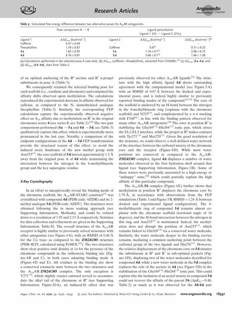
of an optimal anchoring of the R6 acetate and R7 n-propylsubstituents in pose A (Table 3).
We consequently retained the selected binding pose foreach scaffold (i.e., xanthine and chromone) and computed theaffinity shifts observed upon methylation. The calculationsreproduced the experimental decrease in affinity observed forcaffeine, as compared to the N7 demethylated analoguetheophylline (Table 3). Similarly, the corresponding FEPcalculations capture the experimentally observed negativeeffect on A2A affinity due to methylation on R2 in the originalchromones series 4 (see series 5, see Table 2).[30] The two paircomparisons performed (4a ! 5a and 4d ! 5d, see Table 3)qualitatively capture this effect, which is experimentally morepronounced in the last case. Notably, a comparison of theendpoint configurations of the 4d ! 5d FEP transformationprovide the structural reason of this effect: to avoid theinduced steric hindrance of the new methyl group withAsn2536.55, the core scaffold of 5 d moved approximately 1.5 caway from the original pose A of 4 d while maintaining theinteraction between the nitrogen in the 4-methylthiazolegroup and the key asparagine residue.
X-Ray Crystallography
In an effort to unequivocally reveal the binding mode ofthe chromone scaffold, the A2AAR-STAR2 construct[27] wascrystallized with compound 4d (PDB code: 6ZDR) and its 2-methyl analogue 5d (PDB code: 6ZDV). The structures wereobtained following the in meso soaking approach (seeSupporting Information, Methods), and could be refineddown to a resolution of 1.92 and 2.13 c respectively. Statisticsfor data collection and refinement are given in the SupportingInformation, Table S2. The overall structure of the A2AARreceptor is highly similar to previously solved structures withother antagonists (see Figure 4A), with an RMSD of 0.46 cfor the Ca trace as compared to the ZM241385 structure(PDB 4EIY, calculated using PyMOL[42]). The two structuresshow clear positive omit density at 1s for the presence of thechromone compounds in the orthosteric binding site (Fig-ure 4B and C), in both cases adopting binding mode A(Figure 4D and E). All residues in the binding site showa conserved rotameric state between the two structures andthe A2AAR-ZM241385 complex. The only exception isY2717.36, which slightly rotates outward served to accommo-date the alkyl tail of the chromone at R6 (see SupportingInformation, Figure S2A), an induced-fit effect that was
previously observed for other A2AAR ligands.[33] The struc-ture with the high affinity ligand 4 d shows outstandingagreement with the computational model (see Figure 5A)with an RMSD of 0.67 c between the docked and exper-imental poses, and is indeed highly similar to previouslyreported binding modes of the compound.[30,32] The core ofthe scaffold is anchored by an H-bond between the nitrogenin the 4-methylthiazole ring (coplanar with the chromonescaffold) and N2536.55, and complemented by a p-p stackingwith F168EL2, in line with the binding pattern observed formany other A2AAR antagonists.[20] The ester at position R7 isstabilizing the Glu1695.30–His2647.29 ionic pair, which closesthe EL2-EL3 interface, while the propyl at R6 makes contactswith Tyr2717.53 and Met2707.52. Given the good resolution ofthe structure, we could observe a well-defined water networkof the interface between the carbonyl moiety of the chromonecore and the receptor (Figure 4D). While most waterpositions are conserved as compared to the A2AAR-ZM241385 complex, ligand 4d displaces a number of watermolecules observed in the first hydration shell around thatligand (see Supporting Information, Figure 2B). Some ofthese waters were previously associated to a high-energy or“unhappy” state,[30] which could partially explain the highaffinity of this particular compound.
The A2AAR-5b complex (Figure 4E) further shows thatmethylation at position R2 displaces the chromone core by1.79 c, in accordance with observations from the FEPsimulations (Table 3 and Figure 5 B, RMSD = 1.24 c betweendocked and experimental ligand configuration). The 4-methylthiazole ring of compound 5d remains almost co-planar with the chromone scaffold (torsional angle of 16degrees), and the H-bond interaction between the nitrogen inthis ring and Asn2536.55 is maintained. Indeed, the methyl-ation does not disrupt the position of Asn2536.55, whichremains linked to Glu1695.30 via a conserved water molecule.Similarly, the water molecule deeper in the binding creviceremains, mediating a common anchoring point between thecarbonyl group of the two ligands and His2767.43. However,the relative displacement of the chromone core on 5d situatesthe substituents at R6 and R7 in sub-optimal pockets (Fig-ure 4D), displacing two of the water molecules described forcompound 4d, while a new water molecule in the 5d complexreplaces the role of the acetate in 4d (see Figure 5 D) in thestabilization of the Glu1695.30–His2647.29 ionic pair. This couldexplain why the inclusion of an acetyl moiety in compound 5dcould not recover the affinity of the parent 5 b (DpKD = 0.48,Table 2) as much as it was observed for the 4b/4 d pair
Table 3: Calculated free energy difference between two alternative poses for A2AAR antagonists.
Pose comparison A ! B Ligand perturbationLigand 1 (H)! Ligand 2 (CH3)
Ligand 1 DDGcalc [kcalmol@1] Ligand 2 DDGexp [kcalmol@1] DDGcalc [kcalmol@1][a]
Caffeine 0.47:0.49 – – –Theophylline 1.59:0.87 Caffeine 0.6[b] 0.31:0.324a 1.65:0.93 5a 1.16:0.1[c] 2.06:0.724d 8.76:0.81 5d 3.68:0.1[d] 1.66:1.09
[a] Calculations performed in the selected pose A (see text). [b] DGbind (caffeine—theophylline), extracted from ChEMBL.[41] [c] DGbind (5a–4a) and[d] DGbind (5d–4d), data from Table 2.
AngewandteChemieResearch Articles
16541Angew. Chem. Int. Ed. 2020, 59, 16536 – 16543 T 2020 The Authors. Published by Wiley-VCH Verlag GmbH & Co. KGaA, Weinheim www.angewandte.org

(DpKD = 2.6). Finally, the crystal structures offered theopportunity to revise the 4d ! 5d FEP transformation,which was somewhat underestimated (see Table 3). However,the calculated value using the starting pose from theexperimental coordinates did not change significantly,(DDGcalc = 1.54: 1.22 kcalmol@1) indicating that the dockingpose was an accurate enough starting point for the FEPcalculations.
Conclusion
We describe a robust workflow to iteratively improvereceptor-ligand binding models, based on mapping of avail-able experimental data onto structural information via freeenergy calculations. We applied this protocol to provide newinsights in the binding mode of a recent series of A2AARantagonists. An initial binding mode hypothesis was gener-ated based on the exploration of BPM and SAR data of theoriginal chromone series using FEP. This constituted the basisfor the design, synthesis and evaluation of an expanded seriesof chromone derivatives. The experimental results wereconveniently interpreted with the aid of a second iterationof FEP calculations, which reinforced the binding hypothesis.Finally, X-ray crystallography experimentally confirmed thisbinding mode, supporting the rational design of these com-pounds. These structures, combined with the FEP calcula-tions, provide structural and energetic insights in the deter-minants of high affinity binding of the chromone scaffoldseries. We expect the presented workflow to be of generalapplicability in structure-based drug design, particularly inthe case of GPCRs where structures of receptor-ligandcomplexes are increasingly available.
Figure 4. A) Crystal structures of the A2AAR and compound 4d (PDB code: 6ZDR), ligand shown in sticks and sodium ion shown as a sphere.Electron densities of chromones 4d (B) and 5d (C; PDB code: 6ZDV). Omit maps are 2Fo@Fc at 1 sigma (light blue mesh) and Fo-Fc at 3 sigma(green mesh). Binding mode of compound 4d (D) and 5d (E); ligands and the conserved residue N2536.55 shown as sticks, water molecules inred spheres.
Figure 5. Crystal structure (orange) and modelled coordinates (cyan)of (A) the highest affinity compound 4d (PDB code: 6ZDR) and (B)the methylated derivative 5d (PDB code: 6ZDV) with the A2AAR. H-bond interactions are indicated in magenta.
AngewandteChemieResearch Articles
16542 www.angewandte.org T 2020 The Authors. Published by Wiley-VCH Verlag GmbH & Co. KGaA, Weinheim Angew. Chem. Int. Ed. 2020, 59, 16536 – 16543
Acknowledgements
This work was financially supported by the Swedish ResearchCouncil (Grant 521-2014-2118); Conseller&a de Cultura, Edu-caciln e Ordenaciln Universitaria of the Galician Govern-ment (Grant ED431B2017/70); Centro Singular de Investiga-ciln de Galicia accreditation 2016–2019 (Grant ED431G/09),and the European Regional Development Fund (ERDF).Additional support from the Swedish strategic research pro-gram eSSENCE is acknowledged. The computations wereperformed on resources provided by the Swedish NationalInfrastructure for Computing (SNIC). This research programhas been developed in the frame of the European COSTactionERNEST (Grant CA 18133) and GLISTEN (Grant CA 1207).
Conflict of interest
The authors declare no conflict of interest.
Keywords: adenosine receptors · biophysical mapping (BPM) ·free energy perturbation (FEP) ·G protein-coupled receptor (GPCR)
[1] Z. Cournia, B. Allen, W. Sherman, J. Chem. Inf. Model. 2017, 57,2911–2937.
[2] S. N. Rao, U. C. Singh, P. A. Bash, P. A. Kollman, Nature 1987, 328,551– 554.
[3] W. Jespers, G. V. Isaksen, T. A. H. Andberg, S. Vasile, A. Van Veen,J. cqvist, B. O. Brandsdal, H. Guti8rrez-De-Ter#n, J. Chem. TheoryComput. 2019, 15, 5461–5473.
[4] T. B. Steinbrecher, M. Dahlgren, D. Cappel, T. Lin, L. Wang, G.Krilov, R. Abel, R. Friesner, W. Sherman, J. Chem. Inf. Model.2015, 55, 2411– 2420.
[5] V. Gapsys, S. Michielssens, D. Seeliger, B. L. de Groot, J. Comput.Chem. 2015, 36, 348–354.
[6] W. Jespers, M. Esguerra, J. cqvist, H. Guti8rrez-de-Ter#n, J.Cheminf. 2019, 11, 1 –16.
[7] J. Marelius, K. Kolmodin, I. Feierberg, J. cqvist, J. Aqvist, J. Mol.Graphics Modell. 1998, 16, 213–225.
[8] P. Bauer, A. Barrozo, M. Purg, B. A. Amrein, M. Esguerra, P. B.Wilson, D. T. Major, J. Aqvist, S. C. L. Kamerlin, SoftwareX 2018, 7,388– 395.
[9] A. L. Hopkins, C. R. Groom, Nat. Rev. Drug Discovery 2002, 1,727– 730.
[10] A. S. Hauser, M. M. Attwood, M. Rask-Andersen, H. B. Schiçth,D. E. Gloriam, Nat. Rev. Drug Discovery 2017, 16, 829– 842.
[11] L. Boukharta, H. Guti8rrez-de-Ter#n, J. cqvist, PLoS Comput.Biol. 2014, 10, e1003585.
[12] B. Xu, S. Vasile, S. Østergaard, J. F. Paulsson, J. Pruner, J. cqvist,B. S. Wulff, H. Guti8rrez-De-Ter#n, D. Larhammar, Mol. Pharma-col. 2018, 93, 323 –334.
[13] A. C. Nøhr, W. Jespers, M. A. Shehata, L. Floryan, V. Isberg, K. B.Andersen, J. cqvist, H. Guti8rrez-de-Ter#n, H. Br-uner-Osborne,D. E. Gloriam, Sci. Rep. 2017, 7, 1–9.
[14] H. Ker-nen, H. Guti8rrez-de-Ter#n, J. cqvist, PLoS One 2014, 9,e108492.
[15] H. Ker-nen, J. cqvist, H. Guti8rrez-de-Ter#n, Chem. Commun.2015, 51, 3522 –3525.
[16] W. Jespers, A. Oliveira, R. Prieto-D&az, M. Majellaro, J. cqvist, E.Sotelo, H. Guti8rrez-De-Ter#n, Molecules 2017, 22, 1945.
[17] V.-P. Jaakola, M. T. Griffith, M. A. Hanson, V. Cherezov, E. Y. T.Chien, J. R. Lane, A. P. Ijzerman, R. C. Stevens, Science 2008, 322,1211–1217.
[18] A. Jazayeri, S. P. Andrews, F. H. Marshall, Chem. Rev. 2017, 117,21–37.
[19] H. Guti8rrez-de-Ter#n, J. Sallander, E. Sotelo, Curr. Top. Med.Chem. 2017, 17, 40–58.
[20] B. B. Fredholm, A. P. IJzerman, K. A. Jacobson, J. Linden, C. E.Mgller, Pharmacol. Rev. 2011, 63, 1 –34.
[21] C. E. Mgller, K. A. Jacobson, Biochim. Biophys. Acta Biomembr.2011, 1808, 1290–1308.
[22] J.-F. Chen, H. K. Eltzschig, B. B. Fredholm, Nat. Rev. DrugDiscovery 2013, 12, 265–286.
[23] W. Jespers, A. C. Schiedel, L. H. Heitman, R. M. Cooke, L. Kleene,G. J. P. van Westen, D. E. Gloriam, C. E. Mgller, E. Sotelo, H.Guti8rrez-de-Ter#n, Trends Pharmacol. Sci. 2018, 39, 75 –89.
[24] C. M. Richardson, R. J. Gillespie, D. S. Williamson, A. M. Jordan,A. Fink, A. R. Knight, D. M. Sellwood, A. Misra, Bioorg. Med.Chem. Lett. 2006, 16, 5993–5997.
[25] V. Lounnas, T. Ritschel, J. Kelder, R. McGuire, R. P. Bywater, N.Foloppe, Comput. Struct. Biotechnol. J. 2013, 5, e201302011.
[26] W. Liu, E. Chun, A. A. Thompson, P. Chubukov, F. Xu, V. Katritch,G. W. Han, C. B. Roth, L. H. Heitman, A. P. IJzerman, et al.,Science 2012, 337, 232–236.
[27] A. Zhukov, S. P. Andrews, J. C. Errey, N. Robertson, B. Tehan, J. S.Mason, F. H. Marshall, M. Weir, M. Congreve, J. Med. Chem. 2011,54, 4312–4323.
[28] M. Congreve, C. J. Langmead, J. S. Mason, F. H. Marshall, J. Med.Chem. 2011, 54, 4283–4311.
[29] M. Congreve, S. P. Andrews, A. S. Dore, K. Hollenstein, E. Hurrell,C. J. Langmead, J. S. Mason, I. W. Ng, B. Tehan, A. Zhukov, et al., J.Med. Chem. 2012, 55, 1898–1903.
[30] S. P. Andrews, J. S. Mason, E. Hurrell, M. Congreve, MedChem-Comm 2014, 5, 571.
[31] J. A. Ballesteros, H. Weinstein, Methods Neurosci. 1995, 25, 366 –428.
[32] C. J. Langmead, S. P. Andrews, M. Congreve, J. C. Errey, E. Hurrell,F. H. Marshall, J. S. Mason, C. M. Richardson, N. Robertson, A.Zhukov, et al., J. Med. Chem. 2012, 55, 1904–1909.
[33] P. Rucktooa, R. K. Y. Cheng, E. Segala, T. Geng, J. C. Errey, G. A.Brown, R. M. Cooke, F. H. Marshall, A. S. Dor8, Sci. Rep. 2018, 8,41.
[34] A. Bortolato, B. G. Tehan, M. S. Bodnarchuk, J. W. Essex, J. S.Mason, J. Chem. Inf. Model. 2013, 53, 1700–1713.
[35] A. Bortolato, B. G. Tehan, R. T. Smith, J. S. Mason in Computa-tional Methods for GPCR Drug Discovery. (Eds.: A. Heifetz),Methods in Molecular Biology, Vol. 1705, Humana Press, NewYork, 2018, pp. 207– 232.
[36] K. S. Gudmundsson, B. A. Johns, S. H. Allen, Bioorg. Med. Chem.Lett. 2008, 18, 1157–1161.
[37] M. Deodhar, K. Wood, D. S. Black, N. Kumar, Tetrahedron Lett.2012, 53, 6697 –6700.
[38] Y. Liu, S. K. Burger, P. W. Ayers, E. Vçhringer-Martinez, J. Phys.Chem. B 2011, 115, 13880–13890.
[39] R. K. Y. Cheng, E. Segala, N. Robertson, F. Deflorian, A. S. Dor8,J. C. Errey, C. Fiez-Vandal, F. H. Marshall, R. M. Cooke, Structure2017, 25, 1275 –1285.e4.
[40] A. S. Dor8, N. Robertson, J. C. Errey, I. Ng, K. Hollenstein, B.Tehan, E. Hurrell, K. Bennett, M. Congreve, F. Magnani, et al.,Structure 2011, 19, 1283–1293.
[41] A. Gaulton, L. J. Bellis, P. Bento, J. Chambers, M. Davies, A.Hersey, Y. Light, S. McGlinchey, D. Michalovich, B. Al-Lazikani,et al., Nucleic Acids Res. 2012, 40, D1100-7.
[42] The PyMOL Molecular Graphics System, Version 1.4 Schrçdinger,LLC.
Manuscript received: March 13, 2020Revised manuscript received: May 18, 2020Accepted manuscript online: June 16, 2020Version of record online: July 22, 2020
AngewandteChemieResearch Articles
16543Angew. Chem. Int. Ed. 2020, 59, 16536 – 16543 T 2020 The Authors. Published by Wiley-VCH Verlag GmbH & Co. KGaA, Weinheim www.angewandte.org


![Crystallography and the Semantic Web...•Crystallography Open Database and Crystaleye •Recommendations for Open Crystallography Funding includes JISC, Unilever, EPSRC. “[we] owe](https://img.pdfslide.us/doc/110x75/5fe49f82811aa75e5f5c0fce/crystallography-and-the-semantic-web-acrystallography-open-database-and-crystaleye.jpg)