Embed Size (px)
Citation preview

www.soran.edu.iq 2
Integumentary Structure/Function
• What are the Integumentary System• Components?– Cutaneous membrane• Epidermis• Dermis• Accessory structures
– Subcutaneous layer (hypodermis)

www.soran.edu.iq 3
• What are the Main Functions of the Integument?– Protection– Temperature maintenance– Synthesis and storage of nutrients– Sensory reception– Excretion and secretion

www.soran.edu.iq 5
• What Makes Up the Epidermis?– Stratified squamous epithelium– Several distinct cell layers• Thick skin—five layers
– On palms and soles
• Thin skin—four layers– On rest of body

www.soran.edu.iq 6
• What are the Cell Layers of The Epidermis?– Stratum germinativum– Stratum spinosum– Stratum granulosum– Stratum lucidum (in thick skin)– Stratum corneum• Dying superficial layer• Keratin accumulation
www.soran.edu.iq 8
• What are the Cell Layers of The Epidermis?– Stratum germinativum• Basal layer• Stem cells
– Cell division layer– Source of replacement cells
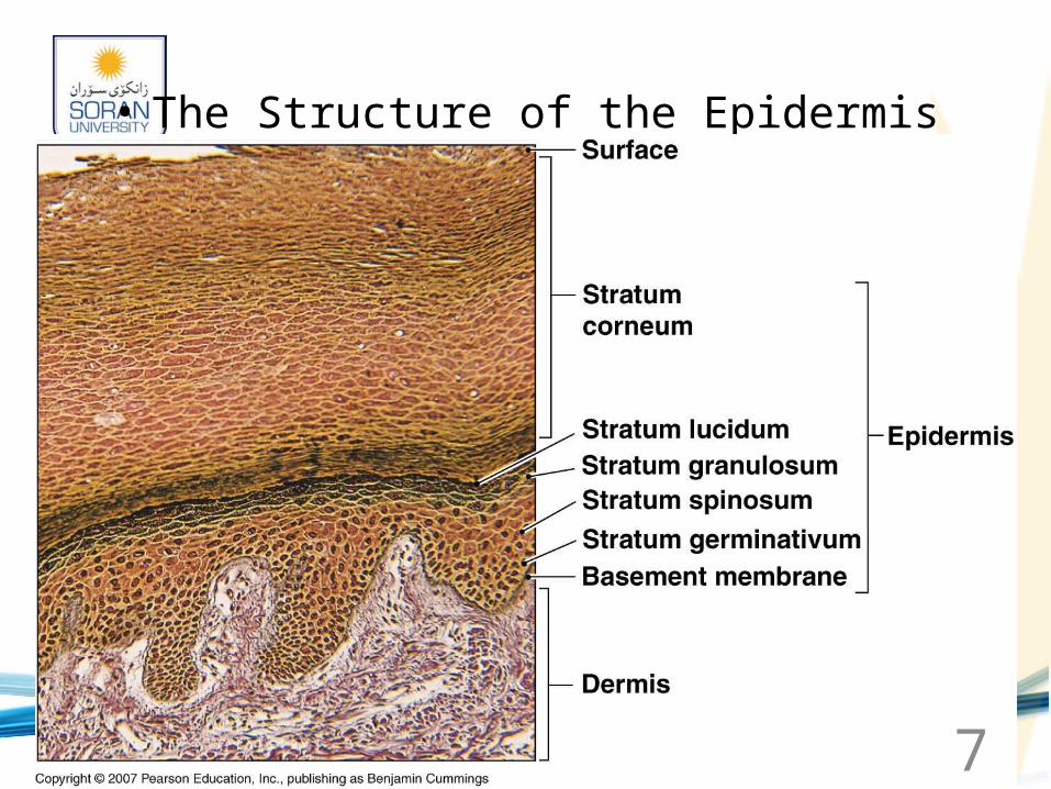
• Melanocytes– Synthesize melanin

www.soran.edu.iq 9
• What are the Cell Layers of the Epidermis? (continued)– Intermediate strata• Stratum spinosum (spiny layer)
– Superficial to stratum germinativum
• Stratum granulosum (grainy layer)– Keratin granules in cytoplasm– No cell division
• Stratum lucidum (clear layer)
www.soran.edu.iq 10
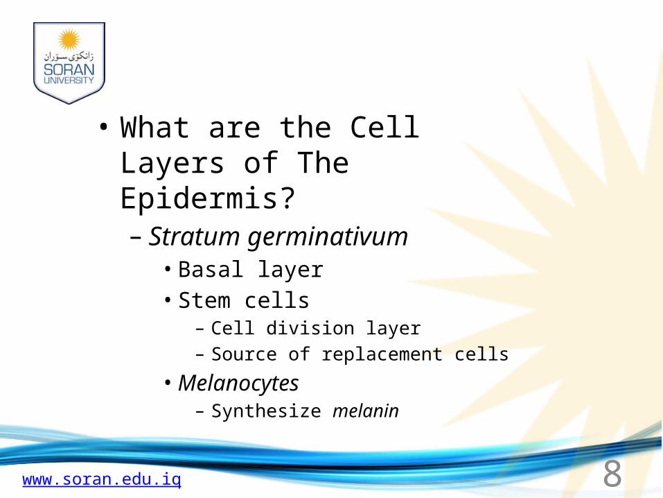
• What are the Cell Layers of the Epidermis? (continued)– Stratum corneum• Most superficial layer• Flattened (squamous) cells• Dead cells• Abundant keratin
– Keratinized (also, cornified)– Tough, water-resistant protein

www.soran.edu.iq 11
• What are the Sources of Skin Color?– Melanocytes• Make melanin• Melanin provides UV protection• Gives reddish-brown to brown-black color
– Carotene• Contributes orange-yellow color• Provided from diet
– Hemoglobin• Blood pigment

www.soran.edu.iq 13
• What are the Effects of UV Radiation?– Beneficial effect• Activates synthesis of vitamin D3
– Harmful effects• Sun burn• Wrinkles, premature aging• Malignant melanoma• Basal cell carcinoma

www.soran.edu.iq 15
• Key Note•The epidermis is a multi-layered, flexible, self-repairing barrier that prevents fluid loss, provides protection from UV radiation, produces vitamin D3, and resists damage from abrasion, chemicals, and pathogens

www.soran.edu.iq 16
•Key Note•The dermis provides mechanical strength, flexibility, and protection for underlying tissues. It is highly vascular and contains a variety of sensory receptors that provide information about the external environment.

www.soran.edu.iq 17
• What is the Subcutaneous Layer?– Composed of loose connective tissue– Stabilizes skin position• Loosely attached to dermis• Loosely attached to muscle
– Contains many fat cells• Provides thermal insulation• Cushions underlying organs
– Safely receives hypodermic needles

www.soran.edu.iq 18
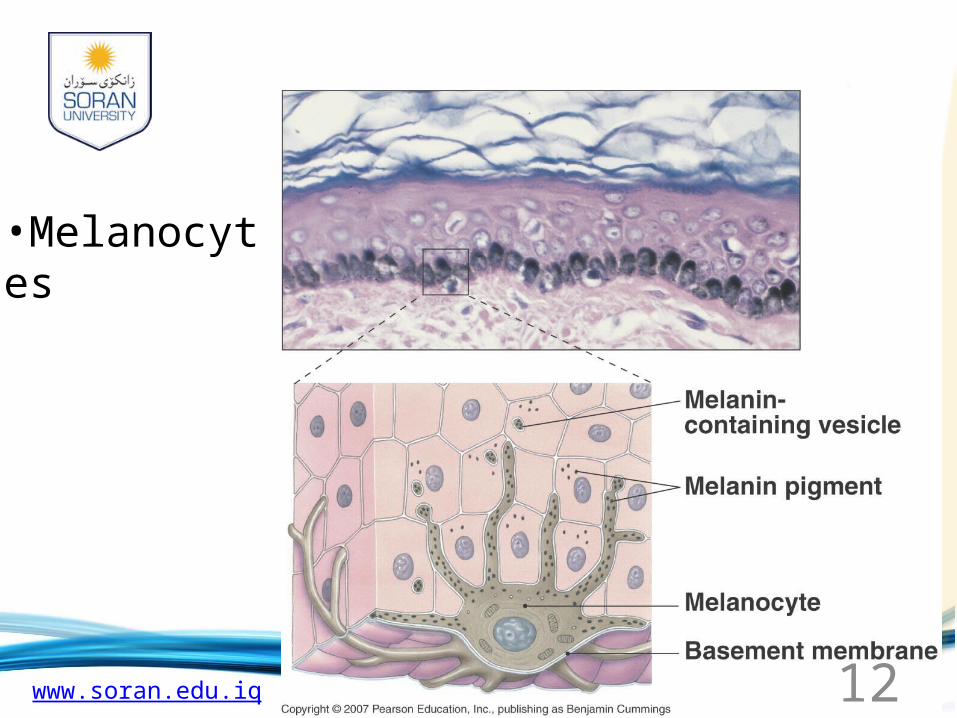
• What are the Accessory Structures?– Hair follicle• A hair
– Shaft– Medulla– Cortex– Cuticle– Arrector pili muscle
» “Goose bumps”

www.soran.edu.iq 22
• What are the Accessory Structures?– Hair growth cycle• 0.3 mm/day growth rate• 2–5 years growth
www.soran.edu.iq 23
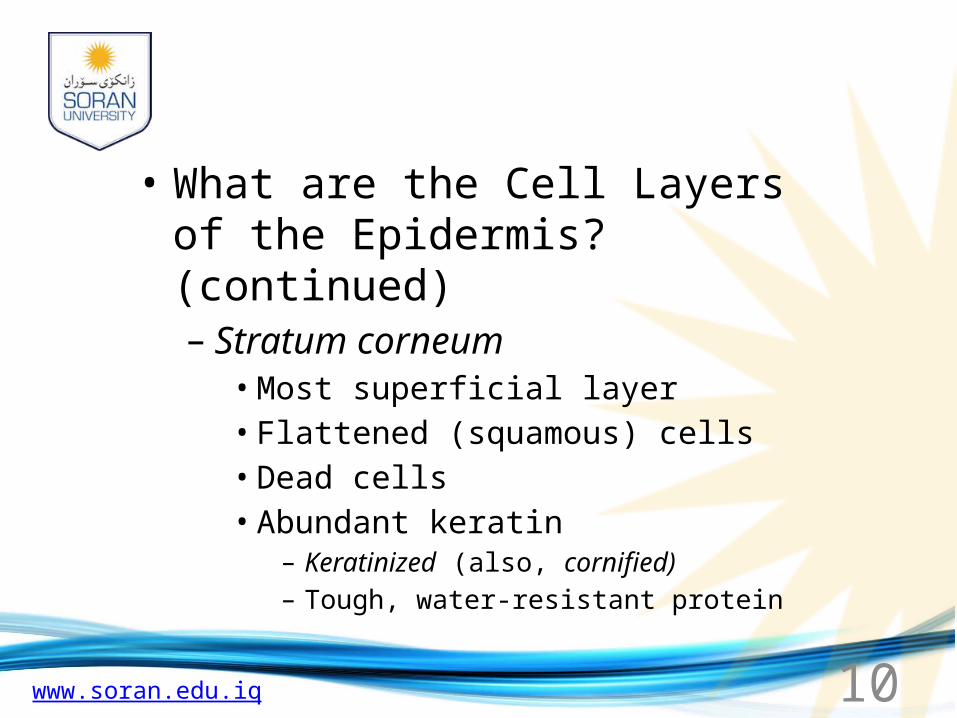
• What are the Accessory Structures?– Sebaceous glands (oil glands)• Holocrine gland• Oily secretion
– Sebum– Hair shaft lubricant
• Sebaceous follicle– Skin lubricant– Skin waterproofing

www.soran.edu.iq 24
•The Structure of Sebaceous Glands and Their Relationship to Hair Follicles

www.soran.edu.iq 25
• What are the Two Types of Sweat Glands?– Apocrine• Odorous secretion (“funky”)• Present in axilla, areola, groin
– Merocrine• Watery sweat (~1% NaCl)• For heat loss

www.soran.edu.iq 27
•Key Note
•The skin plays a major role in controlling body temperature. It acts as a radiator, with the heat being delivered by the dermal circulation and removed primarily by the evaporation of sweat or perspiration.
www.soran.edu.iq 28
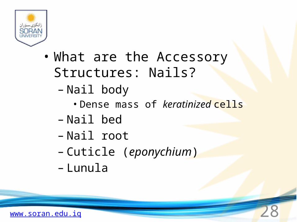
• What are the Accessory Structures: Nails?– Nail body• Dense mass of keratinized cells
– Nail bed– Nail root– Cuticle (eponychium)– Lunula

www.soran.edu.iq 30
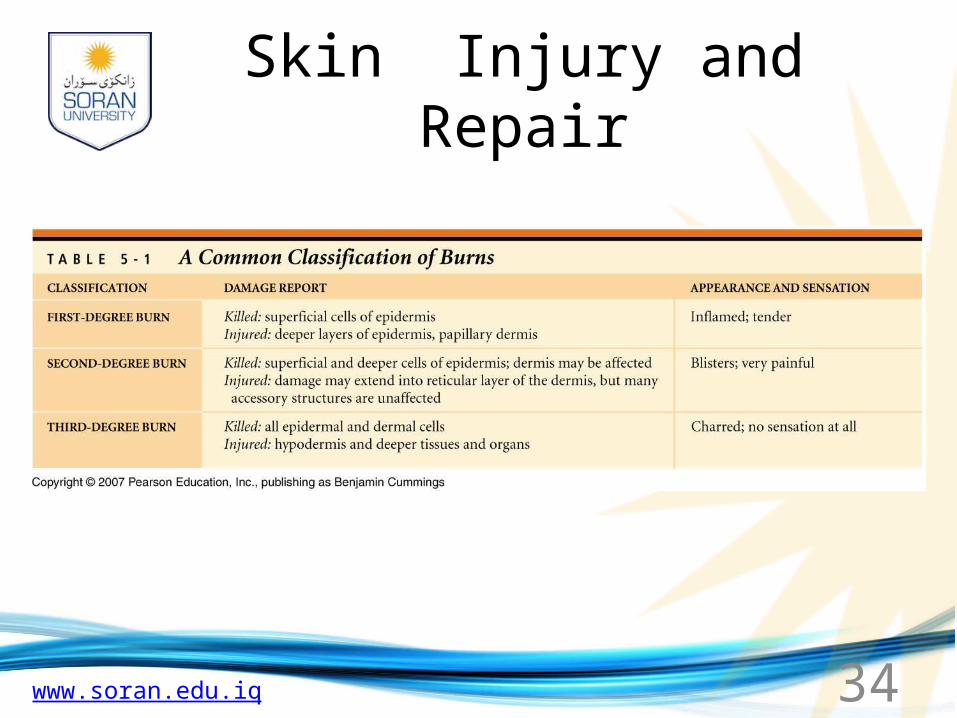
Skin Injury and Repair• What are the Four Stages in
Skin Healing?– Inflammation
• Blood flow increases• Phagocytes attracted
– Scab formation– Cell division and migration– Scar formation

www.soran.edu.iq 31
Bleeding occurs at the site of injury immediately after the injury, and mast cells in the region trigger an inflammatory response.
Epidermis
Dermis
One week after the injury, the scab has been undermined by epidermal cells migrating over the meshwork produced by fibroblastactivity. Phagocytic activity around the site has almost ended, and the fibrin clot is disintegrating.
Fibroblasts
After several hours, a scab has formed and cells of the stratum germinativum are migrating along the edges of the wound. Phagocytic cells are removing debris, andmore of these cells are arriving with the enhanced circulation in the area. Clotting around the edges of the affected area partially isolates the region.
Sweat gland
Scab
Macrophagesand
fibroblasts
Migratoryepithelialcells
Granulationtissue
After several weeks, the scab has been shed, and the epidermis is complete. A shallow depression marks the injury site, but fibroblasts in the dermis continue to create scar tissue that will gradually elevate the overlying epidermis.
Scartissue

www.soran.edu.iq 32
Bleeding occurs at the site of injury immediately after the injury, and mast cells in the region trigger an inflammatory response.
Epidermis
Dermis
After several hours, a scab has formed and cells of the stratum germinativum are migrating along the edges of the wound. Phagocytic cells are removing debris, and more of these cells are arriving with the enhanced circulationin the area. Clotting around the edgesof the affected area partially isolatesthe region.
Sweat gland
Scab
Macrophagesand
fibroblasts
Migratoryepithelialcells
Granulationtissue

www.soran.edu.iq 33
One week after the injury, the scab has been undermined by epidermal cells migrating over the meshwork produced by fibroblast activity. Phagocytic activity around the site has almost ended, and the fibrin clot is disintegrating.
Fibroblasts
After several weeks, the scab has been shed, and the epidermis is complete. A shallow depression marks the injury site, but fibroblasts in the dermis continue to create scar tissue that will gradually elevate the overlying epidermis.
Scartissue

www.soran.edu.iq 35
Aging of the Skin• What are the Major Age-
Related Changes?– Injury and infection increase– Immune cells decrease– Sun protection diminishes– Skin becomes dry, scaly– Hair thins, grays– Sagging, wrinkles occur– Heat loss decreases– Repair slows