Embed Size (px)
Citation preview
Wound Care
Historical Perspective
1867 first antiseptic dressing 1900 true sterilization WW I nonadherent dressings WW II more absorptive dressings 1960’s and 70’s moisture 1980’s moisture acceptance
Goals of Wound Care
Minimizing infective risks Removing dead and devitalized
tissue Allowing for wound drainage Promoting wound epithelialization
and contraction Tissue perfusion Adequate nutrition
Factors That Delay Wound Healing:
Intrinsic Factors Extrinsic Factors
Factors That Delay Wound Healing: Intrinsic
Wound infection
- Bacterial count- Colonization VS
infection- Assessment of
infection
Foreign bodies
Adequacy of blood supply
Factors That Delay Wound Healing: Extrinsic Factors
Smoking
Elderly
Malnutrition
Diabetes
Medication
Obesity
Nutrition and Wound Healing
Anabolic process
Immune response
Vitamins C, A, B6
B1, B2, zinc, and copper, fatty acids
Acceleration of Wound Healing
Wound dressing Oxygenation Adequate nutrition Preparation of the wound Future
“Three Healing Gestures”
Washing the wound
Making plasters-herbs,oils and ointments
Bandaging the wound
Shearing (perpendicular division of tissue) Tearing (<90 degree angle) Compressive (perpendicular with ragged edges)
Mechanism
Household – generally “clean”, but not “sterile”Outdoor – contaminated in varying degrees (the barn, industrial machinery) Bites (human, animal) – highly contaminated
Environment
Age of wound: Rule of Thumb +/ - 12 hr. Wound: Type (mechanism, sharp vs blunt object) Location and vascularity (face, scalp >12hr.?)ContaminationComorbid factors
Modifying Factors
Age Medical hx. – anemia, nutrition, DM, PVD, ETOH, uremia, immuno- compromised Medications – steroids, NSAIDS, anticoagulants, anti-neoplastics
Co morbid Factors
>5yr. < 10yr. Hx. primary series, Need: toxoid > 10yr. Need: toxoid, homotet and toxoid in 60da.No primary series, Need:toxoid,homotet, and toxoid in 60da.
Tetanus Status
Wound Healing
Neovascularization Inflammation Epithelialization Granulation Contraction Remodeling
Phases of Wound Healing
Hemostasis 0-3 hours
Inflammatory 0- 3 days
Proliferation 3-21 days
Maturation 21 days to 1.5 years
Preoperative Management
Debridement & Irrigation Instrumentation Anesthesia Incision planning Patient consultation
Intraoperative Precautions
Incision placement Undermine where necessary Meticulous hemostasis Dead space obliteration **Dermal closure** Suture type & placement Anti-tension taping of wound
Postoperative wound care
Topical emollients for moisture Frequent cleaning with H2O2 Early dermabrasion of irregular
wounds Avoidance of sun, water Steroid creams, retinoids, etc.
Goals of scar revision
Flat scar, level with surrounding skin
Good color match with local tissue Narrow Parallel to the patient’s RSTL Absence of straight, unbroken lines

ASSESSMENT
NeurovascularPulses, capillary refill, motor/sensoryMusculoskeletalMuscle, bone, tendon, jointForeign BodyVisualize/x-ray (radiopaque materials)
Hair Clip, not shaveShaving increases incidence of wound infectionNEVER SHAVE EYEBROWS
PREPARATION
Volume 250 – 1000 + ml. NS60ml. Syringe and 16 – 18 ga. intracath
Irrigation
Do not scrub wounds or use full strength Betadine for irrigation (denatures protein, impairs wound healing) 10 : 1 solution for irrigation or temporary dressing
Irrigation
Repair Sutures
Act as splints Should be Passive Aim to Return Tissues to
Original Position New preplanned Position
Sutures Immobilize Tissues to Allow
Rapid healing Primary intention Less bleeding Reduced haematoma Reduced oedema Reduced discomfort Reduced risk of infection
Sutures May Aid haemostasis
By direct vessel ligation By compression of vessel against
bone edge By retaining a pack or dressing
Suture Needles Eyed Swaged Straight/Curved Large/Micro Taper/Spatula Round Bodied/Cutting/Reverse
Cutting

Sutures Physical Properties
Size Strength Elongation Elasticity Torsional Stiffness Flexibility Surface Capilliarity
Selection of Sutures How long is a suture to be
responsible for wound strength? Is absolute fixation required? Is there a risk of infection? How does the choice of sutures
affect the tissues?
Selection of Sutures How does the suture affect the
healing process? What size of suture
Is strong enough? Provides adequate fixation?
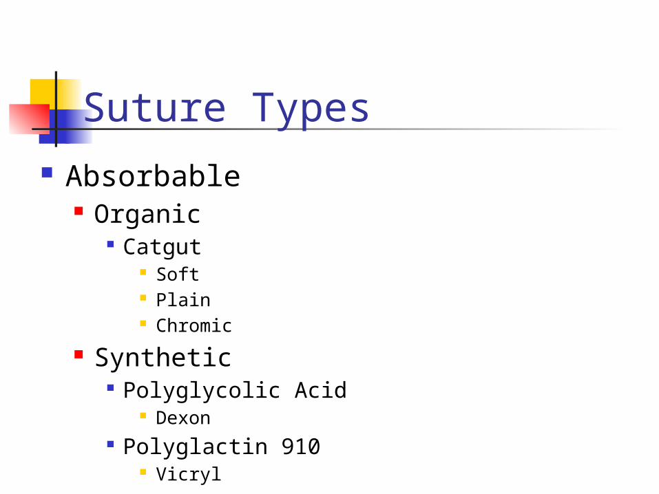
Suture Types Absorbable
Organic Catgut
Soft Plain Chromic
Synthetic Polyglycolic Acid
Dexon Polyglactin 910
Vicryl
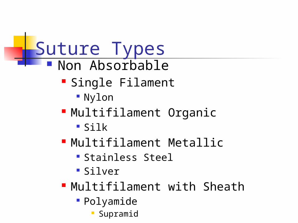
Suture Types Non Absorbable
Single Filament Nylon
Multifilament Organic Silk
Multifilament Metallic Stainless Steel Silver
Multifilament with Sheath Polyamide
Supramid
Biological Properties of Sutures
Tissue Reaction depends on Material Organic > Synthetic
Absorbable Materials Catgut
Proteolytic absorbtion Vicryl
Hydrolytic absorbtion
Non Absorbable Natural but have considerable tissue
reaction Synthetic have little tissue response

Suture Sterilization Gamma Radiation
Cobalt 90 Electron Radiation
Linear Accelerator Ethylene Oxide
Gaseous Liquid
Suturing Techniques Continuous
Subcuticular Blanket Stitch
Over and Under
Interlocking Purse String
Interrupted Simple Mattress
VerticalHorizontal
Suture Tying Techniques
Hand Ties One Handed Two Handed
Instrument Ties Minimise trauma by
Delicate handling of tissues Not constricting tissues Avoidance of dead space Close but not over approximation of tissue
edges
Lidocaine Inject in sub-q tissue ( 21 – 25ga. needle)
Anesthesia
Lidocaine with epinephrine (if you must), butNever in digits, nose, ear, penis
Skin Prep
Betadine (not in wound)Always prep more area than you think you need
Anesthesia
Secondary – granulation and re-epitheliazationDelayed primary closure – closure after 48 – 72hr.Interrupted sutures in ED
Primary – suture, staples, glue

DRESSINGS
Dry sterile dressing – avoid ointments(tend to macerate) Avoid tape on skin if possible Paint skin with tincture of benzoin if you must use tape
DRESSINGS
Encircling dressing ( ACE) Do not wrap tightly Immobilization Excessive motion impairs wound healing Splinting may be necessary
DRESSINGS
Characteristics of Dressings
Protect wound from bacteria and foreign material
Absorb exudates Prevent compression to
minimize edema an obliterate dead space
Dressings Be nonadherent to limit wound
disruption
Create a warm, moist occluded environment to maximize epithelialization and minimize pain
Be esthetically attractive

ANTIBIOTICS
Indications Contaminated wound
Areas of marginal viability
Wounds involving joints, open fractures
All human bite wounds
Most animal bite wounds
Generally, wounds > 12hr. old
SPECIAL WOUNDS
High risk of infection with involvement of bones, joints, tendons, vessels, nervesPuncture wounds (difficult to irrigate and decontaminate)
Bite Wounds
75% involve the extremities Most dog bites in children involve an extremity Severe facial lacerations involve the cheeks and lips as they try to "kiss the doggie”
Dog Bites
Dog Bites Closure
Dog bites – scalp, face, trunk, proximal extremities may be closed if superficial Human bites – “ never” close primarily (delay48 –72hr.)
Never close Irrigate drain, if necessary Foot – shoe on or barefoot? Increased infection risk if shoe on
Puncture Wounds
Abscesses Incise, drain, irrigate, loosely
pack with Iodoform gauzeReturn at 24 hrs. for irrigation fresh packReturn at 48 hrs. for pack removal and healing by granulation
New onset DM may present with abcess Antibiotics may be indicated in addition to I&D
Abscesses
Nail / Nail Bed Injury Subungual hematoma, < 40
% nail area, nail bed injury unlikely, but distal phalanx fx. might be present Treatment: Battery cautery to make drainage hole in nail, irrigate with 25ga. needle and 1% lidocaine Nail Bed - requires surgical repair
Foreign Bodies
Inert – (glass, metal), may leave unremoved if necessaryOrganic – (wood), must be removed