Embed Size (px)
Citation preview

*Corresponding Author Address: Dr. Asha K.R.T, Department of Biochemistry, Government Arts College, Paramakudi, Ramanathapuram
District, Tamil Nadu – 623 707; E-mail: [email protected]
World Journal of Pharmaceutical Sciences ISSN (Print): 2321-3310; ISSN (Online): 2321-3086
Published by Atom and Cell Publishers © All Rights Reserved
Available online at: http://www.wjpsonline.org/
Original Article
In-vitro pharmacological applications of pigment producing halophilic microorganism
from the marine region of India
Asha K.R.T1,*, Joseph Selvin2
1Department of Biochemistry, Government Arts College, Paramakudi, Ramanathapuram District, Tamil Nadu –
623 707 2Department of Microbiology, Pondicherry University, Kalapet, India
Received: 29-05-2016 / Revised: 06-10-2016 / Accepted: 01-11-2016 / Published: 26-11-2016
ABSTRACT
In the present study, two pigment producing promising strains were identified and characterized based on the
biochemical properties from the halophilic environment. Based on the biochemical and physiological
characteristics the strain BQ31 and BV32 were selected and both the strains exhibited showed high similarity
towards the Haloarcula species. It is identified that the strains showed wide range of carbon utilization, able to
tolerate different salts with multiple antimicrobial susceptibility pattern. The extracellular pigment was
identified by various chromatographic techniques and the molecular weight of the pigment was characterized by
MASS spectrum. Interestingly, the pigment revealed significant liver lipid peroxidase activity and
haematological parameters under different body weight of albino rats. The biochemical parameter of the
pigment under different body weight of Albino rats is an additional importance. Thus the pigment could be used
for the various pharmaceutical applications.
Keywords: Halophilic microbes, Pigment; MS spectrum, In-vitro biological applications
INTRODUCTION
The microorganisms exist in extreme environments
such as high salt, high temperature and high
atmospheric pressures are defined as the
extremophiles. These extremophilic microbes
possess unique properties of biotechnological and
commercial significance. They also possess special
adaptation strategies that make them interesting not
only for the fundamental research but also for the
industrial applications [1]. Among the
extrmophiles, the studies on ecology, physiology
and taxonomy of halophiles have revealed an
impressive diversity in hyper saline and alkaline
lakes [2,3]. Halophiles are belonged to the order
Halobacteriales and the family Halobacteriaceae
[4]. Halophilic bacteria also categorized as
psychrophilic, thermophilic, alkaliphilic,
mesophilic, aerobic and anaerobic halophilic based
on the environments [5]. They are mainly aerobic
and exist in the hypersaline regions such as
salterns, salt lakes, sub surface salt formation, and
solar salts. Halophiles are mainly involved in the
biogeochemistry of phosphorus, carbon and other
elements in hypersaline environments [6]. Few
reports claimed that halophiles also play in the
degradation of organic pollutants as hydrocarbons,
pesticides and crude oil [7]. In addition to
degradation of organic pollutants, halophiles also
used as a biocontrol agents against certain
pathogenic fungi [8,9]. Traditionally, halophiles
have been used in the food and nutraceutical
industries as the fermentation of soy and fish
sauces and β-carotene production, also, recently
looked into many novel and unique molecules such
as the compatible solutes, biopolymers or
carotenoids, novel extracellular polysaccharides;
exoenzymes such as cellulase, amylase, lipase,
proteases and xylanase, and biodegradable plastics,
bio-surfactants, bioemulsifiers and
bacteriorhodopsins for molecular biotechnological
applications [10,11]. Novel protein isolated from
from Halobacterium Salinarum used in the
treatment of cancer. Hypersaline environments are
commonly present in the southern parts of the
India. Until now, no reports on the characterization
of halophilic archaeal communities from
Kanyakumari district, India. The present study
aimed in the isolation and identification of the
halophilc bacteria from the southeast coastal region
Asha and Joseph, World J Pharm Sci 2016; 4(12): 235-247
236
of India and investigates its potential application
the medical field.
MATERIALS AND METHODS
Isolation of halophilic strains: Sediment samples
from seawater, salt depositing area and salt
disposals areas were collected and aseptically
transferred in to the laboratory. One gram of the
sample was mixed with sterile water and were
serially diluted and plated on the nutrient agar
plates supplemented with saturated level of 35 %
(w/v) NaCl and other ingredients such as
casaminoacids (7.5g/l); KCl (2g/l); NaCl (25 g/l);
trisodium citrate (3 g/l); magnesium sulphate (20
g/l); manganese sulphate (0.05 g/l); ferrous
sulphate (0.5 g/l); yeast extract (10 g/l); agar (10
g/l). The pH was adjusted to 7.4. The plates were
incubated at 37ºC incubated up to developing
visible colonies. The colonies were purified to
obtain the single colony in the same medium,
labeled and stored in the glycerol stock (-200 C) for
long storage, whereas for the routine laboratory
experiments the purified strains were cultivated in
the slants and stored in the refrigerator at (40 C).
Based on the growth characteristics, pigment
production ability and antimicrobial activity, strain
BQ31 and BV32 were selected for the further
characterization.
Biochemical and physiological characterization
of halophilic strains: To test the biochemical and
physiological properties, the strains were freshly
cultivated in the nutrient broth and mid log phase
strains were routinely used. Phenotypic
characteristics of the strains BQ31 and BV32 were
evaluated by determining the Gram staining,
colonial morphology, size and motility by
comparing with the standard reference strains. The
appearance of the individual colony appearance and
pigment production capability were determined
after growth for seven days. The biochemical and
physiological properties of the isolate were
analysed using routine methods [12-14]. API
50CHB test kits were used to characterize
phenotypes [14]. The API test strips were prepared
according to the manufacturer’s instructions and
scored after 96 h incubation at 37°C. To evaluate
the starch hydrolysis, the strains were streaked onto
nutrient medium with 1% (w/v) soluble starch and
plates were flooded with freshly prepared iodine
solution after growth was obtained. For catalse test
freshly prepared H2O2 solution to colonies on
medium. The presence of oxidase was determined
with tetramethyl p-phenylendiamine-HCl. Casein
hydrolysis was determined by observing the
formation of clear zones around colonies on agar
medium with 0.15% (w/v) skimmed milk powder.
The results were compared with suitable positive
and negative control. Each experiment was
conducted in triplicate.
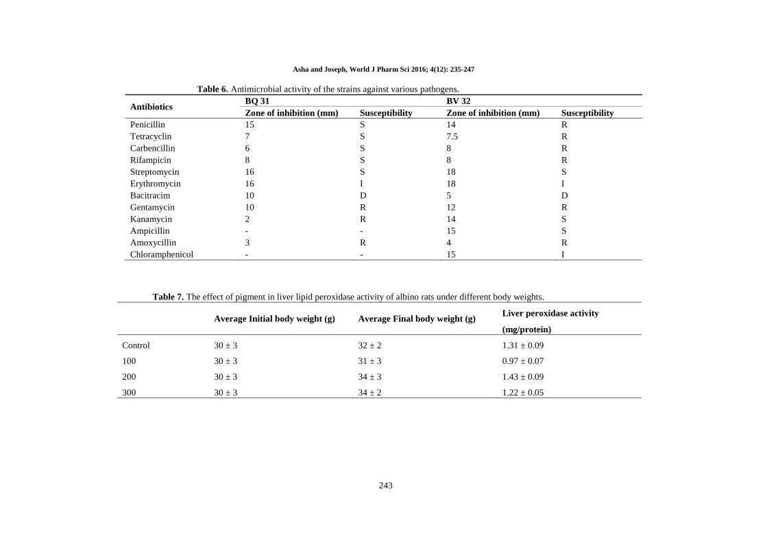
Antibiotic susceptibility pattern of the halophilic
strains BQ31 and BV32: Antibiotic sensitivity
pattern of the halophilic strains BQ31 and BV32
were determined by the disc diffusion method of
Balachandren et al. (2015) [12]. Briefly, the freshly
prepared strains were spread on the top of the
solidified media and allowed to dry for 15 min at
room temperature. Antibiotic discs, penicillin (10
µg), tetracyclin (15 µg), carbencillin (5 µg),
rifampicin (15 µg), streptomycin (5 µg),
erythromycin (20 µg), bacitracim (25 µg),
gentamycin (5 µg), kanamycin (15 µg), ampicillin
(15 µg), amoxycillin (15 µg) and chloramphenicol
(10 µg) were placed on the surface of the medium
and left for 30 min at room temperature for the
antibiotics to diffuse. After that, the plates were
incubated for 72 h at 37°C and inhibition were
measured in millimetres, and the experiment was
performed in triplicate.
Optimizing the parameters for the better
growth and pigment production: The effect of
different pH (5, 6, 7, 8 and 9), different temperature
(20, 30, 40 and 50°C) and different salt
concentration (5, 10, 15, 20, 25 and 30%) were
evaluated for the better growth and pigment
production [15]. Briefly, the freshly prepared
strains (0.01 cell density) were transferred in the
250 ml flask containing DSM-97 medium and
incubated for three weeks. After incubation the
cells were separated by centrifuging and the cell
growth was determined by taking the absorbance at
600 nm and the supernatant were investigated for
the pigment level. The level of pigments was
determined by calorimetric method.
Extraction and identification of the pigment:
The pigments were extracted by following the
method of Blig and Dyer method. To obtain a lipid
extract free of retinol, the polar lipids were
isolated by precipitation in ice-cold acetone. The
isolated polar lipids were dissolved in chloroform
and stored at -20 ºC. Further, mass spectrometric
analysis was carried out for the confirmation of the
pigment. The pigments were dissolved in the
mixture of chloroform- methanol (1:1v/v), and The
experimental conditions of the mass spectrometry
were as follows: range, start (100 amu), stop (1300
amu), and scan time (4.8 s); curtain gas, 20 psi
(N2); heating gas temperature, 5500C; nebulizing
gas, 50 psi; heating gas, 50 psi; ion spray voltage,
5500 V; declustering potential, 100 V; and entrance
potential, 10 V.
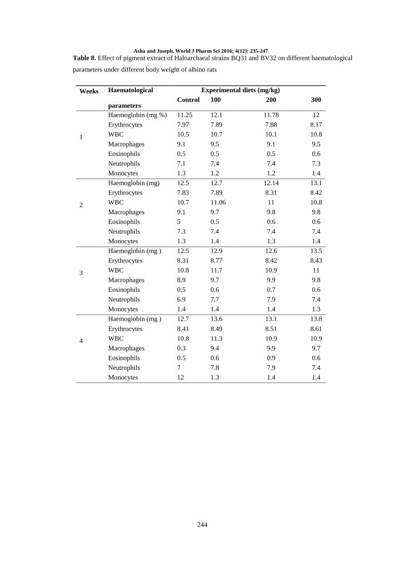
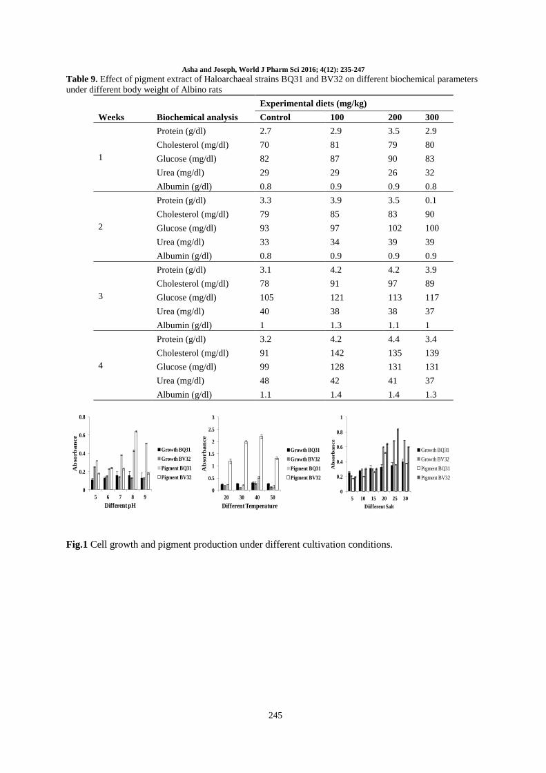
Effect of the pigment on hematological and
biochemical properties: Different concentrations
Asha and Joseph, World J Pharm Sci 2016; 4(12): 235-247
237
of the pigment (100, 200 and 300 mg) was mixed
with the diet and fed to the Swiss albino rats
weighing about 30 g. Feed was administrated orally
for 4 week. Each week blood was removed from
each experimental and control group of rats and
hematological parameters such as hemoglobin (%),
RBC content WBC content, macrophages,
eosinophils, neutrophils and monocytes were
determined. Then serum samples were analyzed for
various biochemical parameters such as protein,
cholesterol, glucose, urea, albumin etc. and the
obtained results were recorded. After 4 weeks, final
body weight was measured and the rats were
anesthetized and sacrificed at the final day of
experiment. The liver tissues were homogenized
with thiobarbituric acid and the entire homogenate
was used for lipid peroxidase activity. Briefly, the
reaction mixture consists of 0.1 ml of liver extract,
0.2 M of tris HCl buffer of pH 7, 0.3 mM of
ascorbic acid and ferrous ammonium sulphate (0.8
mM), whereas 0.1 ml of one water acts as the
control. The reaction mixture was allowed to
incubate for 12 h and then mixed with solution
containing 0.2 ml of TCA (4 %), 2 ml of TBA
(0.8%) and 0.2 ml butylated hydroxy toluene
(BHT) (0.4%). This mixture was incubated for 30
minutes in a boiling water bath and then allowed to
cool for 10 min under ice cold conditions and 2 ml
of chloroform was added and the absorbance was
measured at 532 nm.
RESULTS
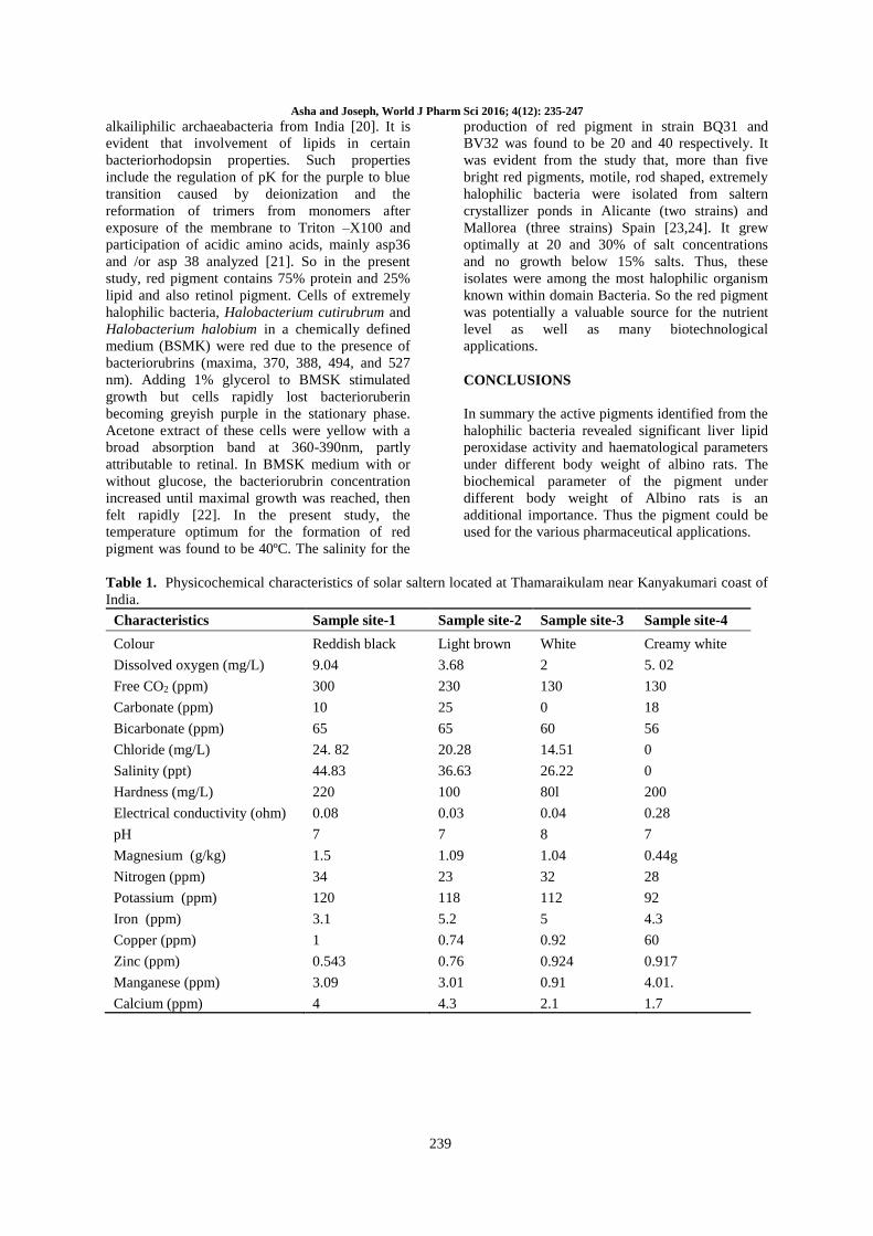
Physiochemical properties of the samples: The
physico-chemical characteristics of the water
content of the sediment samples are presented in
Table 1. The average temperature at the sampling
sites was found to be 27ºC at morning and 32ºC at
noon. Color of the soil collected from four sites
were observed as reddish black, light brown,
slightly reddish and creamy white respectively for
the selected isolation sports. Interestingly the
chloride content of the seawater was found to be
more in all the samples. Copper content was ranged
from 4.01 to 4.3 ppm ppm in all the samples.
Isolation and characterization of halophilic
strains: The results indicated that the total number
of extremely halophilic bacteria in saltern samples
was found to be 10-3CFU/g. Among the isolated
strains, BQ31 and BV32 noted as the promising
strains with regards to the morphology and
appearances (Table 2). Therefore, the two strains
were studied further for its applications. The
colonial pigmentation of these strains were
documented as orange red to pale pink, yellowish
cream and certain colonies were irregular and
spread colonies. The strain BQ31 was found to be
square shaped morphology. Colonies on agar plates
were small, smooth red orange coloured and entire
type. It was a pleomorphic flat cell and measured
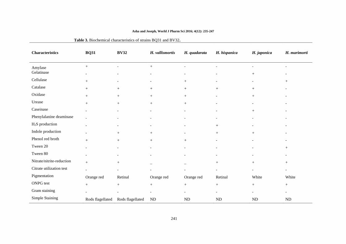
between 2 µm and 3 µm in diameter (Table 3).
Cells were motile by means of flagella. It was
noted that the optimal growth temperature of BQ31
strain was be 52ºC and BV32 strain showed
optimum temperature at 42ºC. No growth was
obtained below 40 ºC and above 60ºC. Biochemical
studies confirmed that the strains were secrete extra
cellular enzymes such as amylase, gelatinase,
cellulase, catalase, oxidase and urease. Simmilarly,
the standard strains such as H. vallismortis, H.
quadarata, H. hispanica, H. japonica and H.
marimorti also expressed similar profile in terms of
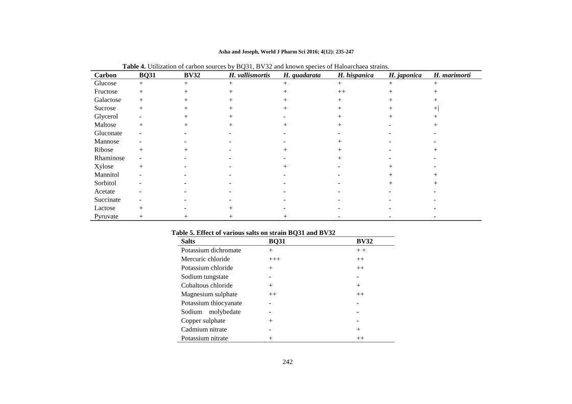
extracellular enzyme secretion. The carbohydrate
fermentation efficiency of these strains was
interesting. The results indicated that the strains
able to ferment wide range of sugars such glucose,
fructose and sucrose etc., whereas the it cannot able
to ferment glycerol and acetate salts (Table 4). The
growth pattern of strain BQ31 and BV32 under
various salts such as Potassium dichromate,
Mercuric chloride, Potassium chloride, Cobaltous
chloride, Magnesium sulphate, Copper sulphate
and Potassium nitrate indicated that the strain have
the ability survive under various stress conditions
(Table 5). In general the identified strains could be
susceptible to various commercially available
antibiotics. Similarly, the selected strains also
revealed antimicrobial sensitivity towards various
commercially available antibiotics such as
penicillin, carbencillin, streptomycin and
gentamycin (Table 5). Overall, the micro-
morphological, physiological, biochemical and
carbohydrate fermentation of the strains confirmed
that the strains were belonged to the halophilic in
nature. Further medical applications of these strains
were studied by checking various parameters.
Optimization of growth and pigment production
pattern of strains: Cell growth and pigment
production status of the selected strains were
optimized by cultivating the strains under different
pH, temperature and salt concentrations. Among
the pH, 8 and 9m were ideal for both growth and
pigment production. Whereas, both the strains
comparatively showed similar growth and pigment
production profile under temperature 30 and 40.
The growth pattern of strains under different
concentration of salt is attractive. The results
indicated that the strains can able to survive and
withstand fewer than 30% of salt. However, the
maximum cell growth and pigment production
profile was detected fewer than 25% of salt
concentration (Figure 1).
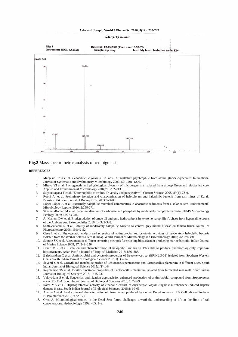
Characterization of pigment: The red color
pigment isolated by the organic solvent method
was further confirmed by MASS spectrum analysis.
Asha and Joseph, World J Pharm Sci 2016; 4(12): 235-247
238
The chromatographic spectrum revealed that the
maximum peak values were observed at 124. The
fragmentation pattern of the red color pigment was
observed at 124. Therefore, it is concluded that the
molecular weight of the pigment was 124.
Biological activity of the pigment: The lipid
peroxidase activity was mentioned in table 7. The
body weight of rats was maintained normally.
There was no significant change in the body weight
of rats within two weeks. There was significant rise
in the body weight of rats in medium 200 mg/kg,
300 mg/kg higher sets of rats. Lipid peroxidase
activity/mg of protein in control was found to be
1.31 ± 0.09 mg/protein. In the case of least (100
mg/kg), the value was found to be 0.97 ± 0.07
mg/protein. In the medium type rats (200 mg/kg),
the peroxidase effect was found to be 1.43 ± 0.09
mg/protein and in the higher type rats (300 mg/kg)
the effect of lipid peroxidase was found to be 1.22
± 0.05 mg/protein comparing four results.
The results indicated that the haematological
parameters like RBC, WBC, Hb, eosinophiles,
neutrophiles and monocytes were increased (Table
8). In the first week control group showed
haemoglobin 11.25 mg%, RBC in the range of
7.97cu/mm and WBC in the range of 10.5 cu/mm
and macrophages 9.1 x 10⁶ when compared with
control. In the first week, all the haematological
parameters were gradually increased.
The effect of the pigment of the biochemical
parameters under different body weight of Albino
rats were displayed in table 9. In the fourth week,
nearly one month of treatment control group animal
showed more value than the other three weeks.
This may be due to gradual growth of the animal.
But in least group animal, cholesterol level raised
142 mg/dl, glucose level raised to 128 mg/dl and
the urea was found to be 42 g/dl and albumin was
found to be 1.4 mg/dl. In median group animals
(200 mg/kg) there was no change in protein. But
cholesterol, glucose, urea and albumin were
increased. In higher weight group animals the
cholesterol level was higher than the other two
groups of rats.
DISCUSSION
The isolated microbial strains documented different
colonial pigments, including red, yellow and
orange were observed. Results confirmed that
most of isolates were Gram negative rods.
Literature study evidenced that the strains were
abundantly found in saline soil samples,
concentration of salts and organic compounds
enhanced the growth and reproduction of
haloarchaea strains. So, the presence of salinity and
other organic components were analyzed to study
the growth and development of haloarchaea. The
present study evidenced that the strains might be
gradually concentrated by brine evaporation. So,
the high content of salt and organic substances in
the soil samples might support the growth of the
extreme haloarchaea. The most abundant extreme
halophilic organism in the saltern soil was red,
yellow and brick red strains. The pleomorphic cells
were predominant type. The most characteristic
inhabitants of hypersaline waters are red due to
carotenoids that serve as protective agents. Other
retinal proteins permit complex photactic
behaviour. The peculiar carotenoids of the
halobacteria, bacteriorubrins act primarily as
photoprotectants but other roles have not been
suggested. For instance, they are involved in an
energy transfer capacity that facilitates
photoreactivation of DNA damaged by exposure to
ultraviolet light [16]. It has been also suggested that
the red colour of halobacteria act as an infrared
(IR) trap increasing the heat accumulation of the
water, which is favorable to these slightly
thermophilic organisms [17].
In the present study, the red pigment of both strains
shows resistant against certain antibiotics. And
some antibiotics are sensitive to red producing
strains. The strain isolate from brine pool in Sinai
Peninsula (Egypt) was characterized. This strain
showed sensitive to bacitracin, novobiocin and
anisomycin (25 g ml-1) and resistant to pencillin,
ampicillin, rifampicin, chloromphenicol, neomycin
and erythromycin (all at 50 g ml-1)) [18].
Bacteriocins and Halocins like molecule are a
diverse collection of proteinacious compounds
often referred as “protein Antibiotics” or
Bacteriocidal protein with molecular weight
ranging from 1kDA-100 kDA. The extremely
halophilic archaeon Haloferax mediterraneii was
able to grow in a minimal medium with optimum
pH 7.5 [19]. In the present study, the haloarchaeal
strain (BQ31) was found to grow at pH 7. Another
isolate (BV32) the pH was found to be 8.
Present study, the result showed that red pigment
contains some lipid associated with it but the exact
structure was not determined. The molecular
weight description was identified by means of
Mass spectrometric studies indicated that lipid are
also necessary for the synthesis of red pigmented
substance. By the use of chemical analysis
chromatography, spectral analysis (including FTIR,
NMR, FAB-MS and CI-MS) in determining the
chemical structure of phospholipid and glycolipids
in extremely halophilic archaeal bacteria is
reviewed in the presence of phospholipid in
halobacteria, novel bis–bis sulfated glycolipid in an
extreme halophile from Japan, an unusual
Asha and Joseph, World J Pharm Sci 2016; 4(12): 235-247
239
alkailiphilic archaeabacteria from India [20]. It is
evident that involvement of lipids in certain
bacteriorhodopsin properties. Such properties
include the regulation of pK for the purple to blue
transition caused by deionization and the
reformation of trimers from monomers after
exposure of the membrane to Triton –X100 and
participation of acidic amino acids, mainly asp36
and /or asp 38 analyzed [21]. So in the present
study, red pigment contains 75% protein and 25%
lipid and also retinol pigment. Cells of extremely
halophilic bacteria, Halobacterium cutirubrum and
Halobacterium halobium in a chemically defined
medium (BSMK) were red due to the presence of
bacteriorubrins (maxima, 370, 388, 494, and 527
nm). Adding 1% glycerol to BMSK stimulated
growth but cells rapidly lost bacterioruberin
becoming greyish purple in the stationary phase.
Acetone extract of these cells were yellow with a
broad absorption band at 360-390nm, partly
attributable to retinal. In BMSK medium with or
without glucose, the bacteriorubrin concentration
increased until maximal growth was reached, then
felt rapidly [22]. In the present study, the
temperature optimum for the formation of red
pigment was found to be 40ºC. The salinity for the
production of red pigment in strain BQ31 and
BV32 was found to be 20 and 40 respectively. It
was evident from the study that, more than five
bright red pigments, motile, rod shaped, extremely
halophilic bacteria were isolated from saltern
crystallizer ponds in Alicante (two strains) and
Mallorea (three strains) Spain [23,24]. It grew
optimally at 20 and 30% of salt concentrations
and no growth below 15% salts. Thus, these
isolates were among the most halophilic organism
known within domain Bacteria. So the red pigment
was potentially a valuable source for the nutrient
level as well as many biotechnological
applications.
CONCLUSIONS
In summary the active pigments identified from the
halophilic bacteria revealed significant liver lipid
peroxidase activity and haematological parameters
under different body weight of albino rats. The
biochemical parameter of the pigment under
different body weight of Albino rats is an
additional importance. Thus the pigment could be
used for the various pharmaceutical applications.
Table 1. Physicochemical characteristics of solar saltern located at Thamaraikulam near Kanyakumari coast of
India.
Characteristics Sample site-1 Sample site-2 Sample site-3 Sample site-4
Colour Reddish black Light brown White Creamy white
Dissolved oxygen (mg/L) 9.04 3.68 2 5. 02
Free CO2 (ppm) 300 230 130 130
Carbonate (ppm) 10 25 0 18
Bicarbonate (ppm) 65 65 60 56
Chloride (mg/L) 24. 82 20.28 14.51 0
Salinity (ppt) 44.83 36.63 26.22 0
Hardness (mg/L) 220 100 80l 200
Electrical conductivity (ohm) 0.08 0.03 0.04 0.28
pH 7 7 8 7
Magnesium (g/kg) 1.5 1.09 1.04 0.44g
Nitrogen (ppm) 34 23 32 28
Potassium (ppm) 120 118 112 92
Iron (ppm) 3.1 5.2 5 4.3
Copper (ppm) 1 0.74 0.92 60
Zinc (ppm) 0.543 0.76 0.924 0.917
Manganese (ppm) 3.09 3.01 0.91 4.01.
Calcium (ppm) 4 4.3 2.1 1.7

Asha and Joseph, World J Pharm Sci 2016; 4(12): 235-247
240
Table 2. Phenotypic characteristic of haloarchaeal strains BQ31 and BV32
Characteristics BQ31 BV32
Reference strains
Haloarcula
vallismortis Haloarcula quadrata
Haloarcula
hispanica
Haloarcula
japonica
Haloarcula
marimorti
Colonial
morphology Circular Circular Circular Circular Circular Circular Circular
Colonial size 3-5 3-5 3-5 2-3 2-3 2-5 3.5
Colony edge Entire Entire Entire Entire Entire Entire Entire
Cell shape Pleo-morphic
flat
Pleo-morphic
rod
Pleo-morphic
square flat
Pleo-morphic
triangular flat
Pleo-morphic
flat disc
Pleo-morphic
rod Pleo-morphic rod
Square end + – + + + + +
Gram stain – – – – – – –
Motile Motile Motile Motile Motile Motile Motile Motile
Pigmentation Orange red Brown Brick red Yellow White Red Light yellow
pH 6.5-7.2 7-7.4 7.4-7.5 6.5-7 7 7-7.5 7-7.2
NaCl (%) 4.3-4.37 4-4.2 4-4.3 3.4-4.3 3.3-4.2 2.5-5 -7.3
Asha and Joseph, World J Pharm Sci 2016; 4(12): 235-247
241
Table 3. Biochemical characteristics of strains BQ31 and BV32.
Characteristics BQ31 BV32 H. vallismortis H. quadarata H. hispanica H. japonica H. marimorti
Amylase + - + - - - -
Gelatinase - - - - - + -
Cellulase + - - + - - +
Catalase + + + + + + -
Oxidase + + + + - + -
Urease + + + + - - -
Caseinase - - - - - + -
Phenylalanine deaminase - - - - - - -
H2S production - - - - + - -
Indole production - + + - + + -
Phenol red broth + + + + - - -
Tween 20 - - - - - - +
Tween 80 - - - - - - -
Nitrate/nitrite-reduction + + _ _ + + +
Citrate utilization test - - - - - - -
Pigmentation Orange red Retinal Orange red Orange red Retinal White White
ONPG test + + + + + + +
Gram staining - - - - - - -
Simple Staining Rods flagellated Rods flagellated ND ND ND ND ND
Asha and Joseph, World J Pharm Sci 2016; 4(12): 235-247
242
Table 4. Utilization of carbon sources by BQ31, BV32 and known species of Haloarchaea strains.
Carbon BQ31 BV32 H. vallismortis H. quadarata H. hispanica H. japonica H. marimorti
Glucose + + + + + + +
Fructose + + + + ++ + +
Galactose + + + + + + +
Sucrose + + + + + + +|
Glycerol - + + - + + +
Maltose + + + + + - +
Gluconate - - - - - - -
Mannose - - - - + - -
Ribose + + - + + - +
Rhaminose - - - - + - -
Xylose + - - + - + -
Mannitol - - - - - + +
Sorbitol - - - - - + +
Acetate - - - - - - -
Succinate - - - - - - -
Lactose + - + - - - -
Pyruvate + + + + - - -
Table 5. Effect of various salts on strain BQ31 and BV32
Salts BQ31 BV32
Potassium dichromate + + +
Mercuric chloride +++ ++
Potassium chloride + ++
Sodium tungstate - -
Cobaltous chloride + +
Magnesium sulphate ++ ++
Potassium thiocyanate - -
Sodium molybedate - -
Copper sulphate + -
Cadmium nitrate - +
Potassium nitrate + ++
Asha and Joseph, World J Pharm Sci 2016; 4(12): 235-247
243
Table 6. Antimicrobial activity of the strains against various pathogens.
Antibiotics BQ 31 BV 32
Zone of inhibition (mm) Susceptibility Zone of inhibition (mm) Susceptibility
Penicillin 15 S 14 R
Tetracyclin 7 S 7.5 R
Carbencillin 6 S 8 R
Rifampicin 8 S 8 R
Streptomycin 16 S 18 S
Erythromycin 16 I 18 I
Bacitracim 10 D 5 D
Gentamycin 10 R 12 R
Kanamycin 2 R 14 S
Ampicillin - - 15 S
Amoxycillin 3 R 4 R
Chloramphenicol - - 15 I
Table 7. The effect of pigment in liver lipid peroxidase activity of albino rats under different body weights.
Average Initial body weight (g) Average Final body weight (g) Liver peroxidase activity
(mg/protein)
Control 30 ± 3 32 ± 2 1.31 ± 0.09
100 30 ± 3 31 ± 3 0.97 ± 0.07
200 30 ± 3 34 ± 3 1.43 ± 0.09
300 30 ± 3 34 ± 2 1.22 ± 0.05
Asha and Joseph, World J Pharm Sci 2016; 4(12): 235-247
244
Table 8. Effect of pigment extract of Haloarchaeal strains BQ31 and BV32 on different haematological
parameters under different body weight of albino rats
Weeks Haematological
parameters
Experimental diets (mg/kg)
Control 100 200 300
1
Haemoglobin (mg %) 11.25 12.1 11.78 12
Erythrocytes 7.97 7.89 7.88 8.17
WBC 10.5 10.7 10.1 10.8
Macrophages 9.1 9.5 9.1 9.5
Eosinophils 0.5 0.5 0.5 0.6
Neutrophils 7.1 7.4 7.4 7.3
Monocytes 1.3 1.2 1.2 1.4
2
Haemoglobin (mg) 12.5 12.7 12.14 13.1
Erythrocytes 7.83 7.89 8.31 8.42
WBC 10.7 11.06 11 10.8
Macrophages 9.1 9.7 9.8 9.8
Eosinophils 5 0.5 0.6 0.6
Neutrophils 7.3 7.4 7.4 7.4
Monocytes 1.3 1.4 1.3 1.4
3
Haemoglobin (mg ) 12.5 12.9 12.6 13.5
Erythrocytes 8.31 8.77 8.42 8.43
WBC 10.8 11.7 10.9 11
Macrophages 8.9 9.7 9.9 9.8
Eosinophils 0.5 0.6 0.7 0.6
Neutrophils 6.9 7.7 7.9 7.4
Monocytes 1.4 1.4 1.4 1.3
4
Haemoglobin (mg ) 12.7 13.6 13.1 13.8
Erythrocytes 8.41 8.49 8.51 8.61
WBC 10.8 11.3 10.9 10.9
Macrophages 0.3 9.4 9.9 9.7
Eosinophils 0.5 0.6 0.9 0.6
Neutrophils 7 7.8 7.9 7.4
Monocytes 12 1.3 1.4 1.4
Asha and Joseph, World J Pharm Sci 2016; 4(12): 235-247
245
Table 9. Effect of pigment extract of Haloarchaeal strains BQ31 and BV32 on different biochemical parameters
under different body weight of Albino rats
Weeks Biochemical analysis
Experimental diets (mg/kg)
Control 100 200 300
1
Protein (g/dl) 2.7 2.9 3.5 2.9
Cholesterol (mg/dl) 70 81 79 80
Glucose (mg/dl) 82 87 90 83
Urea (mg/dl) 29 29 26 32
Albumin (g/dl) 0.8 0.9 0.9 0.8
2
Protein (g/dl) 3.3 3.9 3.5 0.1
Cholesterol (mg/dl) 79 85 83 90
Glucose (mg/dl) 93 97 102 100
Urea (mg/dl) 33 34 39 39
Albumin (g/dl) 0.8 0.9 0.9 0.9
3
Protein (g/dl) 3.1 4.2 4.2 3.9
Cholesterol (mg/dl) 78 91 97 89
Glucose (mg/dl) 105 121 113 117
Urea (mg/dl) 40 38 38 37
Albumin (g/dl) 1 1.3 1.1 1
4
Protein (g/dl) 3.2 4.2 4.4 3.4
Cholesterol (mg/dl) 91 142 135 139
Glucose (mg/dl) 99 128 131 131
Urea (mg/dl) 48 42 41 37
Albumin (g/dl) 1.1 1.4 1.4 1.3
0
0.2
0.4
0.6
0.8
5 6 7 8 9
Ab
sorb
an
ce
Different pH
Growth BQ31
Growth BV32
Pigment BQ31
Pigment BV32
0
0.5
1
1.5
2
2.5
3
20 30 40 50
Ab
sorb
an
ce
Different Temperature
Growth BQ31
Growth BV32
Pigment BQ31
Pigment BV32
0
0.2
0.4
0.6
0.8
1
5 10 15 20 25 30
Ab
sorb
an
ce
Diifferent Salt
Growth BQ31
Growth BV32
Pigment BQ31
Pigment BV32
Fig.1 Cell growth and pigment production under different cultivation conditions.
Asha and Joseph, World J Pharm Sci 2016; 4(12): 235-247
246
Fig.2 Mass spectrometric analysis of red pigment
REFERENCES
1. Margesin Rosa et al. Pedobacter cryoconitis sp. nov., a facultative psychrophile from alpine glacier cryoconite. International
Journal of Systematic and Evolutionary Microbiology 2003; 53: 1291-1296. 2. Miteva VI et al. Phylogenetic and physiological diversity of microorganisms isolated from a deep Greenland glacier ice core.
Applied and Environmental Microbiology 2004;70: 202-213. 3. Satyanarayana T et al. "Extremophilic microbes: Diversity and perspectives". Current Science, 2005; 89(1): 78-9.
4. Roohi A et al. Preliminary isolation and characterization of halotolerant and halophilic bacteria from salt mines of Karak,
Pakistan. Pakistan Journal of Botany 2012; 44:365-370. 5. López-López A et al. Extremely halophilic microbial communities in anaerobic sediments from a solar saltern. Environmental
Microbiology Reports 2010; 2:258-271.
6. Sánchez-Román M et al. Biomineralization of carbonate and phosphate by moderately halophilic bacteria. FEMS Microbiology Ecology 2007; 61:273-284.
7. Al-Mailem DM et al. Biodegradation of crude oil and pure hydrocarbons by extreme halophilic Archaea from hypersaline coasts
of the Arabian Sea. Extremophiles 2010; 14:321-328. 8. Sadfi-Zouaoui N et al. Ability of moderately halophilic bacteria to control grey mould disease on tomato fruits. Journal of
Phytopathology 2008; 156:42-52.
9. Chen L et al. Phylogenetic analysis and screening of antimicrobial and cytotoxic activities of moderately halophilic bacteria isolated from the Weihai Solar Saltern (China). World Journal of Microbiology and Biotechnology 2010; 26:879-888.
10. Satpute SK et al. Assessment of different screening methods for selecting biosurfactant producing marine bacteria. Indian Journal
of Marine Science 2008; 37: 243–250 11. Donio MBS et al. Isolation and characterization of halophilic Bacillus sp. BS3 able to produce pharmacologically important
biosurfactants. Asian Pacific Journal of Tropical Medicine 2013; 876–883.
12. Balachandran C et al. Antimicrobial and cytotoxic properties of Streptomyces sp. (ERINLG-51) isolated from Southern Western Ghats. South Indian Journal of Biological Science 2015;1(1):7-14.
13. Ilavenil S et al. Growth and metabolite profile of Pediococcus pentosaceus and Lactobacillus plantarum in different juice. South
Indian Journal of Biological Science 2015;1(1):1-6. 14. Rejiniemon TS et al. In-vitro functional properties of Lactobacillus plantarum isolated from fermented ragi malt. South Indian
Journal of Biological Sciences 2015; 1: 15-23.
15. Velayudam S et al. Sequential optimization approach for enhanced production of antimicrobial compound from Streptomyces rochei BKM-4. South Indian Journal of Biological Sciences 2015; 1: 72-79.
16. Rathi MA et al. Hepatoprotective activity of ethanolic extract of Alysicarpus vaginalisagainst nitrobenzene-induced hepatic
damage in rats. South Indian Journal of Biological Sciences 2015;1: 60-65. 17. Aparna A et al. Production and characterization of biosurfactant produced by a novel Pseudomonas sp. 2B. Colloids and Surfaces
B: Biointerfaces 2012; 95:23–29
18. Oren A. Microbiological studies in the Dead Sea: future challenges toward the understanding of life at the limit of salt concentrations. Hydrobiologia 1999; 405: 1–9.
Asha and Joseph, World J Pharm Sci 2016; 4(12): 235-247
247
19. Martinez LR et al. Cryptococcus neoformans var. neoformans (serotype D) strains are more susceptible to heat than C. neoformans var. grubii (serotype A) strains. Journal of Clinical Microbiology 2001;39: 3365–3367.
20. Sehgal SN et al. Lipids of Halobacterium cutirubrum. Canadian Journal of Biochemistry and Physiology 1962; 40: 69–81.
21. Dracheva S et al. Chemical and functional studies on the importance of purple membrane lipids in bacteriorhodopsin photocycle behavior. FEBS Letters 1996; 382: 209–212.
22. Hezayen FF et al. Characterization of a novel halophilic archaeon, Halobiforma haloterrestris gen. nov., sp. nov., and transfer of
Natronobacterium nitratireducens to Halobiforma nitratireducens comb. nov. International Journal of Systematic and Evolutionary Microbiology 2002;52:2271–2280.
23. Hescox MA, et al. Photoreactivation in Halobacterium cutirubrum. Canadian Journal of Microbiology1972;18(7):981-5.
24. Khanafari A et al. Solar salt lake as natural environmental source for extraction halophilic pigments. Archive of "Iranian Journal of Microbiology 2010; 2(2): 103–109.


![Pfizer/BioNTech COVID-19 mRNA vaccine...- 10 µg, 20 µg, 30 µg, 100 µg - Immunized on Day 1 and a boost dose on Day 21 [No boost for 100µg cohort] Germany Phase 1/2 Study** (BNT162-01](https://img.pdfslide.us/doc/110x75/60a064ae2ff07627e1303b25/pfizerbiontech-covid-19-mrna-vaccine-10-g-20-g-30-g-100-g-immunized.jpg)