Embed Size (px)
Citation preview

191
WHO Drug Information Vol 21, No. 3, 2007
WHO Drug Information
Contents
World Health Organization
Pharmacovigilance FocusChallenges of pharmacovigilance in
Ukraine 193Adverse reaction reporting in Canada 197Educational modules for health
professionals reporting adversereactions 197
WHO pharmacovigilance capacitybuilding 202
Safety and Efficacy IssuesRosiglitazone: cardiac safety 203Rosiglitazone: cardiac safety profile 204Bevacizumab: tracheoesophageal
fistula 204NSAIDS and cardiovascular risk 205Rosiglitazone and parotid gland
enlargement 206Cinacalcet: indication changes 206Omalizumab and anaphylaxis 207Vinca alkaloids: intravenous
administration 207Pioglitazone: fractures in women 207
Regulatory Action and NewsViracept®: suspension pending quality
and safety assessment 208Nimesulide: suspension following liver
failure 209Tegeserod temporarily suspended 210Withdrawal of products containing
veralipride 210
Current TopicsGlobal strategy to prevent transmission
of Chagas disease 211
Consultation DocumentsInternational PharmacopoeiaAmodiaquine hydrochloride tablets 212Chloroquine sulfate oral solution 214Paediatric artemether and lumefantrine
oral suspension 216Sulfadoxine and pyrimethamine tablets 221Quinine sulfate tablets 223Rifampicin, isoniazid and ethambutol
hydrochloride tablets 226Rifampicin and isoniazid dispersible
tablets 229Rifampicin, isoniazid and pyrazinamide
dispersible tablets 232Efavirenz capsules 236Nevirapine oral solution 238Nevirapine tablets 241
Recent Publications,Information and EventsPharmacogenomics guidance from
Health Canada 245Untangling the web: tenth edition 245Priorities for dengue control 246PLoS Neglected Tropical Disease open
access journal 246
Recommended InternationalNonproprietary Names: List 58
247
192
WHO Drug Information Vol 21, No. 3, 2007World Health Organization
Announcement
The 13th International Conferenceof Drug Regulatory Authorities (ICDRA)will be hosted by the Swiss Agency forTherapeutic Products (Swissmedic) incollaboration with the World Health
Organization
The ICDRA will take placein Berne, Switzerland
from 16 to 19 September 2008
Updated information will be provided regularly at:http://www.icdra.ch
or
http://www.who.int/medicines/icdra/en/index/html
193
WHO Drug Information Vol 21, No. 3, 2007
Pharmacovigilance Focus
Challenges of pharmaco-vigilance in UkraineAssuring the safety of medicinal productsis a key component of Ukraine’s nationalmedicines policy. Activities involvingsafety and monitoring of adverse reac-tions are part of a pharmacovigilancesystem operated by the State Pharmaco-logical Centre. Quality assurance ofmedicinal products remains the responsi-bility of the State Inspectorate for QualityControl of Medicinal Products.
In Ukraine, information provided by thepharmacovigilance system allows for thecollection and scientific assessment ofadverse reactions data reported followinguse of medicinal products. A fully func-tioning pharmacovigilance system isessential for providing scientific andevidence-based information to supportregulatory decisions.
The pharmacovigilance system in Ukrainehas been operational since 1996 and,over time, monitoring and reporting ofadverse reactions has largely beenintegrated into the health care systemthrough relevant legislation. The StatePharmacological Centre became a fullmember of the WHO Programme forInternational Drug Monitoring in 2002.Pharmacovigilance in Ukraine is currentlybased on European Union guidelines andrules governing medicinal products in theEuropean Union which have been devel-oped as a result of the InternationalConference on Harmonization (ICH)process (1, 2). These regulations have
been translated and published in a bookentitled “Pharmaceutical Sector: Pharma-covigilance of Medicinal Products forHuman Use” (3). Regulatory guidancedocuments have also been issued toprovide a framework for pharmacovigi-lance practices (4).
The early existence of an operationalpharmacovigilance system in Ukrainegreatly facilitated harmonization andintroduction of European Union stand-ards. A Ministry of Health Order (5) laiddown future requirements for pharmaco-vigilance procedures with the aim ofreinforcing medicines safety and ensuringaccess to effective and safe products forthe population.
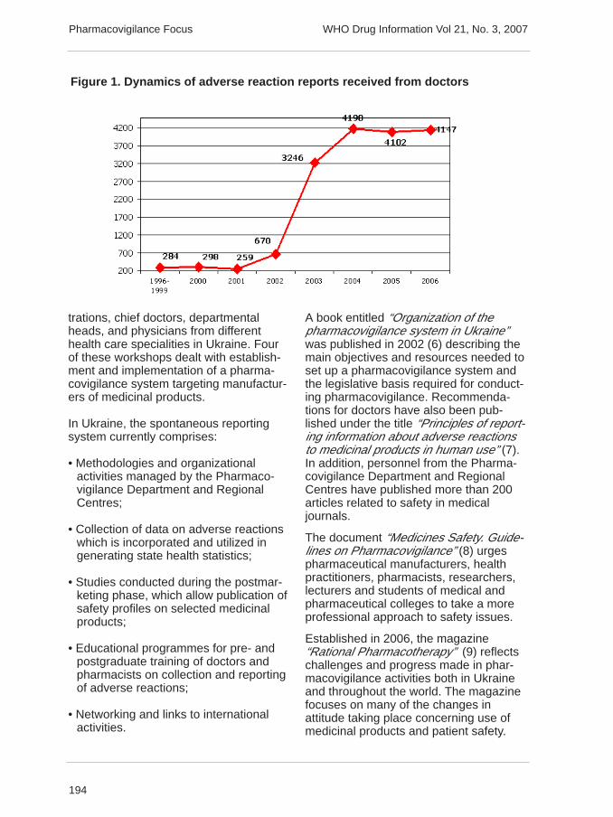
The State Pharmacological Centrecoordinates all pharmacovigilance-relatedactivities. Information on adverse reac-tions to medicinal products is processedand analysed by the PharmacovigilanceDepartment. This Department is alsolinked to Regional Centres where staffwork with health care programmes andmedical doctors throughout Ukraine.Department staff handle over 4000 casesof adverse reaction reports annually andduring the period 1996–2006, the Phar-macovigilance Department received over18 600 reports of adverse reactions tomedicinal products (Figure 1, next page).
The Pharmacovigilance Department isresponsible for the organization, metho-dology and educational activities relatedto pharmacovigilance. Since 2001,twenty-two workshops on organizationand management have been held inUkraine and were attended by over 2000health care professionals. Participantsincluded full-time specialists, chiefs ofhealth departments of the State Adminis-
Article prepared by O.P. Viktorov, O.V.Matveyeva, I.O. Logvina. State Pharmacologi-cal Centre, Ministry of Health, Ukraine
194
WHO Drug Information Vol 21, No. 3, 2007
trations, chief doctors, departmentalheads, and physicians from differenthealth care specialities in Ukraine. Fourof these workshops dealt with establish-ment and implementation of a pharma-covigilance system targeting manufactur-ers of medicinal products.
In Ukraine, the spontaneous reportingsystem currently comprises:
• Methodologies and organizationalactivities managed by the Pharmaco-vigilance Department and RegionalCentres;
• Collection of data on adverse reactionswhich is incorporated and utilized ingenerating state health statistics;
• Studies conducted during the postmar-keting phase, which allow publication ofsafety profiles on selected medicinalproducts;
• Educational programmes for pre- andpostgraduate training of doctors andpharmacists on collection and reportingof adverse reactions;
• Networking and links to internationalactivities.
A book entitled “Organization of thepharmacovigilance system in Ukraine”was published in 2002 (6) describing themain objectives and resources needed toset up a pharmacovigilance system andthe legislative basis required for conduct-ing pharmacovigilance. Recommenda-tions for doctors have also been pub-lished under the title “Principles of report-ing information about adverse reactionsto medicinal products in human use” (7).In addition, personnel from the Pharma-covigilance Department and RegionalCentres have published more than 200articles related to safety in medicaljournals.
The document “Medicines Safety. Guide-lines on Pharmacovigilance” (8) urgespharmaceutical manufacturers, healthpractitioners, pharmacists, researchers,lecturers and students of medical andpharmaceutical colleges to take a moreprofessional approach to safety issues.
Established in 2006, the magazine“Rational Pharmacotherapy” (9) reflectschallenges and progress made in phar-macovigilance activities both in Ukraineand throughout the world. The magazinefocuses on many of the changes inattitude taking place concerning use ofmedicinal products and patient safety.
Figure 1. Dynamics of adverse reaction reports received from doctors
Pharmacovigilance Focus
195
WHO Drug Information Vol 21, No. 3, 2007
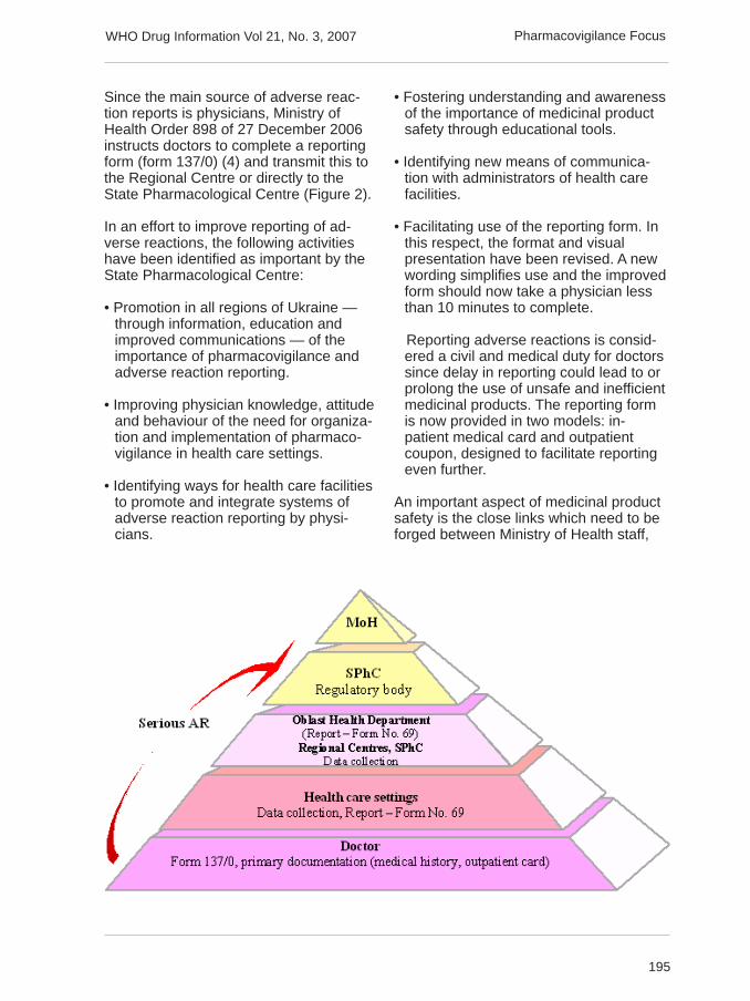
Since the main source of adverse reac-tion reports is physicians, Ministry ofHealth Order 898 of 27 December 2006instructs doctors to complete a reportingform (form 137/0) (4) and transmit this tothe Regional Centre or directly to theState Pharmacological Centre (Figure 2).
In an effort to improve reporting of ad-verse reactions, the following activitieshave been identified as important by theState Pharmacological Centre:
• Promotion in all regions of Ukraine —through information, education andimproved communications — of theimportance of pharmacovigilance andadverse reaction reporting.
• Improving physician knowledge, attitudeand behaviour of the need for organiza-tion and implementation of pharmaco-vigilance in health care settings.
• Identifying ways for health care facilitiesto promote and integrate systems ofadverse reaction reporting by physi-cians.
• Fostering understanding and awarenessof the importance of medicinal productsafety through educational tools.
• Identifying new means of communica-tion with administrators of health carefacilities.
• Facilitating use of the reporting form. Inthis respect, the format and visualpresentation have been revised. A newwording simplifies use and the improvedform should now take a physician lessthan 10 minutes to complete.
Reporting adverse reactions is consid-ered a civil and medical duty for doctorssince delay in reporting could lead to orprolong the use of unsafe and inefficientmedicinal products. The reporting formis now provided in two models: in-patient medical card and outpatientcoupon, designed to facilitate reportingeven further.
An important aspect of medicinal productsafety is the close links which need to beforged between Ministry of Health staff,
Pharmacovigilance Focus

196
WHO Drug Information Vol 21, No. 3, 2007
city health departments and health carefacilities. Order 898 (4) also provides forchiefs of health care units to supportpharmacovigilance activities of RegionalCentres and the State PharmacologicalCentre through training of health careworkers in both regional and local set-tings. This means that, as a priority,heads of Regional Centres should bemembers of the health department reviewboard and that heads of health depart-ments and chief medical officers shouldbe supportive of pharmacovigilance bothat policy and implementation levels.
It should also be mentioned that Order898 (4) sets out detailed requirements onpharmacovigilance for medicinal productmanufacturers and their representatives,thus promoting a responsible attitude tosafety of medicinal products. Compliancewith these requirements is an obligatorycondition for marketing the product in thecountry.
Year Name Decision
1996 Phenacetin Medical use banned1996 Cimetidine Medical use banned1999 Hemodez Medical use banned2201 Phenylbutazone Medical use limited2002 Gentamycin Medical use limited2003 Nitrofural Medical use of oral dosage
forms (tablets) banned2003 Nitrofurans Medical use limited2003 Preparations containing kava-kava Medical use banned
Disintoxic solutions containinglow-molecular polyvinylpyrrolidone Medical use limited
2005 Metamizole sodium Medical use limited2005 Rofecoxib Medical use limited2005 Thioridazine Medical use limited2005 Euphylline Medical use of dosage forms
with ethylendiamine stabilizer banned
Table 1. Regulatory decisions taken in Ukraine on safety of medicinal products
In Ukraine, the importance of safe medici-nal products has now been addressedand legislative tools enacted. Establish-ment of structures to support regulatorymechanisms is under way and everyeffort is made for systems and processesto be compliant with European Unionstandards. It is hoped that the objectivesof supporting rational pharmacotherapywill be promptly accomplished andexpected improvements and outcomesachieved within an evidence-based,scientific context.
References
1. Directive of the European Parliament andEuropean Council of 06.11.2001, 2001/83/EC.Regulation of the European Council of22.07.93, 2309/93/EC.
2. The Rules Governing Medicinal Products inthe European Union, Vol. 9 - Pharmacovigi-lance, Medicinal Products for Human Use andVeterinary Medicinal Products.
Pharmacovigilance Focus

197
WHO Drug Information Vol 21, No. 3, 2007
3. N.A. Liapunov, L.I. Kovtun, O.P.Besugla etal., Kyiv, Morion. Pharmaceutical Sector:Pharmacovigilance of Medicinal Products forHuman Use. Eds. O.V. Stefanov, T.A.Bukhtiarova, V.I. Maltsev, V.G. Varchenko,O.P.Viktorov. 2003, p.216.
4. Ministry of Health, Ukraine, Order of 19.12.2000 No. 347 registered in the Ministry ofJustice, Ukraine, 26.12.2000. Instructions forsurveillance of adverse reactions/effects ofmedicinal products. Related Orders of16.07.2001 No. 292; 08.02.2001 No. 51 & No.52; 12.12.2001; and No. 497 registered in theMinistry of Justice, Ukraine, 28.12.2001.
5. Ministry of Health, Ukraine, Order of28.12.2006 No. 898 registered in the Ministryof Justice, Ukraine, 29.01.2007.
6. O.V. Stefanov, O.P. Victorov, V.I. Maltsev,L.I. Kovtun, I.O. Logvina. Organization of thepharmacovigilance system in Ukraine. Kyiv,Avicena, 2002, p. 68.
7. V.M. Kovalenko, O.P. Viktorov, V.I. Maltsevet al. Principles of reporting adverse reactionsto medicinal products in human ue. Methodo-logical recommendations. Kyiv, Avicena, 2004,p.55.
8. O.P. Viktorov, V.I. Maltsev, Yu.B. Belousova,Medicines Safety. Guidelines on Pharma-covigilance, Kyiv, Morion, 2007, p.240.
9. Rational pharmacotherapy. Scientific andpractial journal for physicians. Kyiv, ZdorovjeUkrainy — XXI stoletije, LTD, 2006.
Adverse reaction reporting in Canada
Educational modules forhealth professionals reportingadverse reactionsHealth Professional Reporting of AdverseDrug Reactions is a narrated, electronic(online) presentation to help health profes-sionals recognize and report adverse re-actions to Health Canada.
Reporting adverse reactions contributes to:
• The identification of previously unrecog-nized rare, or serious adverse reactions;
• Changes in product safety information;
• International data collection and dis-semination regarding benefits, risks,and/or effectiveness of products.
Health Canada provides health professionals, consumers and patients with educa-tional tools for reporting adverse reactions to drugs and other marketed health prod-ucts, including prescription drugs; non-prescription drugs (including natural healthproducts); biological products (including products derived from blood, biotech drugs,and therapeutic and diagnostic vaccines); and radiopharmaceuticals.
Health Canada has now developed two new educational modules on adverse reac-tion (AR) reporting of marketed health products. In addition to the existing modulefor naturopathic doctors, modules for other health professionals and for consumersare now available at the Learning Centre on the MedEffect website at http://www..hc-sc.gc.ca
The online module Health Professional Reporting of Adverse (Drug) Reactions pro-vides an interactive tool to help health professionals report suspected adverse reac-tions. The online module for consumers, Reporting Side Effects from Your Medi-cine: What You Need to Know, comes with a guidebook and provides comprehen-sive learning tools to help consumers and patients report side effects.
Pharmacovigilance Focus
198
WHO Drug Information Vol 21, No. 3, 2007
Since 1965, health professionals havebeen reporting suspected adversereactions to marketed health products tothe Canadian Adverse Drug ReactionMonitoring Program.
Learning tools have now been developedto make it easier to report adversereactions by increasing awareness about
how to report, what to report and howadverse reaction reports contribute tohealth safety.
A summary of the modules is set outbelow. The modules are available at theMedeffect website in the form of narrated,electronic presentations.
A vital link in the patient safety chainAfter completing the learning modules and presentations, health professionals shouldbe more knowledgeable about adverse reaction reporting and better able to:
Describe an adverse reactionAssociate adverse reaction reporting with your current work practiceDescribe the adverse reaction reporting processReport an adverse reaction
Some physicians have discontinued or changed their patients’ medication regime dueto adverse reactions associated with a particular drug. While this information is veryvaluable, it is often not reported because the physician or a member of the patient’shealth care team does not realize that the reaction should be reported, is unfamiliarwith the reporting process or falsely believes that only proven adverse reactionsshould be reported.
With over 50% of newly approved therapeutic health products having serious sideeffects that are discovered only after the product is on the market, the need to report iscritical. It has been estimated that for every adverse reaction that gets reported, up to10 go unreported. It is for these reasons that there is a need to stimulate the reportingof adverse reactions by health professionals.
The module and reporting mechanism is presented in the form of a case presentation.
The caseA 32-year old male patient was diagnosed with depression. He was started on sertra-line 50 mg daily. A week after starting therapy, he developed severe diarrhoea. He lostconsciousness, fell and was admitted to hospital for rehydration.
While in hospital, sertraline was discontinued and the diarrhoea resolved. There areactually two problems here to deal with. First, what should be done for the patient andsecondly how Health Canada is involved in the adverse reaction of this patient.
Before using another medical product, it is important to report this suspected adversereaction. This patient is not taking any other prescribed medications and is not regu-larly using any OTC or natural health product. He is a young healthy man, with anunremarkable recent annual health exam. There is no relevant past history of concern.This is his first presentation of depression and there are a variety of therapeuticchoices available for treatment.
Pharmacovigilance Focus

199
WHO Drug Information Vol 21, No. 3, 2007
Could this be an ADR? Health Canada describes an adverse reaction as a harmfuland unintended response to a health product. This includes any undesirable effectsuspected to be associated with the use of a health product. Examples of reportableundesirable effects include:
Any unintended effectHealth product abuseOverdoseInteraction (including drug-drug and drug-food interactions)Any unusual lack of therapeutic efficacy
Suspected adverse reactions may be:
seriousunexpectedin recently marketed products (< 5 years)
Reporting reactions, includes any reaction that is serious or seems unexpected, thatis, if it’s not consistent with the product information or labelling regardless of severity;and particularly any adverse reaction to a product that has been available for less than5 years. A serious effect would require hospitalization or prolong an existing hospitalstay; it may have caused congenital malformation, persistent or significant disability orincapacity; have required an intervention to prevent damage or permanent impairment,was life-threatening or resulted in death.
Where to startThere are a number of ways to report. There is a written form to fill in and send toHealth Canada. The form is also available on-line at the MedEffect website. In additionto faxing or mailing in the form, a health professional can phone a toll free number andspeak to someone who can take the information from the caller.
There are 4 pieces of information that must be entered on the form; an identifiablepatient in Section A: a description of the reaction in Section B; a suspect healthproduct in Section C, and an identifiable reporter in Section D .
Information on the identity of the patient and the reporter of the adverse reaction iskept strictly confidential. Personal information will not be disclosed to anyone exceptHealth Canada personnel who need the information to carry out their responsibilities.Any requests for disclosure of the personal information would be dealt with accordingto the federal Privacy Act.
Patient information. The ‘patient identifier’ is reserved for the health professional inorder to identify their patient in the event that follow-up is needed. A code, recogniz-able to the patient’s health professional only, is best put here – no names.
For the present case patient, the adverse reaction is “severe diarrhoea with syncoperequiring hospitalization”. The words used to describe the adverse reaction are veryimportant here as this description will get a medical code so the information can beretrieved from the database as needed.
Pharmacovigilance Focus
200
WHO Drug Information Vol 21, No. 3, 2007
Additional Information: The remaining fields add value to the report. They becomeindicators of causality to help Health Canada detect early warning signs.
Time-lines are very important – when the reaction took place, and when the patientstarted the drug. The evaluators working at Health Canada look at these reports todecide whether the suspected drug is likely to have caused the reaction. Indications ofa temporal relationship are important. Also, the patient’s health condition in the past,any relevant lab data, what happened when the patient stopped the drug, or if theystarted the drug again. All this information is very important to note. If a patient wastaking other drugs or health products during the time of the adverse reaction, theseproducts should be listed in the section calling for concomitant drugs. It is also good towrite ‘ no other drugs taken’ rather than leaving this space blank.
The form asks whether the reaction abated after use stopped or dose reduced. Thesevere diarrhoea did resolve once the patient was admitted and the sertraline wasdiscontinued. This information field does not imply that the patient should be put backon the drug just to see if the reaction reappears. However, if the patient ends upcoming back to this drug some time later, it would be useful follow-up information.
What does Health Canada do with the AR report form?The adverse reaction report forms received are screened for the 4 minimum criteriamentioned earlier. When all 4 are present, the report is entered into the CanadianAdverse Drug Reaction Information System database.
The report is further assessed regarding the seriousness of the reaction, and a stand-ard medical terminology is used to classify the reported information. Assigning stand-ard medical terminology helps ensure that the same term is applied in similar situa-tions reported.
Signals may be identified through the systematic review of adverse reaction reportsplus any other additional information on product safety, and may be early indicators ofa product-related issue. Detecting a safety signal triggers the need to investigatefurther a potential association between the product and the adverse reaction.
Medical evaluators use adverse reaction reports from the database (among otherrelevant sources of information) to determine if the suspect drug could have causedthe reported reaction. Health professionals are encouraged to report their cases in asmuch detail as possible because it makes the information that much more valuable tothe medical evaluators.
Products reported on the AR form: The type of health product to which this formapplies include:
prescription and non-prescription drugsnatural health productsradiopharmaceuticals, andbiologics
Pharmacovigilance Focus
201
WHO Drug Information Vol 21, No. 3, 2007
Products not reported on this form
Vaccines (prevention of infectious disease)Medical Devices
Preventative vaccines are monitored by the Public Health Agency of Canada. Forthese vaccines, health professionals should report to their local public health depart-ment.
An adverse reaction to a medical device gets reported to another part of HealthCanada: the Health Products and Food Branch Inspectorate.
After reporting this reaction, an acknowledgment letter is sent thanking the reporter fortheir report and a reference or tracking number is given should any follow-up informa-tion become available or be needed at a later date, e.g., a biopsy or autopsy report, orany other clarification where needed.
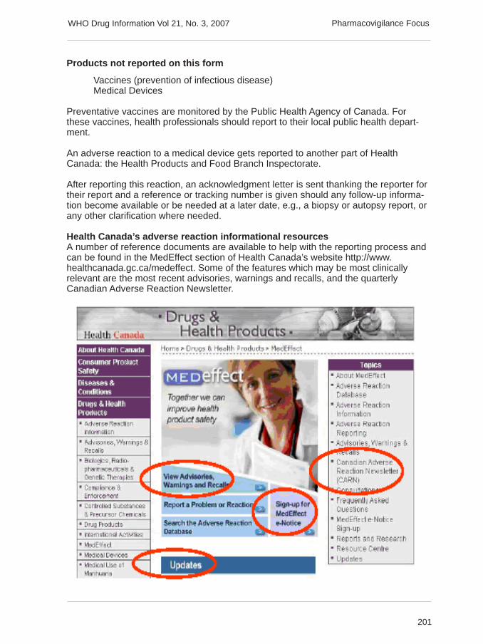
Health Canada’s adverse reaction informational resourcesA number of reference documents are available to help with the reporting process andcan be found in the MedEffect section of Health Canada’s website http://www.healthcanada.gc.ca/medeffect. Some of the features which may be most clinicallyrelevant are the most recent advisories, warnings and recalls, and the quarterlyCanadian Adverse Reaction Newsletter.
Pharmacovigilance Focus
202
WHO Drug Information Vol 21, No. 3, 2007
WHO pharmacovigilancecapacity buildingThe WHO Programme for InternationalDrug Monitoring is supporting a three-wayapproach to international pharmacovigi-lance training by providing:
1. Generic pharmacovigilance training forparticipants from different parts of theworld.
2. National training courses, tailored tothe specific needs of each country, andcustomized to include safety surveillanceof specific disease programmes.
3. A synthesis of the two approaches toinclude capacity building for a group ofcountries with very similar diseaseburdens and pharmacovigilance interests.This would have the advantage of provid-ing a regional pool of expertise.
Recent activities
Botswana — A pharmacovigilancetraining course was organized in Septem-ber 2006 in Gaborone. Ten drug regula-tory officials attended the week-longcourse and were trained in the basicprinciples of pharmacovigilance, reportingsystems, adverse drug reactions (ADR)database, tools for ADR data manage-ment, causality assessment, signaldetection, preventing and managingADRs, cohort-event monitoring andcommunications in pharmacovigilance.
The course included practical methods forsetting up a pharmacovigilance systemand liaising with public health pro-grammes. Working groups comparedvarious national reporting forms.
Morocco — A pharmacovigilance trainingcourse was organized in February 2007for Francophone countries and includedparticipants from Algeria, Benin, BurkinaFaso, Cameroun, Côte d’Ivoire, Demo-cratic Republic of Congo, Gabon, Mada-gascar, Mali, Morocco, Sao Tomé-et-Principe, Senegal, Togo and Tunisia.
Zambia — A follow-up course was held inFebruary 2007 with participants fromBurundi, Democratic Republic of Congo,Mozambique, Zambia and Zanzibar. Thisfollowed on from a course in 2003 whichintroduced pharmacovigilance to coun-tries about to introduce artemisinin-basedcombination therapies (ACTs).
At this meeting, countries identified threeareas for action:
• the need to prioritize and strengthenpharmacovigilance activities;
• the need to advocate pharmacovigi-lance issues at regional level throughregional economic bodies; and
• the need to harmonize national ADRreporting forms and guidelines forspontaneous reporting, in particular inMalaria Case Management trainingmanuals and guidelines.
WHO is currently preparing two trainingprogrammes in Ghana devoted to teach-ing the basic principles of cohort eventmonitoring and developing a final protocolto collect ADR reports in the nationalmalaria programmes. Additionally, partici-pants will learn about other problems thatcould arise in pharmacovigilance.
Reference: World Health Organization.Feature. WHO Pharmaceuticals Newsletter,No. 3, 2007.
Pharmacovigilance Focus
203
WHO Drug Information Vol 21, No. 3, 2007
Safety and Efficacy Issues
Rosiglitazone: cardiac safetyCanada — An article recently publishedin the New England Journal of Medicine(NEJM) has generated significant publicattention on the cardiac safety of theantidiabetic rosiglitazone (Avandia®,Avandamet® and AvandarylTM).The article(1), based on a meta analysis of 42clinical studies, noted a statisticallysignificant increased risk of myocardialinfarction and a statistically non-signifi-cant increase in the risk of cardiovasculardeath associated with the use of rosiglita-zone in comparison to placebo or otheranti-diabetic therapies. The conclusionsreached require confirmation. Analysis ofall currently available data is ongoing andfindings will be communicated when areview is complete.
Some of the studies in the NEJM articleincluded patients using rosiglitazone incombination with other anti-diabetictherapies. Some of these combinations,specifically rosiglitazone + metformin +sulfonylurea or rosiglitazone + insulin arenot approved for use in Canada.
In Canada, Avandia® is NOT approvedfor use with insulin therapy; with thecombination of metformin AND asulfonylurea; or in patients with pre-diabetes (2).
Avandia® is contraindicated in patientswith Class III and IV cardiac status.Avandia® should be used with caution inany patient with NYHA Class I and IIcardiac status. All patients should bemonitored for signs and symptoms of fluidretention, edema, and rapid weight gain.The dose of Avandia® used in combina-tion with a sulfonylurea should not exceed4 mg daily.
In Canada, Avandia® is indicated for:
• use as monotherapy in patients notcontrolled by diet and exercise alone, toreduce insulin resistance and lowerelevated blood glucose in patients withtype 2 diabetes mellitus.
• use in combination with metformin or asulfonylurea when diet and exerciseplus the single agent do not result inadequate glycemic control. For patientsinadequately controlled on metformin ora sulfonylurea, Avandia® should beadded to, not substituted for, metforminor the sulfonylurea.
Treatment with thiazolidinediones hasbeen associated with cases of congestiveheart failure, some of which were difficultto treat unless the medication was dis-continued. Avandia® should be discontin-ued if any deterioration in cardiac statusoccurs.
References
1. Nussen SE, Wolski K. Effect of rosiglita-zone on the risk of myocardial infarction anddeath from cardiovascular causes. NewEngland Journal of Medicine, 356: 2457-71,2007.
2. Communication from GlaxoSmithKline, 1June 2007 at http://www.hc-sc.gc.ca
European Union — The EuropeanMedicines Evaluation Agency (EMEA)has reminded physicians that whenrosiglitazone was first authorized in theEuropean Union in 2000, it was contrain-dicated in patients with a history ofcardiac failure. Since then, EMEA hasclosely monitored rosiglitazone for cardio-vascular effects.
204
WHO Drug Information Vol 21, No. 3, 2007
The EU rosiglitazone product informationwas updated in September 2006 withinformation about the risk of ischaemicevents. Prescribers are reminded toadhere to the restrictions for use inpatients with cardiac disease as set out inthe product information. Patients areadvised not to stop treatment with rosigli-tazone, but to discuss the medication withtheir doctor.
Reference: Press release. EMEA, 23 May2007 at http://www.emea.europa.eu
Rosiglitazone: cardiovascularsafety profileSingapore — Rosiglitazone (Avandia®)is an oral agent for the treatment of type 2diabetes mellitus registered by the HealthSciences Agency (HSA) since 2000 foruse as an adjunct to diet and exercise, asmonotherapy, or in combination withmetformin or a sulfonylurea to reduceinsulin resistance and lower elevatedblood glucose in patients with type 2diabetes mellitus. Avandamet® is anotherregistered product containing a combina-tion of two active ingredients, rosiglita-zone and metformin.
The risk of cardiac adverse events (i.e.heart failure, fluid retention, oedema) isknown to be associated with the thiazolid-inediones class of drugs. Recently,concerns have been raised about thepossible elevation of ischaemic cardio-vascular (CV) risk with rosiglitazonetherapy.
The findings of a study published in theNew England Journal of Medicine on 21May 2007 have caused considerabledebate. Two additional studies have beenconducted. The various studies providecontradictory findings on the ischaemicCV risk of rosiglitazone (1–3). HSA willcontinue to keep rosiglitazone underclose surveillance for CV effects (i.e.cardiac failure, myocardial infarction) andmonitor the international developments in
this area. Healthcare professionals will beupdated on new developments in thisarea when the data becomes clearer.
In the interim, prescribers should continueto carefully make individualised treatmentdecisions for patients with diabetesmellitus. It is advised that patients onAvandia® should be monitored for signsand symptoms of heart failure, fluidretention, oedema and rapid increases inweight.
Source: Product Safety Alert, 26 July 2007.http://www.hsa.gov.sg/cda/safetyalerts
References
1. N Engl J Med 2007 Jun 14; 356(24):2457-71 (Epub May 21).
2. N Engl J Med 2007 (Epub Jun 5).
3. N Engl J Med 2006 Dec 7; 355(23):2427-43.
4. Lancet 2006 Sept 23; 368:1096-105.
Bevacizumab: trache-oesophageal fistulaCanada — Important new safety informa-tion regarding use of bevacizumab(Avastin®) has been released. Bevacizu-mab is a recombinant humanized mono-clonal antibody that is directed againstthe vascular endothelial growth factor(VEGF). It is authorized for first-linetreatment of patients with metastaticcarcinoma of the colon or rectum incombination with fluoropyrimidine basedchemotherapy.
Based on a review of post market andclinical trial reports serious adverseevents, including fatal events, of trache-oesophageal (TE) fistula have beenreported in association with use ofbevacizumab in clinical trials of small celllung cancer (SCLC), non small cell lungcancer (NSCLC) and esophageal cancer.Bevacizumab should be permanently
Safety and Efficacy Issues
205
WHO Drug Information Vol 21, No. 3, 2007
discontinued in patients with tracheo-esophageal (TE) fistula or anygastrointestinal fistula. There is limitedinformation on the continued use ofbevacizumab in patients with otherfistulas. In cases of internal fistula notarising in the GI tract, discontinuation ofbevacizumab should be considered.
Reference: Medeffect 1 June 2007 at http://www.hc-sc.gc.ca
NSAIDS and cardiovascular riskSingapore — Following worldwide volun-tary withdrawal of rofecoxib (Vioxx®) inOctober 2004 due to concerns of anincreased cardiovascular (CV) risk, HSAand its Expert Advisory Committee hasreviewed the CV risks of the entire classof non-steroidal anti-inflammatory drugs(NSAIDs).
Based on the available data in April 2005,HSA concluded that the coxibs or thenewer COX-2-selective inhibitors, namelyrofecoxib, celecoxib, and etoroicoxib,were associated with an increased risk ofCV events and that the risk increasedwith higher dose and duration of use.HSA also strengthened the local packageinserts of these products to contraindicatethe perioperative use of these drugs inpatients who have recently undergonecoronary artery bypass graft (CABG)surgery and revascularisation proce-dures. At that time, due to the lack ofinformation on the CV risks for the olderNSAIDs, HSA concluded that the possibil-ity of similar CV risk could not be ruledout although there was insufficient data toshow that the older NSAIDs pose similarCV risks as the coxibs.
Recently, the availability of several wellconducted meta-analyses of randomisedtrials, case-control and cohort studiesinvolving the older NSAIDs have providedregulators with a better means of assess-ing the safety profile of these drugs. HSA
and its Pharmacovigilance AdvisoryCommittee (PVAC) have reviewed thesedata and taking into account internationalregulatory developments, arrived at thefollowing recommendations on the CVrisks and the use of this class of drugs:
• Non-selective NSAIDs are importanttreatments for arthritis and other anti-inflammatory and painful conditions.
• Non-selective NSAIDs may be associ-ated with a small increase in the abso-lute risk of cardiovascular events (e.g.myocardial infarction and stroke),especially when used at high doses forlong-term treatment.
• All NSAIDs should be prescribed at thelowest effective dose and the durationof treatment should be periodicallyreviewed and kept as short as possible.
• All NSAIDs should not be used peri-operatively in patients who have re-cently undergone coronary arterybypass graft (CABG) surgery andrevascularisation procedures.
HSA will be working with the relevantdrug companies to strengthen the label-ling information in the package inserts ofthe older NSAIDs to reflect the CV safetyconcerns.
Source: Product Safety Alert, 26 July 2007.http://www.hsa.gov.sg/cda/safetyalerts
References
1. BMJ 2006 Jun; 332:1302-08.
2. JAMA 2006 Oct; 296:E1-E12.
3. Basic Clinic Pharmacol Toxicol. 2006 Mar;98(3):266-74.
4. Pharmacotherapy 2006 Oct; 26:1379–87.
5. Eur Heart J 2006 Jul; 27:1657-63.
Safety and Efficacy Issues
206
WHO Drug Information Vol 21, No. 3, 2007
Rosiglitazone and parotidgland enlargementCanada — The thiazolidinedione rosigli-tazone (Avandia®) is an insulin sensitizerthat is indicated for use either as mono-therapy or in combination with metforminor a sulfonylurea in patients whose type 2diabetes is inadequately controlled bydiet and exercise alone (1). In the Janu-ary 2006 issue of the Canadian AdverseReaction Newsletter, it was reported thatHealth Canada had received 5 domesticreports of parotid gland enlargementsuspected of being associated with theuse of rosiglitazone (2). As of December2006, 1 additional domestic report wasreceived. All 6 cases involved patientswho experienced visibly evident enlarge-ment of one or both parotid glands whiletaking the drug. In 4 cases, the adversereaction was alleviated or resolved whenrosiglitazone therapy was stopped; in theremaining 2 cases this information wasnot provided.
The parotid glands are the largest of thesalivary glands and are located in thefacial subcutaneous tissue, over theposterior aspect of each mandibularramus. There is considerable variation inthe size of parotid glands of healthyindividuals (3), but on clinical examinationthey are not visible and are not readilypalpable. Nontender parotid gland en-largement has been associated with anumber of medical disorders and medi-cations (4–7).
Rosiglitazone is a highly selective andpotent agonist for the peroxisome pro-liferator-activated receptor gamma (1)and binding of rosiglitazone seems to bean important component of its mechanismof action (8).
Extracted from Canadian Adverse ReactionNewsletter, Volume 17(3), July 2007.
References
1. Avandia (rosiglitazone) [product mono-graph]. Mississauga (ON): GlaxoSmithKlineInc.; 2006.
2. Health Canada. Rosiglitazone (Avandia):suspected association with parotid glandenlargement. Can Adverse Reaction Newsl2006;16(1):3.
3. Medbery R, Yousem DM, Needham MF, etal. Variation in parotid gland size, configura-tion, and anatomic relations. Radiother Oncol2000;54(1):87–9.
4. Daniels TE. Diseases of the mouth andsalivary glands. In: Goldman L, Ausiello Deditors. Cecil textbook of medicine. 22ndedition. Philadelphia: Saunders; 2004. p.2426–32.
5. Russotto SB. Asymptomatic parotid glandenlargement in diabetes mellitus. Oral SurgOral Med Oral Pathol 1981;52(6):594–8.
6. Gill G. Metabolic and endocrine influenceson the salivary glands. Otolaryngol Clin NorthAm 1977;10(2):363–9.
7. Infectious diseases, Mumps. In: Porter RS,Kaplan JL, eds. The Merck manual online.Available: www.merck.com/mmpe/index.html.
8. Wagstaff AJ, Goa KL. Rosiglitazone: areview of its use in the management of type 2diabetes mellitus. Drugs 2002;62(12):1805–37.
Cinacalcet: indication changesCanada — The manufacturer ofcinacalcet hydrochloride (Sensipar®) hasannounced that the product is no longerindicated for chronic kidney diseasepatients (stages 3 and 4) not receivingdialysis.
Sensipar® was authorized by HealthCanada on 9 August 2004 for the treat-ment of secondary hyperparathyroidismin patients with Chronic Kidney Disease(CKD). Cinacalcet controls parathyroidhormone levels, calcium and phospho-
Safety and Efficacy Issues
207
WHO Drug Information Vol 21, No. 3, 2007 Safety and Efficacy Issues
Between 2000 and 2006, the Agencyreceived seven reports of administrationerrors where vinca alkaloids were givenby intrathecal route. Six of these errorswere reported in adult patients and one ina child of 23 months. The patients diedin all seven cases.
Reference: Communication to healthcareprofessionals. Afssaps, May 2007 http://www.afssaps.sante.fr
Pioglitazone: fracturesin womenSwitzerland/Canada,/France — Themanufacturers of pioglitazone haveinformed health care professionals ofthe increased incidence of fractures inwomen receiving long-term treatment withpioglitazone for type 2 diabetes. Pioglita-zone belongs to the thiazolidinedione(TZD) group of antidiabetic medicines.
An analysis of the pioglitazone clinicaltrial database, with a special focus onfractures reported as adverse events, hasshown that significantly more pioglita-zone-treated female patients experiencedat least one event of bone fracture thanpatients treated with non-TZD comparatordrugs (other diabetes medicines such asmetformin or sulfonylureas or placebo).The majority of the fractures involveddistal lower limb: ankle, foot, or distalupper limb: hand, forearm and wrist.
References
1. Switzerland. Communication from Takeda,5 April 2007. http://www.swissmedic.ch
2. Canada. Communication from Eli Lilly, 18April 2007. http://www.hc-sc.gc.ca
3. France. Communication from Takeda, 19April 2007. http://www.afssaps.fr
rous levels, and the serum calcium-phosphorous product in patients withCKD. This indication included patientswith CKD receiving dialysis and notreceiving dialysis.
Use of Sensipar® in patients with sec-ondary hyperparathyroidism and CKD notreceiving dialysis is no longer indicated.
Reference: Communication to healthcareprofessionals. 19 June 2007 at http://www.hc-sc.gc.ca
Omalizumab and anaphylaxisUnited States of America — A NewMedication Guide on the risk of anaphy-laxis following administration of omalizu-mab (Xolair®) has been distributed.Omalizumab is approved to treat adultsand adolescents (12 years of age andabove) with moderate to severe persist-ent asthma who have a positive skin testor in vitro reactivity to a perennial aero-allergen and whose symptoms areinadequately controlled with inhaledcorticosteroids.
Reference: FDA Press Release: FDA Pro-poses to Strengthen Label Warning for Xolairand FDA Alert, 21 February 2007; updated 5July 2007
Vinca alkaloids: intravenousadministrationFrance — The French Agency for Safetyof Medicines and Health Products(AFSSAPS) has reminded healthcareprofessionals that the chemotherapeuticagents vindesine, vincristine and vinblast-ine are to be given only by intravenousand not intrathecal route.
Spontaneous monitoring systems are useful in detecting signals of relatively rare, serious and unexpected adversedrug reactions. A signal is defined as "reported information on a possible causal relationship between an adverse eventand a drug, the relationship being unknown or incompletely documented previously. Usually, more than a single reportis required to generate a signal, depending upon the seriousness of the event and the quality of the information". Allsignals must be validated before any regulatory decision can be made.

208
WHO Drug Information Vol 21, No. 3, 2007
Regulatory Action and News
Viracept®: suspensionpending quality and safetyassessmentEuropean Union — The EuropeanCommission has suspended the market-ing authorization for Viracept® (INN -nelfinavir) (1). Nelfinavir is an antiretrovi-ral medicine used to treat HIV-1 infectedadults, adolescents and children of threeyears of age and older.
The current suspension follows a PressRelease from EMEA (2) that Viracept®was being recalled by the manufacturerdue to the presence of a genotoxicsubstance, ethyl mesylate, in somebatches of the product. In agreement withthe manufacturer, EMEA has outlined aspecific action plan to follow-up patientsexposed to the contaminated product.
As the contamination may have affectedall strengths and presentations ofViracept®, the company has undertakena recall of this product in all territoriesexcept USA, Canada and Japan. Patientsreceiving this product should contact theirdoctor immediately for advice on appro-priate treatment alternatives.
References
1. Press Release. European MedicinesAgency, EMEA/275367/2007, 21 June 2007.http://www.emea.europa.eu and CommissionDecision C(2007)3865 6 August 2007.
2. Press Release. European, MedicinesAgency, EMEA/251283/2007, 6 June 2007http://www.emea.europa.eu
World Health Organization — Viracept®was prequalified by WHO based on ascientific evaluation of the EuropeanMedicines Agency (EMEA). Following the
recall of Viracept® (nelfinavir) by themanufacturer (3), WHO has temporarilysuspended Viracept® from the list ofprequalified products and has issued thefollowing advice.
Nelfinavir belongs to the Protease Inhibi-tor (PI) class of antiretroviral medicines.Current stocks of Viracept® should bequarantined and all remaining formula-tions returned to the manufacturer (4).
Countries that have included Viracept® inpost-exposure prophylaxis packs shouldremove and replace it with a suitableboosted protease inhibitor. If no boostedPI is available, dual nucleoside therapywithout a PI will remain effective. Adultsor children currently taking Viracept®should not interrupt their antiretroviraltherapy. However, they should see theirantiretroviral provider as soon as possi-ble, to change to a suitable alternative.Updated information can be obtained at:http://www.who.int/prequal
The following protease inhibitors areadequate within-class substitutions fornelfinavir: lopinavir/ritonavir (LPV/r),indinavir/ritonavir (IDV/r), saquinavir/ritonavir (SQV/r), atazanavir/ritonavir(ATV/r), and fosamprenavir/ritonavir(FPV/r). LPV/r is the preferred boostedPI, as it has the advantage of beingavailable as a fixed-dose combinationand has recently been approved in a newheat-stable formulation, which eliminatesthe need for refrigeration. In children,SQV is only licensed for use in childrenover 25 kg. ATV and FPV are not yetapproved for use in children.
Nelfinavir is sometimes used as part ofinitial therapy for pregnant women. If thisis the case and no other PI is available,then substitution of Viracept® by another

209
WHO Drug Information Vol 21, No. 3, 2007 Regulatory Action and News
active ARV from another class of drugsshould be considered. Non-nucleosidereverse transcriptase inhibitors need tobe used with caution in pregnant women.
In summary, for patients needing tosubstitute Viracept®, options include:
• a boosted protease inhibitor;
• nevirapine – with close observation fortoxicity if CD4 is above 250 cells/mm3;
• efavirenz – unless in the first trimesterof pregnancy;
• triple nucleoside therapy.
References
1. WHO Statement on Roche’s Viracept®recall. WHO Prequalification Programme, 14June 2007. http://mednet3.who.int/prequal
2. WHO Information Exchange System AlertNo. 114, 11 June 2007. http://www.who.int/medicines/publications/drugalerts/ en/index.html
3. Suspension of Viracept® from the list ofWHO prequalified products. WHO Prequalifi-cation Programme, 21 June 2007. http://mednet3.who.int/prequal
4. Demand Manager F.Hoffmann-La RocheLtd., Basel, Building 237/2.17; CH 4303Switzerland Phone: +41 61 688 9390; Fax:+41 61 687 1815; e-mail: [email protected]
Nimesulide: suspensionfollowing liver failureIreland —The Irish Medicines Board(IMB) has announced suspension ofnimesulide containing medicinal productsfor oral use with immediate effect (1).The suspended products includeAulin®, Mesulid® and Mesine®. Thedecision was based on information from aNational Liver Transplant Unit concerningsix patients requiring liver transplantfollowing treatment with nimesulide.
Nimesulide is a nonsteroidal anti-inflam-matory agent for the treatment of acutepain, symptomatic treatment of painfulosteoarthritis and primary dysmenor-rhoea. Liver damage is a serious and rareevent known to occur with nimesulide.IMB had previously issued advice tohealthcare professionals on this risk.
The IMB has received 53 liver-relatedadverse reaction reports with nimesulidesince the product was first approved foruse in Ireland in 1995.
Reference: Press Release from the IrishMedicines Board, 15 May 2007 http://www.imb.ie
World Health Organization — WHO hasissued a Drug Alert based on the IMBdecision to suspend oral nimesulide-containing products from the Irish market.In 2003 the Committee for ProprietaryMedicinal Products (CPMP) of the Euro-pean medicines Agency (EMEA) consid-ered the benefit-risk profile of nimesulide-containing products.
The WHO Adverse Reactions Databasereports a total number of 320 cases ofliver and biliary system disorders inpatients who received nimesulide. Ofthese, 18 cases have been associatedwith nimesulide use.
Reference: WHO Information ExchangeSystem Alert No. 113, http://www.who.int/medicines/publications/drugalerts).
Singapore — The Health SciencesAuthority (HSA) is suspending sale of oralpreparations containing the nonsteroidalanti-inflammatory drug, nimesulide.
This suspension is being taken as aprecautionary measure in view of newinformation suggesting an increased riskof liver toxicity compared to other drugs inthe same class. HSA will be conducting amore in-depth risk-benefit assessment ofthe drug following this suspension.
210
WHO Drug Information Vol 21, No. 3, 2007Regulatory Action and News
Nimesulide is a nonsteroidal anti-inflam-matory drug (NSAID) available since1999. Currently there are five nimesulide-containing products registered locally,namely Nidol® Tablet, Nidol® Satchet,Nimotas-CD® Tablet, Nise® Tablet andQnim MD® Tablet. They are regulated asprescription only medicines and arelicensed for the treatment of acute pain,symptomatic treatment of painful osteoar-thritis and primary dysmenorrhoea.
The local adverse drug reaction databasenoted isolated reports of liver toxicityassociated with nimesulide. However, theadverse reactions could not be attributeddirectly to the drug because of the pres-ence of concurrent medications. Asnimesulide is not a widely prescribed drugin Singapore and there are many otheralternative treatment options available,HSA has assessed that it would beprudent to suspend the sales of theseproducts pending a review by the Euro-pean Medicines Agency, EMEA.
Reference: HSA Press Release, 15 June2007 at http://www.hsa.gov.sg/cda/safetyalerts
Tegaserod temporarilysuspendedSingapore — The Health SciencesAuthority (HSA) has requested themanufacturer (Singapore) to temporarilywithhold sales of tegaserod (Zelmac®)(1). This measure has been taken topermit further evaluation of new safetyinformation submitted by the manufac-turer and reassessment of risk/benefit.Marketing and sales of tegaserod havealso been suspended in the US andCanada (2).
HSA advises patients taking tegaserod tostop taking the drug and consult theirdoctor to discuss alternative treatmentoptions for their condition. Patients whoare taking tegaserod and experiencing
severe chest pain, shortness of breath,dizziness, sudden onset of weakness ordifficulty walking or talking or othersymptoms of heart attack or stroke,should seek immediate medical attention.
Reference: HSA Product Safety Alert,3 April 2007 at http://www.hsa.gov.sg/cda/safetyalerts
Withdrawal of productscontaining veraliprideEuropean Union — The EuropeanMedicines Agency (EMEA) has recom-mended the withdrawal of all medicinalproducts containing veralipride. EMEAmade this recommendation followingadvice from the Committee for MedicinalProducts for Human Use (CHMP) that therisks of veralipride in the treatment of hotflushes associated with menopause aregreater than its benefits.
The CHMP has assessed all availableinformation on the safety and efficacy ofveralipride and has concluded that whileveralipride shows limited efficacy, it isassociated with side-effects, includingdepression, anxiety, and tardive dyski-nesia (a movement disorder which maybe long lasting or irreversible), bothduring and after treatment. The CHMPundertook this assessment following arequest from the European Commissionin September 2006 when veralipride waswithdrawn from the Spanish Market dueto reports of serious nervous systemdisorders.
EMEA advises patients taking veralipridefor the treatment of hot flushes to consulttheir doctor. Veralipride treatment shouldnot be stopped abruptly but the dosereduced gradually.
Reference: EMEA Press Release, EuropeanMedicines Agency Press Office, 19 July 2007.http://www.emea.europa.eu
211
WHO Drug Information Vol 21, No. 3, 2007
Current Topics
Global strategy to preventtransmission of Chagas diseaseChagas disease is a serious, potentiallylife-threatening illness caused by theprotozoan parasite Trypanosomacruzi. It is mainly transmitted by largeblood-sucking insects. The parasite canalso be transmitted by blood transfusionor organ transplant from infected donors,and occasionally by transplacentalpassage from infected mother to newbornbaby. Early symptoms include fever,fatigue, swollen glands and heart pain,but in later years the infection can lead tochronic debilitation caused by progressivedestruction of the heart muscle.
Latin American countries have madeenormous efforts to control the infection,and current estimates suggest that under8 million people remain infected. In someregions of South America, chronic infec-tion can also give rise to severe intestinalproblems requiring corrective surgery.
However, because of blood transmissionand organ transplantation, the infection isno longer confined to the Americas.Cases have been identified in non-endemic countries in Europe, Canadaand the United States.
During a meeting of experts and partnersheld in Geneva in July 2007, WHO haslaunched a new effort to eliminate Cha-gas disease by 2010. The WHO GlobalNetwork for Chagas Elimination has beenestablished to coordinate global activitiesand a strategy has been outlined ontreatment and control. The network, iscomprised of technical groups of expertswho will develop a five-pillar strategy andfocus on several key aspects of transmis-sion, including:
• strengthening epidemiological surveil-lance and information systems;
• preventing transmission through bloodtransfusion and organ transplantation inendemic and non-endemic countries;
• identifying a diagnostic test(s) forscreening and diagnosis of infections;
• expanding secondary prevention ofcongenital transmission and casemanagement of congenital and non-congenital infections; and
• promoting a consensus on adequatecase management.
Efforts to eliminate Chagas disease willbe enhanced by the pharmaceuticalindustry, which is providing financialsupport to the network and provision ofone of the medicines used for the dis-ease.
The establishment of the network occurswithin the broader context of WHO’s goalto address neglected tropical diseases.Cases identified in non-endemic countrieshave demonstrated the need to globalizeefforts. The WHO Region of the Americashas achieved success in eliminatingvector-borne transmission of Chagasdisease. Now, however, it is important toreduce risk of transmission from blood orblood products obtained from migrantsfrom areas endemic for Chagas disease,and to ensure screening and diagnosis ofcongenital Chagas disease.
Reference: Weekly Epidemiological Record,82(28/29): 259–261 (2007). http://www.who.int/wer

212
WHO Drug Information Vol 21, No. 3, 2007
International Pharmacopoeia
Amodiaquine hydrochloride tablets
Draft proposal for the International Pharmocopoeia (June 2007). Pleaseaddress any comments to Quality Assurance and Safety: Medicines, Medi-cines Policy and Standards, World Health Organization, 1211 Geneva 27,Switzerand. Fax +4122791 4730 or e-mail to [email protected]
Category. Antimalarial.
Storage. Amodiaquine hydrochloride tablets should be kept in a well-closed container.
Labelling. The designation of the container of Amodiaquine hydrochloride tabletsshould state that the active ingredient is in the hydrochloride form and the quantityshould be indicated in terms of the equivalent amount of amodiaquine.
Additional information. Strength in the current WHO Model list of essential medi-cines: 153 mg and 200 mg of amodiaquine (as hydrochloride).
153 mg of amodiaquine is approximately equivalent to 200 mg of amodiaquine hydro-chloride (2HCl, 2H
2O); 200 mg of amodiaquine is approximately equivalent to 260 mg
of amodiaquine hydrochloride (2HCl, 2H2O).
REQUIREMENTS
Comply with the monograph for “Tablets”.
Definition. Amodiaquine hydrochloride tablets contain Amodiaquine hydrochloride.They contain not less than 90.0% and not more than 110.0% of the amount of amodi-aquine (C
20H
22ClN
3O) stated on the label.
Identity tests
Either tests A, B and D, or test C and D may be applied.
A. Carry out test A.1 or, where UV detection is not available, test A.2.
A.1. Carry out the test as described under 1.14.1 Thin-layer chromatography. Preparea solution of chloroform saturated with ammonia by shaking chloroform R with ammo-nia (~260 g/l) TS and separate the chloroform layer. Use silica gel R6 as the coatingsubstance and a mixture of 9 volumes of chloroform saturated with ammonia, and 1volume of dehydrated ethanol R as the mobile phase. Apply separately to the plate 2 ìl
Consultation Documents
213
WHO Drug Information Vol 21, No. 3, 2007 International Pharmacopoeia
of each of the following two solutions in chloroform saturated with ammonia. Forsolution (A) shake a quantity of the powdered tablets containing the equivalent ofabout 0.15 g amodiaquine with 10 ml vigorously for 2 minutes in a glass-stopperedtest-tube, filter through a 0.45 filter and use the filtrate. For solution (B) shake 20 mg ofamodiaquine hydrochloride RS per ml vigorously for 2 minutes in a glass stopperedtest-tube, allow the solids to settle and use the clear supernatant. After removing theplate from the chromatographic chamber, allow it to dry in air or in a current of cool airand examine the chromatogram in ultraviolet light (254 nm).
The principal spot obtained with solution A corresponds in position, appearance andintensity to that obtained with solution B.
A.2. Carry out the test as described under 1.14.1 Thin-layer chromatography, usingthe conditions described above under test A.1 but using silica gel R5 as the coatingsubstance and applying 10 ìl of each of solutions (A) and (B). Examine the chromato-gram in daylight.
The principal spot obtained with solution A corresponds in position, appearance andintensity to that obtained with solution B.
B. The absorption spectrum of the final solution prepared for the Assay, when ob-served between 300 nm and 400 nm, exhibits one maximum at about 342 nm.
C. Shake a quantity of powdered tablets containing the equivalent of about 50 mg ofamodiaquine with 20 ml of water and transfer to a separating funnel. Add 1 ml ofammonia (~260 g/l) TS and 25 ml of dichloromethane R and shake well. Let the layersseparate and filter the dichloromethane extract through glass-fibre paper or a cottonplug previously washed and kept moistened with dichloromethane R. Evaporate thedichloromethane and dry the residue at 105˚C for one hour. Carry out the examinationas described under 1.7 Spectrophotometry in the infrared region. The infrared absorp-tion spectrum is concordant with the spectrum obtained from amodiaquine hydrochlo-ride RS, treated in the same way as the test substance, or with the reference spec-trum of amodiaquine.
D. To a quantity of powdered tablets containing the equivalent of about 0.15 g ofamodiaquine add 10 ml of water R, shake well, and filter. The filtrate yields reaction Bdescribed under 2.1 General identification tests as characteristic of chlorides.
Related substances. Carry out the test as described under 1.14.1 Thin-layer chroma-tography. Prepare a solution of chloroform saturated with ammonia by shaking chloro-form R with ammonia (~260 g/l) TS and separate the chloroform layer. Use silica gelR6 as the coating substance and a mixture of 9 volumes of chloroform saturated withammonia, and 1 volume of dehydrated ethanol R as the mobile phase. Apply sepa-rately to the plate 10 ìl of each of the following two solutions in chloroform saturatedwith ammonia. For solution (1) shake a quantity of the powdered tablets containing theequivalent of 0.15 g amodiaquine with 10 ml vigorously for 2 minutes in a glass-stoppered test-tube, filter through a 0.45 filter and use the filtrate. For solution (2)dilute 1.0 ml of solution (1) to 200 ml. After removing the plate from the chromato-graphic chamber, allow it to dry in air or in a current of cool air and examine thechromatogram in ultraviolet light (254 nm).
214
WHO Drug Information Vol 21, No. 3, 2007International Pharmacopoeia
Any spot obtained with solution (1), other than the principal spot, is not more intensethan that obtained with solution (2).
Assay. Weigh and powder 20 tablets. Transfer a quantity of the powdered tabletscontaining the equivalent of about 0.3 g of amodiaquine, accurately weighed, to a 200ml volumetric flask. Add about 150 ml of hydrochloric acid (~4 g/l) TS, sonicate for 15minutes, allow to cool to room temperature, and make up to volume using the samesolvent. Transfer 10.0 ml of the clear supernatant to a separating funnel. Add about 10ml of hydrochloric acid (~4 g/l) TS, shake gently with 20 ml of chloroform R, let thelayers separate well and discard the chloroform layer, retaining the aqueous layerquantitatively in the separating funnel. Add 5 ml of sodium hydroxide (~40 g/l) TS andextract with four 25 ml quantities of chloroform R. Combine the chloroform extracts andextract the resulting chloroform solution with three 50 ml quantities of hydrochloric acid(~4 g/l) TS. Combine the acid extracts and dilute to 200 ml with hydrochloric acid (~4g/l) TS. Filter a portion of this solution through a 0.45 mm filter, discarding the first fewml of the filtrate. Dilute 20.0 ml of the clear filtrate to 100 ml with hydrochloric acid (~4g/l) TS. Measure the absorbance of this solution in a 1 cm layer at the maximum atabout 342 nm against a solvent cell containing hydrochloric acid (~4 g/l) TS. Calculatethe content of amodiaquine, C20H22ClN3O, using an absorptivity value of 47.8 (A1%
1cm =478).
Reagents
Hydrochloric acid (~4 g/l) TS
Dilute 10 ml of hydrochloric acid (~420 g/l) TS with sufficient water to produce 1000 ml(approximately 0.1 mol/l)
Chloroquine sulfate oral solution
Draft proposal for the International Pharmocopoeia (June 2007). Pleaseaddress any comments to Quality Assurance and Safety: Medicines, Medi-cines Policy and Standards, World Health Organization, 1211 Geneva 27,Switzerand. Fax +4122791 4730 or e-mail to [email protected]
Category. Antimalarial.
Storage. Chloroquine sulfate oral solution should be kept in a well-closed container,protected from light.
Labelling. The designation of the container of Chloroquine sulfate oral solution shouldstate that the active ingredient is in the sulfate form and the quantity should be indi-cated in terms of the equivalent amount of chloroquine.
Additional information. Strength in the current WHO Model List of Essential Medi-cines: 50 mg of chloroquine (as sulfate) per 5 ml (10 mg per ml).
50 mg of chloroquine is equivalent to approximately 68 mg of chloroquine sulfate.
215
WHO Drug Information Vol 21, No. 3, 2007 International Pharmacopoeia
REQUIREMENTS
Complies with the monograph for “Liquids for Oral Use”.
Definition. Chloroquine sulfate oral solution is a solution of Chloroquine sulfate in asuitable flavoured vehicle. It contains not less than 90.0% and not more than 110.0%of the amount of chloroquine (C
18H
26ClN
3) stated on the label.
Identity tests
A. Carry out test A.1 or, where UV detection is not available, test A.2.
A.1 Carry out the test as described under 1.14.1 Thin-layer chromatography, usingsilica gel R6 as the coating substance and a mixture of 5 volumes of chloroform R, 4volumes of cyclohexane R and 1 volume of diethylamine R as the mobile phase.Apply separately to the plate 2 ìl of each of the following two solutions. For solution(A) dilute a quantity of the oral solution containing the equivalent of 50 mg of chloro-quine with 50 ml of water and add 4 ml of sodium hydroxide (~40 g/l) TS. Transfer to aseparating funnel and extract with two 5 ml quantities of dichloromethane R. Use thecombined dichloromethane extracts. For solution (B) use 6.8 mg chloroquine sulfateRS per ml water R. After removing the plate from the chromatographic chamber, allowit to dry exhaustively in air or a current of cool air and examine the chromatogram inultraviolet light (254 nm).
The principal spot obtained with solution A corresponds in position, appearance andintensity to that obtained with solution B.
A.2 Carry out the test as described under 1.14.1 Thin-layer chromatography, usingthe conditions described above under test A.1 but using silica gel R5 as the coatingsubstance. Dip the plate in modified Dragendorff reagent TS. Examine the chromato-gram in daylight.
The principal spot obtained with solution A corresponds in position, appearance andintensity to that obtained with solution B.
B. The absorption spectrum of the final solution prepared for the Assay, when ob-served between 210 nm and 370 nm, exhibits maxima at about 220 nm, 235 nm, 256nm, 329 nm and 342 nm.
C. To a quantity of the oral solution containing the equivalent of 50 mg of chloroquineadd 10 ml of water and 1 ml of hydrochloric acid (~70 g/l) TS, and filter if necessary.To the filtrate add 1 ml of barium chloride (50 g/l) TS; a white precipitate is produced.
pH value. pH of the oral solution: 4.0 – 6.5.
Related substances. Carry out the test as described under 1.14.1 Thin-layer chro-matography, using silica gel R6 as the coating substance and a mixture of 5 volumesof chloroform R, 4 volumes of cyclohexane R and 1 volume of diethylamine R as themobile phase. Apply separately to the plate 2 ìl of each of the following three solu-tions. For solution (1) dilute a quantity of the oral solution containing the equivalent of
216
WHO Drug Information Vol 21, No. 3, 2007International Pharmacopoeia
0.20 g of chloroquine with 50 ml of water and add 4 ml of sodium hydroxide (~40 g/l)TS. Transfer to a separating funnel and extract with two 5 ml quantities dichlorometh-ane. Use the combined dichloromethane extracts. For solution (2) dilute 2 ml ofsolution (1) to 200 ml with dichloromethane. For solution (3) dilute 5 ml of solution (2)to 10 ml with dichloromethane. After removing the plate from the chromatographicchamber, allow it to dry exhaustively in air or a current of cool air and examine thechromatogram in ultraviolet light (254 nm).
Any spot in the chromatogram obtained with solution (1), other than the principle spot,is not more intense than that in the chromatogram obtained with solution (2) (1.0%),and no more than one such spot is more intense than that obtained with solution (3)(0.5%).
[Note from the Secretariat: The related substances limits proposed above are inaccordance with those in the BP monograph for Chloroquine Sulphate Oral Solution;the same limits are applied in the BP monograph for the tablets and for the API. Thecorresponding limits in the Ph.Int. monographs are 5% and 2.5% for Chloroquinesulfate tablets and a single limit of 2% for the API. It is suggested that the limits in thepublished Ph Int monographs should be reviewed.]
Assay. Dilute an accurately measured quantity of the oral solution containing theequivalent of 50 mg of chloroquine to 100 ml with water R. Dilute 2 ml to 100 ml withwater. Filter a portion of this solution through a 0.45ìm filter, discarding the first few mlof the filtrate. Measure the absorbance of the filtrate in a 1-cm layer at the maximumat about 342 nm against a solvent cell containing water R. Calculate the content ofchloroquine, C
18H
26ClN
3, in the oral solution using an absorptivity value of 60.1 (A1%
1cm= 601).
[Note from the Secretariat : A1%1cm
value subject to confirmation.]
Paediatric artemether and lumefantrineoral suspension
Draft proposal for the International Pharmocopoeia (June 2007). Pleaseaddress any comments to Quality Assurance and Safety: Medicines, Medi-cines Policy and Standards, World Health Organization, 1211 Geneva27, Switzerand. Fax +41 22 791 4730 or e-mail to [email protected]
Category. Antimalarial.
Storage. Paediatric artemether and lumefantrine powder for oral suspension shouldbe kept in a well-closed container, protected from light.
Additional information. Not yet included in the current WHO Model List of EssentialMedicines (EML). EML Note: : “Not recommended in children below 5 kg”.
217
WHO Drug Information Vol 21, No. 3, 2007 International Pharmacopoeia
REQUIREMENTS
Complies with the monograph for “Liquids for Oral Use”; the powder for oral suspen-sion complies with the section of the monograph entitled “Powders and Granules fororal solutions and suspension” and with the requirements below.
Definition. Paediatric artemether and lumefantrine oral suspension is a suspension ofArtemether and Lumefantrine in a suitable flavoured vehicle. It is prepared by sus-pending the powder for oral suspension in the specified volume of the liquid stated onthe label.
The powder for oral suspension contains not less than 90.0% and not more than110.0% of the amounts of artemether (C
16H
26O
5) and lumefantrine (C
30H
32Cl
3NO) stated
on the label.
Identity tests
A. Carry out test A.1 or, where UV detection is not available, test A.2.
A.1 Carry out the test as described under 1.14.1 Thin-layer chromatography, usingsilica gel R6 as the coating substance and a mixture of 40 volumes of light petroleumR1, 10 volumes of ethyl acetate R and 5 volumes of glacial acetic acid R as the mobilephase. Apply separately to the plate 10 µl of each of the following 2 solutions in ac-etone R. For solution (A) shake a quantity of the powder for oral suspension equivalentto about 10 mg Artemether for 5 minutes with 10 ml, filter, and use the clear filtrate. Forsolution (B) use 1 mg artemether RS and a proportional quantity (according to the ratioin the powder for suspension) of lumefantrine RS per ml. After removing the plate fromthe chromatographic chamber, allow it to dry exhaustively in air or in a current of coolair.
(i) Examine the chromatogram in ultraviolet light (254 nm).
The principal spot obtained with solution A corresponds in position, appearance, andintensity to that obtained with solution B (identifying Lumefantrine).
(ii) Spray the plate with sulfuric acid/methanol TS. Heat the plate for 10 minutes at140˚C. Examine the chromatogram in daylight.
The principal spot obtained with solution A corresponds in position, appearance, andintensity to that obtained with solution B (identifying Artemether).
A.2 Carry out the test as described under 1.14.1 Thin-layer chromatography, usingsilica gel R5 as the coating substance and a mixture of 40 volumes of light petroleumR1, 10 volumes of ethyl acetate R and 5 volumes of glacial acetic acid R as the mobilephase. Apply separately to the plate 10 µl of each of the following 2 solutions in ac-etone R. For solution (A) shake a quantity of the powder for oral suspension equivalentto about 10 mg Artemether for 5 minutes with 10 ml, filter, and use the clear filtrate. Forsolution (B) use 1 mg artemether RS and a proportional quantity (according to the ratioin the powder for suspension) of lumefantrine RS per ml. After removing the plate fromthe chromatographic chamber, allow it to dry exhaustively in air or in a current of cool
218
WHO Drug Information Vol 21, No. 3, 2007International Pharmacopoeia
air. Spray with sulfuric acid/methanol TS. Heat the plate for 10 minutes at 140˚C, allowit to cool and expose to iodine vapours for 20 minutes. Examine the chromatogramimmediately in daylight.
The principal spots obtained with solution A corresponds in position, appearance, andintensity to those obtained with solution B.
B. See the test described below under Assay. The retention times of the two principalpeaks in the chromatogram obtained with solution (1) are similar to those in the chro-matogram obtained with solution (2).
Artemether-related substances
Protect samples from light, also during chromatography.
Carry out the test as described under 1.14.1 Thin-layer chromatography, using silicagel R5 as the coating substance and a mixture of 40 volumes of light petroleum R1, 10volumes of ethyl acetate R and 5 volumes of glacial acetic acid R as the mobile phase.
Prepare the following solutions in the solvent consisting of 1 volume of purified waterand 1 volume of acetonitrile R. For solution (1), shake a quantity of the powder for oralsuspension containing 100 mg Artemether with a mixture of 100 ml water and 4 mlsodium hydroxide (~40 g/l) TS. Extract with four 15 ml quantities of dichloromethane Rand evaporate the combined extracts to dryness. Add 10 ml of hexane R and evapo-rate to dryness. Sonicate the residue with 20 ml of the solvent for 15 minutes, centri-fuge and use the clear supernatant. For solution (2) dissolve 2 mg of each of arte-mether RS, dihydroartemisinin (artenimol RS) and α-artemether RS in 20 ml of thesolvent. For solution (3) dilute 2.0 ml of solution (2) to 20 ml with the solvent. Forsolution (4) dilute 3.0 ml of solution (2) to 20 ml with the solvent. For solution (5) dilute5.0 ml of solution (2) to 20 ml with the solvent. For solution (6) dilute 1.0 ml of solution(2) to 2 ml with the solvent. For solution (7) dilute 3.0 ml of solution (2) to 4 ml with thesolvent.
Apply separately to the plate 20 µl of each of the solution (1), (3), (4), (5), (6) and (7).After application allow the spots to dry for 15 minutes in a current of cool air. Developover a path of 12 cm. After removing the plate from the chromatographic chamber,allow it to dry exhaustively in air or in a current of cool air. Dip the plate in sulfuric acid/methanol TS. Heat the plate for 10 minutes at 140˚C. Examine the chromatogram indaylight.
Artemether and related substances have the following Rf values: impurity A about 0.25;
dihydroartemisinin about 0.3; impurity B about 0.35; α-artemether about 0.4; arte-mether about 0.55.
In the chromatogram obtained with solution (1):
– any spot corresponding in Rf value to impurity A is not more intense than the spot
corresponding to artemether obtained with solution (7) (1.5%),
– any spot corresponding in Rf value to dihydroartemisinin is not more intense than the
spot corresponding to dihydroartemisinin obtained with solution (6) (1.0%),
219
WHO Drug Information Vol 21, No. 3, 2007 International Pharmacopoeia
– any spot corresponding in Rf value to impurity B is not more intense than the spotcorresponding to artemether obtained with solution (5) (0.5%),
– any spot corresponding in Rf value to á-artemether is not more intense than the spotcorresponding to á-artemether obtained with solution (4) (0.3%),
– the spot of any other impurity is not more intense than the spot corresponding toartemether obtained with solution (3) (0.2%). Disregard any spot remaining at thepoint of application.
Assay. Carry out the test as described under 1.14.4 High-performance liquid chroma-tography, using a stainless steel column (15 cm x 3.9 mm) packed with particles ofsilica gel, the surface of which has been modified with chemically bonded octa-decylsilyl groups (5 ìm) (Symmetry is suitable).
Use the following conditions for gradient elution:
Mobile phase A: 700 volumes of ion pair reagent and 300 volumes of acetonitrile R.
Mobile phase B: 300 volumes of ion pair reagent and 700 volumes of acetonitrile R.
Prepare the ion pair reagent by dissolving 5.65 g of sodium hexanesulfonate R and2.75 g of sodium dihydrogen phosphate R in about 900 ml of purified water. Adjust thepH to 2.3 using phosphoric acid (~105 g/l) TS, dilute to 1000 ml and filter through a0.45 µm filter.
Time Mobile phase A Mobile phase B Comments(min) (% v/v) (% v/v)
0–28 60 40 Isocratic28–29 60 to 0 40 to 100 Linear gradient29–45 0 100 Isocratic45–46 0 to 60 100 to 40 Linear gradient46–55 60 40 Isocratic re-equilibration
Prepare the following solutions in the solvent which is obtained by mixing 200 ml of ionpair reagent, 60 ml of purified water and 200 ml of 1-propanol R and diluting to 1000ml with acetonitrile R. For solution (1), transfer a quantity of the powder for oral sus-pension containing about 20 mg of Artemether, accurately weighed, to a 200 mlvolumetric flask. Add 100 ml of the solvent, sonicate for 20 minutes, allow to cool toroom temperature. Filter through a 0.45 µm filter, discarding the first few ml of thefiltered solution. For solution (2), accurately weigh 20 mg artemether RS and a propor-tional quantity (according to the ratio in the powder for suspension) of lumefantrine RSin a 100 ml volumetric flask. Add approximately 85 ml of solvent, sonicate until dis-solved, allow to cool to room temperature and dilute to volume.
Operate with a flow rate of 1.3 ml per minute. As a detector use an ultraviolet spectro-photometer set at a wavelength of about 210 nm for the first 28 minutes and thenswitch to about 380 nm.
220
WHO Drug Information Vol 21, No. 3, 2007International Pharmacopoeia
Inject alternately 20 µl each of solutions (1) and (2). (The peak for artemether is elutedat a retention time of approximately 19 minutes, and that for lumefantrine at a reten-tion time of approximately 34 minutes.)
Measure the areas of the peak responses obtained in the chromatograms fromsolutions (1) and (2), and calculate the content of artemether (C
16H
26O
5) and lumefan-
trine (C30
H32
Cl3NO) in the powder for oral suspension.
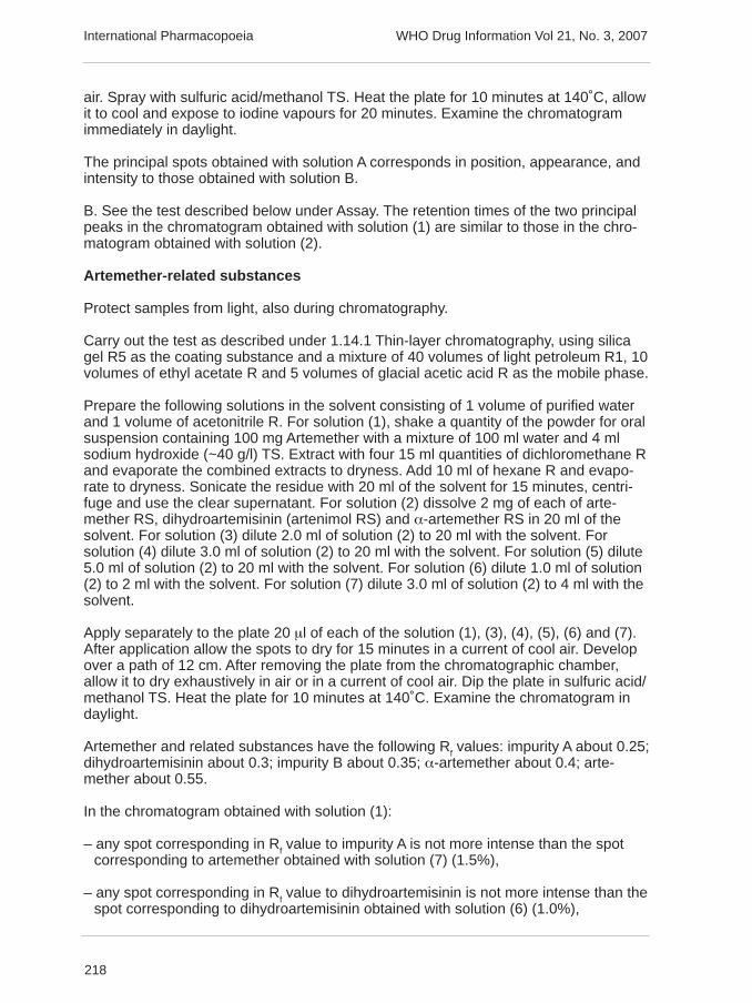
Impurities (artemether-related)
Dihydroartemisinin (artenimol) 284.4 C15
H24
O5
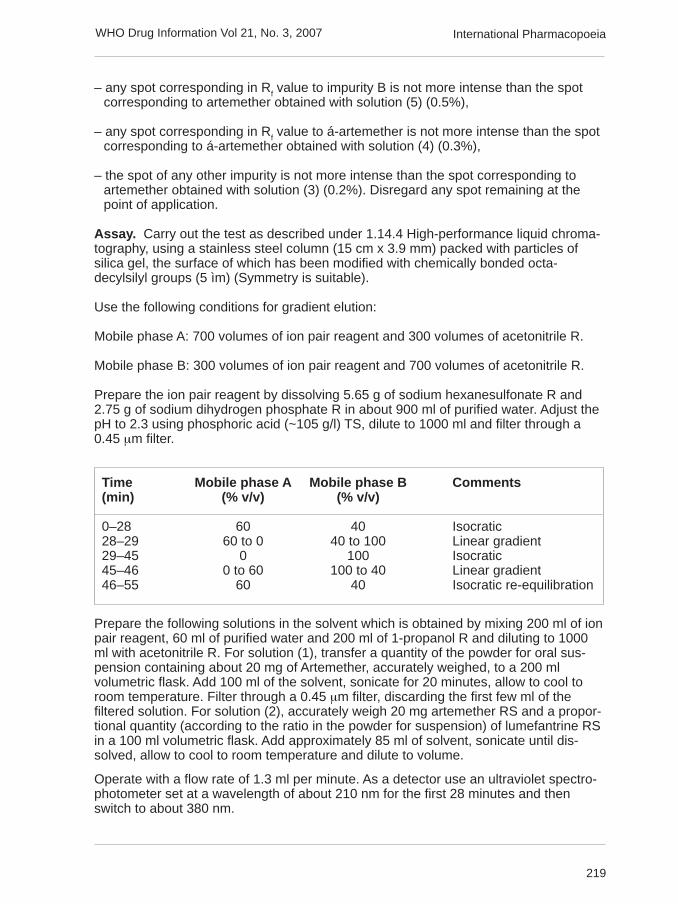
α-artemether 298.4 C16
H26
O5
221
WHO Drug Information Vol 21, No. 3, 2007 International Pharmacopoeia
Sulfadoxine and pyrimethamine tablets
Draft proposal for the International Pharmocopoeia (June 2007). Pleaseaddress any comments to Quality Assurance and Safety: Medicines, Medi-cines Policy and Standards, World Health Organization, 1211 Geneva 27,Switzerand. Fax +41 22 791 4730 or e-mail to [email protected]
Category. Antimalarial.
Storage. Sulfadoxine and Pyrimethamine tablets should be kept in a well-closedcontainer, protected from light.
Additional information. Strength in the current WHO Model list of essential medi-cines: 500 mg sulfadoxine and 25 mg pyrimethamine.
B. 298.4 C16H26O5
A. 298.3 C16
H26
O5
[Names to be provided for A and B]
222
WHO Drug Information Vol 21, No. 3, 2007International Pharmacopoeia
REQUIREMENTS
Comply with the monograph for “Tablets”.
Definition. Sulfadoxine and Pyrimethamine tablets contain Sulfadoxine and Pyrimeth-amine. They contain not less than 90.0% and not more than 110.0% of the amounts ofsulfadoxine (C12H14N4O4S) and pyrimethamine (C12H13ClN4) stated on the label.
Identity tests
A. Carry out test A.1 or, where UV detection is not available, test A.2.
A.1 Carry out the test as described under 1.14.1 Thin-layer chromatography, usingsilica gel R6 as the coating substance and a mixture of 75 volumes of ethylacetate R,25 volumes of methanol R and 1 volume of glacial acetic acid R as the mobile phase.Apply separately to the plate 10 ìl of each of the following two solutions in methanol R.For solution (A) shake a quantity of the powdered tablets containing about 100 mgsulfadoxine for 5 minutes with 20 ml, filter, and use the filtrate. For solution (B) use 5mg sulfadoxine RS and 0.25 mg pyrimethamine RS per ml. After removing the platefrom the chromatographic chamber, allow it to dry in a current of air and examine thechromatogram in ultraviolet light (254 nm).
The two principal spots obtained with solution A correspond in position, appearanceand intensity to those obtained with solution B.
A.2 Carry out the test as described under 1.14.1 Thin-layer chromatography, using theconditions described above under test A.1 but using silica gel R5 as the coatingsubstance. Dip the plate in modified Dragendorff reagent TS. Examine the chromato-gram in daylight.
The two principal spots obtained with solution A correspond in position, appearance,and intensity to those obtained with solution B (the spot for pyrimethamine is faintlyvisible).
B. See the test described below under Assay. The retention times of the two principalpeaks in the chromatogram obtained with solution (1) are similar to those in thechromatogram obtained with solution (4).
Related Substances
[Note from Secretariat: The test for related substances is under investigation.]
Assay. Carry out the test as described under 1.14.4 High-performance liquid chroma-tography, using a stainless steel column (25 cm x 4.6 mm) packed with particles ofsilica gel, the surface of which has been modified with chemically bondedoctadecylsilyl groups (5 ìm) (Phenomenex is suitable).
As the mobile phase, use a solution prepared as follows: mix 200 volumes of ac-etonitrile R, 800 volumes of acetic acid (~10 g/l) TS and 0.5 volume of triethylamine R;adjust the pH to 4.2 by adding sodium hydroxide (~400 g/l) TS.
223
WHO Drug Information Vol 21, No. 3, 2007 International Pharmacopoeia
For solution (1) weigh and powder 20 tablets, and transfer a quantity of the powdercontaining about 0.50 g of Sulfadoxine, accurately weighed, into a 200 ml volumetricflask. Add about 70 ml of acetonitrile R and sonicate for 10 minutes. Allow to cool toroom temperature, make up to volume using the mobile phase and sonicate for 10minutes. Dilute 5 ml to 25 ml with mobile phase and filter a portion of this solutionthrough a 0.45 ìm filter, discarding the first few ml of the filtered solution. For solution(2), transfer 25 mg sulfadoxine RS, accurately weighed, to a 25ml volumetric flask,add about 10 ml acetonitrile R, sonicate until dissolved and dilute to volume with themobile phase. For solution (3), transfer 25 mg pyrimethamine RS, accurately weighed,to a 25ml volumetric flask, add about 35 ml acetonitrile R, sonicate until dissolved anddilute to volume with the mobile phase. For solution (4) transfer 10 ml of solution (2)and 2 ml of solution (3) to a 20 ml volumetric flask and make up to volume with themobile phase.
Operate with a flow rate of 2 ml per minute. As a detector use an ultraviolet spectro-photometer set at a wavelength of about 254 nm.
Inject 20 µl of solution (4). The assay is not valid unless the resolution between thesulfadoxine and pyrimethamine peaks, eluting in this order, is at least 3. The run timefor the analyses is not less than 25 min.
Inject alternately 20 µl each of solutions (1) and (4).
Measure the areas of the peak responses obtained in the chromatograms fromsolutions (1) and (4), and calculate the content of sulfadoxine, C
12H
14N
4O
4S and
pyrimethamine, C12
H13
ClN4 in the tablets.
Reagents
Acetic acid (~10 g/l) TS.
Acetic acid (~300 g/l) TS, diluted with water to contain about 10 g of C2H
4O
2 per litre.
Quinine sulfate tablets
Draft proposal for the International Pharmocopoeia (June 2007). Pleaseaddress any comments to Quality Assurance and Safety: Medicines, Medi-cines Policy and Standards, World Health Organization, 1211 Geneva27, Switzerand. Fax +41 22 791 4730 or e-mail to [email protected]
Category. Antimalarial.
Storage. Quinine sulfate tablets should be kept in a well-closed container, protectedfrom light.
Additional information. Strength in the current WHO Model list of essential medi-cines: 300 mg of quinine sulfate. The tablets are coated.
224
WHO Drug Information Vol 21, No. 3, 2007
300 mg of quinine sulfate is equivalent to approximately 248.6 mg of anhydrousquinine.
REQUIREMENTS
Comply with the monograph for “Tablets”.
Definition. Quinine sulfate tablets contain Quinine sulfate. They contain not less than90.0% and not more than 110.0% of the amount of (C
20H
24N
2O
2)
2,H
2O
4S,2H
2O stated
on the label.
Identity tests
A. Carry out test A.1 or, where UV detection is not available, test A.2.
A.1 Carry out the test as described under 1.14.1 Thin-layer chromatography, usingsilica gel R6 as the coating substance and a mixture of 20 volumes of toluene R, 12volumes of ether R and 5 volumes of diethylamine R as the mobile phase. Applyseparately to the plate 2 µl of each of the following two solutions in a mixture of 2volumes of chloroform R and 1 volume of ethanol (~750 g/l) TS. For solution (A)shake a quantity of the powdered tablets containing about 0.1 g Quinine sulfate with10 ml, filter, and use the filtrate (if the tablets are sugar coated, the coating should beremoved prior to powdering thereof). For solution (B) use 10 mg of quinine sulfate RSper ml and for solution (C) use 10 mg of quinine sulfate RS and 10 mg of quinidinesulfate RS per ml. After removing the plate from the chromatographic chamber, allow itto dry in air or in a current of cool air and examine the chromatogram in ultraviolet light(254 nm).
The test is not valid unless the chromatogram obtained with solution C shows twoclearly separated spots.
The principal spot obtained with solution A corresponds in position, appearance andintensity to that obtained with solution B.
A.2 Carry out the test as described under 1.14.1 Thin-layer chromatography, using theconditions described above under test A.1 but using silica gel R5 as the coatingsubstance. Spray with sulfuric acid/ethanol (~0.05 mol/l) TS. and then with potassiumiodobismuthate TS2. Examine the chromatogram in daylight.
The test is not valid unless the chromatogram obtained with solution C shows twoclearly separated spots.
The principal spot obtained with solution A corresponds in position, appearance andintensity to that obtained with solution B.
B. To a quantity of powdered tablets containing 50 mg of Quinine sulfate add 100 mlof water R, shake, and filter. To 10 ml of the filtrate add one drop of sulfuric acid(~100 g/l) TS. When examined in direct daylight or in ultraviolet light (366 nm) a strongblue fluorescence appears; it disappears almost completely on the addition of a fewdrops of hydrochloric acid (~70 g/l) TS.
International Pharmacopoeia
225
WHO Drug Information Vol 21, No. 3, 2007
C. To a quantity of powdered tablets containing 0.1 g of Quinine sulfate add 10 ml ofwater R, shake, and filter. The filtrate yields reaction A described under 2.1 Generalidentification tests as characteristic of sulfates.
Related cinchona alkaloids. Carry out the test as described under 1.14.1 Thin-layerchromatography, using silica gel R5 as the coating substance and a mixture of 20volumes of toluene R, 12 volumes of ether R, and 5 volumes of diethylamine R as themobile phase. Apply separately to the plate 4 ìl of each of the following four solutionsin methanol R. For solution (1) sonicate a quantity of the powdered tablets containing0.10 g Quinine sulfate with 10 ml for 10 minutes, filter, and use the filtrate (if thetablets are sugar coated, the coating should be removed prior to powdering thereof).For solution (2) use 0.31 mg of quinine sulphate R per ml and for solution (3) 0.25 mgof cinchonidine R per ml. For solution (4) dissolve 10 mg of quinine sulfate R in 1 ml ofsolution (3). After removing the plate from the chromatographic chamber, allow it to dryin a current of air for 15 minutes and repeat the development. Heat the plate at 105˚Cfor 30 minutes, allow it to cool, spray it with potassium iodoplatinate TS, and examinethe chromatogram in daylight.
The test is not valid unless the chromatogram obtained with solution (4) shows twoclearly separated spots.
Any spot obtained with solution (1), other than the principal spot, is not more intensethan that obtained with solution (2) or solution (3). Disregard any spot obtained withsolution (1) immediately below the principal spot.
Dihydroquinine. Weigh and powder 20 tablets. Sonicate a quantity of the powderequivalent to about 0.20 g of Quinine sulfate, accurately weighed, in 20 ml of water Rfor 10 minutes. Add 0.5 g of potassium bromide R, 15 ml of hydrochloric acid (~70 g/l)TS and 0.1 ml of methyl red/ethanol TS. Titrate with potassium bromate (0.0167 mol/l)VS until a yellow colour is produced. Add 0.5 g of potassium iodide R in 200 ml ofwater, stopper the flask, and allow to stand in the dark for 5 minutes. Titrate the iodineliberated by excess potassium bromate in the solution with sodium thiosulfate (0.1mol/l) VS, adding 2 ml of starch TS when the solution has reached a light yellowcoloration. Each ml of potassium bromate (0.0167 mol/l) VS is equivalent to 26.10 mgof (C
20H
24N
2O
2)
2,H
2SO
4,2H
2O. Express the results of both the above determination and
the assay in percentages, calculated with reference to the (C20H24N2O2)2,H2SO4,2H2O.The difference between the two is not more than 10%.
Assay. Weigh and powder 20 tablets. Gently stir a quantity of the powder equivalentto about 0.40 g of Quinine sulfate, accurately weighed, in 40 ml of acetic acid R for 15minutes. Titrate with perchloric acid (0.1 mol/l) VS, determine the end point potentio-metrically as described under 2.6 Non-aqueous titration, Method A. Each ml of per-chloric acid (0.1 mol/l) VS is equivalent to 26.10 mg of (C
20H
24N
2O
2)
2,H
2O
4S,2H
2O.
Reagents
Sulfuric acid/ethanol (~0.05 mol/l)
Carefully add 4.9 g of sulfuric acid (~1760 g/l) TS to about 800 ml ethanol (~750 g/l)TS, while mixing gently, and dilute to 1000 ml with ethanol (~750 g/l) TS.
International Pharmacopoeia
226
WHO Drug Information Vol 21, No. 3, 2007
Rifampicin, isoniazid and ethambutol hydrochloride tablets
Draft proposal for the International Pharmocopoeia (June 2007). Pleaseaddress any comments to Quality Assurance and Safety: Medicines, Medi-cines Policy and Standards, World Health Organization, 1211 Geneva 27,Switzerand. Fax +41 22 791 4730 or e-mail to [email protected]
Category. Antituberculosis drugs.
Storage. Rifampicin, Isoniazid and Ethambutol hydrochloride tablets should be kept ina tightly closed container, protected from light.
Additional information. Strength in the current WHO Model list of essential medi-cines : 150 mg Rifampicin, 75 mg Isoniazid and 275 mg Ethambutol hydrochloride.The tablets are coated.
REQUIREMENTS
Comply with the monograph for “Tablets”.
Definition. Rifampicin, Isoniazid and Ethambutol hydrochloride tablets contain Ri-fampicin, Isoniazid and Ethambutol Hydrochloride. They contain not less than 90.0%and not more than 110.0% of the amounts of rifampicin (C
43H
58N
4O
12), isoniazid
(C6H7N3O) and ethambutol hydrochloride (C10H24N2O2, 2HCl) stated on the label.
Manufacture. The manufacturing process and the product packaging are designedand controlled so as to minimize the moisture content of the tablets. They ensure that,if tested, the tablets would comply with a loss on drying limit of not more than 30 mg/gwhen determined by drying freshly powdered tablets to constant mass under vacuumat 60 ˚.
Identity tests
Either tests A and B or test C may be applied.
A. See the test described below under Assay method A. The retention times of the twoprincipal peaks in the chromatogram obtained with solution (1) correspond to those ofthe principal peaks in the chromatogram obtained with solution (2).
B. See the test described below under Assay method B. The retention time of theprincipal peak in the chromatogram obtained with solution (1) corresponds to that ofthe principal peak in the chromatogram obtained with solution (2).
C. Carry out test C.1. or, where UV detection is not available, test C.2.
C.1 Carry out the test as described under 1.14.1 Thin-layer chromatography, usingsilica gel R6 as the coating substance and a mixture of 100 volumes of methanol Rand 1.5 volumes of strong ammonia solution R as the mobile phase. Apply separatelyto the plate 5 ml of each of the following two solutions in methanol R. For solution (A)shake a quantity of the powdered tablets equivalent to about 5 mg Isoniazid for 15minutes with 5 ml of methanol R, filter, and use the filtrate. For solution (B) use 1 mg
International Pharmacopoeia
227
WHO Drug Information Vol 21, No. 3, 2007
isoniazid RS and proportional quantities (according to the ratio in the tablet) of ri-fampicin RS, and ethambutol hydrochloride RS per ml of methanol R. After removingthe plate from the chromatographic chamber, allow it to dry in a current of air, place ina chamber with iodine vapours, and allow to stand for 20 minutes. Examine the chro-matogram immediately in ultraviolet light (254 nm).
The principal spots obtained with solution A correspond in position, appearance andintensity to those obtained with solution B.
C.2 Carry out the test as described under 1.14.1 Thin-layer chromatography, using theconditions described above under test A.1 but using silica gel R5 as the coatingsubstance. Examine the chromatogram immediately in daylight.
The principal spots obtained with solution A correspond in position, appearance andintensity to those obtained with solution B.
Rifampicin-related substances. Carry out the test as described under 1.14.4 High-performance liquid chromatography, preparing the solutions and using the conditionsgiven below under Assay method B.
Inject 20 µl each of solutions (1), (3) (4) and (5). The test is not valid unless in thechromatogram obtained with solution (4) the resolution between the peaks is at least 4.
In the chromatograms obtained with solutions (4) and (5) the following impurity peaksare eluted at the following relative retention with reference to rifampicin (retention timeabout 25 minutes): 3-(isonicotinoylhydrazinomethyl)rifamycin [the “hydrazone” result-ing from reaction between 3-formylrifamycin and isoniazid] about 0.5; rifampicinquinone about 0.7.
In the chromatogram obtained with solution (1), the area of any peak corresponding tothe hydrazone impurity is not greater than the area of the principal peak in the chroma-togram obtained with solution (3) (5.0%), the area of any peak corresponding torifampicin quinone is not more than 0.8 times the area of the principal peak in thechromatogram obtained with solution (3) (4.0%) and the area of any other peak is notgreater than 0.3 times the area of the principal peak in the chromatogram obtainedwith solution (3) (1.5%). The sum of the areas of all the peaks, other than the principalpeak, is not greater than twice the area of the principal peak obtained with solution (3)(10.0% with reference to the content of rifampicin). Disregard any peak with an arealess than 0.02 times the area of the principal peak in the chromatogram obtained withsolution (3) (0.1%) and any peak with relative retention time less than 0.23 with refer-ence to rifampicin.
[Note from the Secretariat: As agreed by the WHO Expert Committee on Specifica-tions for Pharmaceutical Preparations for the finalized texts for TB dosage form mono-graphs published on the Medicines web site (QSM/EC/06.13), solution (5) (see underAssay method B) describes the in situ preparation of the hydrazone impurity. Theagreed change will be made to the finalized texts before inclusion in the first Supple-ment to the 4th edition.]
International Pharmacopoeia
228
WHO Drug Information Vol 21, No. 3, 2007
Assay
A. For isoniazid and ethambutol hydrochlorideDetermine by 1.14.4 High-performance liquid chromatography, using a stainless steelcolumn (15 cm x 4.6 mm) packed with particles of silica gel, the surface of which hasbeen modified with chemically bonded octadecylsilyl groups, (5 µm) (Luna® is suit-able). As the mobile phase, use a solution prepared as follows: dissolve 50 g ammo-nium acetate R and 0.2 g copper(II) acetate R in 1000 ml of water and adjust to pH5.0 with glacial acetic acid R. Mix 940 ml of this solution with 60 ml methanol R.
Prepare the following solutions in water. For solution (1) weigh and powder 20 tablets.Transfer a quantity of the powder equivalent to about 100 mg Ethambutol hydrochlo-ride, accurately weighed, to a 500 ml volumetric flask. Dissolve in about 400 ml waterby shaking for about 15 minutes. [If foaming occurs, use 400 ml of a 4% solution ofmethanol R in place of the water.] Dilute to 500 ml with water. Filter a portion of thissolution through a 0.45 µm filter, discarding the first few ml of the filtered solution. Forsolution (2) dissolve 27.3 mg of isoniazid RS and 100 mg ethambutol hydrochlorideRS in 500 ml water.
Operate with a flow rate of 2.0 ml per minute. As a detector use an ultraviolet spectro-photometer set at a wavelength of about 270 nm.
Inject alternately 20 µl each of solutions (1) and (2). (The peak for isoniazid is elutedat a retention time of approximately 1.6 minutes, and that for ethambutol hydrochlorideat a retention time of approximately 6 minutes.)
Measure the areas of the peak responses obtained in the chromatograms from solu-tions (1) and (2), and calculate the content of isoniazid, C
6H
7N
3O and ethambutol
hydrochloride, C10
H24
N2O
2, 2HCl.
B. For rifampicinPrepare fresh solutions and perform the assay without delay. Low-actininc glasswareis recommended.
Determine by 1.14.4 High-performance liquid chromatography, using a stainless steelcolumn (25 cm x 4.6 mm) packed with particles of silica gel, the surface of which hasbeen modified with chemically bonded octadecylsilyl groups, (5 µm) (Luna® is suit-able). As the mobile phase, use a mixture of 6 volumes of methanol R and 4 volumesof phosphate buffer pH 7.0 (potassium dihydrogen phosphate R (0.01 mol/l), adjustedwith sodium hydroxide (0.1 mol/l)VS).
Prepare the following solutions in a mixture of 4 volumes of methanol R and 6 vol-umes of phosphate buffer pH 7.0. For solution (1) weigh and powder 20 tablets.Without delay, shake a quantity of the powder equivalent to about 40 mg Rifampicin in200 ml and filter. Solution (2) contains 0.20 mg rifampicin RS per ml. For solution (3)dilute a suitable volume of solution (1) to obtain a concentration equivalent to 10 µgRifampicin per ml. Solution (4) contains 0.2 mg rifampicin RS per ml and 0.2 mgrifampicin quinone RS per ml. For solution (5) dissolve 4 mg of rifampicin RS and 2mg of isoniazid RS in 25.0 ml of acetic acid (~60g/l) TS and keep the solution at roomtemperature for 30 minutes.
International Pharmacopoeia
229
WHO Drug Information Vol 21, No. 3, 2007
Operate with a flow rate of 1.0 ml per minute. As a detector use an ultraviolet spectro-photometer set at a wavelength of about 254 nm.
Inject 20 µl of solution (4). The assay is not valid unless the resolution between thepeaks is at least 4.
Inject alternately 20 µl each of solutions (1) and (2). Measure the areas of the peakresponses obtained in the chromatograms from solutions (1) and (2), and calculatethe content of rifampicin, C
43H
58N
4O
12 in the tablets.
[Note from the Secretariat: The preparation of solutions (1) to (4) has been modifiedfrom that described in the finalized texts for TB dosage form monographs publishedon the Medicines website in order to improve the stability of the test solution. It isintended to make corresponding changes to the finalized texts before inclusion in thefirst Supplement to the 4th edition.]
Dissolution test. To be added for rifampicin.
Rifampicin and Isoniazid dispersible tablets
Draft proposal for the International Pharmocopoeia (June 2007). Pleaseaddress any comments to Quality Assurance and Safety: Medicines, Medi-cines Policy and Standards, World Health Organization, 1211 Geneva27, Switzerand. Fax +41 22 791 4730 or e-mail to [email protected]
Category. Antituberculosis drugs.
Storage. Rifampicin and Isoniazid dispersible tablets should be kept in a tightlyclosed container, protected from light.
Additional information. [ Dispersible tablets are not included in the current WHOModel List of Essential Medicines. It is understood that the following strengths areavailable as dispersible tablets: 60 mg Rifampicin and 30 mg Isoniazid,60 mg Rifampicin and 60 mg Isoniazid.]
REQUIREMENTS
Comply with the monograph for “Tablets”.
Definition. Rifampicin and Isoniazid dispersible tablets contain Rifampicin and Isoni-azid in a suitable dispersible basis that may contain suitable flavouring agents. Theycontain not less than 90.0% and not more than 110.0% of the amounts of rifampicin(C
43H
58N
4O
12) and isoniazid (C
6H
7N
3O) stated on the label.
Identity tests
Either tests A and B or test C may be applied.
International Pharmacopoeia

230
WHO Drug Information Vol 21, No. 3, 2007
A. See Assay method A described below. The retention time of the principal peak inthe chromatogram obtained with solution (1) corresponds to that of the principal peakin the chromatogram obtained with solution (2).
B. See Assay method B described below. The retention time of the principal peak inthe chromatogram obtained with solution (1) corresponds to that of the principal peakin the chromatogram obtained with solution (2).
C. Carry out test C.1. or, where UV detection is not available, test C.2.
C.1 Carry out the test as described under 1.14.1 Thin-layer chromatography, usingsilica gel R6 as the coating substance and a mixture of 100 volumes of methanol Rand 1.5 volumes of strong ammonia solution R as the mobile phase. Apply separatelyto the plate 5 ml of each of the following two solutions in methanol R. For solution (A)shake a quantity of the powdered tablets containing about 5 mg Isoniazid for 15minutes with 5 ml, filter, and use the filtrate. For solution (B) use 1 mg isoniazid RSand a proportional quantity (according to the ratio in the tablet) of rifampicin RS per ml.After removing the plate from the chromatographic chamber, allow it to dry in a currentof air and examine the chromatogram in ultraviolet light (254 nm).
The principal spots obtained with solution A correspond in position, appearance andintensity to those obtained with solution B.
C.2 Carry out the test as described under 1.14.1 Thin-layer chromatography, using theconditions described above under test A.1 but using silica gel R5 as the coatingsubstance. Examine the chromatogram immediately in daylight. Add detection byiodine vapour…
The principal spots obtained with solution A correspond in position, appearance andintensity to those obtained with solution B.
Disintegration. Disintegrate within 3 minutes when examined by 5.3 Disintegrationtest for tablets and capsules, but using water R at 15 to 25 ˚C.
Dissolution test. [To be added for rifampicin.]
Rifampicin-related substances. Carry out the test as described under 1.14.4 High-performance liquid chromatography, using the conditions given below under Assaymethod B.
Inject alternately 20 µl each of solutions (1), (3), (4) and (5). The test is not validunless in the chromatogram obtained with solution (4) the resolution between thepeaks is at least 4.
In the chromatograms obtained with solutions (4) and (5) the following impurity peaksare eluted at the following relative retention with reference to rifampicin (retention timeabout 25 minutes): 3-(isonicotinoylhydrazinomethyl)rifamycin [the “hydrazone” result-ing from reaction between 3-formylrifamycin and isoniazid] about 0.5; rifampicinquinone about 0.7.
International Pharmacopoeia
231
WHO Drug Information Vol 21, No. 3, 2007
In the chromatogram obtained with solution (1), the area of any peak corresponding tothe hydrazone impurity is not greater than the area of the principal peak in the chro-matogram obtained with solution (3) (5.0%), the area of any peak corresponding torifampicin quinone is not greater than 0.8 times the area of the principal peak in thechromatogram obtained with solution (3) (4.0 %) and the area of any other peak is notgreater than 0.3 times the area of the principal peak in the chromatogram obtainedwith solution (3) (1.5%). The sum of the areas of all the peaks, other than the principalpeak, is not greater than twice the area of the principal peak in the chromatogramobtained with solution (3) (10.0%). Disregard any peak with an area less than 0.02times the area of the principal peak in the chromatogram obtained with solution (3)(0.1%) and any peak with a relative retention less than 0.23 with reference to ri-fampicin.
Assay
A. For isoniazid. Determine by 1.14.4 High-performance liquid chromatography,using a stainless steel column (15 cm x 4.6 mm) packed with particles of silica gel, thesurface of which has been modified with chemically bonded octadecylsilyl groups (5µm) (Luna® is suitable). As the mobile phase, use a solution prepared as follows:dissolve 50 g ammonium acetate R in 1000 ml of water and adjust to pH 5.0 withglacial acetic acid R. Mix 940 ml of this solution with 60 ml methanol R.
Prepare the following solutions in water. For solution (1) weigh and powder 20 tablets.Transfer a quantity of the powder containing about 30 mg Isoniazid, accuratelyweighed, to a 500 ml volumetric flask. Dissolve in about 400 ml water by shaking forabout 15 minutes. [If foaming occurs, use 400 ml of a 4% solution of methanol R inplace of the water.] Dilute to 500 ml with water. Filter a portion of this solution througha 0.45 µm filter, discarding the first few ml of the filtered solution. For solution (2)dissolve 30 mg isoniazid RS in 500 ml water.
Operate with a flow rate of 2.0 ml per minute. As a detector use an ultraviolet spectro-photometer set at a wavelength of about 240 nm.
Inject alternately 20 µl each of solutions (1) and (2). The peak for isoniazid is eluted ata retention time of about 1.6 minutes.
Measure the areas of the peak responses obtained in the chromatograms from solu-tions (1) and (2), and calculate the content of isoniazid, C
6H
7N
3O, in the tablets.
B. For rifampicin. Prepare fresh solutions and perform the assay without delay. Low-actinic glassware is recommended.
Determine by 1.14.4 High-performance liquid chromatography, using a stainless steelcolumn (25 cm x 4.6 mm) packed with particles of silica gel, the surface of which hasbeen modified with chemically bonded octadecylsilyl groups (5 µm) (Luna® is suitable).As the mobile phase, use a mixture of 6 volumes of methanol R and 4 volumes ofphosphate buffer pH 7.0 (potassium dihydrogen phosphate R (0.01 mol/l), adjustedwith sodium hydroxide (0.1 mol/l)VS).
Prepare the following solutions in a mixture of 4 volumes of methanol R and 6 vol-umes of phosphate buffer pH 7.0. For solution (1) weigh and powder 20 tablets.Without delay, shake a quantity of the powder containing about 40 mg Rifampicin in
International Pharmacopoeia
232
WHO Drug Information Vol 21, No. 3, 2007
200 ml and filter. Solution (2) contains 0.20 mg rifampicin RS per ml. For solution (3)dilute a suitable volume of solution (1) to obtain a concentration equivalent to 10 µgRifampicin per ml. Solution (4) contains 0.2 mg rifampicin RS per ml and 0.2 mgrifampicin quinone RS per ml. For solution (5) dissolve 4 mg of rifampicin RS and 2mg of isoniazid RS in 25 ml of acetic acid (~60 g/l) TS and keep the solution at roomtemperature for 30 minutes.
Operate with a flow rate of 1.0 ml per minute. As a detector use an ultraviolet spectro-photometer set at a wavelength of about 254 nm.
Inject 20 µl of solution (4). The assay is not valid unless the resolution between thepeaks is at least 4.
Inject alternately 20 µl each of solutions (1) and (2). Measure the areas of the peakresponses obtained in the chromatograms from solutions (1) and (2), and calculatethe content of rifampicin, C
43H
58N
4O
12, in the tablets.
Rifampicin, isoniazid and pyrazinamide dispersible tablets
Draft proposal for the International Pharmocopoeia (June 2007). Pleaseaddress any comments to Quality Assurance and Safety: Medicines, Medi-cines Policy and Standards, World Health Organization, 1211 Geneva27, Switzerand. Fax +41 22 791 4730 or e-mail to [email protected]
Category. Antituberculosis drugs.
Storage. Rifampicin, Isoniazid and Pyrazinamide dispersible tablets should be kept ina tightly closed container, protected from light.
Additional information. Not included in the current WHO Model List of EssentialMedicines. It is understood that the following strengths are available as dispersibletablets: 60 mg Rifampicin, 30 mg Isoniazid and 150 mg Pyrazinamide.
REQUIREMENTS
Comply with the monograph for “Tablets”.
Definition. Rifampicin, Isoniazid and Pyrazinamide dispersible tablets contain Ri-fampicin, Isoniazid and Pyrazinamide in a suitable dispersible basis that may containsuitable flavouring agents. They contain not less than 90.0% and not more than110.0% of the amounts of rifampicin (C
43H
58N
4O
12), isoniazid (C
6H
7N
3O) and pyrazina-
mide (C5H5N3O) stated on the label.
Identity tests
Either tests A and B or test C may be applied.
A. See Assay method A described below. The retention times of the two principalpeaks in the chromatogram obtained with solution (1) correspond to those of theprincipal peaks in the chromatogram obtained with solution (2).
B. See Assay method B described below. The retention time of the principal peak in
International Pharmacopoeia
233
WHO Drug Information Vol 21, No. 3, 2007
the chromatogram obtained with solution (1) corresponds to that of the principal peakin the chromatogram obtained with solution (2).
C. Carry out test C.1 or, where UV detection is not available, test C.2.
C.1 Carry out the test as described under 1.14.1 Thin-layer chromatography, usingsilica gel R6 as the coating substance and a mixture of 100 volumes of methanol Rand 1.5 volumes of strong ammonia solution R as the mobile phase. Apply separatelyto the plate 5 ml of each of the following two solutions in methanol R. For solution (A)shake a quantity of the powdered tablets containing about 5 mg Isoniazid for 15minutes with 5 ml, filter, and use the filtrate. For solution (B) use 1 mg isoniazid RSand proportional quantities (according to the ratio in the tablet) of rifampicin RS andpyrazinamide RS per ml. After removing the plate from the chromatographic chamber,allow it to dry in a current of air and examine the chromatogram in ultraviolet light (254nm).
The principal spots obtained with solution A correspond in position, appearance andintensity to those obtained with solution B.
C.2 Carry out the test as described under 1.14.1 Thin-layer chromatography, using theconditions described above under test A.1 but using silica gel R5 as the coatingsubstance. Examine the chromatogram immediately in daylight.Add detection by iodine vapour…
The principal spots obtained with solution A correspond in position, appearance andintensity to those obtained with solution B.
Disintegration. Disintegrate within 3 minutes when examined by 5.3 Disintegrationtest for tablets and capsules, but using water R at 15-25 ˚C.
Dissolution test. [ To be added for rifampicin.]
Rifampicin-related substances. Carry out the test as described under 1.14.4 High-performance liquid chromatography, using the conditions given below under Assaymethod B.
Inject alternately 20 µl each of solutions (1), (3), (4) and (5). The test is not validunless in the chromatogram obtained with solution (4) the resolution between thepeaks is at least 4.
In the chromatograms obtained with solutions (4) and (5) the following impurity peaksare eluted at the following relative retention with reference to rifampicin (retention timeabout 25 minutes): 3-(isonicotinoylhydrazinomethyl)rifamycin [the “hydrazone” result-ing from reaction between 3-formylrifamycin and isoniazid] about 0.5; rifampicinquinone about 0.7.
In the chromatogram obtained with solution (1), the area of any peak corresponding tothe hydrazone impurity is not greater than the area of the principal peak in the chro-matogram obtained with solution (3) (5.0%), the area of any peak corresponding torifampicin quinone is not greater than 0.8 times the area of the principal peak in thechromatogram obtained with solution (3) (4.0 %) and the area of any other peak is notgreater than 0.3 times the area of the principal peak in the chromatogram obtained
International Pharmacopoeia
234
WHO Drug Information Vol 21, No. 3, 2007
with solution (3) (1.5%). The sum of the areas of all the peaks, other than the principalpeak, is not greater than twice the area of the principal peak in the chromatogramobtained with solution (3) (10.0%). Disregard any peak with an area less than 0.02times the area of the principal peak in the chromatogram obtained with solution (3)(0.1%) and any peak with a relative retention less than 0.23 with reference to ri-fampicin.
Assay
A. For isoniazid and pyrazinamide. Determine by 1.14.4 High-performance liquidchromatography, using a stainless steel column (15 cm x 4.6 mm) packed with parti-cles of silica gel, the surface of which has been modified with chemically bondedoctadecylsilyl groups (5 µm) Luna® is suitable). As the mobile phase, use a solutionprepared as follows: dissolve 50 g ammonium acetate R in 1000 ml of water andadjust to pH 5.0 with glacial acetic acid R. Mix 940 ml of this solution with 60 mlmethanol R.
Prepare the following solutions in water. For solution (1) weigh and powder 20 tablets.Transfer a quantity of the powder containing about 30 mg Isoniazid, accuratelyweighed, to a 500 ml volumetric flask. Dissolve in about 400 ml water by shaking forabout 15 minutes. [If foaming occurs, use 400 ml of a 4% solution of methanol R inplace of the water.] Dilute to 500 ml with water. Filter a portion of this solution througha 0.45 µm filter, discarding the first few ml of the filtered solution. For solution (2)dissolve 30 mg isoniazid RS and a proportional quantity (according to the ratio in thetablet) of pyrazinamide RS in 500 ml water.
Operate with a flow rate of 2.0 ml per minute. As a detector use an ultraviolet spectro-photometer set at a wavelength of about 240 nm.
Inject 20 µl of solution (2). The assay is not valid unless the resolution between theisoniazid and pyrazinamide peaks, eluting in this order, is at least 2.
Inject alternately 20 µl each of solutions (1) and (2). Measure the areas of the peakresponses obtained in the chromatograms from solutions (1) and (2), and calculate thecontent of isoniazid, C
6H
7N
3O, and pyrazinamide, C
5H
5N
3O, in the tablets.
B. For rifampicin. Prepare fresh solutions and perform the assay without delay. Low-actinic glassware is recommended.
Determine by 1.14.4 High-performance liquid chromatography, using a stainless steelcolumn (25 cm x 4.6 mm) packed with particles of silica gel, the surface of which hasbeen modified with chemically bonded octadecylsilyl groups (5 µm) Luna® is suitable).As the mobile phase, use a mixture of 6 volumes of methanol R and 4 volumes ofphosphate buffer pH 7.0 (potassium dihydrogen phosphate R (0.01 mol/l), adjustedwith sodium hydroxide (0.1 mol/l)VS).
Prepare the following solutions in a mixture of 4 volumes of methanol R and 6 volumesof phosphate buffer pH 7.0. For solution (1) weigh and powder 20 tablets. Withoutdelay, shake a quantity of the powder containing about 40 mg Rifampicin in 200 mland filter. Solution (2) contains 0.20 mg rifampicin RS per ml. For solution (3) dilute a
International Pharmacopoeia
235
WHO Drug Information Vol 21, No. 3, 2007
suitable volume of solution (1) to obtain a concentration equivalent to 10 µg Rifampicinper ml. Solution (4) contains 0.2 mg rifampicin RS per ml and 0.2 mg rifampicinquinone RS per ml. For solution (5) dissolve 4 mg of rifampicin RS and 2 mg ofisoniazid RS in 25 ml of acetic acid (~60 g/l) TS and keep the solution at room tem-perature for 30 minutes.
Operate with a flow rate of 1.0 ml per minute. As a detector use an ultraviolet spectro-photometer set at a wavelength of about 254 nm.
Inject 20 µl of solution (4). The assay is not valid unless the resolution between thepeaks is at least 4.
Inject alternately 20 µl each of solutions (1) and (2). Measure the areas of the peakresponses obtained in the chromatograms from solutions (1) and (2), and calculatethe content of rifampicin, C
43H
58N
4O
12, in the tablets.
Efavirenz capsules
Draft proposal for the International Pharmocopoeia (May 2007). Pleaseaddress any comments to Quality Assurance and Safety: Medicines, Medi-cines Policy and Standards, World Health Organization, 1211 Geneva27, Switzerand. Fax +41 22 791 4730 or e-mail to [email protected]
Category. Antiretroviral (Non-nucleoside Reverse Transcriptase Inhibitor).
Storage. Efavirenz capsules should be kept in a well-closed container, protected fromlight.
Additional information. Strength in the current WHO Model List of Essential Medi-cines: 50 mg
REQUIREMENTS
Comply with the monograph for “Capsules”.
Definition. Efavirenz capsules contain Efavirenz. They contain not less than 90.0%and not more than 110.0% of the amount of C
14H
9CIF
3NO
2 stated on the label.
Identity tests
Either tests A and B or tests B and C or test D alone may be applied.
A. Carry out test A.1 or, where UV detection is not available, test A.2.
A.1 Carry out the test as described under 1.14.1 Thin-layer chromatography, usingsilica gel R6 as the coating substance and a mixture of 90 volumes of dichlorometh-ane R, 10 volumes of methanol R and 3 volumes of glacial acetic acid R as the mobilephase. Apply separately to the plate 5 ìl of each of 2 solutions in methanol R contain-ing (A) shake a quantity of the content of the capsules containing 5 mg of Efavirenz
International Pharmacopoeia
236
WHO Drug Information Vol 21, No. 3, 2007
with 5 ml, filter and use the clear filtrate and (B) 1 mg of efavirenz RS per ml. Afterremoving the plate from the chromatographic chamber, allow it to dry exhaustively inair or in a current of cool air. Examine the chromatogram in ultraviolet light (254 nm).
The principal spot obtained with solution A corresponds in position, appearance andintensity with that obtained with solution B.
A.2 Carry out the test as described under 1.14.1 Thin-layer chromatography, usingthe conditions described above under test A.1 but using silica gel R5 as the coatingsubstance. Spray the plate with basic potassium permanganate (~ 1 g/l) TS. Examinethe chromatogram in daylight.
The principal spot obtained with solution A corresponds in position, appearance andintensity with that obtained with solution B.
B. See the test described below under Assay method A. The retention time of theprincipal peak in the chromatogram obtained with solution (1) is similar to that in thechromatogram obtained with solution (2).
C. The absorption spectrum of the final solution prepared for Assay method B, whenobserved between 210 nm and 300 nm, exhibits one maximum at about 247 nm.
D. To a quantity of the contents of the capsules containing 25 mg of Efavirenz, add 10ml of methanol R, shake to dissolve and filter. Evaporate the filter to dryness. Carryout the examination as described under 1.7 Spectrophotometry in the infrared region.The infrared absorption spectrum is concordant with the spectrum obtained fromefavirenz RS or with the reference spectrum of efavirenz.
If the spectra thus obtained are not concordant, repeat the test using the test residueand the residue obtained by dissolving efavirenz RS in a small amount of methanol Rand evaporating to dryness. The infrared absorption spectrum is concordant with thespectrum obtained from efavirenz RS.
Related substances
Note: Prepare fresh solutions and perform the test without delay.
Carry out the test as described under 1.14.4 High-performance liquid chromatography,using the conditions given under Assay method A.
Prepare the following solutions in a mixture of 50% acetonitrile R and 50% water R(dissolution solvent). For solution (1) transfer a quantity of the content of the capsulescontaining 25 mg of Efavirenz into the dissolution solvent and dilute to 25.0 ml with thesame solvent. For solution (2) dilute 1.0 ml of solution (1) to 50.0 ml with the dissolu-tion solvent and dilute 5.0 ml of the resulting solution to 100.0 ml with the samesolvent. For solution (3) dissolve about 5 mg of efavirenz for system suitability RS(containing efavirenz and impurities A to F) in 5 ml of the dissolution solvent.
Inject separately 35 µl of each solution (1), (2) and (3) and of the dissolution solvent inthe chromatographic system. Examine the blank chromatogram for any extraneous
International Pharmacopoeia
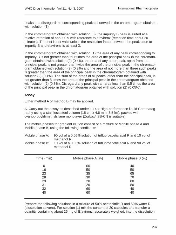
237
WHO Drug Information Vol 21, No. 3, 2007
peaks and disregard the corresponding peaks observed in the chromatogram obtainedwith solution (1).
In the chromatogram obtained with solution (3), the impurity B peak is eluted at arelative retention of about 0.9 with reference to efavirenz (retention time about 20minutes). The test is not valid unless the resolution factor between the peaks due toimpurity B and efavirenz is at least 3.
In the chromatogram obtained with solution (1) the area of any peak corresponding toimpurity B is not greater than four times the area of the principal peak in the chromato-gram obtained with solution (2) (0.4%), the area of any other peak, apart from theprincipal peak, is not greater than twice the area of the principal peak in the chromato-gram obtained with solution (2) (0.2%) and the area of not more than three such peaksis greater than the area of the principal peak in the chromatogram obtained withsolution (2) (0.1%). The sum of the areas of all peaks, other than the principal peak, isnot greater than 8 times the area of the principal peak in the chromatogram obtainedwith solution (2) (0.8%). Disregard any peak with an area less than 0.5 times the areaof the principal peak in the chromatogram obtained with solution (2) (0.05%).
Assay
Either method A or method B may be applied.
A. Carry out the assay as described under 1.14.4 High-performance liquid Chromatog-raphy using a stainless steel column (15 cm x 4.6 mm, 3.5 ìm), packed withcyanopropyldimethylsilane monolayer (Zorbax® SB-CN is suitable).
The mobile phases for gradient elution consist of a mixture of Mobile phase A andMobile phase B, using the following conditions:
Mobile phase A: 90 vol of a 0.05% solution of trifluoroacetic acid R and 10 vol ofmethanol R.
Mobile phase B: 10 vol of a 0.05% solution of trifluoroacetic acid R and 90 vol ofmethanol R.
Time (min) Mobile phase A (%) Mobile phase B (%)
0 60 4016 50 5023 35 6528 30 7029 20 8031 20 8032 60 4040 60 40
Prepare the following solutions in a mixture of 50% acetonitrile R and 50% water R(dissolution solvent). For solution (1) mix the content of 20 capsules and transfer aquantity containing about 25 mg of Efavirenz, accurately weighed, into the dissolution
International Pharmacopoeia
238
WHO Drug Information Vol 21, No. 3, 2007
solvent and dilute to 25.0 ml with the same solvent. Dilute 1.0 ml of the resultingsolution to 100.0 ml with the dissolution solvent For solution (2) dissolve 25 mg ofefavirenz RS in the dissolution solvent and dilute to 25.0 ml with the same solvent.Dilute 1.0 ml of the resulting solution to 100.0 ml with the dissolution solvent. Forsolution (3) dissolve about 5 mg of efavirenz for system suitability RS (containingefavirenz and impurities A to F) in 5 ml of the dissolution solvent.
Operate with a flow rate of 1.5 ml per minute. as a detector use an ultraviolet spectro-photometer set at a wavelength of about 250 nm.
Inject separately 35 µl of solutions (1), (2) and (3). In the chromatogram obtained withsolution (3), the impurity B peak is eluted at a relative retention of about 0.9 withreference to efavirenz (retention time about 20 minutes). The assay is not valid unlessthe resolution factor between the peaks due to impurity B and efavirenz is at least 3.
Measure the areas of the peaks responses obtained in the chromatogram from solu-tions (1) and (2), and calculate the content of C
14H
9ClF
3NO
2 in the capsules.
B. Mix the content of 20 capsules and transfer a quantity containing about 25 mg ofEfavirenz , accurately weighed, to a 50.0 ml volumetric flask. Add about 25 ml ofmethanol R, sonicate for about 5 minutes, allow to cool to room temperatureand make up the volume using the same solvent. Filter a portion of thissolution through a 0.45 ìm filter, discarding the first few ml of the filtrate.Dilute 1.0 of this solution to 50.0 ml with the same solvent. Measure theabsorbance (1.6) of 1-cm layer of the diluted solution at the maximumat about 247 nm. Calculate the amount of C
14H
9CIF
3NO
2 in the capsules
using an absorptivity value of 47.3 (specific absorbance A 1%1cm
= 473).
Impurities. The impurities limited by the requirements of this monograph includethose listed in the monograph for Efavirenz.
Nevirapine oral solution
Draft proposal for the International Pharmocopoeia (April 2007). Pleaseaddress any comments to Quality Assurance and Safety: Medicines, Medi-cines Policy and Standards, World Health Organization, 1211 Geneva 27,Switzerand. Fax +41 22 791 4730 or e-mail to [email protected]
Category. Antiretroviral
Storage. Nevirapine oral solution should be kept in a well-closed container,
Labelling. The designation of the container of nevirapine oral solution should statethat the active ingredient is the hemihydrate form and the quantity should be indicatedin terms of the equivalent amount of nevirapine.
Additional information. Strength in the current WHO Model List of Essential Medi-cines: 50 mg/5 ml.
International Pharmacopoeia
239
WHO Drug Information Vol 21, No. 3, 2007
REQUIREMENTS
Complies with the monograph for “Liquids for oral use”.
Definition. Nevirapine oral suspension contains Nevirapine as the hemihydrate. Itcontains not less than 90.0% and not more than 110.0% of the amount of C
15H
14N
4O
stated on the label.
Identity tests
Either tests A and C or tests B and C may be applied.
A. Carry out test A.1 or, where UV detection is not available, test A.2.
A.1 Carry out the test as described under 1.14.1 Thin-layer chromatography, usingsilica gel R6 as the coating substance and a mixture of 90 volumes of dichlorometh-ane R, 10 volumes of methanol R and 3 volumes of glacial acetic acid R as the mobilephase. Apply separately to the plate 5 ìl of each of 2 solutions in methanol: (A) transferan accurately measured volume of the oral suspension equivalent to 5 mg of nevirap-ine with 5 ml and (B) 1 mg of anhydrous nevirapine RS per ml. After removing theplate from the chromatographic chamber, allow it to dry exhaustively in air or in acurrent of cool air. Examine the chromatogram in ultraviolet light (254 nm).
The principal spot obtained with solution A corresponds in position, appearance andintensity with that obtained with solution B.
A.2. Carry out the test as described under 1.14.1 Thin-layer chromatography, usingthe conditions described above under test A.1 but using silica gel R5 as the coatingsubstance. Spray the plate with dilute basic potassium permanganate (1 g/l) TS.Examine the chromatogram in daylight.The principal spot obtained with solution A corresponds in position, appearance andintensity with that obtained with solution B.
B. See the test described below under Assay. The retention time of the principal peakin the chromatogram obtained with solution (1) is similar to that obtained with solution(2).
C. To a volume of the oral suspension equivalent to about 100 mg of nevirapineadd 8 ml methanol R, shake and filter. The absorption spectrum, when observedbetween 220 nm and 350 nm, exhibits a maximum at about 283 nm.
Related substances
Note: Prepare fresh solutions and perform the tests without delay
Carry out the test as described under 1.14.4 High-performance liquid chromatography,using the conditions given under Assay.
Prepare the following solutions. For solution (1) transfer a volume of the oral suspen-sion equivalent to about 24 mg of nevirapine in 4 ml of acetonitrile R, add 80 ml of the
International Pharmacopoeia
240
WHO Drug Information Vol 21, No. 3, 2007
mobile phase and sonicate. Dilute to 100.0 ml with the mobile phase. For solution (2)dilute 5.0 ml of solution (1) to 50.0 ml with the mobile phase. Dilute 5.0 ml of theresulting solution to 50.0 ml with the same solvent. For solution (3) dissolve 6 mg ofnevirapine impurity B RS in a mixture of 25 ml of acetonitrile R and 55 ml of mobilephase, sonicate for 15 min and dilute to 100.0 ml with the mobile phase. Mix 6.0 ml ofthe resulting solution with 3.0 ml of solution (1) and dilute to 50.0 ml with the mobilephase.
Inject separately 50 ìl each of solutions (1), (2) and (3) and of mobile phase in thechromatographic system and record the chromatograms for 9 times the retention timeof nevirapine. Examine the mobile phase chromatogram for any extraneous peaks anddisregard the corresponding peaks observed in the chromatogram obtained withsolution (1).
In the chromatogram obtained with solution (3), the following impurity peaks are elutedat the following retention ratios with reference to nevirapine (retention time = about 7.6min): impurity B = about 0.7; impurity A = about 1.5; impurity C = about 2.8. The test isnot valid unless the resolution factor between the peaks due to nevirapine and nevi-rapine impurity B is at least 5.
In the chromatogram obtained with solution (1) the area of any individual peak corre-sponding to impurity A or C is not greater than 0.2 times the area of the principal peakobtained with solution (2) (0.2%) and 0.77 times the area of any individual peakcorresponding to impurity B is not greater than 0.2 times the area of the principal peakobtained with solution (2) (0.2%). The area of any other impurity peak is not greaterthan 0.1 times the area of the principal peak obtained with solution (2) (0.1%). Thesum of the areas of all peaks, other than the principal peak, is not greater than 0.6times the area of the principal peak obtained with solution (2) (0.6%). Disregard anypeak with an area less than 0.05 times the area of the principal peak obtained withsolution (2) (0.05%)
Assay. Carry out the assay as described under 1.14.4 High-performance liquid Chro-matography using a stainless steel column (15 cm x 4.6 mm), packed with particles ofsilica gel, the surface of which has been modified with chemically bonded hexadecyl-amidylsilyl groups (5 µm) (Supelcosil LC-ABZ is suitable).
Maintain the column temperature at 35 ˚C.
The mobile phase consists of a filtered and degassed mixture of 20 volumes of aceto-nitrile R and 80 volumes of a 3.6 g/l solution of ammonium dihydrogen phosphate Radjusted to pH 5.0 using ammonia (~260 g/l) TS.
Operate with a flow rate of 1.0 ml per minute. As a detector use an ultraviolet spectro-photometer set at a wavelength of about 220 nm.
Prepare the following solutions. For solution (1) transfer an accurately weighed volumeof the oral suspension equivalent to about 24 mg of nevirapine in 4 ml of acetonitrile R,add 80 ml of the mobile phase and sonicate. Dilute to 100.0 ml with the mobile phase(solution A). Dilute 3.0 ml of solution A to 50.0 ml with the mobile phase. For solution(2) dissolve 12 mg of anhydrous nevirapine RS in 2 ml of acetonitrile R and 40 ml of
International Pharmacopoeia
241
WHO Drug Information Vol 21, No. 3, 2007
the mobile phase and sonicate. Dilute to 50.0 ml with the mobile phase and dilute 3.0ml of the resulting solution to 50.0 ml with the mobile phase. For solution (3) dissolve 6mg of nevirapine impurity B RS in a mixture of 25 ml of acetonitrile R and 55 ml ofmobile phase, sonicate for 15 min and dilute to 100.0 ml with the mobile phase. Mix6.0 ml of the resulting solution with 3.0 ml of solution A and dilute to 50.0 ml with themobile phase.
Inject separately 50 ìl of solutions (1), (2) and (3). In the chromatogram obtained withsolution (3), the impurity B peak is eluted at a relative retention of about 0.7 withreference to nevirapine (retention time about 7.6 minutes). The assay is not validunless the resolution between nevirapine and nevirapine impurity B is at least 5.
Measure the areas of the peak responses obtained in the chromatograms from solu-tions (1) and (2). Determine the weight per ml (1.3.1) of the oral suspension andcalculate the content of C
15H
14N
4O, weight in volume, in the oral suspension.
[Note from the Secretariat: method text 1.3.1 attached for information.]
Impurities. The impurities limited by the requirements of this monograph includethose listed in the monograph for Nevirapine.
Nevirapine tablets
Draft proposal for the International Pharmocopoeia (April 2007). Pleaseaddress any comments to Quality Assurance and Safety: Medicines, Medi-cines Policy and Standards, World Health Organization, 1211 Geneva 27,Switzerand. Fax +41 22 791 4730 or e-mail to [email protected]
Category. Antiretroviral
Storage. Nevirapine tablets should be kept in a well-closed container,
Labelling. The designation of the container of nevirapine tablets should state that theactive ingredient is the anhydrous form.
Additional information. Strength in the current WHO Model List of Essential Medi-cines: 200 mg
REQUIREMENTS
Comply with the monograph for “Tablets”.
Definition. Nevirapine tablets contain Nevirapine in the anhydrous form. They containnot less than 90.0% and not more than 110.0% of the amount of C
15H
14N
4O stated on
the label.
Identity tests
Either tests A and B or test C may be applied.
International Pharmacopoeia
242
WHO Drug Information Vol 21, No. 3, 2007
A. Carry out test A.1. or, where UV detection is not available, test A.2.
A.1. Carry out the test as described under 1.14.1 Thin-layer chromatography, usingsilica gel R6 as the coating substance and a mixture of 90 volumes of dichlorometh-ane R, 10 volumes of methanol R and 3 volumes of glacial acetic acid R as the mobilephase. Apply separately to the plate 5 ìl of each of 2 solutions in methanol R: (A)shake a quantity of the powdered tablets containing 5 mg of nevirapine with 5 ml, filterand use the clear filtrate and (B) 1 mg of anhydrous nevirapine RS per ml. Afterremoving the plate from the chromatographic chamber, allow it to dry exhaustively inair or in a current of cool air. Examine the chromatogram in ultraviolet light (254 nm).
The principal spot obtained with solution A corresponds in position, appearance andintensity with that obtained with solution B.
A.2. Carry out the test as described under 1.14.1 Thin-layer chromatography, usingthe conditions described above under test A.1 but using silica gel R5 as the coatingsubstance. Spray the plate with dilute basic potassium permanganate (1 g/l) TS.Examine the chromatogram in daylight.
The principal spot obtained with solution A corresponds in position, appearance andintensity with that obtained with solution B.
B. The absorption spectrum of the final solution prepared for the assay, when ob-served between 220 nm and 350 nm, exhibits a maximum at about 283 nm.
C. To a quantity of the powdered tablets containing 50 mg of nevirapine add 10 ml ofmethanol R, shake to dissolve and filter, Evaporate the filtrate to dryness. Carry outthe examination as described under 1.7 Spectrophotometry in the infrared region. Theinfrared absorption spectrum is concordant with the spectrum obtained from anhy-drous nevirapine RS or with the reference spectrum of anhydrous nevirapine.
Related substances
Note: Prepare fresh solutions and perform the tests without delay
Carry out the test as described under 1.14.4 High-performance liquid chromatography,using the conditions given under Assay.
Prepare the following solutions. For solution (1) weigh and powder 20 tablets andtransfer a quantity of the powder equivalent to about 24 mg of nevirapine in 4 ml ofacetonitrile R, add 80 ml of the mobile phase and sonicate. Dilute to 100.0 ml with themobile phase and filter if necessary. For solution (2) dilute 5.0 ml of solution (1) to50.0 ml with the mobile phase. Then dilute 5.0 ml of this solution to 50.0 ml with thesame solvent. For solution (3) dissolve 6 mg of nevirapine impurity B RS in a mixtureof 25 ml of acetonitrile R and 55 ml of mobile phase, sonicate for 15 min and dilute to100.0 ml with the mobile phase. Then mix 6.0 ml of this solution with 3.0 ml of solution(1) and dilute to 50.0 ml with the mobile phase.
Inject separately 50 ìl each of solution (1) (2) and (3) and of mobile phase in thechromatographic system and record the chromatograms for 6 times the retention timeof nevirapine. Examine the mobile phase chromatogram for any extraneous peaks
International Pharmacopoeia
243
WHO Drug Information Vol 21, No. 3, 2007
and disregard the corresponding peaks observed in the chromatogram obtained withsolution (1).
In the chromatogram obtained with solution (3), the following impurity peaks are elutedat the following retention ratios with reference to nevirapine (retention time = about 7.6min): impurity B = about 0.7; impurity A = about 1.5; impurity C = about 2.8. The test isnot valid unless the resolution factor between the peaks due to nevirapine and nevi-rapine impurity B is at least than 5.
In the chromatogram obtained with solution (1) the area of any individual peak corre-sponding to impurity A or C is not greater than 0.2 times the area of the principal peakobtained with solution (2) (0.2%) and 0.77 times the area of any individual peakcorresponding to impurity B is not greater than 0.2 times the area of the principal peakobtained with solution (2) (0.2%). The area of any other impurity peak is not greaterthan 0.1 times the area of the principal peak obtained with solution (2) (0.1%). Thesum of the areas of all peaks, other than the principal peak, is not greater than 0.6times the area of the principal peak obtained with solution (2) (0.6%). Disregard anypeak with an area less than 0.05 times the area of the principal peak obtained withsolution (2) (0.05%)
Assay
Either method A or method B may be applied.
A. Carry out the assay as described under 1.14.4 High-performance liquid chromatog-raphy, using a stainless steel column (15 cm x 4.6 mm), packed with particles of silicagel, the surface of which has been modified with chemically bondedhexadecylamidylsilyl groups (5 µm) (Supelcosil LC-ABZ is suitable).
Maintain the column temperature at 35˚C.
The mobile phase consists of a filtered and degassed mixture of 20 volumes of ac-etonitrile R and 80 volumes of a 3.6 g/l solution of ammonium dihydrogen phosphateR adjusted to pH 5.0 using ammonia (~260 g/l) TS.
Operate with a flow rate of 1.0 ml per minute. As a detector use an ultraviolet spectro-photometer set at a wavelength of about 220 nm.
Prepare the following solutions. For solution (1) weigh and powder 20 tablets andtransfer a quantity of the powder equivalent to about 24 mg of nevirapine, accuratelyweighed, in 4 ml of acetonitrile R, add 80 ml of the mobile phase and sonicate. Diluteto 100.0 ml with the mobile phase and filter if necessary (solution A). Dilute 3.0 ml ofsolution A to 50.0 ml with the mobile phase. For solution (2) dissolve 12 mg of anhy-drous nevirapine RS in 2 ml of acetonitrile R and 40 ml of the mobile phase andsonicate. Dilute to 50.0 ml with the mobile phase and dilute 3.0 ml of the resultingsolution to 50.0 ml with the mobile phase.For solution (3) dissolve 6 mg of nevirapineimpurity B RS in a mixture of 25 ml of acetonitrile R and 55 ml of mobile phase,sonicate for 15 min and dilute to 100.0 ml with the mobile phase. Mix 6.0 ml of thissolution with 3.0 ml of solution A and dilute to 50.0 ml with the mobile phase.
International Pharmacopoeia
244
WHO Drug Information Vol 21, No. 3, 2007
Inject separately 50 ìl of solutions (1, (2) and (3). In the chromatogram obtained withsolution (3), the impurity B peak is eluted at a relative retention of about 0.7 withreference to nevirapine (retention time about 7.6 minutes). The assay is not validunless the resolution factor between the peaks due to nevirapine and nevirapineimpurity B is at least 5.
Measure the areas of the peak responses obtained in the chromatograms from solu-tions (1) and (2), and calculate the content of C
15H
14N
4O in the tablets.
B. Weigh and powder 20 tablets. Transfer a quantity of the powder containing about20 mg of nevirapine, accurately weighed, to a 100 ml volumetric flask. Add about 25ml of methanol R, sonicate for about 5 minutes, allow to cool to room temperature,and make up the volume using the same solvent. Filter a portion of this solutionthrough a 0.45 ìm filter, discarding the first few ml of the filtrate. Dilute 5.0 ml of thefiltrate to 100.0 ml with the same solvent. Measure the absorbance of this solution in a1 cm layer at the maximum at about 283 nm against a solvent cell containing metha-nol R.
Calculate the content of C15H14N4O in the tablets using an absorptivity value of 26.7(A1%
1cm = 267).
Impurities. The impurities limited by the requirements of this monograph includethose listed in the monograph for Nevirapine.
International Pharmacopoeia

245
WHO Drug Information Vol 21, No. 3, 2007
Recent Publications,Information and EventsPharmacogenomics guidancefrom Health CanadaHealth Canada has released Submissionof Pharmacogenomic Information, thefinal version of a regulatory guidancedocument issued following public consul-tation in 2006. It provides guidance tosponsors on how and when to submitpharmacogenomic information to HealthCanada.
The document was prepared for sponsorsintending to submit pharmacogenomicinformation to Health Canada in supportof an application or submission for a drug,biologic drug, or pharmacogenomic testat the clinical trial and new drug submis-sion stages. The document containspolicy statements and regulatory guid-ance on the following topics:
• clinical trials and new drug submissionsinvolving pharmacogenomic tests;
• pharmacogenomic testing for explora-tory research purposes;
• informed consent for pharmacogenomictesting; and
• labelling considerations.
The document also provides guidance onthe submission of pharmacogenomicinformation as part of ongoing postmar-keting activities.
Pharmacogenomics is an evolving fieldand this guidance document is intendedto promote dialogue between sponsorsand regulators on submissions containingpharmacogenomic information or datasupporting the development of a thera-peutic agent. As opposed to other agen-cies such as the US Food and Drug
Administration (FDA) and the EuropeanMedicines Evaluation Agency (EMEA),Health Canada is not requesting spon-sors to submit voluntary genomic data.Instead, they are encouraged to requestpre-submission consultation meetingsprior to filing pharmacogenomic applica-tions or submissions.
Additionally, the International Conferenceon Harmonization (ICH) is in the finalstages of developing a Guideline onPharmacogenomic Terminology (TopicE15). Health Canada intends to updateits guidance document over time toincorporate ICH terminology and to reflectexperience gained. As an active observerand contributor to the E15 Expert Work-ing Group, Health Canada is committedto adopting ICH guidelines and to con-tinue contributing to ICH harmonizationefforts.
Reference: http://www.hc-sc.gc.ca/dhp-mps/brgtherap/applic-demande/guides/pharmaco/index_e.html
Untangling the web:tenth editionUntangling the web of price reductions isa pricing guide for the purchase of anti-retroviral medicines for developingcountries, prepared by the MedecinsSans Frontieres Campaign for Access toEssential Medicines. The publicationprovides information in simple tableformat about the prices and suppliers ofantiretroviral drugs in developing coun-tries.
The prices listed are selling prices asquoted from the manufacturers. Inaddition, the guide provides informationabout where the particular price offers by
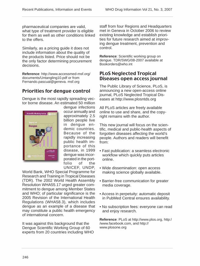
246
WHO Drug Information Vol 21, No. 3, 2007Recent Publications, Information and Events
pharmaceutical companies are valid,what type of treatment provider is eligiblefor them as well as other conditions linkedto the offers.
Similarly, as a pricing guide it does notinclude information about the quality ofthe products listed. Price should not bethe only factor determining procurementdecisions.
Reference: http://www.accessmed-msf.org/documents/Untangling10.pdf or fromFernando.pascual@geneva. msf.org
Priorities for dengue controlDengue is the most rapidly spreading vec-tor borne disease. An estimated 50 million
dengue infectionsoccur annually andapproximately 2.5billion people livein dengue en-demic countries.Because of therapidly increasingpublic health im-portance of thisdisease, in 1999dengue was incor-porated in the port-folio of theUNICEF, UNDP,
World Bank, WHO Special Programme forResearch and Training in Tropical Diseases(TDR). The 2002 World Health AssemblyResolution WHA55.17 urged greater com-mitment to dengue among Member Statesand WHO; of particular significance is the2005 Revision of the International HealthRegulations (WHA58.3), which includesdengue as an example of a disease thatmay constitute a public health emergencyof international concern.
It was against this background that theDengue Scientific Working Group of 60experts from 20 countries including WHO
staff from four Regions and Headquartersmet in Geneva in October 2006 to reviewexisting knowledge and establish priori-ties for future research aimed at improv-ing dengue treatment, prevention andcontrol.
Reference: Scientific working group ondengue. TDR/SWG/08-2007 available [email protected]
PLoS Neglected TropicalDiseases open access journalThe Public Library of Science, PLoS, isannouncing a new open-access onlinejournal, PLoS Neglected Tropical Dis-eases at http://www.plosntds.org
All PLoS articles are freely availableonline to use and share, and the copy-right remains with the author.
This new journal will focus on the scien-tific, medical and public-health aspects offorgotten diseases affecting the world’speople. Authors and readers will benefitfrom:
• Fast publication: a seamless electronicworkflow which quickly puts articlesonline.
• Wide dissemination: open accessmaking science globally available.
• Barrier-free communication for greatermedia coverage.
• Access in perpetuity: automatic depositin PubMed Central ensures availability.
• No subscription fees: everyone can readand enjoy research.
Reference: PLoS at http://www.plos.org, http://www.facebook.com, and http://www.plosone.org


![Pharmaco Latin Resep Key 01]](https://img.pdfslide.us/doc/110x75/55cf8cb45503462b138f1920/pharmaco-latin-resep-key-01.jpg)