Embed Size (px)
Citation preview
1
WHITE PAPER ON RETROSPECTIVE STUDIES IN ARCHIVE TISSUES
WORKSHOP
“Tissue-based Biomarkers for Advancement of Personalized Cancer Treatment”
Graz, 28th – 29th March 2014
Giorgio Stanta1,4, Kurt Zatloukal2, Peter Riegman3
1OECI – ESP, University of Trieste, Italy. 2BBMRI.at, Medical University of Graz, Austria. 3Department of Pathology, Erasmus Medical Center Rotterdam, The Netherlands. 4Giorgio Stanta, Medical Science Dep., University of Trieste, [email protected] c/o Ospedale di Cattinara, Strada di Fiume 447, 34149 Trieste, Italy.

2
Executive Summary On 28th and 29th March many European Organizations interested in cancer met in Graz to explore the possibilities to improve the quality of clinical research, to shorten the time needed for verification and validation of tissue-based biomarkers and to accelerate clinical application. The use of fixed and paraffin-embedded tissues (archive tissues - AT) – which are the residues of clinical activities stored in hospital pathology archives sometimes for decades and which represent the entire clinical variability – can be the solution. Retrospective study designs are suggested and different models of tissue and clinical information collections suitable for different purposes are analyzed. The recent improvements in tissue preservation during the pre-analytical phase and the capability to standardize the levels of molecular degradation do allow performing any kind of molecular analysis in AT. This way, thanks to the standardization of tissue collections and analysis methods, diagnostic and research capabilities are taken to a higher level. Importantly, method standardization needs standard operating procedures and proper controls to obtain the high level of reproducibility that is still missing in most of today’s studies, not only in diagnostics but also in clinical research. Research in primary and metastatic tumor tissues is necessary to better establish therapy target and intrinsic and acquired resistance predictive biomarkers. It is also crucial to validate any new diagnostic approach like liquid biopsies. This first document highlights the necessity to develop organized networks for archive tissue biobanking and to improve the quality of clinical studies. Established working groups and a technical platform of reference are necessary to perform multicentric European projects and to obtain reproducible and faster results for patients. This can be done with a collaboration among different European Organizations interested in tissue molecular analysis and clinical research and with the help of Patients’ Associations. Table of Contents: 1-Introduction …………………………………………………………………………………………………………………………… 3 Box 1: Participating Organizations…………………………………………………………………………………………….. 5 Box 2: Why retrospective survival studies? ………………………………………………………………………………. 6 2-Workshop preliminary evidence …………………………………………………………………………………………… 6 Box 3: Why archive tissues? ……………………………………………………………………………………………………... 7 3-Characteristics and necessities for improvement ………………………………………………………………….. 7 Box 4: How to organize the network? ………………………………………………………………………………………. 8 4-Design of retrospective studies ……………………………………………………………………………………………… 8 5-Models for cases and tissues collection …………………………………………………………………………………. 9 6-Pre-analytics and degradation standardization ………………………………………………………………………. 9 7-Heterogeneity and standardization procedures for microdissection …………………………………….. 10 8-Standardization of molecular analysis …………………………………………………………………………………... 10 Box 5: How to obtain reproducibility in clinical research? ……………………………………………………….. 11 Box 6: How to improve it? ……………………………………………………………………………………………………….. 12 Box 7: Why including AT research organization in a European infrastructure?………………………….. 12 Box 8: Conclusions …………………………………………………………………………………………………………………… 12 9-References ……………………………………………………………………………………………………………………………. 13
3
1-Introduction
Many recent papers show that results reported in medical scientific literature cannot be reproduced. Important institutions and scientific committees believe that a large part of many clinical biomarker research studies are “deemed to be of fair to marginal quality” and are not sufficient to make serious decisions on diagnostic application (EGAPP Working Group, 2013; Ioannidis, 2005). Furthermore, many researchers complain that today it takes too long to develop diagnostic and predictive biomarkers and that the procedure is too complex, which damages patients’ opportunities (Blanke et al., 2011). Prospective approaches in clinical trials are usually very expensive and the follow-up is too short. On the other hand retrospective studies called “convenience studies” are mostly not reproducible for many reasons related to the choice of patients, the design of the studies and technical performance (Simon et al., 2009).
We should consider that in the pathology archives of hospitals huge collections of fixed and paraffin- embedded tissues are stored. These archives are kept as part of the diagnostic documentation and require storage for technical and juridical purposes (Bevilacqua et al. 2010). Such archive tissues (AT) are related to clinical records and follow-up information and can be utilized for medical research under further use. Those tissues represent the most complete coverage of clinical diversity of human diseases available. It is estimated that every year in Europe 100 million new tissue samples are collected and stored for 10 years or longer in most European countries. In some academic institutions they are even preserved without any time limit. European groups (IMPACTS, SPIDIA, EurocanPlatform) studied the process and therewith developed methods and circumstances that enable the use of AT for any type of molecular analysis in the best and most reproducible way. This enormously enlarges not only molecular diagnostic capabilities, but also medical research skills. Nowadays we can perform most types of molecular analysis in such tissues and to this purpose European working groups focusing on pre-analytical conditions, tissue heterogeneity, DNA and RNA extraction standardization, Next Generation Sequencing, proteomics of ATs have been set up.
We can now improve the quality of retrospective studies in ATs with population based study designs and protocols similar to prospective studies, as well as with standardization of tissue conditions and reproducible analytical methods. ATs can be readily used for affordable clinical research, verification and, in some cases, even validation of clinical biomarkers. These Retrospective Survival Studies (RSS) will reduce the time lapse for effective clinical application of predictive and prognostic biomarkers, which will allow lower costs and will enable to prepare well-oriented and efficient prospective trials.
Research in primary and metastatic tumor tissues must be compared with any type of developing clinical platform to validate any new diagnostic approach like liquid biopsies. It is also necessary to better establish new generation therapy targets and biomarkers to monitor intrinsic and acquired resistance to therapy. The new information acquired from the clinical level can then be used on a basic research level to confirm biological functions with an improvement in translational and reverse translational research. Even more, with NGS procedures the information at the DNA level in cancer is almost completed, but we need now to evaluate it at the functional level to better understand biology and to better classify the driving impact of the alterations. Coding and not coding RNA and protein analyses in tissues are almost at the starting point of their exploration.

4
Analysis of therapy outcomes is today one of the most compelling necessities. This type of analysis is usually performed through epidemiological procedures often based on cancer registries data or other kinds of retrospective studies. ATs can be used to correlate those data with further bio-molecular information acquired through molecular analyses of patients’ tissues. In this way it is possible to stratify patients into homogeneous molecularly defined sub-groups with different outcomes and give further information on the predictive value of therapy and resistance biomarkers. This discussion is already ongoing within the EPAAC organization (EPAAC).
Biomarker and outcome studies in ATs can also be an effective European contribution to drafting a new taxonomy of diseases based on molecular evidence. This taxonomy was recently proposed in a strategy document on precision medicine by the US National Research Council and will be developed in the next few decades. For this reason, a substantial contribution on frequency detection of molecular alterations will be needed not only at the genetic, but also at the functional level (National Research Council, 2011).
All those activities will require collaboration between basic researchers, pathologists and oncologists with a holistic effect in developing more comprehensive health institutions and in setting common goals shared by stakeholders such as patients and industries. At the European level, this will result in better dissemination of knowledge in molecular medicine with better training for new generations of researchers and clinicians.
The first general discussion on this issue was held in Graz on 28th and 29th of March 2014 with the joint participation of most European Organizations interested in this kind of clinical research development and with Stakeholder Associations. The outcome of this discussion should lay the foundation for this white paper on the use of archive tissues to develop tissue-based biomarkers for the advancement of personalized medicine.
5
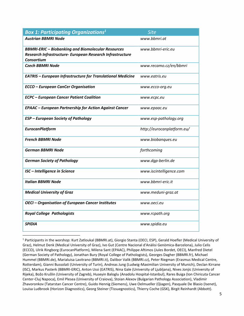
Box 1: Participating Organizations1 Site Austrian BBMRI Node www.bbmri.at
BBMRI-ERIC – Biobanking and Biomolecular Resources Research Infrastructure- European Research Infrastructure Consortium
www.bbmri-eric.eu
Czech BBMRI Node www.recamo.cz/en/bbmri
EATRIS – European Infrastructure for Translational Medicine www.eatris.eu
ECCO – European CanCer Organisation www.ecco-org.eu
ECPC – European Cancer Patient Coalition www.ecpc.eu
EPAAC – European Partnership for Action Against Cancer www.epaac.eu
ESP – European Society of Pathology www.esp-pathology.org
EurocanPlatform http://eurocanplatform.eu/
French BBMRI Node www.biobanques.eu
German BBMRI Node forthcoming
German Society of Pathology www.dgp-berlin.de
ISC – Intelligence in Science www.iscintelligence.com
Italian BBMRI Node www.bbmri-eric.it
Medical University of Graz www.meduni-graz.at
OECI – Organisation of European Cancer Institutes www.oeci.eu
Royal College Pathologists www.rcpath.org
SPIDIA www.spidia.eu
1 Participants in the worshop: Kurt Zatloukal (BBMRI.at), Giorgio Stanta (OECI, ESP), Gerald Hoefler (Medical University of Graz), Helmut Denk (Medical University of Graz), Ivo Gut (Centre Nacional d'Anàlisi Genòmica-Barcelona), Julio Celis (ECCO), Ulrik Ringborg (EurocanPlatform), Milena Sant (EPAAC), Philippe Aftimos (Jules Bordet, OECI), Manfred Dietel (German Society of Pathology), Jonathan Bury (Royal College of Pathologists), Georges Dagher (BBMRI.fr), Michael Hummel (BBMRI.de), Marialuisa Lavitrano (BBMRI.it), Dalibor Valik (BBMRI.cz), Peter Riegman (Erasmus Medical Centre, Rotterdam), Gianni Bussolati (University of Turin), Andreas Jung (Ludwig-Maximilian University of Munich), Declan Kirrane (ISC), Markus Pasterk (BBMRI-ERIC), Anton Ussi (EATRIS), Nina Gale (University of Ljubljana), Nives Jonjic (University of Rijeka), Božo Krušlin (University of Zagreb), Huseyin Baloglu (Anadolu Hospital-Istanbul), Rares Buiga (Ion Chiricuta Cancer Center-Cluj Napoca), Emil Plesea (University of Craiova), Stoian Alexov (Bulgarian Pathology Association), Vladimir Zhavoronkov (Tatarstan Cancer Centre), Guido Hennig (Siemens), Uwe Oelmueller (Qiagen), Pasquale De Blasio (Isenet), Louisa Ludbrook (Horizon Diagnostics), Georg Steiner (Tissuegnostics), Thierry Coche (GSK), Birgit Reinhardt (Abbott).

6
Box 2: Why retrospective survival studies? -Clinical research today can and should become an undistinguishable part of applied medicine strictly related to medical excellence. A large part of these studies can be easily performed with a retrospective type design. -Reproducibility of clinical research can be increased by better organization and standardization of retrospective clinical studies. -Retrospective survival studies can accelerate the clinical use of important biomarkers. -These studies can also be preparative to obtain better oriented prospective studies.
2-Workshop preliminary evidence We are increasingly aware that AT samples are a key resource for the advancement of medicine and in particular of personalized medicine. This is how medicine has developed since Virchow in the 19th
century. Research carried out on this material is a direct continuum with the developing experience of physicians in everyday practical clinics and it is part of the continuous process to develop medicine excellence. Clinical research should be considered as part of today’s medicine and absolutely not separable from it. European Organizations and stakeholders should support such process which in today’s complexity cannot r e l y m e r e l y o n individual researchers but should be better organized through extensive cooperation among basic researchers, oncologists, pathologists, biotechnologists, patients and industry. A European network for “biobanking” of those tissues generated in medical diagnosis should be made available for medical research with the direct and effective collaboration of pathologists, the custodians of those clinical samples. On the other hand, pathologists as well as other stakeholders like clinical researchers should not consider these tissues as for their personal use only, but as an important resource for the entire medical community with the same attitude of solidarity and altruism as demonstrated by patients who made their samples available for research. This network should be one of the primary goals of European scientific organizations and infrastructures like BBMRI-ERIC, and it requires a European strategy to harmonize ethical, legal and regulatory issues with the important contribution of European patients associations. ATs are used in morphological and molecular diagnostics, as well as in clinical biomarker development and commercial kits. For such reasons industry is an important counterpart for any cooperation and should be involved in this strategy. Many companies presented their interests and their solutions for this kind of clinical research in archive tissues. Organizations like the European Society of Pathology (ESP) and the Organisation of European Cancer Institutes (OECI) are conducting multicentric studies to improve technical and preservation characteristics in ATs with working groups open to further collaboration. Common standard operating procedures and quality controls will also be developed not only for diagnostics but also to be compelling for clinical research. CEN (the European Committee for Standardization) is developing technical specifications to ISO 15189 which might become instrumental. Several important European/global initiatives should work jointly with biological and medical science research infrastructures of the roadmap of the European Strategy Forum for Research Infrastructures, such as BBMRI-ERIC, EATRIS, ECRIN, ELIXIR. Along with the collaboration of ESP, OECI, ESMO, ECCO, EuroCanPlatform, EPAAC, SPIDIA and other organizations, it should be possible to accelerate the process to develop clinical application derived from new scientific knowledge not only for the benefit

7
of future patients but also for today’s patients, increasing their life expectancy as well as their quality of life.
Box 3: Why archive tissues? -Pathology diagnosis is performed on tissues that are formalin fixed, paraffin-embedded and then stored in archives. -They are available for almost every patient for clinical research and diagnostics. -They represent the major collection of human tissues with the entire clinical heterogeneity and with available clinical and follow-up information. -Today we can perform most kinds of molecular analyses in those tissues. -Cases are easy to recruit and the study can be conducted at a lower cost. -The time period to conclude the research project is shorter because the tissues have already been collected. -After carrying out an experiment, the results can be validated immediately and used for routine patient care shortly after. -The same tissues can be used instead of fresh collected tissues also for prospective clinical trials.
3-Characteristics and necessities for improvement Prospective approaches in clinical trials are usually very expensive, therefore the time devoted to follow-up is often very short. One of the longest, most demanding and most costly phases of prospective clinical trials is recruitment of patients. This is the reason why, when possible, retrospective case studies should be the first choice due to easy inclusion of patients and tissues, and low organizational commitment. Moreover, this can be done in a very short time. Even the bioethical complexity is lower because patients have already given their consent for ablation and examination of those tissues, as such tissues are residues of p re v iou s surgical in tervent ion s and clinical diagnostics . As already mentioned, when doctors involved in a diagnostic or therapeutic process - like
surgeons, oncologists or pathologists – use clinical material to extend their knowledge in the specific
disease affecting their patients, this is part of a process which is strictly related to medicine itself and it should be accepted as part of the increasing experience and the developing excellence of health care professionals. Of course, the cost of this kind of case studies is very low because the material is already available. At the same time, accessibility to clinical records and to the follow-up data stored in many institutions and geographical areas is also of help to develop the studies. The involvement of pathologists is also related to their function as doctors participating in the clinical process and, therefore, able to retrieve clinical and follow-up information. They are also the firewall of patient privacy because they are bound by professional secrecy, whose rules are stricter than any other general “privacy” law.
Improved analytical methods in this kind of tissues make this material increasingly accessible to perform expression functional studies. Today we know that next generation sequencing can be performed on archive tissues with results that are very similar to those obtained in fresh frozen samples (Schweiger, 2009). These tissues also have some characteristics that give advantages as compared to fresh frozen tissues: it is easier to have a high level of morphological analysis and all the diagnostic antibodies work in those tissues in a safe and reproducible way, making results easier to be interpreted. Microdissection can be better performed with mechanical, laser or tissue array methods, this would yield well-localized and defined samples to analyze with a high level of reproducibility. Those

8
aspects are really important if we consider the relevance of the well-known problem of tumour tissue heterogeneity.
Box 4: How to organize the network? -The network is set up as a virtual network of pathology archives for clinical research. -Participation in the network and in the projects is voluntary and collaborative (these materials are residues from clinical procedures with specific requirements). -Clinical and follow-up information can be directly collected from pathologists because they also deal with the diagnostic procedures. -Privacy is guaranteed by the pathologists’ professional secrecy, who have the duty to anonymize the cases. -There can be different network models for different strategies. -The network may become associated with BBMRI-ERIC for sustainable development and operation.
4-Design of retrospective studies There can be many types of retrospective studies with different design characteristics and in which ATs can be the biological material of interest. Examples of such studies are: -Studies related to verification of unusual clinical cases. This means, for example, that patients might not react to a treatment despite the positive presence of specific predictive biomarkers, or the opposite could happen. -Clinical research with the use of large case studies to subdivide patients into homogeneous subgroups with specific molecular characteristics that can be useful to improve treatments. -Verification projects performed to verify rapidly biomarkers or fingerprints that were proposed by preliminary studies. -Validation projects in which already verified biomarkers can be validated for clinical use with an established analytical and clinical validity and also demonstrated clinical utility using different patient populations. -On ATs continuous performance evaluation could be done in the clinical use of predictive and prognostic biomarkers, and in monitoring treatment outcomes. Looking at the design of the studies, it is possible to carry out studies based on populations belonging to different geographical areas. There are indeed many European regions in which health treatment standards are very high, medical databases are very well developed, it is possible to have careful follow-up information and the level of patient migration is low. This situation is quite peculiar in Europe: it applies to specific geographical areas in large countries or to entire small countries like Slovenia and the Netherlands, whose situation was presented during the workshop. The study design must be well-defined before starting the project and collection and inclusion of cases and tissues must be performed without prior collection of follow-up data and outcome definition. This gives to this kind of studies characteristics that are similar to those of prospective studies. Even molecular analysis should be performed before cases are connected with specific outcomes. Molecular analysis should be done with well-defined standard operating procedures and with internal quality controls that must already be established during the study design. In the final analysis of the project, we should also consider the loss of cases due to the loss of patients during the follow-up, because such information is connected with the molecular results only at the end of the study. As already

9
defined, the positive results must be verified in different populations and the validation process can also be developed with retrospective material, as we will report in the following paragraph. 5-Models for cases and tissue collection Many models for tissue collection can be devised and some of those were preliminarily proposed during the workshop. One of those models had already been discussed in the past meetings held by EPAAC (European Partnership for Action Against Cancer). In this case the outcomes of cancer treatments are analyzed by using tumor registry data, and new high-resolution studies were proposed adding more detailed and specific characteristics. To perform those studies, in specific geographical areas it is possible to address the hospitals that treated the patients to collect tissues from the pathology archives. This will be especially useful to define groups that did not respond to therapy and to find the specific molecular characteristics of those subgroups. This kind of design can be very useful to increase knowledge of intrinsic and acquired resistance to therapy. A second model can be developed in a large clinical network like the Organisation of European Cancer Institutes (OECI), in which biomarker validation studies are possible. In retrospective designs we should consider developing equal parallel projects in two or three big cancer institutions at the same time (for example, retrospective validation of a specific biomarker in a large unselected number of cases for a defined period of time with well-established tumor stage and treatment characteristics). Validation comes from the positive comparison of the study results. The third model can be developed by the Biobanking and Biomolecular Resources Research Infrastructure (BBMRI-ERIC) and can be used for retrospective clinical research in large case studies or for rare entities. The idea is to organize virtual networks of pathology archives from hospitals that have well-developed computerized databases, from which it is possible to collect large case studies of even rare tumor subgroups. This must be done with the direct collaboration of the pathologists who are in charge of the tissues and participated in the diagnostic process. In this way clinical and follow-up data can be collected and precise micro-dissection of tissues can be performed. The fourth model presented is called “area model”. In this case, research is developed in specific geographical areas whose health care system is of high level and migration of resident patients is low. Sometimes those areas have tumor registries and well-developed clinical databases. 6-Pre-analytics and degradation standardization One of the major problems in archive tissues, especially for diagnostics, is to have standardized conditions for specimens and biological macromolecules preservation from human tissues collected for diagnostic and surgical purposes. This is a basic requirement for clinical research reproducibility but also for molecular diagnostics reliability. Much progress has recently been made through the studies first conducted by the IMPACTS group (www.impactsnetwork.eu), by the SPIDIA project (www.spidia.eu) and by the ESP group (European Society of Pathology), who recently started to develop further activities in this field. Recommendations for specific rules will also be given as common quality standards by technical specifications to ISO 15189 of CEN (www.cen.eu). It is interesting to observe how some technical developments are already improving the quality of tissues in many large European and North American hospitals, with the use of vacuum storage at low temperatures for transport and conservation of tissues before pathology sampling. On the other hand, the degradation level of the macromolecules can be standardized during nucleic acid or protein analysis. For DNA analysis, in most cases the quality of DNA extracted from FFPE is
10
assessed by testing the maximum size of an amplifiable PCR product, either in single or multiple runs. Generally, the size distribution of DNA obtained from FFPE is an adequate predictor for the success of most downstream assays. Nonetheless, quality in terms of maximum length should be equal or superior to the length of the performed test. The same can be done for RNA after reverse transcription (Bonin, 2013; Kashofer, 2013). As for extracted proteins, their amount and quantity could be detected by using serial dilutions detected with specific antibodies (Gundisch et al., 2012). 7-Heterogeneity and standardization procedures for microdissection One of the emerging problems in tissue analysis, and especially in cancer, is heterogeneity. We have different types of heterogeneity at the clinical level, morphological/histological level and molecular level. Recognizing such characteristics is of paramount importance to obtain reproducible results in tissue molecular analysis after accurate micro-dissection. Clinical heterogeneity is related to the choice and design of studies. It is the only characteristic that is usually correctly performed in those studies, mostly depending on the design of the study. Morphological/histological heterogeneity is linked to general tissue characteristics like fibrosis, inflammatory infiltration, necrosis, normal tissue residues etc. A second type of tissue heterogeneity refers to the functional area of the analyzed tumor chosen for the analysis. It was very well shown that the expression signature of the infiltrative border of cancer is different from that of the central part of the tumor (Hlubek et al, 2007). Heterogeneity could also refer to the type of tumor showing different histological patterns possibly corresponding to different molecular processes. Micro-dissection can be especially useful to overcome all this morphological heterogeneity by choosing carefully the area of the tumor, avoiding fibrosis, necrosis and inflammation. Recently we have proposed to standardize also the functional area considered for analysis. The border of tumors with the invasion front is the most aggressive part and we suggest using it for research and clinical diagnostics, as a first step for further standardization. Molecular heterogeneity is the most complex one and we are still far from understanding its high complexity. We know that this can be related to genetic clonal evolution or epigenetic clonal development or, at the functional level, wide phenomena of phenotypic plasticity, or heterotypic interaction with autocrine, paracrine phenomena. Research is still being carried out and the use of new technologies like next generation sequencing can help us to better understand the clinical meaning of this heterogeneity. We already know that this can be for example the basis of secondary resistance to cancer treatment. Within the European Society of Pathology an inter working group (groups performing clinical research in different type of tumours) study is in development, to enhance knowledge and strategies for cancer heterogeneity. 8-Standardization of molecular analysis If we only consider extraction methods for nucleic acids, we should take into consideration a very high number of variables, from the type of dewaxing of tissues to protein digestion and nucleic acid extraction and isolation. Each method has very different characteristics and a simple change in one of the buffers can modify quality and quantity of the nucleic acids obtained (Bonin and Stanta, 2013). Different extraction methods can be more suitable for further specific analysis than others (Kashofer, 2013). Comparison of the quantity of nucleic acids extracted by different experienced laboratories can vary enormously especially for RNA (Bonin et al., 2010), even if we use the same commercial kit. This is

11
mostly related to the lack of strict standard operating procedures and of adequate controls. Besides the quantity of the extracted molecules, we must take into consideration the level of degradation that we always find in this kind of tissues. Quality assessment of DNA, RNA and proteins is crucial to obtain reproducible analysis results. The basic criterion for nucleic acids is their level of amplifiability, which is the maximum length that can be amplified by PCR. Many quantitative and qualitative methods used today are not mirroring the real state of the nucleic acid availability for analysis (Bonin and Stanta, 2013; Kashofer, 2013). Up to now, application of molecular tests on DNA extracts from FFPE has become quite frequent, mostly because DNA is better conserved than RNA in FFPE, and secondarily because most tests are qualitative and not quantitative. Even though in the past decade a huge amount of studies was conducted on expression profiles and signature of cancer subgroups, only a few diagnostic tests were developed. In RNA, although the selected genes in a specific assay may be the same, differences in specimen preservation, along with differences in RNA extraction, reverse transcription (Nardon, 2009), amplification and analysis, may produce highly altered ratios of detectable genes and thereby lead to poor reproducible qRT-PCR results (Bonin and Stanta, 2013). The possibility to use an algorithm, which could correct and standardize the threshold cycle value due to molecules degradation, will be a great advancement towards molecular “functional” pathology. Nowadays, some researchers are investigating methods to improve normalization thus making quantification more reliable. There is the necessity for a technical platform to increase abilities in clinical research with standardized and reproducible methods. The IMPACTS group, together with ESP, OECI and other organizations, is already participating in the development of working groups to standardize nucleic acid and protein extraction from archive tissues and their analysis with specific operating procedures and quality controls in tissues selected according to their heterogeneous composition. These working groups are:
-DNA and RNA extraction SOPs and IQC rules in AT (OECI, ESP) -NGS in diagnostics and clinical research (ESP, OECI) -Pre-analytical conditions in tissues (ESP, CEN) -Proteomics SOPs in AT (OECI, ESP) -Heterogeneity (inter-WGs of ESP)
These groups will allow achieving a high level of confidence in the clinical studies performed in archive tissues for faster application of the results to patients.
Box 5: How to obtain reproducibility in clinical research?
-Well defined research designs with prospective like characteristics and choice of suitable outcomes.
-Technical support with standard operating procedures and internal quality controls also in research with the development of a technical platform.
-Dissemination of information through courses and training.
12
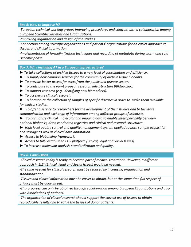
Box 6: How to improve it?
-European technical working groups improving procedures and controls with a collaboration among European Scientific Societies and Organizations.
-Improving organization and design of the studies.
-Connection among scientific organizations and patients’ organizations for an easier approach to tissues and clinical information.
-Implementation of formalin fixation techniques and recording of metadata during warm and cold ischemic phase.
Box 7: Why including AT in a European infrastructure?
► To take collections of archive tissues to a new level of coordination and efficiency. ► To supply new common services for the community of archive tissue biobanks. ► To provide better access for users from the public and private sector. ► To contribute to the pan-European research infrastructure BBMRI-ERIC. ► To support research (e.g. identifying new biomarkers). ► To accelerate clinical research. ► To harmonize the collection of samples of specific diseases in order to make them available for clinical studies. ► To offer a service to researchers for the development of their studies and to facilitate communication and exchange of information among different groups of scientists. ► To harmonize clinical, molecular and imaging data to enable interoperability between national biobanks, disease oriented registries and clinical and research structures. ► High level quality control and quality management system applied to both sample acquisition and storage as well as clinical data annotation. ► Access to biobanking framework. ► Access to fully established ELSI platform (Ethical, legal and Social Issues). ► To increase molecular analysis standardization and quality.
Box 8: Conclusions
-Clinical research today is ready to become part of medical treatment. However, a different approach in ELSI (Ethical, legal and Social Issues) would be needed.
-The time needed for clinical research must be reduced by increasing organization and standardization.
-Tissues and clinical information must be easier to obtain, but at the same time full respect of privacy must be guaranteed.
-This progress can only be obtained through collaboration among European Organizations and also with Associations of patients.
-The organization of clinical research should support the correct use of tissues to obtain reproducible results and to value the tissues of donor patients.

13
9-References -Bevilacqua G., Bosman F., Dassesse T., Höfler H., Janin A., Langer R., Larsimont D., Morente M.M., Riegman P., Schirmacher P., Stanta G., Zatloukal K., Caboux E., Hainaut P. (2010) The role of the pathologist in tissue banking: European Consensus Expert Group Report. Virchows Arch 456, 449-54. -Blanke C.D., Goldberg R.M., Grothey A., Mooney M., Roach N., Saltz L.B., Welch J.J., Wood W.A., Meropol N.J., Force N.G.S.C.C.C.T. (2011). KRAS and colorectal cancer: ethical and pragmatic issues in effecting real-time change in oncology clinical trials and practice. The oncologist 16, 1061-1068. -Bonin S., Hlubek F., Benhattar J., Denkert C., Dietel M., Fernandez P.L., Hofler G., Kothmaier H., Kruslin B., Mazzanti C.M. et al. (2010). Multicentre validation study of nucleic acids extraction from FFPE tissues. Virchows Arch 457, 309-317. -Bonin S., Stanta, G. (2013). Nucleic acid extraction methods from fixed and paraffin-embedded tissues in cancer diagnostics. Expert review of molecular diagnostics 13, 271-282. -EGAAP, Evaluation of Genomic Applications and Prevention Working Group (2013). Recommendations from the EGAPP Working Group: can testing of tumor tissue for mutations in EGFR pathway downstream effector genes in patients with metastatic colorectal cancer improve health outcomes by guiding decisions regarding anti-EGFR therapy? Genetics in medicine : Official Journal of the American College of Medical Genetics 15, 517-527. -Gundisch S., Hauck S., Sarioglu H., Schott C., Viertler C., Kap M., Schuster T., Reischauer B., Rosenberg R., Verhoef C., Mischinger H.J., Riegman P., Zatloukal K., Becker K.F. (2012). Variability of protein and phosphoprotein levels in clinical tissue specimens during the preanalytical phase. J Proteome Res. 11, 5748-62. -Hlubek F., Brabletz T., Budczies J., Pfeiffer S., Jung A., Kirchner T. (2007). Heterogeneous expression of Wnt/beta-catenin target genes within colorectal cancer. Int J Cancer 1, 1941-8. -Ioannidis J.P. (2005). Why most published research findings are false. PLoS medicine 2, e124. -Kashofer K., Viertler C., Pichler M., Zatloukal K. (2013) Quality control of RNA preservation and
extraction from paraffin-embedded tissue: implications for RT-PCR and microarray analysis. PLoS One 8, e70714 -Nardon E., Donada M., Bonin S., Dotti I., Stanta G. (2009). Higher random oligos concentration improves reverse transcription yield of cDNA from bioptic tissues and quantitative RT-PCR reliability. Exp Mol Pathol 87, 146–151. -National Research Council (2011). Toward Precision Medicine: Building a Knowledge Network for Biomedical Research and a New Taxonomy of Disease. The National Academies Press. -Simon R.M., Paik S., Hayes D.F. (2009). Use of archived specimens in evaluation of prognostic and predictive biomarkers. Journal of the National Cancer Institute 101, 1446-1452. -Schweiger M.R., Kerick M., Timmermann B., Albrecht M.W., Borodina T., Parkhomchuk D., Zatloukal K., Lehrach H. (2009) Genome-wide massively parallel sequencing of formaldehyde fixed-paraffin embedded (FFPE) tumor tissues for copy-number- and mutation-analysis. PLoS One 4, e5548. Websites BBMRI-ERIC (Biobanking and Biomolecular Resources Research Infrastructure- European Research Infrastructure Consortium – www.bbmri-eric.eu) CEN (European Committee for Standardization – www.cen.eu) EATRIS (European Infrastructure for Translational Medicine – www.eatris.eu) ECCO (European CanCer Organisation – www.ecco-org.eu)

14
ECPC (European Cancer Patients Coalition – www.ecpc.eu) ECRIN (European Clinical Research and Infrastructure Network – www.ecrin.org) ELIXIR (www.elixir-europe.org) EPAAC (European Partnership Action Against Cancer – www.epaac.eu) ESMO (European Society of Medical Oncology – www.esmo.org), ESP (European Society of Pathology – www.esp-pathology.org) – Molecular Pathology Working Group and in collaboration with the Organisation of European Cancer Institutes. EuroCanPlatform (http://eurocanplatform.eu) IMPACTS Group (Archive Tissues: Improving Molecular Medicine Research and Clinical Practice – www.impactsnetwork.eu) ISC (Intelligence in Science – www.iscintelligence.com) OECI (Organisation of European Cancer Institutes OECI- www.oeci.eu) – Biobanking and Molecular Pathobiology Working Group. SPIDIA (Standardisation and improvement of generic pre-analytical tools and procedures for in-vitro diagnostics – www.spidia.eu)