Embed Size (px)
Citation preview

Neurobiology of Aging 31 (2010) 244–256
White matter pathology isolates the hippocampal formation inAlzheimer’s disease
D.H. Salat a,b,d,!, D.S. Tuch a,b, A.J.W. van der Kouwe a,b, D.N. Greve a,b,V. Pappu a,c, S.Y. Lee a,c, N.D. Hevelone a,c, A.K. Zaleta a,c,J.H. Growdon c, S. Corkin a,d, B. Fischl a,b, H.D. Rosas a,c
a MGH/MIT/HMS Athinoula A. Martinos Center for Biomedical Imaging, Charlestown, MA, United Statesb Department of Radiology, Massachusetts General Hospital, Boston, MA, United Statesc Department of Neurology, Massachusetts General Hospital, Boston, MA, United States
d Department of Brain and Cognitive Sciences, Massachusetts Institute of Technology, Cambridge, MA, United States
Received 17 October 2007; received in revised form 17 March 2008; accepted 22 March 2008Available online 5 May 2008
Abstract
Prior work has demonstrated that the memory dysfunction of Alzheimer’s disease (AD) is accompanied by marked cortical pathology inmedial temporal lobe (MTL) gray matter. In contrast, changes in white matter (WM) of pathways associated with the MTL have rarely beenstudied. We used diffusion tensor imaging (DTI) to examine regional patterns of WM tissue changes in individuals with AD. Alterationsof diffusion properties with AD were found in several regions including parahippocampal WM, and in regions with direct and secondaryconnections to the MTL. A portion of the changes measured, including effects in the parahippocampal WM, were independent of gray matterdegeneration as measured by hippocampal volume. Examination of regional changes in unique diffusion parameters including anisotropy andaxial and radial diffusivity demonstrated distinct zones of alterations, potentially stemming from differences in underlying pathology, with apotential myelin specific pathology in the parahippocampal WM. These results demonstrate that deterioration of neocortical connections tothe hippocampal formation results in part from the degeneration of critical MTL and associated fiber pathways.© 2008 Elsevier Inc. All rights reserved.
Keywords: Alzheimer; Aging; Diffusion tensor imaging; Fractional anisotropy; Diffusivity; MRI; White matter; Dementia; Hippocampus; Entorhinal; Memory;Axial diffusivity; Radial diffusivity; Tractography; Volume; Hyperintensities; T2; Myelin; Axon
1. Introduction
Careful histological examination of the brains of indi-viduals with Alzheimer’s disease (AD) has uncovered aprobable substrate for the memory impairment in this condi-tion. Specifically, layer-preferred degeneration in perirhinaland entorhinal cortices likely impedes the transfer of infor-mation from the neocortex to the hippocampus (Ball, 1978;Braak and Braak, 1991; Gomez-Isla et al., 1996; Hyman et
! Corresponding author at: MGH/MIT/HMS Athinoula A. Martinos Cen-ter for Biomedical Imaging, MGH Department of Radiology, Building 149,13th St., Mail Code 149 (2301), Charlestown, MA 02129-2060, UnitedStates. Tel.: +1 617 726 4704; fax: +1 617 726 7422.
E-mail address: [email protected] (D.H. Salat).
al., 1984, 1986; Van Hoesen et al., 1991), thereby degrad-ing the processing and storage of sensory input. Layer II ofthe entorhinal cortex shows profound alterations, includingsubstantial loss of neurons even in the early stages of AD(Gomez-Isla et al., 1996). The projection termination zone ofthese fibers in the dentate gyrus of the hippocampal formationis also marked by degenerative changes, effectively resultingin a ‘disconnection’ between association and limbic cortices(Hyman et al., 1984, 1986). Given these important patho-logic signatures, the majority of studies of mechanisms of ADsymptomology have focused on medial temporal lobe (MTL)cortical degeneration. Nevertheless, these prior findings alsoimplicate regional connectivity as a factor contributing tocognitive deterioration. Histological research demonstratesthat brain white matter (WM) also degenerates in AD (Brun
0197-4580/$ – see front matter © 2008 Elsevier Inc. All rights reserved.doi:10.1016/j.neurobiolaging.2008.03.013

D.H. Salat et al. / Neurobiology of Aging 31 (2010) 244–256 245
and Englund, 1986; Englund and Brun, 1990; Englund etal., 1988; Hyman et al., 1986). Brun and Englund (1986)reported a syndrome in 60% of AD patients of demyelina-tion and axonal and oligodendroglial loss with accompanyinggliosis in the deep WM that was independent from graymatter lesions. The authors suggested that the degenerationwas potentially due to comorbid factors such as hyperten-sion (Brun and Englund, 1986). WM disease, however, hasbeen reported at autopsy in individuals with pure AD with nocomponents of vascular brain disease (Sjobeck et al., 2006).Further, myelin staining is reduced in the perforant pathway,the main input fibers projecting neocortical information fromthe entorhinal cortex to the granule cells of the dentate gyrusin the hippocampal formation (Hyman et al., 1986). Thesefindings suggest that at least some of the WM changes in ADare not due simply to comorbid factors, but are likely associ-ated with AD pathological processes including MTL corticalpathology. The pathologic signatures spanning the perforantpathway, and the reduction in myelin integrity of this fascicle,underscores the potential influence of regional connectivityin the putative propagation of neurodegenerative events. Anopen question is whether such changes could be detected inpatients in vivo, and whether this principal of degeneration inanatomically connected regions extends beyond the findingsin the perforant pathway.
Neuroimaging studies have attempted to understand pat-terns and mechanisms of WM pathology in AD, and theclinical significance of such changes (de Leeuw et al., 2006),but the regional patterns and potential mechanisms of thisWM pathology are still unclear. Moreover, whether WMchanges are independent of classically described AD cor-tical pathology, such as hippocampal atrophy, is completelyunknown. Total and regional WM volume is reduced in AD(Fotenos et al., 2005; Jernigan et al., 1991; Salat et al.,1999a,b, 2001; Stout et al., 1996), and WM signal abnor-malities are associated with risk for cognitive decline (Auet al., 2006) and dementia (Prins et al., 2004), as well asan enhanced clinical syndrome in specific cognitive domains(Hirono et al., 2000). The use of WM signal abnormalitiesas a clinically relevant measure of WM pathology remainscontroversial because a number of studies report little conse-quence of this marker on clinical status (Mungas et al., 2005;Schmidt et al., 2002). Additionally, WM volume measuresare limited because of the need to define regionally identi-fiable borders using morphometric landmarks, a particularlydifficult task given the complex anatomy of WM and the lim-ited information provided about this anatomy from a standardstructural MR image.
Diffusion tensor imaging (DTI) has been applied exten-sively to understand the regional basis of tissue degenerationin a variety of clinical conditions including the study ofnormal aging (Moseley, 2002; Pfefferbaum et al., 2000,2005; Pfefferbaum and Sullivan, 2003; Salat et al., 2005a,b;Sullivan et al., 2001, 2006; Sullivan and Pfefferbaum, 2006).Two primary metrics of the diffusion properties within avoxel, termed diffusivity and fractional anisotropy (FA)
(Basser, 1995; Basser and Pierpaoli, 1996; Pierpaoli andBasser, 1996), have been commonly employed as indicesof tissue pathology. More recently, studies by Song andcolleagues utilizing animal models demonstrate that thediffusivity measure can be further subdivided in to axialand radial components, which could provide informationon axonal and myelin pathology selectively (Budde et al.,2007; Song et al., 2002, 2003). Rose and colleagues (Roseet al., 2000) demonstrated altered diffusion measures in thesplenium of the corpus callosum, the superior longitudinalfasciculus, cingulum, and internal capsule in patients withAD, and in parahippocampal, thalamic, and cingulate WMin individuals with mild cognitive impairment (Rose et al.,2006). Diffusion measures were related to indices of diseaseseverity and cognitive ability and the specific association withepisodic memory presents a potential clinical role for DTI toindex WM degeneration and track AD symptoms. Other stud-ies have found altered diffusion measures in patients withAD in the uncinate and inferior occipital fasciculi (Taokaet al., 2006), and in the corpus callosum and WM of thefrontal, temporal, and parietal lobes (Bozzali et al., 2002).Two studies in AD (Head et al., 2004; Medina et al., 2006)demonstrated generalized alterations in diffusion measures ofposterior lobar WM that differed from those seen in normalaging. Two recent studies provide preliminary investigationinto mechanisms of WM alteration in AD through the exami-nation of axial and radial diffusivity (Choi et al., 2005; Huanget al., 2007). These studies examined selected regions of inter-est in small participant samples (10 AD in the former, and6 AD in the latter study), and reached different conclusionswith one focusing on compromised myelin (Choi et al., 2005)and the other suggesting loss of axonal processes as a primarypathologic mechanism (Huang et al., 2007). These prior stud-ies provide important information about the regional patternsof AD pathology, yet questions remain about the whole brainpatterns of WM change in these various diffusion parametersin AD. Additionally, no prior study has examined how clas-sically described measures of pathology such as white mattersignal abnormalities and hippocampal atrophy contribute tothe changes measured.
The current study aimed to elucidate regional patterns ofalterations in diffusion parameters in AD through a com-prehensive, whole brain analysis of commonly and recentlydescribed DTI measures of tissue integrity. These analysesincluded the examination of anisotropy and axial and radialdiffusivity components, and whether changes in these dif-fusion parameters provide information beyond traditionalMRI measures of gray and WM degeneration. We usedrecently developed procedures in the FSL image analy-sis suite (http://www.fmrib.ox.ac.uk/fsl/) for interparticipantregistration, reducing potential confounds in spatial normal-ization. We additionally utilized tractography procedures todefine a path of interest (POI) in the native space of eachindividual to confirm voxel-based results. We find complexregional patterns of alterations in diffusion parameters withAD, with prominent changes in pathways associated with

246 D.H. Salat et al. / Neurobiology of Aging 31 (2010) 244–256
Table 1Participant demographics
OA AD
N 54 (45 F/9 M) 20 (16 F/4 M)Age 75.8 (5.6) 77.8 (4.9)MMSE (n = 26) 28.8 (1.2)a 20.0 (5.4)d
BDS (n = 49) 0.86 (1.0)b 12.9 (6.4)TICS (n = 22) 34.4 (2.0)c NACDR global (n = 20) NA 11 (.5)/7 (1)/2 (2)CDR sum of boxes (n = 20) NA 4.9 (2.9)
Data presented as mean and standard deviation where applicable. MMSE,Mini Mental State Examination; BDS, Blessed Dementia Scale; TICS,Telephone Interview of Cognitive Status; CDR, Clinical Dementia Rating(Hughes et al., 1982; Morris, 1997); Higher scores indicate better perfor-mance on MMSE (0–30) and TICS (0–39), whereas lower scores indicatebetter performance on BDS (0–37), global CDR (0–3), and CDR sum ofboxes (0–18).
a n = 20.b n = 29.c n = 22.d n = 6.
the hippocampal formation. These changes are beyond whatcan be explained by classically described AD pathology, andsuggest that multiple pathologies may disrupt the transfer ofneocortical information to limbic structures important for arange of cognitive processes.
2. Methods
2.1. Participants
Images were obtained for 74 participants (Table 1).Twenty patients with probable Alzheimer’s disease (meanage 77.8 ± 4.9 years) were recruited through the Mas-sachusetts General Hospital Memory Disorders Unit(MGH-MDU) and 54 non-demented older adults (OA, meanage 75.8 ± 5.6 years) through the Harvard CooperativeProgram on Aging (http://www.hebrewrehab.org/homeinstitute.cfm?id=90) and the Nurses’ Health Study(http://www.channing.harvard.edu/nhs/) at Harvard MedicalSchool and Brigham and Women’s Hospital. OA werescreened for dementia using one of the following mentalstatus examinations: the Mini Mental Status Exam (MMSE)(Folstein et al., 1975), the Blessed Dementia Scale (BDS)(Blessed et al., 1968; Stern et al., 1990), or the TelephoneInterview of Cognitive Status (TICS) (de Jager et al., 2003;Lipton et al., 2003). AD patients were assessed by theClinical Dementia Rating scale (CDR) (Hughes et al., 1982;Morris, 1997; Morris et al., 1997) which yields a calculatedglobal score (CDR rating) as well as a summated score ofindividual CDR domains (sum of boxes). All patients withAD were assessed by a memory disorders neurologist fromthe MGH-MDU. AD diagnoses as determined by CDR scorewere very mild to mild AD in 90% and moderate dementiain 10%. Participants were excluded if they had a historyof significant neurologic or psychiatric disorder (other
than AD), or serious cerebrovascular conditions. Groupswere matched for proportion of individuals with controlledelevated blood pressure.
2.2. DTI acquisition
Global and regional WM integrity was assessed using DTImeasures of FA and diffusivity (comprised of axial and radialcomponents (Budde et al., 2007; Song et al., 2002, 2003)),as well as through T2 image intensity measured throughthe B = 0 DTI volume. Image acquisition employed singleshot echo planar imaging with a twice-refocused spin echopulse sequence, optimized to minimize eddy current-inducedimage distortions (Reese et al., 2003) (Siemens Avanto;TR/TE = 7400/89 ms, b = 700 s mm"2, 256 mm # 256 mmFOV, 128 # 128 matrix, 2 mm slice thickness with 0-mmgap, 10 T2 + 60 DWI, total acquisition time 8 min, 38 s).We acquired 64 slices in the AC-PC plane. The 60 diffu-sion weighted directions were obtained using the electrostaticshell method (Jones et al., 1999), providing a high signal-to-noise diffusion volume. The diffusion tensor was calculatedon a voxel-by-voxel basis with conventional reconstructionmethods (Basser et al., 1994) using tools developed at theMartinos Center at MGH.
2.3. DTI preprocessing and analysis: motion and eddycurrent correction
Image preprocessing was performed as described in ourprevious work (Salat et al., 2005a,b). Diffusion volumeswere motion corrected and averaged using FLIRT (FMRIB’sLinear Image Registration Tool; http://www.fmrib.ox.ac.uk/analysis/research/flirt/) (Jenkinson et al., 2002) with mutualinformation cost function to register each direction to theminimally eddy current distorted T2-weighted DTI volumethat had no diffusion weighting.
2.4. Fractional anisotropy (FA) and diffusivity mapcalculation
The primary measures acquired from the DTI data weretwo common scalar metrics describing the WM microstruc-ture. FA, which is dependent on the orientational coherenceof the diffusion compartments within a voxel (Pierpaoli andBasser, 1996), was considered the primary metric of inter-est given the use of this parameter in a number of recentstudies of tissue deterioration. FA was calculated using thestandard formula defined previously (Basser, 1997). Diffusiv-ity is a scalar measure of the total amount of diffusion withina voxel calculated as described in previous work (Basserand Pierpaoli, 1996). We additionally examined measures ofaxial (!1) and radial [(!2 + !3)/2] diffusivity as described inprior work (Budde et al., 2007; Song et al., 2002, 2003). T2images were obtained using the exact parameters as the diffu-sion sensitive images except without any diffusion weighting.The T2 images were analyzed to determine whether changes

D.H. Salat et al. / Neurobiology of Aging 31 (2010) 244–256 247
other than those in tissue microstructure contributed to theobserved effects because T2 differences would reveal tech-nical artifact such as image registration or atrophy, as well aslarge scale signal changes such as WM signal abnormalities(e.g. hyperintensities).
2.5. Nonlinear registration and tract-based spatialstatistics (TBSS) (Rueckert et al., 1999; Smith et al.,2006; Smith et al., 2004)
Voxelwise statistical analysis of the FA data was car-ried out using TBSS (Tract-Based Spatial Statistics (Smithet al., 2006)), part of FSL (Smith et al., 2004). All par-ticipants’ diffusion data were first aligned into a commonspace using the nonlinear registration IRTK (Rueckert et al.,1999) (http://www.doc.ic.ac.uk/$dr/software). Next, a meanFA image was created across all participants, and this meanimage was thinned to create a mean FA skeleton which rep-resents the centers of all tracts common to the group. Eachparticipant’s aligned FA data were then projected onto thisskeleton and the resulting data fed into voxelwise groupstatistics. Data along the skeleton were smoothed utilizing ananatomical constraint to limit the smoothing to neighboringdata within adjacent voxels along the skeleton. All analyseswere masked to only display regions with FA values of >0.2to avoid examination of regions that are likely comprised ofmultiple tissue types or fiber orientations. The exact trans-formations derived for the anisotropy maps were applied tothe T2 and diffusivity volumes for matched processing of allimage volumes. Statistical maps were dilated from the TBSSskeleton for visualization purposes.
2.6. Hippocampal, WM, and WM signal abnormalityvolume measurements
Hippocampal, WM, and WM signal abnormality volumemeasurements were calculated through an automated pro-cedure, using probabilistic information estimated from amanually labeled training set as described in our prior work(Fischl et al., 2002; Han et al., 2006; Walhovd et al., 2005).All volumetric measures were corrected as a percentage ofintracranial volume (ICV).
2.7. POI analyses
We created highly constrained region of interest mea-sures in parahippocampal WM across multiple participantsusing a procedure termed POI to reconstruct optimal path-ways from a DTI image. This procedure was performed toobtain native space measurements across individuals in ahomologous region of WM spanning the anterior to pos-terior parahippocampal gyrus. This region is comprised ofmultiple important fiber systems, including the perforantpathway, the parahippocampal/cingulum fibers, and fibersprojecting from the amygdala to the parahippocampal region(the anatomy of the cortical projections recently summa-
rized in (Mohedano-Moriano et al., 2007; Schmahmann andPandya, 2006)). Anterior-posterior fiber systems dominatethe tensor directionality in this region. However, it is impor-tant to note that additional fiber systems can contribute tothe diffusion parameters of tissue integrity within this path.The POI was created by first transforming each participant’sT2 and anisotropy maps to standardized space to facilitateidentification of manually placed homologous initiation andtermination points for the POI in each individual’s DTI vol-ume. Based on the tensor data, the optimal (strongest) pathamong all voxels in the labeled start and end points in the3D volume was identified, and a path was created betweenthose two points. FA was then sampled from the central vox-els along the entire path, and the values were interpolatedso that the number of samples along the path was the sameacross participants for point-by-point FA comparisons. Com-pared to region of interest approaches, the POI method allowssampling over extended WM pathways by simply specifyingthe initial and terminal points. The path construction algo-rithm is based on the replica exchange Monte Carlo (REMC)method (Habeck et al., 2005; Rieping et al., 2005; Swendsenand Wang, 1986), a recently developed improvement tothe Metropolis–Hastings algorithm. The REMC algorithmoperates by simulating multiple replica of the system simul-taneously. Each system undergoes optimization according tothe classical Metropolis–Hastings procedure, but the differ-ent replica can exchange temperatures through a Metropolisstep. The multiple replica can search the configuration spaceefficiently, and the temperature exchange step enables thereplica to overcome local minima. The REMC algorithmmodels the WM pathway as a trilinear spline with a sparsenumber of control points. The energy of the path is definedas the negative log product integral of the diffusion orienta-tion distribution function (Tuch, 2004) along the path (Fig. 4).Data were smoothed using an anatomical constraint along thepath by obtaining the mean of each set of three neighboringvoxels.
3. Results
3.1. Voxel-based group comparisons of DTI measures
Fig. 1 demonstrates a coronal slice showing the mean FAmap of each group from the spatially normalized FA volumesusing the IRTK nonlinear registration step (Rueckert et al.,1999) of the tract-based spatial statistics (TBSS) (Smith etal., 2006) procedure (top), and a representative individual FAmap (bottom) from the OA (left) and the AD (right) groups.Importantly, much of the anatomic detail of the individualparticipant volumes was retained in this initial processing ofthe TBSS procedure.
Table 2 lists regions showing reduced FA in AD com-pared to OA without the use of any nuisance regressors. FAwas reduced in numerous regions including bilateral reduc-tions in lateral occipital WM, middle and inferior temporal

248 D.H. Salat et al. / Neurobiology of Aging 31 (2010) 244–256
Fig. 1. Top panel. Example of a mean FA map in non-demented older adults (A) and patients with AD (B) resulting from the spatial normalization of theFA volumes using the IRTK nonlinear registration procedure (Rueckert et al., 1999) (http://www.doc.ic.ac.uk/$dr/software) from tract-based spatial statistics(TBSS) (Smith et al., 2006). Bottom panel. Example of an individual FA map in a non-demented older adult (C) and in a patient with AD (D). Importantly, muchof the anatomic detail in the individual participant volumes is retained in the group averaged volumes, and these averages do not qualitatively differ substantiallybetween the control and AD groups, demonstrating the robustness of the nonlinear procedure. Alterations in parahippocampal FA can be qualitatively seen inthe group average as well as in the individual participant comparison (reduced FA intensity between groups at the white arrows).
WM, inferior parietal/supramarginal WM, precuneus WM,and parahippocampal WM. Fig. 2 demonstrates the TBSS-based statistical comparison of FA and diffusivity betweenOA and AD controlling for T2 intensity at each voxel (Fig. 2,panel A). When controlling for T2 intensity, changes in FAwere most prominent in parahippocampal and temporal, pre-cuneus, and ventromedial frontal WM (Fig. 2). Diffusivityincreased with AD in regions similar to those reported for FAyet were somewhat more widespread with additional changesin the corpus callosum, cingulum, occipital, and periventric-ular regions. Overlap in FA and diffusivity changes (resultswith a p value <0.01) was greatest in medial temporal, pre-cuneus, and ventromedial frontal WM (Fig. 2; bottom leftpanel). Analyses in Fig. 2 controlled for T2 intensity at eachvoxel, and therefore the changes measured exceeded thoseof T2 which would be affected by partial volume and WMhyperintensities.
Increases were apparent in the radial as well as axialcomponents of diffusivity (Fig. 2, panel B) however, thesecomponents were affected in an almost completely region-ally distinct manner. Increases in axial diffusivity were mostapparent in periventricular, occipital, and callosal regions
whereas increases in radial diffusivity were more selectivelylocalized to medial temporal, occipital, and precuneus WM.Overlap of changes in the two components was in smallportions of occipital and temporal WM (Fig. 2, right panel,bottom).
3.2. Comparisons of DTI measures controlling forhippocampal volume and T2
Because hippocampal atrophy is a neuroimaging hall-mark (although not perfectly specific) of AD degenerativeprocesses (Jack et al., 1992), we next examined whetherchanges in the diffusion measures remained when controllingfor hippocampal volume in addition to T2 intensity. Con-trolling for these parameters reduced the statistical effect onFA and diffusivity in certain areas including the precuneusand medial frontal WM (Fig. 3). However, the statisticaleffect on temporal lobe WM, and in particular, temporal andparahippocampal WM remained. Differences in thalamic andinternal capsule WM were also highlighted in this analysis.Overlap in changes in FA and in diffusivity was apparent inmedial temporal, thalamic, and temporal stem and pyrami-

D.H. Salat et al. / Neurobiology of Aging 31 (2010) 244–256 249
Table 2Regions of altered FA in AD
Region Size (mm3) Minimum p value (10"x) Weighta
TemporalLh-inferior/middle temporal 1467 5.81 8523.30Lh-parahippocampal 534 5.41 2888.90Rh-middle temporal 502 4.75 2384.50Rh-fusiform/inferior temporal/middle temporal 579 3.81 2206.00Lh-medial temporal pole 358 4.66 1668.30Lh-middle temporal 270 4.70 1269.00Rh-temporal stem 304 3.28 997.12Rh-inferior temporal 204 4.16 848.64Rh-inferior temporal 181 3.97 718.57Rh-middle temporal 170 3.98 676.60Rh-parahippocampal 131 4.44 581.64Lh-fusiform 161 "3.43 552.23Lh-parahippocampal 87 3.71 322.77
ParietalLh-precuneus 1219 4.18 5095.40Lh-inferior parietal 620 4.74 2938.80Lh-inferiorparietal/supramarginal 446 4.80 2140.80Rh-supramarginal 366 3.59 1313.90Lh-cuneus/precuneus 274 4.69 1285.10Rh-precuneus 343 3.32 1138.80Rh-inferior parietal 231 4.03 930.93Lh-inferior parietal 176 4.48 788.48Rh-precuneus 108 3.16 341.28Lh-inferior parietal 109 3.07 334.63Lh-precuneus 90 3.62 325.80
FrontalRh-medial/lateral orbitofrontal 421 3.71 1561.90Rh-superior frontal 181 4.30 778.30Lh-rostral middle frontal 89 3.66 325.74
OccipitalLh-lateral occipital/lingual 851 4.58 3897.60Rh-lateraloccipital 516 5.78 2982.50
Deep/otherLh-periventricular 1046 "5.00 5230.00Lh-pulvinar 433 5.68 2459.40Fornix 269 4.73 1272.40Lh-anterior callosum 271 3.04 823.84Rh-anterior capsule 210 3.02 634.20Rh-pulvinar 133 4.71 626.43Lh-posterior callosum 151 3.16 477.16Rh-cerebellum 97 4.21 408.37Lh-ventral diencephalon 83 3.80 315.40
Clusters with a p value of %0.01 and a cluster size >40 voxels.a Regions were ordered by region/lobe and by a weighting calculated as the product of the cluster size by the minimum p value (expressed as 10"x). The final
weighting was somewhat arbitrary due to specifics of the processing procedures. Clusters with <300 weight were omitted from the table. Regional definitionswere based on proximity to neural labels described in (Desikan et al., 2006; Fischl et al., 2002).
dal WM when controlling for T2 and hippocampal volume.These results demonstrate that diffusion measures in theseregions provide unique information compared to hippocam-pal volume and T2 signal intensity alone.
3.3. POI analysis
The results of the voxel-based analyses in parahip-pocampal WM were confirmed using the POI tractographytechnique to extract native space values from each individ-
ual. Fig. 4 demonstrates this procedure and results from thisanalysis. FA was reduced in patients on a point-by-point basisalong the majority of the path, confirming the findings fromthe voxel-based results with stronger effects in the more ante-rior portions of the pathway (the points on the right side ofthe plot; bottom panel). Total path length did not significantlydiffer between the two groups. Mean values of all points fromalong the path demonstrated highly significant reduction inFA (Fig. 5). For comparison, the effects of AD on hippocam-pal volume, whole brain WM volume, and white matter signal

250 D.H. Salat et al. / Neurobiology of Aging 31 (2010) 244–256
Fig. 2. TBSS-based statistical comparison of FA and diffusivity between OA and AD, regressing out T2 signal intensity. Left panel. When controlling forT2 intensity, changes in FA were most prominent in parahippocampal, temporal, precuneus, and ventromedial frontal WM. Diffusivity increased with AD inregions similar to those reported for FA yet somewhat more widespread with additional changes in the corpus callosum, cingulum, occipital, and periventricularregions. Composite map of the statistical patterns of diffusion changes with group comparisons (bottom panel). Classes were distinguished based on the uniquecombination of each direction of the statistical results for each map for any results with a p value of 0.01 or lower (i.e. an increase or a decrease [two classes]in FA or diffusivity and each potential unique combination of classes). Overlap in FA and diffusivity changes was greatest in medial temporal, precuneus, andventromedial frontal WM (bottom left panel). Analyses controlled for T2 intensity at each voxel, and therefore the changes measured exceeded those of T2which would be affected by partial volume and/or WM hyperintensities. Right panel. Alterations in axial (right top) and radial (right middle) diffusivity in AD,and the composite of these effects (right bottom).
abnormality volume (Fischl et al., 2002; Han et al., 2006;Walhovd et al., 2005) are also presented (Fig. 5).
4. Discussion
These data demonstrate for the first time, the distinct andoverlapping anatomy of whole brain changes in anisotropyand diffusivity, as well as the differential patterns of alter-ations in axial and radial diffusivity. These different diffusionparameters provide unique information, and the resultsdemonstrate the complex anatomical basis of DTI changes inAD. To a certain extent, the most prominent regional tissuechanges in overlapping diffusion parameters resembled theanatomic connectivity of MTL structures that are importantfor memory processes, with alterations in parahippocam-pal and ventromedial frontal WM. Of note were bilateralalterations in FA in the precuneus, which is connected tothe MTL by way of retrosplenial cortex, and is consid-ered by some to be part of the limbic system (Cavanna and
Trimble, 2006). This region is of great current interest inthe study of AD because the precuneus is likely an exten-sion of the MTL memory system (Vincent et al., 2006),and is functionally and structurally altered by AD pathol-ogy (Hirono et al., 2004; Ishii et al., 2005; Lustig et al.,2003; Mintun et al., 2006; Rombouts et al., 2005; Wanget al., 2006). Several regions outside of this medial tempo-ral network were also affected, however, including variousportions of occipital, temporal, and parietal WM, and moreminimal changes in frontal WM. Greater changes in radialcompared to axial diffusivity were apparent in the parahip-pocampal WM, suggesting that the pathology in this regionincludes some form of myelin degradation and the currentdata demonstrate the first whole brain regional description ofthis effect.
Prior voxel-based morphometry studies have demon-strated alterations in parahippocampal WM in AD (Stoubet al., 2006). Our work is also in accord with previous workdemonstrating altered diffusion properties in parahippocam-pal, thalamic, and cingulate WM (Rose et al., 2006) and in
D.H. Salat et al. / Neurobiology of Aging 31 (2010) 244–256 251
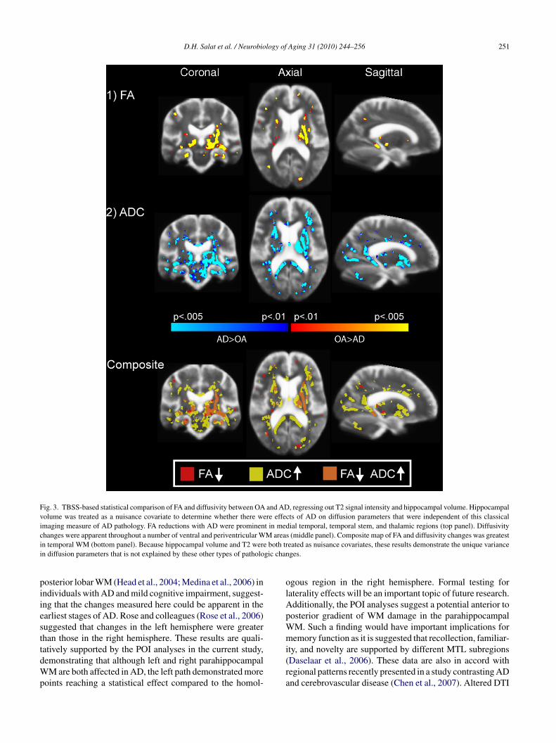
Fig. 3. TBSS-based statistical comparison of FA and diffusivity between OA and AD, regressing out T2 signal intensity and hippocampal volume. Hippocampalvolume was treated as a nuisance covariate to determine whether there were effects of AD on diffusion parameters that were independent of this classicalimaging measure of AD pathology. FA reductions with AD were prominent in medial temporal, temporal stem, and thalamic regions (top panel). Diffusivitychanges were apparent throughout a number of ventral and periventricular WM areas (middle panel). Composite map of FA and diffusivity changes was greatestin temporal WM (bottom panel). Because hippocampal volume and T2 were both treated as nuisance covariates, these results demonstrate the unique variancein diffusion parameters that is not explained by these other types of pathologic changes.
posterior lobar WM (Head et al., 2004; Medina et al., 2006) inindividuals with AD and mild cognitive impairment, suggest-ing that the changes measured here could be apparent in theearliest stages of AD. Rose and colleagues (Rose et al., 2006)suggested that changes in the left hemisphere were greaterthan those in the right hemisphere. These results are quali-tatively supported by the POI analyses in the current study,demonstrating that although left and right parahippocampalWM are both affected in AD, the left path demonstrated morepoints reaching a statistical effect compared to the homol-
ogous region in the right hemisphere. Formal testing forlaterality effects will be an important topic of future research.Additionally, the POI analyses suggest a potential anterior toposterior gradient of WM damage in the parahippocampalWM. Such a finding would have important implications formemory function as it is suggested that recollection, familiar-ity, and novelty are supported by different MTL subregions(Daselaar et al., 2006). These data are also in accord withregional patterns recently presented in a study contrasting ADand cerebrovascular disease (Chen et al., 2007). Altered DTI

252 D.H. Salat et al. / Neurobiology of Aging 31 (2010) 244–256
Fig. 4. Demonstration of the POI technique and analysis of parahippocampal WM. Seed points were manually placed in each individual participant’s diffusionvolumes at anterior and posterior points in the parahippocampal WM defined by morphometric landmarks in the T2 image. Those points were then connectedusing the optimal path calculated from the diffusion tensor information (top panel; see Section 2). The mean of the OA (top left) and AD (top middle) of asingle sagittal slice is presented to demonstrate the visual differences in the raw data between these groups (arrows). FA was sampled from the central voxelsalong the path, providing an individual POI for each participant, and minimizing the potential confounds of partial volume contamination from more peripheralvoxels. FA was most affected in anterior portions of the parahippocampal WM, but significant reductions in FA were found with AD along the majority of thepath when the mean FA across the hemispheres was examined (bottom plot). Overall path length did not differ between the groups.
measures have been related to indices of disease severity andcognitive ability, with particular association with episodicmemory, presenting a potential role for DTI in uncoveringWM degeneration in the clinical presentation of AD (Rose etal., 2006). The present results demonstrate regional changesin parahippocampal and precuneus WM, and degenerationof these regions may result in isolation of the hippocam-pus from neocortical input and related areas that have beenrecently described to comprise part of a functional memorycircuit (Vincent et al., 2006).
The relation between DTI measures and more classicalimaging indices of pathology, including brain volume, isnot clear. Our prior volumetric work demonstrated signif-icantly lower prefrontal WM volume in patients with ADcompared to age-matched control participants (Salat et al.,1999a), and the current data may suggest that this effect
is due to degenerative changes in selective regions. Thecurrent data demonstrate that changes in diffusion metricsprovide independent information beyond hippocampal vol-ume alone. Removing variance due to hippocampal volumedid not reduce the effects measured in parahippocampal WM,and even highlighted effects in thalamic regions that were lessapparent in the group comparisons without consideration ofhippocampal volume. The association between microstruc-tural FA measures and macrostructural volumetric measuresmay be complex (Salat et al., 2005b). Similarly, the patho-logic basis of changes in DTI measures is currently unknown.Prior studies report loss of oligodendrocytes, reactive astrocy-tosis, and reduction in neuropil density that could contributeto WM damage in AD (Sjobeck et al., 2006), and we positthat at least some of the measured effects could be due toWallerian degeneration of axons and the surrounding myelin

D.H. Salat et al. / Neurobiology of Aging 31 (2010) 244–256 253
Fig. 5. Mean anisotropy along the entire path (top left) differed between OA and AD in the left and right hemispheres. For comparison, traditional imagingmeasures of AD pathology are presented including hippocampal volume (top right), total WM volume (bottom left), and white matter signal abnormality(WMSA; bottom right) volume.
sheath. However, the fact that effects remained in the MTLWM after regressing out hippocampal volume and that therewas greater alteration in radial as opposed to axial diffusivityin parahippocampal WM supports prior work demonstratingreduced myelin staining in AD (Hyman et al., 1986), andsuggests that the effects measured are somewhat indepen-dent of regional cortical degeneration and may represent aunique myelin pathology (Budde et al., 2007; Song et al.,2002, 2003). These findings should be interpreted with cau-tion however, as it is important to note that the current dataalso demonstrated a regional increase in axial diffusivity. Thisis not what is expected from the animal models demonstrat-ing that axonal pathology should result in a decrease in axialdiffusivity (Song et al., 2003), and little information exists forthe interpretation of increased axial diffusivity. DTI has beenused to guide pathology studies (Englund et al., 2004), andfurther work with similar methods will likely yield importantnew information about how these novel diffusion measuresrelate to histopathology in AD.
The current results have some limitations. The changesin FA measured were relatively small and regionally local-ized, and thus it is unknown how early such changes couldbe detected. Future work will examine individuals with mildcognitive impairment to determine whether the current pat-tern of results exists in preclinical stages of disease. Althoughadvanced procedures were applied to address common con-cerns of diffusion imaging studies, further improvement isrequired in this field. Methods to enhance spatial (Li et al.,2005; Liu et al., 2004, 2005) and angular (Tuch, 2004; Tuchet al., 2002) resolution will be necessary to fully exclude
concerns about eddy current and susceptibility artifact dis-tortions. Similarly, although the procedures used here aresuperior to standard linear transformations for registrationof DTI data, the potential still exists for further refinementof DTI registration using the full tensor information (Park etal., 2003). In spite of these limitations, the current analysisprocedures provide a set of techniques that address commonconcerns for the analysis of DTI data, and demonstrate thevulnerability of WM to AD pathology. Thus, along with cor-tical degeneration, we suggest that an additional mechanismunderlying AD clinical symptoms is the WM degenerationthat further isolates MTL structures.
Acknowledgements
This work was supported in part by NIH K01AG024898,a Massachusetts Alzheimer’s Disease Research CenterPilot Grant 2001/2002 (AG05886), the National Centerfor Research Resources (P41RR14075), the Mental Illnessand Neuroscience Discovery (MIND) Institute, and a grantfrom the National Alliance for Medical Image Computing(NAMIC U54 EB05149). We thank Dr. Francine Grodsteinand the Nurses’ Health Study for a portion of the participantrecruitment and imaging.
References
Au, R., Massaro, J.M., Wolf, P.A., Young, M.E., Beiser, A., Seshadri, S.,D’Agostino, R.B., DeCarli, C., 2006. Association of white matter hyper-

254 D.H. Salat et al. / Neurobiology of Aging 31 (2010) 244–256
intensity volume with decreased cognitive functioning: the FraminghamHeart Study. Arch. Neurol. 63 (2), 246–250.
Ball, M.J., 1978. Topographic distribution of neurofibrillary tangles andgranulovacuolar degeneration in hippocampal cortex of aging anddemented patients. A quantitative study. Acta Neuropathol. (Berl.) 42(2), 73–80.
Basser, P.J., 1995. Inferring microstructural features and the physiologicalstate of tissues from diffusion-weighted images. NMR Biomed. 8 (7–8),333–344.
Basser, P.J., 1997. New histological and physiological stains derived fromdiffusion-tensor MR images. Ann. N. Y. Acad. Sci. 820, 123–138.
Basser, P.J., Mattiello, J., LeBihan, D., 1994. Estimation of the effectiveself-diffusion tensor from the NMR spin echo. J. Magn. Reson. B 103(3), 247–254.
Basser, P.J., Pierpaoli, C., 1996. Microstructural and physiological featuresof tissues elucidated by quantitative-diffusion-tensor MRI. J. Magn.Reson. B 111 (3), 209–219.
Blessed, G., Tomlinson, B.E., Roth, M., 1968. The association betweenquantitative measures of dementia and of senile change in the cere-bral grey matter of elderly subjects. Br. J. Psychiatry 114 (512),797–811.
Bozzali, M., Falini, A., Franceschi, M., Cercignani, M., Zuffi, M., Scotti, G.,Comi, G., Filippi, M., 2002. White matter damage in Alzheimer’s diseaseassessed in vivo using diffusion tensor magnetic resonance imaging. J.Neurol. Neurosurg. Psychiatry 72 (6), 742–746.
Braak, H., Braak, E., 1991. Neuropathological stageing of Alzheimer-relatedchanges. Acta Neuropathol. (Berl.) 82 (4), 239–259.
Brun, A., Englund, E., 1986. A white matter disorder in dementia of theAlzheimer type: a pathoanatomical study. Ann. Neurol. 19 (3), 253–262.
Budde, M.D., Kim, J.H., Liang, H.F., Schmidt, R.E., Russell, J.H., Cross,A.H., Song, S.K., 2007. Toward accurate diagnosis of white matterpathology using diffusion tensor imaging. Magn. Reson. Med. 57 (4),688–695.
Cavanna, A.E., Trimble, M.R., 2006. The precuneus: a review of its func-tional anatomy and behavioural correlates. Brain 129 (Pt 3), 564–583.
Chen, S.Q., Kang, Z., Hu, X.Q., Hu, B., Zou, Y., 2007. Diffusion tensor imag-ing of the brain in patients with Alzheimer’s disease and cerebrovascularlesions. J. Zhejiang Univ. Sci. B 8 (4), 242–247.
Choi, S.J., Lim, K.O., Monteiro, I., Reisberg, B., 2005. Diffusion tensorimaging of frontal white matter microstructure in early Alzheimer’sdisease: a preliminary study. J. Geriatr. Psychiatry Neurol. 18 (1), 12–19.
Daselaar, S.M., Fleck, M.S., Cabeza, R., 2006. Triple dissociation in themedial temporal lobes: recollection, familiarity, and novelty. J. Neuro-physiol. 96 (4), 1902–1911.
de Jager, C.A., Budge, M.M., Clarke, R., 2003. Utility of TICS-M for theassessment of cognitive function in older adults. Int. J. Geriatr. Psychiatry18 (4), 318–324.
de Leeuw, F.E., Korf, E., Barkhof, F., Scheltens, P., 2006. White matterlesions are associated with progression of medial temporal lobe atrophyin Alzheimer disease. Stroke 37 (9), 2248–2252.
Desikan, R.S., Segonne, F., Fischl, B., Quinn, B.T., Dickerson, B.C., Blacker,D., Buckner, R.L., Dale, A.M., Maguire, R.P., Hyman, B.T., Albert, M.S.,Killiany, R.J., 2006. An automated labeling system for subdividing thehuman cerebral cortex on MRI scans into gyral based regions of interest.Neuroimage 31 (3), 968–980.
Englund, E., Brun, A., 1990. White matter changes in dementia ofAlzheimer’s type: the difference in vulnerability between cell compart-ments. Histopathology 16 (5), 433–439.
Englund, E., Brun, A., Alling, C., 1988. White matter changes in dementia ofAlzheimer’s type. Biochemical and neuropathological correlates. Brain111 (Pt6), 1425–1439.
Englund, E., Sjobeck, M., Brockstedt, S., Latt, J., Larsson, E.M., 2004.Diffusion tensor MRI post mortem demonstrated cerebral white matterpathology. J. Neurol. 251 (3), 350–352.
Fischl, B., Salat, D.H., Busa, E., Albert, M., Dieterich, M., Haselgrove, C.,van der Kouwe, A., Killiany, R., Kennedy, D., Klaveness, S., Montillo,A., Makris, N., Rosen, B., Dale, A.M., 2002. Whole brain segmentation:
automated labeling of neuroanatomical structures in the human brain.Neuron 33 (3), 341–355.
Folstein, M.F., Folstein, S.E., McHugh, P.R., 1975. “Mini-mental state”A practical method for grading the cognitive state of patients for theclinician. J. Psychiatr Res. 12 (3), 189–198.
Fotenos, A.F., Snyder, A.Z., Girton, L.E., Morris, J.C., Buckner, R.L., 2005.Normative estimates of cross-sectional and longitudinal brain volumedecline in aging and AD. Neurology 64 (6), 1032–1039.
Gomez-Isla, T., Price, J.L., McKeel Jr., D.W., Morris, J.C., Growdon, J.H.,Hyman, B.T., 1996. Profound loss of layer II entorhinal cortex neu-rons occurs in very mild Alzheimer’s disease. J. Neurosci. 16 (14),4491–4500.
Habeck, M., Nilges, M., Rieping, W., 2005. Replica-exchange MonteCarlo scheme for Bayesian data analysis. Phys. Rev. Lett. 94 (1),018105.
Han, X., Jovicich, J., Salat, D., van der Kouwe, A., Quinn, B., Czanner, S.,Busa, E., Pacheco, J., Albert, M., Killiany, R., Maguire, P., Rosas, D.,Makris, N., Dale, A., Dickerson, B., Fischl, B., 2006. Reliability of MRI-derived measurements of human cerebral cortical thickness: the effectsof field strength, scanner upgrade and manufacturer. Neuroimage 32 (1),180–194.
Head, D., Buckner, R.L., Shimony, J.S., Williams, L.E., Akbudak, E., Con-turo, T.E., McAvoy, M., Morris, J.C., Snyder, A.Z., 2004. Differentialvulnerability of anterior white matter in nondemented aging with min-imal acceleration in dementia of the Alzheimer type: evidence fromdiffusion tensor imaging. Cereb. Cortex 14 (4), 410–423.
Hirono, N., Hashimoto, M., Ishii, K., Kazui, H., Mori, E., 2004. One-yearchange in cerebral glucose metabolism in patients with Alzheimer’sdisease. J. Neuropsychiatry Clin. Neurosci. 16 (4), 488–492.
Hirono, N., Kitagaki, H., Kazui, H., Hashimoto, M., Mori, E., 2000. Impactof white matter changes on clinical manifestation of Alzheimer’s disease:a quantitative study. Stroke 31 (9), 2182–2188.
Huang, J., Friedland, R.P., Auchus, A.P., 2007. Diffusion tensor imaging ofnormal-appearing white matter in mild cognitive impairment and earlyAlzheimer disease: preliminary evidence of axonal degeneration in thetemporal lobe. AJNR Am. J. Neuroradiol. 28 (10), 1943–1948.
Hughes, C.P., Berg, L., Danziger, W.L., Coben, L.A., Martin, R.L., 1982.A new clinical scale for the staging of dementia. Br. J. Psychiatry 140,566–572.
Hyman, B.T., Van Hoesen, G.W., Damasio, A.R., Barnes, C.L., 1984.Alzheimer’s disease: cell-specific pathology isolates the hippocampalformation. Science 225 (4667), 1168–1170.
Hyman, B.T., Van Hoesen, G.W., Kromer, L.J., Damasio, A.R., 1986. Per-forant pathway changes and the memory impairment of Alzheimer’sdisease. Ann. Neurol. 20 (4), 472–481.
Ishii, K., Kawachi, T., Sasaki, H., Kono, A.K., Fukuda, T., Kojima, Y.,Mori, E., 2005. Voxel-based morphometric comparison between early-and late-onset mild Alzheimer’s disease and assessment of diagnos-tic performance of z score images. AJNR Am. J. Neuroradiol. 26 (2),333–340.
Jack Jr., C.R., Petersen, R.C., O’Brien, P.C., Tangalos, E.G., 1992. MR-based hippocampal volumetry in the diagnosis of Alzheimer’s disease.Neurology 42 (1), 183–188.
Jenkinson, M., Bannister, P., Brady, M., Smith, S., 2002. Improved optimiza-tion for the robust and accurate linear registration and motion correctionof brain images. Neuroimage 17 (2), 825–841.
Jernigan, T.L., Salmon, D.P., Butters, N., Hesselink, J.R., 1991. Cerebralstructure on MRI. Part II: Specific changes in Alzheimer’s and Hunting-ton’s diseases. Biol. Psychiatry 29 (1), 68–81.
Jones, D.K., Horsfield, M.A., Simmons, A., 1999. Optimal strategies formeasuring diffusion in anisotropic systems by magnetic resonance imag-ing. Magn. Reson. Med. 42 (3), 515–525.
Li, T.Q., Kim, D.H., Moseley, M.E., 2005. High-resolution diffusion-weighted imaging with interleaved variable-density spiral acquisitions.J. Magn. Reson. Imaging 21 (4), 468–475.
Lipton, R.B., Katz, M.J., Kuslansky, G., Sliwinski, M.J., Stewart, W.F.,Verghese, J., Crystal, H.A., Buschke, H., 2003. Screening for demen-

D.H. Salat et al. / Neurobiology of Aging 31 (2010) 244–256 255
tia by telephone using the memory impairment screen. J. Am. Geriatr.Soc. 51 (10), 1382–1390.
Liu, C., Bammer, R., Kim, D.H., Moseley, M.E., 2004. Self-navigated inter-leaved spiral (SNAILS): application to high-resolution diffusion tensorimaging. Magn. Reson. Med. 52 (6), 1388–1396.
Liu, C., Moseley, M.E., Bammer, R., 2005. Simultaneous phase correctionand SENSE reconstruction for navigated multi-shot DWI with non-Cartesian k-space sampling. Magn. Reson. Med. 54 (6), 1412–1422.
Lustig, C., Snyder, A.Z., Bhakta, M., O’Brien, K.C., McAvoy, M., Raichle,M.E., Morris, J.C., Buckner, R.L., 2003. Functional deactivations:change with age and dementia of the Alzheimer type. Proc. Natl. Acad.Sci. U. S. A. 100 (24), 14504–14509.
Medina, D., DeToledo-Morrell, L., Urresta, F., Gabrieli, J.D., Moseley, M.,Fleischman, D., Bennett, D.A., Leurgans, S., Turner, D.A., Stebbins,G.T., 2006. White matter changes in mild cognitive impairment and AD:a diffusion tensor imaging study. Neurobiology. Aging 27 (5), 663–672.
Mintun, M.A., Larossa, G.N., Sheline, Y.I., Dence, C.S., Lee, S.Y., Mach,R.H., Klunk, W.E., Mathis, C.A., DeKosky, S.T., Morris, J.C., 2006.[11C]PIB in a nondemented population: potential antecedent marker ofAlzheimer disease. Neurology 67 (3), 446–452.
Mohedano-Moriano, A., Pro-Sistiaga, P., Arroyo-Jimenez, M.M., Artacho-Perula, E., Insausti, A.M., Marcos, P., Cebada-Sanchez, S., Martinez-Ruiz, J., Munoz, M., Blaizot, X., Martinez-Marcos, A., Amaral, D.G.,Insausti, R., 2007. Topographical and laminar distribution of corticalinput to the monkey entorhinal cortex. J. Anat. 211 (2), 250–260.
Morris, J.C., 1997. Clinical dementia rating: a reliable and valid diagnosticand staging measure for dementia of the Alzheimer type. Int. Psychogeri-atr. 9 (Suppl. 1), 173–176, discussion 7–8.
Morris, J.C., Ernesto, C., Schafer, K., Coats, M., Leon, S., Sano, M., Thal,L.J., Woodbury, P., 1997. Clinical dementia rating training and reliabil-ity in multicenter studies: the Alzheimer’s Disease Cooperative Studyexperience. Neurology 48 (6), 1508–1510.
Moseley, M., 2002. Diffusion tensor imaging and aging—a review. NMRBiomed. 15 (7–8), 553–560.
Mungas, D., Harvey, D., Reed, B.R., Jagust, W.J., DeCarli, C., Beckett,L., Mack, W.J., Kramer, J.H., Weiner, M.W., Schuff, N., Chui, H.C.,2005. Longitudinal volumetric MRI change and rate of cognitive decline.Neurology 65 (4), 565–571.
Park, H.J., Kubicki, M., Shenton, M.E., Guimond, A., McCarley, R.W.,Maier, S.E., Kikinis, R., Jolesz, F.A., Westin, C.F., 2003. Spatial normal-ization of diffusion tensor MRI using multiple channels. Neuroimage 20(4), 1995–2009.
Pfefferbaum, A., Adalsteinsson, E., Sullivan, E.V., 2005. Frontal circuitrydegradation marks healthy adult aging: Evidence from diffusion tensorimaging. Neuroimage 26 (3), 891–899.
Pfefferbaum, A., Sullivan, E.V., 2003. Increased brain white matter diffusiv-ity in normal adult aging: relationship to anisotropy and partial voluming.Magn. Reson. Med. 49 (5), 953–961.
Pfefferbaum, A., Sullivan, E.V., Hedehus, M., Lim, K.O., Adalsteinsson, E.,Moseley, M., 2000. Age-related decline in brain white matter anisotropymeasured with spatially corrected echo-planar diffusion tensor imaging.Magn. Reson. Med. 44 (2), 259–268.
Pierpaoli, C., Basser, P.J., 1996. Toward a quantitative assessment of diffu-sion anisotropy. Magn. Reson. Med. 36 (6), 893–906.
Prins, N.D., van Dijk, E.J., den Heijer, T., Vermeer, S.E., Koudstaal, P.J.,Oudkerk, M., Hofman, A., Breteler, M.M., 2004. Cerebral white matterlesions and the risk of dementia. Arch. Neurol. 61 (10), 1531–1534.
Reese, T.G., Heid, O., Weisskoff, R.M., Wedeen, V.J., 2003. Reductionof eddy-current-induced distortion in diffusion MRI using a twice-refocused spin echo. Magn. Reson. Med. 49 (1), 177–182.
Rieping, W., Habeck, M., Nilges, M., 2005. Inferential structure determina-tion. Science 309 (5732), 303–306.
Rombouts, S.A., Barkhof, F., Goekoop, R., Stam, C.J., Scheltens, P., 2005.Altered resting state networks in mild cognitive impairment and mildAlzheimer’s disease: an fMRI study. Hum. Brain Mapp. 26 (4), 231–239.
Rose, S.E., Chen, F., Chalk, J.B., Zelaya, F.O., Strugnell, W.E., Benson, M.,Semple, J., Doddrell, D.M., 2000. Loss of connectivity in Alzheimer’s
disease: an evaluation of white matter tract integrity with colour codedMR diffusion tensor imaging. J. Neurol. Neurosurg. Psychiatry 69 (4),528–530.
Rose, S.E., McMahon, K.L., Janke, A.L., O’Dowd, B., de Zubicaray, G.,Strudwick, M.W., Chalk, J.B., 2006. Diffusion indices on magneticresonance imaging and neuropsychological performance in amnesticmild cognitive impairment. J. Neurol. Neurosurg. Psychiatry 77 (10),1122–1128.
Rueckert, D., Sonoda, L.I., Hayes, C., Hill, D.L., Leach, M.O., Hawkes, D.J.,1999. Nonrigid registration using free-form deformations: application tobreast MR images. IEEE Trans. Med. Imaging 18 (8), 712–721.
Salat, D.H., Kaye, J.A., Janowsky, J.S., 1999a. Prefrontal gray and whitematter volumes in healthy aging and Alzheimer disease. Arch. Neurol.56 (3), 338–344.
Salat, D.H., Kaye, J.A., Janowsky, J.S., 2001. Selective preservation anddegeneration within the prefrontal cortex in aging and Alzheimer disease.Arch. Neurol. 58 (9), 1403–1408.
Salat, D.H., Stangl, P.A., Kaye, J.A., Janowsky, J.S., 1999b. Sex differencesin prefrontal volume with aging and Alzheimer’s disease. Neurobiol.Aging 20 (6), 591–596.
Salat, D.H., Tuch, D.S., Greve, D.N., van der Kouwe, A.J., Hevelone, N.D.,Zaleta, A.K., Rosen, B.R., Fischl, B., Corkin, S., Rosas, H.D., Dale,A.M., 2005a. Age-related alterations in white matter microstructure mea-sured by diffusion tensor imaging. Neurobiol. Aging 26 (8), 1215–1227.
Salat, D.H., Tuch, D.S., Hevelone, N.D., Fischl, B., Corkin, S., Rosas, H.D.,Dale, A.M., 2005b. Age-related changes in prefrontal white matter mea-sured by diffusion tensor imaging. Ann. N. Y. Acad. Sci. 1064, 37–49.
Schmahmann, J., Pandya, D., 2006. Fiber Pathways of the Brain. OxfordUniversity Press, Oxford.
Schmidt, R., Schmidt, H., Kapeller, P., Enzinger, C., Ropele, S., Saurugg,R., Fazekas, F., 2002. The natural course of MRI white matter hyperin-tensities. J. Neurol. Sci. 203–204, 253–257.
Sjobeck, M., Haglund, M., Englund, E., 2006. White matter mapping inAlzheimer’s disease: a neuropathological study. Neurobiol. Aging 27(5), 673–680.
Smith, S.M., Jenkinson, M., Johansen-Berg, H., Rueckert, D., Nichols, T.E.,Mackay, C.E., Watkins, K.E., Ciccarelli, O., Cader, M.Z., Matthews,P.M., Behrens, T.E., 2006. Tract-based spatial statistics: voxelwise anal-ysis of multi-subject diffusion data. Neuroimage 31 (4), 1487–1505.
Smith, S.M., Jenkinson, M., Woolrich, M.W., Beckmann, C.F., Behrens,T.E., Johansen-Berg, H., Bannister, P.R., De Luca, M., Drobnjak, I.,Flitney, D.E., Niazy, R.K., Saunders, J., Vickers, J., Zhang, Y., De Ste-fano, N., Brady, J.M., Matthews, P.M., 2004. Advances in functional andstructural MR image analysis and implementation as FSL. Neuroimage23 (Suppl. 1), S208–S219.
Song, S.K., Sun, S.W., Ju, W.K., Lin, S.J., Cross, A.H., Neufeld, A.H., 2003.Diffusion tensor imaging detects and differentiates axon and myelindegeneration in mouse optic nerve after retinal ischemia. Neuroimage20 (3), 1714–1722.
Song, S.K., Sun, S.W., Ramsbottom, M.J., Chang, C., Russell, J., Cross,A.H., 2002. Dysmyelination revealed through MRI as increased radial(but unchanged axial) diffusion of water. Neuroimage 17 (3), 1429–1436.
Stern, Y., Hesdorffer, D., Sano, M., Mayeux, R., 1990. Measurement andprediction of functional capacity in Alzheimer’s disease. Neurology 40(1), 8–14.
Stoub, T.R., deToledo-Morrell, L., Stebbins, G.T., Leurgans, S., Bennett,D.A., Shah, R.C., 2006. Hippocampal disconnection contributes to mem-ory dysfunction in individuals at risk for Alzheimer’s disease. Proc. Natl.Acad. Sci. U S A 103 (26), 10041–10045.
Stout, J.C., Jernigan, T.L., Archibald, S.L., Salmon, D.P., 1996. Associationof dementia severity with cortical gray matter and abnormal white mat-ter volumes in dementia of the Alzheimer type. Arch. Neurol. 53 (8),742–749.
Sullivan, E.V., Adalsteinsson, E., Hedehus, M., Ju, C., Moseley, M., Lim,K.O., Pfefferbaum, A., 2001. Equivalent disruption of regional whitematter microstructure in ageing healthy men and women. Neuroreport12 (1), 99–104.

256 D.H. Salat et al. / Neurobiology of Aging 31 (2010) 244–256
Sullivan, E.V., Adalsteinsson, E., Pfefferbaum, A., 2006. Selective age-related degradation of anterior callosal fiber bundles quantified in vivowith fiber tracking. Cereb. Cortex 16 (7), 1030–1039.
Sullivan, E.V., Pfefferbaum, A., 2006. Diffusion tensor imaging and aging.Neurosci. Biobehav. Rev. 30 (6), 749–761.
Swendsen, R.H., Wang, J.S., 1986. Replica Monte Carlo simulation of spinglasses. Phys. Rev. Lett. 57 (21), 2607–2609.
Taoka, T., Iwasaki, S., Sakamoto, M., Nakagawa, H., Fukusumi, A.,Myochin, K., Hirohashi, S., Hoshida, T., Kichikawa, K., 2006. Diffu-sion anisotropy and diffusivity of white matter tracts within the temporalstem in Alzheimer disease: evaluation of the “tract of interest” by dif-fusion tensor tractography. AJNR Am. J. Neuroradiol. 27 (5), 1040–1045.
Tuch, D.S., 2004. Q-ball imaging. Magn. Reson. Med. 52 (6), 1358–1372.Tuch, D.S., Reese, T.G., Wiegell, M.R., Makris, N., Belliveau, J.W., Wedeen,
V.J., 2002. High angular resolution diffusion imaging reveals intravoxel
white matter fiber heterogeneity. Magn. Reson. Med. 48 (4), 577–582.
Van Hoesen, G.W., Hyman, B.T., Damasio, A.R., 1991. Entorhinal cortexpathology in Alzheimer’s disease. Hippocampus 1 (1), 1–8.
Vincent, J.L., Snyder, A.Z., Fox, M.D., Shannon, B.J., Andrews, J.R.,Raichle, M.E., Buckner, R.L., 2006. Coherent spontaneous activity iden-tifies a hippocampal-parietal memory network. J. Neurophysiol. 96 (6),3517–3531.
Walhovd, K.B., Fjell, A.M., Reinvang, I., Lundervold, A., Dale, A.M., Eil-ertsen, D.E., Quinn, B.T., Salat, D., Makris, N., Fischl, B., 2005. Effectsof age on volumes of cortex, white matter and subcortical structures.Neurobiol. Aging 26 (9), 1261–1270, discussion 75–8.
Wang, L., Zang, Y., He, Y., Liang, M., Zhang, X., Tian, L., Wu, T., Jiang, T.,Li, K., 2006. Changes in hippocampal connectivity in the early stages ofAlzheimer’s disease: evidence from resting state fMRI. Neuroimage 31(2), 496–504.