Embed Size (px)
Citation preview

1What’s the Diagnosis – Case 43

2What’s the Diagnosis – Case 43

3What’s the Diagnosis – Case 43

4What’s the Diagnosis – Case 43

5What’s the Diagnosis – Case 43
6What’s the Diagnosis – Case 43

7What’s the Diagnosis – Case 43

8What’s the Diagnosis – Case 43
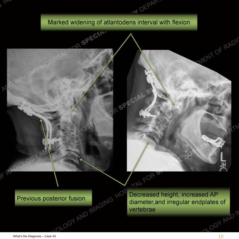
Findings
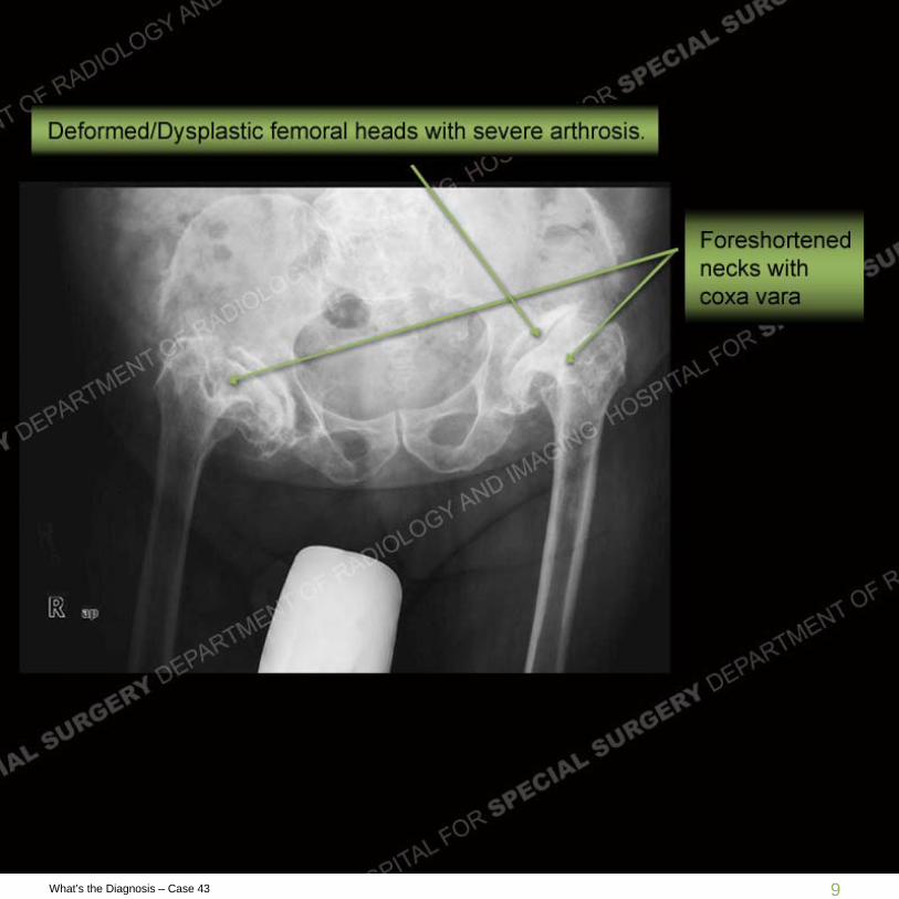
Radiographs of the pelvis demonstrate markedly abnormal/dysplastic femoralheads with severe arthrosis of both hips, remodelled acetabulae, andforeshortened femoral necks with coxa vara deformity. Cervical spineradiographs demonstrate previous occipito-cervical fusion with poordelineation of the upper cervical spine architecture. However, noted is amarked widening of the atlantodental interval when comparing the neutral tothe flexion views.
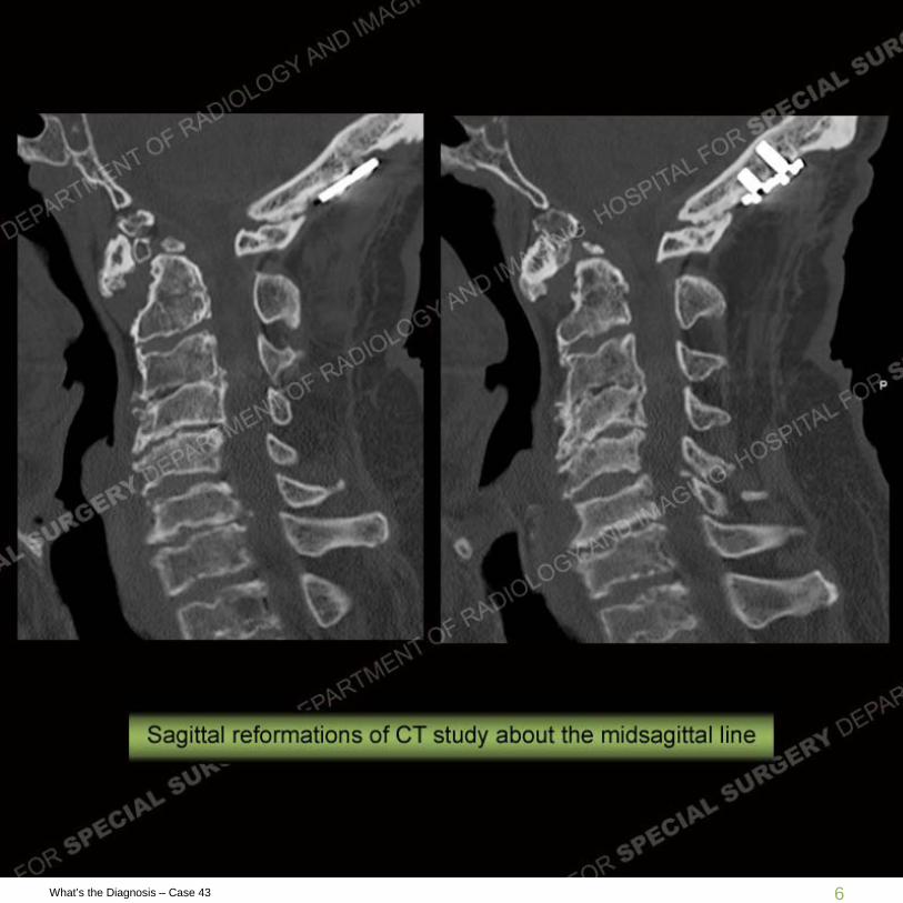
Cross sectional imaging of the cervical spine demonstrates decreased heightof the vertebral bodies with increased AP diameter and prominent end plateirregularities. There are multiple non-fused ossification centers seen at the tipof the dens and with an overall decreased amount of bone at the dens than typically seen. Severe stenosis is seen at the C1/C2 junction with severe compression of the cord and high signal within the cord representing edema/myelomalacia. Underlying developmental spinal stenosis is also seen.
9What’s the Diagnosis – Case 43
10What’s the Diagnosis – Case 43

11What’s the Diagnosis – Case 43

12What’s the Diagnosis – Case 43

13What’s the Diagnosis – Case 43

14What’s the Diagnosis – Case 43
Diagnosis
Spondyloepiphyseal Dysplasia (SED)
SED is an inherited dysplasia that involves the ends of the bones or epiphyses and the spine. It comes in two variants, congenita ( present at birth) and tarda which has a normal appearance at birth and then develops at 4 years of age and older. Given the underlying dysplasia there is premature osteoarthritis which in this patient may have been neglected. In the spine, there is typically a hypoplastic dens which leads to spinal instability and as in this patient leads to fusion to help prevent a catastrophic event. The presence of an os odontoideum or non fused tip of the dens may be seen but is not as typically present.

15What’s the Diagnosis – Case 43
Discussion
he vertebral bodies are decreased in height and at times may be completely flat yielding platyspondyly. Ovoid or trapezoidal bodies in the pediatric patient typically than yield vertebrae in the adult with decreased height, increased AP diameter, and end plate irregularities as seen here. Severe stenosis or C1/C2 kinking may be found as compared to the typical cervicomedullary kinking found in achondroplasia. In this patient, no myelopathic symptoms were present, astonishingly so. Imaging of the other appendicular structures would have shown mutliple areas of epiphyseal dysplasia and advanced arthrosis.

16What’s the Diagnosis – Case 43
Resources
Resnick. Diagnosis of Bone and Joint Disorders. 4th Ed. 2002
http://emedicine.medscape.com/article/1260836-overview