Embed Size (px)
Citation preview
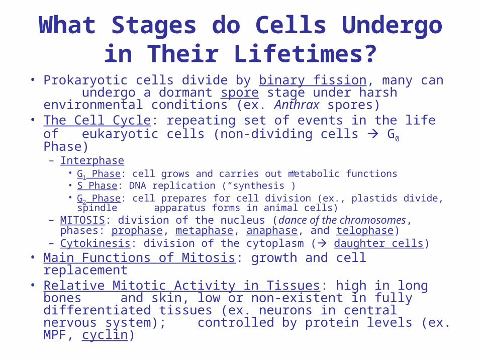
What Stages do Cells Undergo in Their Lifetimes?
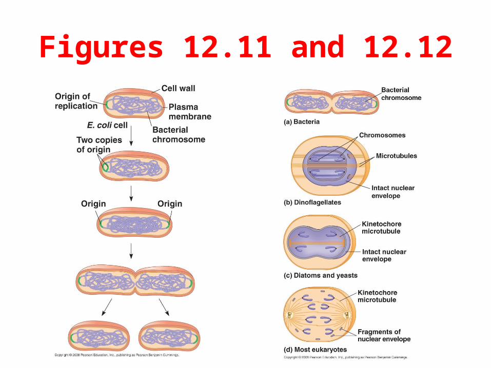
• Prokaryotic cells divide by binary fission, many can undergo a dormant spore stage under harsh environmental conditions (ex. Anthrax spores)
• The Cell Cycle: repeating set of events in the life of eukaryotic cells (non-dividing cells G0 Phase)– Interphase
• G1 Phase: cell grows and carries out metabolic functions • S Phase: DNA replication (“synthesis”) • G2 Phase: cell prepares for cell division (ex., plastids divide, spindle
apparatus forms in animal cells)– MITOSIS: division of the nucleus (dance of the chromosomes,
phases: prophase, metaphase, anaphase, and telophase)– Cytokinesis: division of the cytoplasm ( daughter cells)
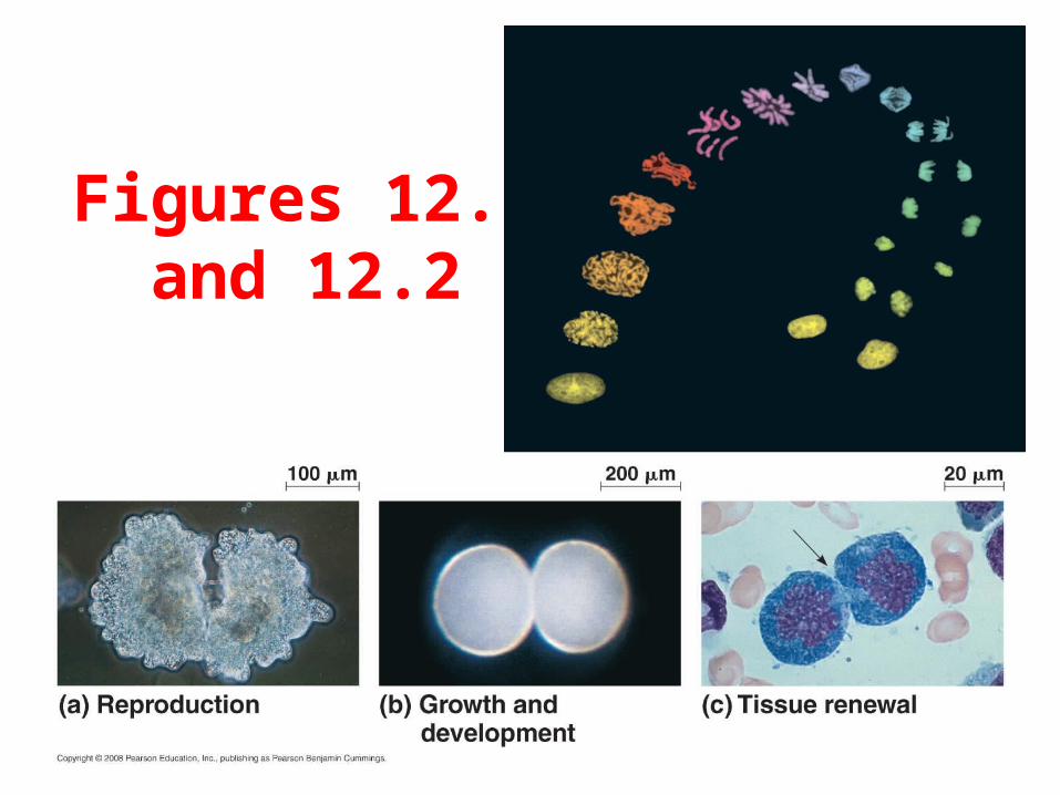
• Main Functions of Mitosis: growth and cell replacement• Relative Mitotic Activity in Tissues: high in long bones
and skin, low or non-existent in fully differentiated tissues (ex. neurons in central nervous system); controlled by protein levels (ex. MPF, cyclin)
Figures 12.11 and 12.12
Figures 12.1 and 12.2

Figure 12.5
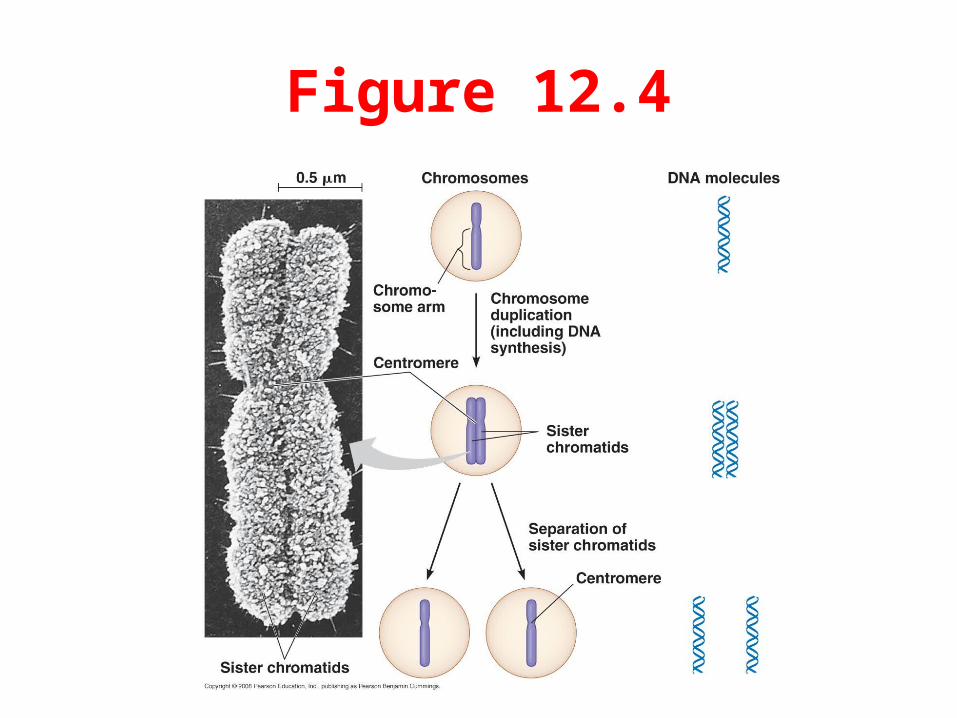
Figure 12.4
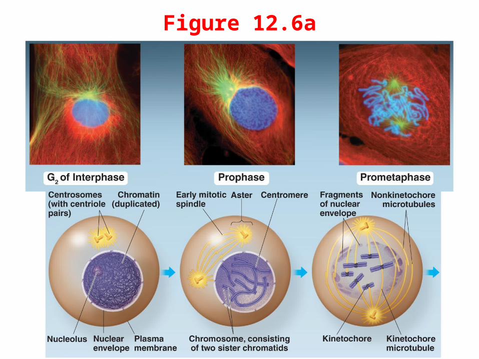
Figure 12.6a
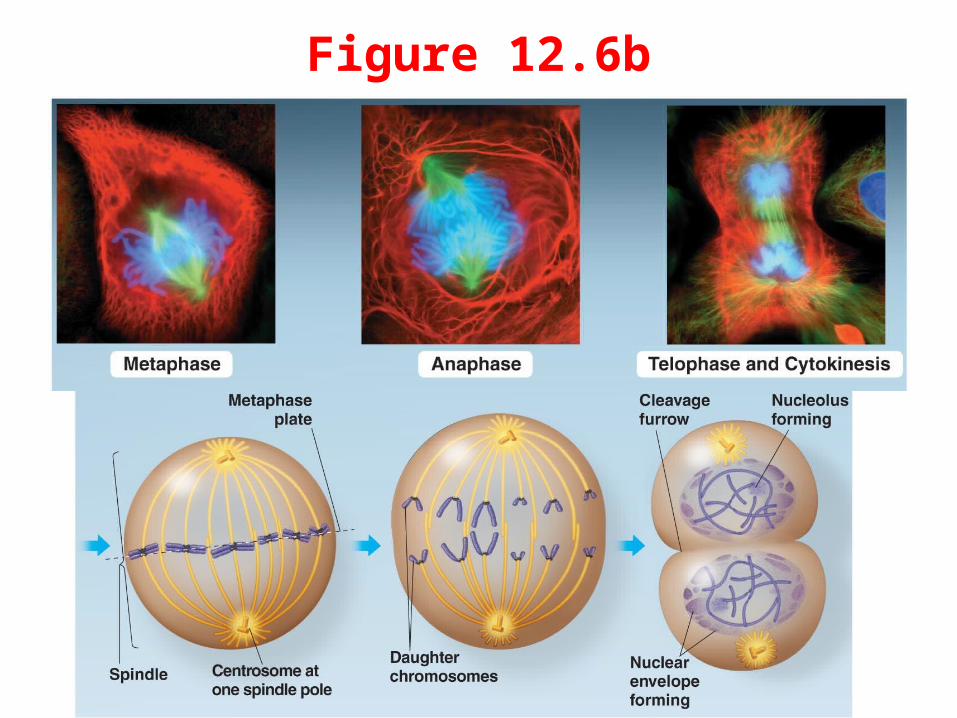
Figure 12.6b
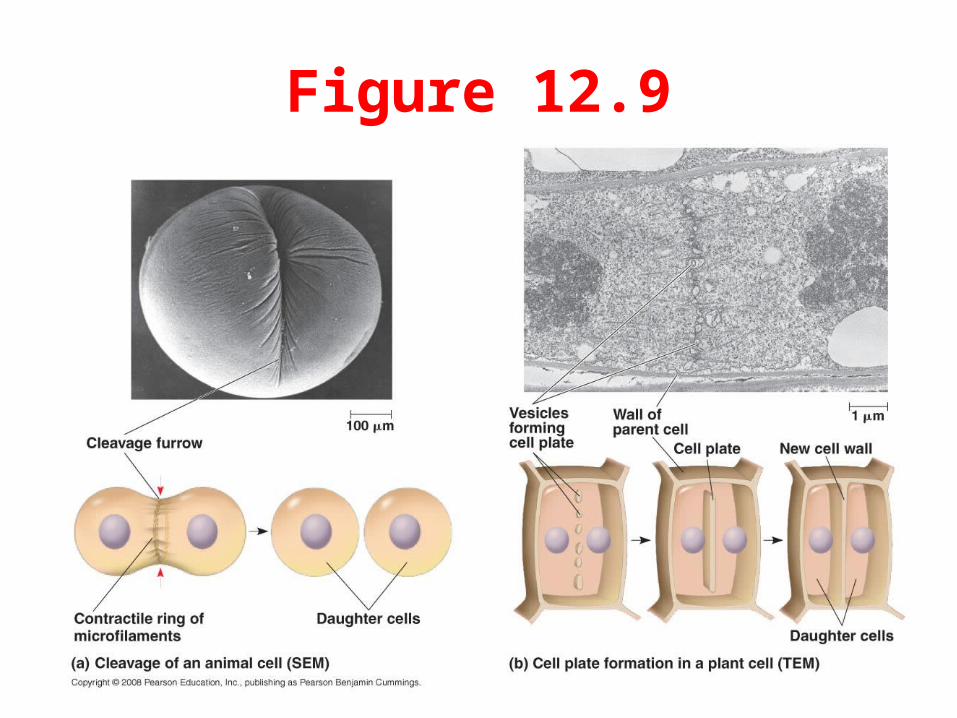
Figure 12.9
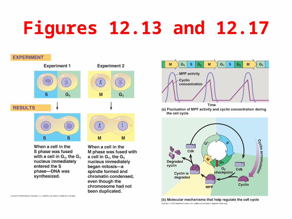
Figures 12.13 and 12.17
What are the Differences Between Sexual and Asexual Reproduction?
• Asexual Reproduction: offspring are clones of a single parent; variation among individuals limited to mutations
– Bacteria divide by binary fission – Spores in many eukaryotes (environmentally resistant
reproductive cells that can develop alone)• Sexual Reproduction: two parents donate genes to
offspring via gametes (sex cells)– Gametes are haploid, must fuse diploid zygote– Several sources of variation in addition to mutation
great physical diversity among individuals’ traits • Many organisms with both asexual and sexual
cycles (alternate, depending on various factors)– Example: parthenogenesis in some fishes and reptiles
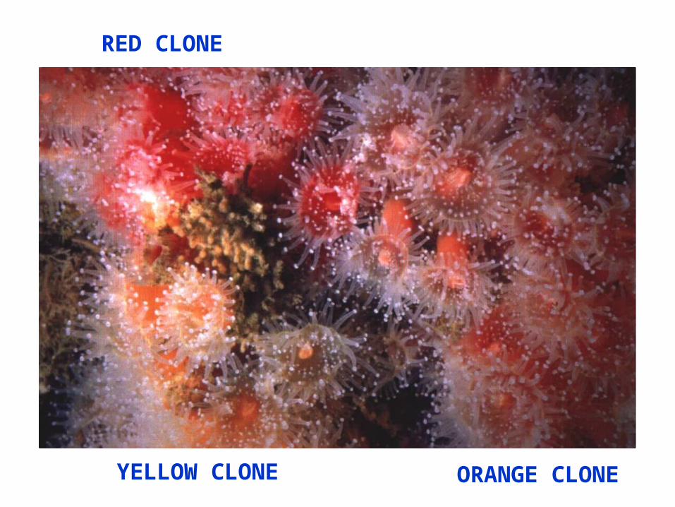
RED CLONE
YELLOW CLONE ORANGE CLONE
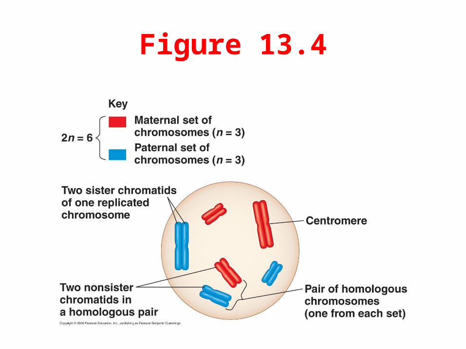
Figure 13.4
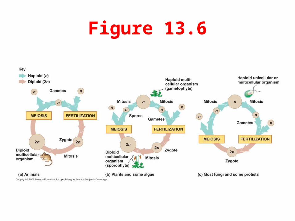
Figure 13.6
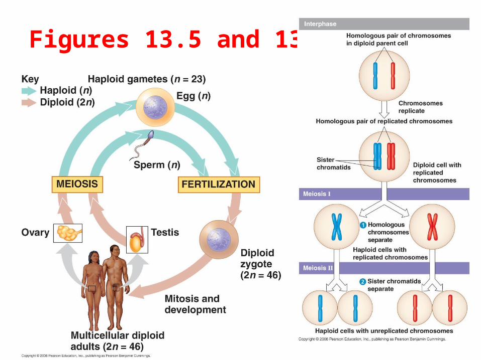
How are Gametes Produced? What are Some Sources of Variation Among Individuals?
• Gametogenesis: formation of gametes from somatic cells (via MEIOSIS, cell differentiation)
– Spermatogenesis: formation of sperm cells; occurs in testes• Haploid spermatids differentiate into sperm cells (with cap and
flagellum)– Oogenesis: formation of egg cells (oocytes); occurs in ovaries
• One of four grand-daughter cells absorbs cytoplasms of others (egg cells are very large cells, with RNA-rich and protein-
rich cytoplasms); polar bodies remain after formation of oocyte
• Important sources of variation for sexually reproducing organisms (other than mutation):
1. Independent assortment of chromosomes in meiosis2. Crossing over (genetic recombination) among homologous
chromosomes; more likely to occur away from centromere 3. Gene duplications, inversions, translocations, and deletions4. Non-disjunction of chromosomes / duplication of chromosomes5. Whole-genome duplications
Figures 13.5 and 13.7
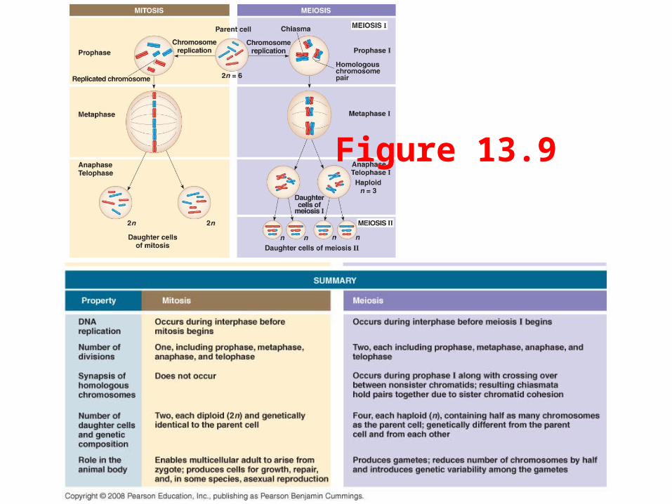
Figure 13.9
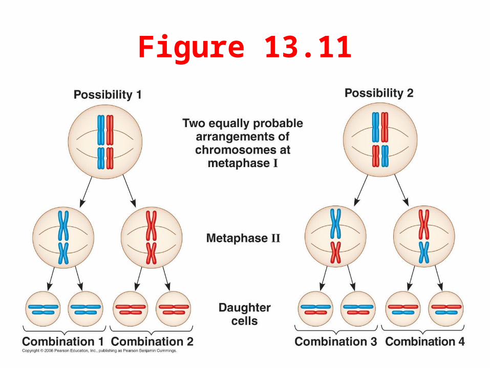
Figure 13.11
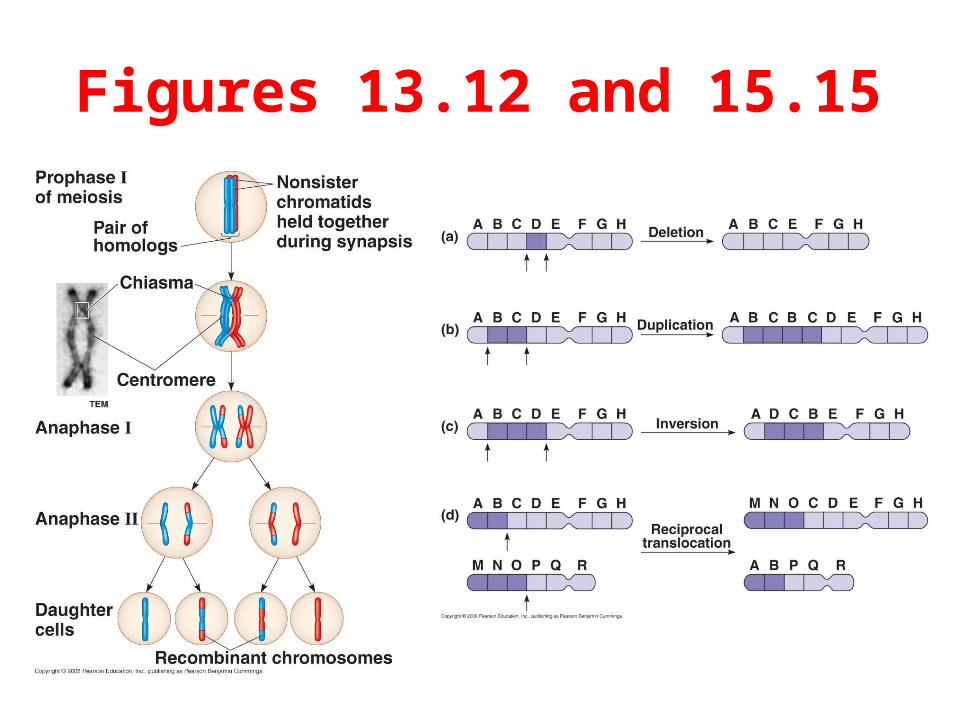
Figures 13.12 and 15.15
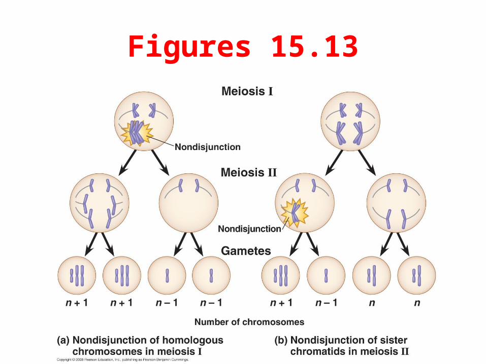
Figures 15.13
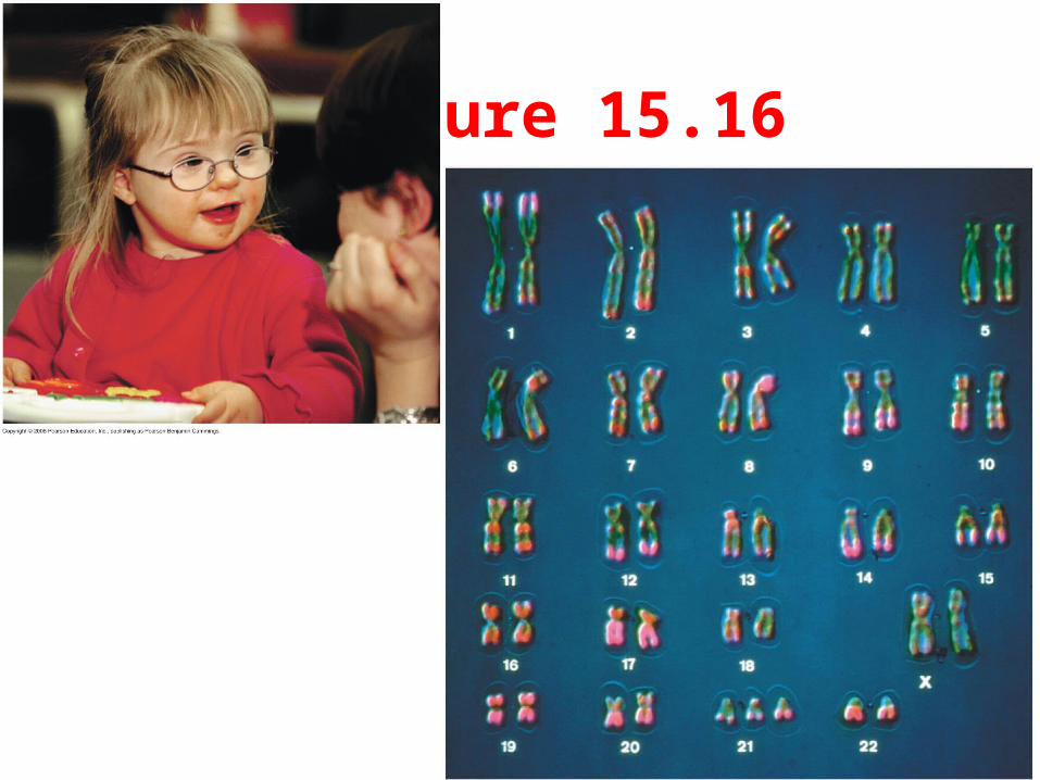
Figure 15.16