Embed Size (px)
Citation preview

Weir PSFMWeir PSFMWeir PSFMWeir PSFM
Fixed Focus AnalysisDataset: blanketfocmon.XLS
Features: Full-wafer exposure, fixed focus
Sections:
• Comparison of PSFM 180, 160 and PGM response
• PGM whole-wafer focus model
• Reticle Scan-stage analysis (Up/Down scan comparison)
• Up-Scan analysis
• Down-Scan analysis of image
• Average Field
• Astigmatic behaviorTEA Systems Corp.
65 Schlossburg St.Alburtis, PA 18011
610 682 [email protected]

0304 Weir PSFM - Fixed Focus Analysis Page -2-TEA Systems Corp. Confidential
Project Summary• Candidate slides for Paper
Comparison of PSFM 180, 160 and PGM response• All 3 features present a lens-characteristic signature response• 3 features can be used to replicate focus behavior of features with increasing density.
– PSFM 180 = isolated– PGM = dense-packed feature performance.
PGM whole-wafer focus model • Modeled focus across wafer shows high-point in central area.• X-Slit (lens) and Y-scan (reticle stage travel) characteristics• Methods of plotting lens signature and reticle scan characteristics are shown
Reticle Scan-stage analysis (Up/Down scan comparison)• While being well balanced, stage is shown to contribute more noise in down-scan• Scan tilt is present and shown• Scan-speed nonlinearity shown to be greater in down-scan• Sensitivity of direction and wafer-edge on field tilt • Plate bowing during scan can be seen• Astigmatism across slit is shown to have a directional component.
– Consistent with the concept of scan-averaging of y-direction aberrations
• Background slides with in-depth analysis Up-Scan analysis Down-Scan analysis of image Average Field Astigmatic behavior

0304 Weir PSFM - Fixed Focus Analysis Page -3-TEA Systems Corp. Confidential
PSFM 180 – data conversion
• Data converted to focus errors

0304 Weir PSFM - Fixed Focus Analysis Page -4-TEA Systems Corp. Confidential
PSFM 180 Raw Focus Data
• Mean Focus contour shown
• Note the high focus errors located at left/right edges & in lower left edge. “7 o’clock” location
Reticle stage scan direction

0304 Weir PSFM - Fixed Focus Analysis Page -5-TEA Systems Corp. Confidential
PSFM 180 – raw Astigmatism
• Data is before conversion to focus
• Used to determine data culling limits Use a cull = 0.013 um base on
quartiles.
• A total of 24 points were removed using this method.

0304 Weir PSFM - Fixed Focus Analysis Page -6-TEA Systems Corp. Confidential
PSFM 160 Raw Focus
0304 Weir PSFM - Fixed Focus Analysis Page -7-TEA Systems Corp. Confidential
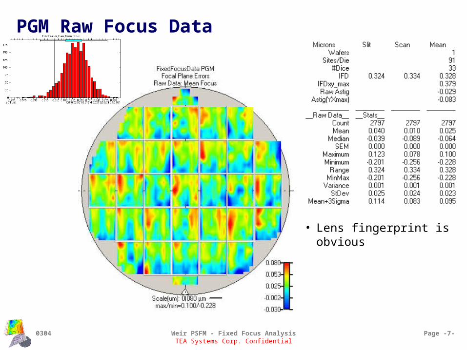
PGM Raw Focus Data
• Lens fingerprint is obvious

0304 Weir PSFM - Fixed Focus Analysis Page -8-TEA Systems Corp. Confidential
Comparison PSFM 180, 160 & PGM
• Ranges change
• Field signatures are very similar
• PGM and PSFM replication is very close.

0304 Weir PSFM - Fixed Focus Analysis Page -9-TEA Systems Corp. Confidential
Comparison PSFM 180, 160 & PGM
• Mean Linear change from –93 to 0 to +25 nm for mean focus Represents progressively increased density in feature packing.
• Range Approximately the same with PSFM 160 being slightly higher Recall that PSFM data used some data culling
• Standard Deviation. Same for PSFM 180,160 Lower for PGM
• Astigmatism Follows mean focus trend
PGMPSFM 160PSFM 180
0304 Weir PSFM - Fixed Focus Analysis Page -10-TEA Systems Corp. Confidential
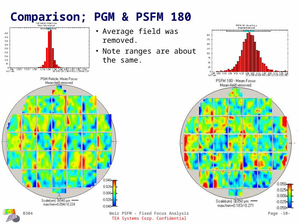
Comparison; PGM & PSFM 180• Average field was removed.
• Note ranges are about the same.

0304 Weir PSFM - Fixed Focus Analysis Page -11-TEA Systems Corp. Confidential
Comparison; PGM & PSFM 180
• PSFM 180 Focus behavior replicates isolated
line performance
• PGM Responds to focus like dense-
packed features
• Differences Range of PSFM 180 is about
150nm greater Astigmatic behavior is different
• Pgm = -29nm, PSFM = 4 nm
Standard deviation about the same

TEA Systems Corp. Confidential
PGM Whole-wafer Analysis

0304 Weir PSFM - Fixed Focus Analysis Page -13-TEA Systems Corp. Confidential
PGM Focus modeled data
• Sites at lower-left exhibit greatest errors on wafer
• Wafer Model X-tilt is equivalent to Field
Slit-tilt and will add

0304 Weir PSFM - Fixed Focus Analysis Page -14-TEA Systems Corp. Confidential
Wafer-based focus variation
• Wafer contributes a total of 39 nm focus error Systematic variations mostly lie with the +5, -7 nm
range. Note high points at top and center as well as tilt.

0304 Weir PSFM - Fixed Focus Analysis Page -15-TEA Systems Corp. Confidential
New Algorithm
0304 Weir PSFM - Fixed Focus Analysis Page -16-TEA Systems Corp. Confidential
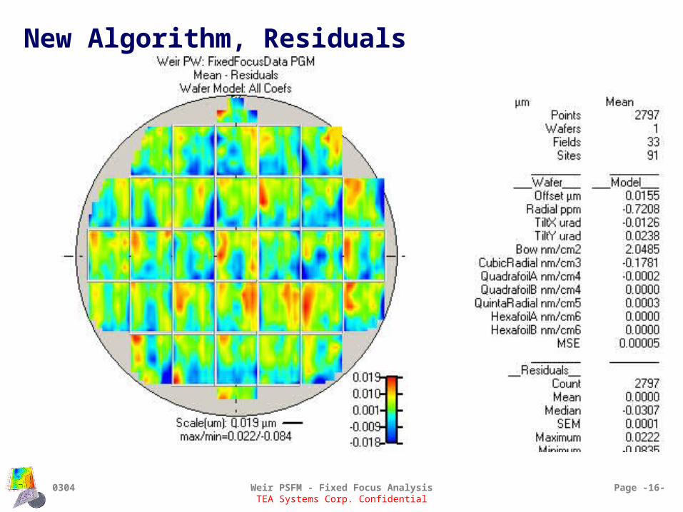
New Algorithm, Residuals

0304 Weir PSFM - Fixed Focus Analysis Page -17-TEA Systems Corp. Confidential
X-slit flight characteristics
• Objective: View X-slit pitch, roll and dip
• Aberrations removed Wafer aberrations Average row X-focus Average column X-focus
• View variation in X-slit focus across wafer up-scan
• tendency to bow the field down-scan
• Greater problems with scan-direction tilt
Down- scan
Up- scan

Down- scan
Up- scan

0304 Weir PSFM - Fixed Focus Analysis Page -19-TEA Systems Corp. Confidential
Y-Scan focus, Modeled field piston
• Y-scan focus• Modeled focus piston (offset) for
each row.• Objective:
View focus-signature across lens-slit

0304 Weir PSFM - Fixed Focus Analysis Page -20-TEA Systems Corp. Confidential
Slit Signature – using Y-scan focus piston
• Note circled row on right All down-scan Alternating field left & right edges
• Field tilt resulting from direction of travel-to-the-field

0304 Weir PSFM - Fixed Focus Analysis Page -21-TEA Systems Corp. Confidential
X-slit roll
• Only the modeled X-slit Tilt is shown Illustrates the roll of the slit as it is scanned
• Majority of tilt lies within the +/- 25 nm range (max=261 nm, min =-56 nm)• Note large tilt on right side of wafer.
Reticle-stageScan direction

TEA Systems Corp. Confidential
Scan Direction Analysis
• Examine the contribution of the reticle-stage scan to the focus-error budget Results in:
• Loss of process window
• Overlay errors in registration
• Asymmetric errors in critical feature size
• Analyze Raw-focus data Variations in modeled field performance

0304 Weir PSFM - Fixed Focus Analysis Page -23-TEA Systems Corp. Confidential
Reticle-Stage Scan Direction
Reticle-stageScan direction

0304 Weir PSFM - Fixed Focus Analysis Page -24-TEA Systems Corp. Confidential
Scan Direction Contours • Mean-focus plotted

0304 Weir PSFM - Fixed Focus Analysis Page -25-TEA Systems Corp. Confidential
Scan Direction Statistics
• Astigmatism Same
• Mean focus Same
• Standard Deviation same
• Range Up scan < Down 194 nm < 328 nm
• Down scan exhibits greater range than UP! We’ll see why in the
next few slides
Up Scan Down Scan

0304 Weir PSFM - Fixed Focus Analysis Page -26-TEA Systems Corp. Confidential
Radial-location Dependence
• Mean-focus and wafer-radial position
• Performance very similar
• Wafer-edge noise increases slightly for both distributions

0304 Weir PSFM - Fixed Focus Analysis Page -27-TEA Systems Corp. Confidential
Modeled Field and scan
• Field Model Every row/column
modeled separately
• Best Focus (BF) Same
• Tilt Down-scan > up
• Curvature Up-scan > down
• Residuals St.Dev. residuals Random variation of
down-scan > up
Down-Scan, Modeled Field
Up-Scan, Modeled Field

0304 Weir PSFM - Fixed Focus Analysis Page -28-TEA Systems Corp. Confidential
Comparison, Scan-stage flight path
• “Stable” fields values are plotted
• Note the increased “noise” of down-scan in 3rd & 4th rows
Down-Scan, X-slit Piston
Up-Scan, X-slit Piston

0304 Weir PSFM - Fixed Focus Analysis Page -29-TEA Systems Corp. Confidential
Comparison, Scan-stage flight path
• “Stable” fields values are plotted
• Note the increased “noise” of down-scan in 3rd & 4th rows
Down-Scan, X-slit Piston
Up-Scan, X-slit Piston

0304 Weir PSFM - Fixed Focus Analysis Page -30-TEA Systems Corp. Confidential
Comparison, X-slit signature
• All fields are collapsed onto one graph
• Plot modeled Y-scan Piston (focus offset) against position in slit
• Fluctuation at edges of lens “flutter” of slit height

0304 Weir PSFM - Fixed Focus Analysis Page -31-TEA Systems Corp. Confidential
Comparison, X-slit focus signature stability
• Down-scan tilts seem to drop toward outer edge of wafer. Auto-leveling
limitation?
• Slit-signature is maintained in most fields
Up Scan
DownScan

Comparison, X-slit focus signature stability
• Down-scan tilts seem to drop toward outer edge of wafer. Auto-leveling
limitation?
• Slit-signature is maintained in most fields
Up Scan
DownScan

0304 Weir PSFM - Fixed Focus Analysis Page -33-TEA Systems Corp. Confidential
Astigmatism – Scan-stage flight comparison• Astigmatism = (Y-X) focus• Y-Focus
Errors are averaged by scan of the slit Sensitive to:
• scan-speed• Pitch (y-tilt) of reticle-scan path
• X-focus Sensitive to:
• lens focus aberrations• Roll (X-tilt) of slit from auto-leveling
• Up-scan (upper graph) Center-field is near-zero
• Constant velocity of scan Scan-start (left side)
• Requires 7 mm of scan to settle Scan stop (right side)
• Last 2 mm exhibit acceleration
• Down-scan (lower graph) Shown to be “noisier” scan direction Scanning top (right side) to bottom (left side) Acceleration is non-zero at only one point
• –7mm from field bottom• Shown to be a point in field where curvature and field tilt
change anomalously Source of down-scan noise!
Same sign, velocity change
Opposite sign, velocity change

Astigmatism – Scan-stage flight comparison
• Astigmatism = (Y-X) focus• Y-Focus
Errors are averaged by scan of the slit Sensitive to:
• scan-speed• Pitch (y-tilt) of reticle-scan path
• X-focus Sensitive to:
• lens focus aberrations• Roll (X-tilt) of slit from auto-leveling
• Up-scan (upper graph) Center-field is near-zero
• Constant velocity of scan Scan-start (left side)
• Requires 7 mm of scan to settle Scan stop (right side)
• Last 2 mm exhibit acceleration
• Down-scan (lower graph) Shown to be “noisier” scan direction Scanning top (right side) to bottom (left side) Acceleration is non-zero at only one point
• –7mm from field bottom• Shown to be a point in field where curvature and field
tilt change anomalously Source of down-scan noise!
Same sign, velocity change
Opposite sign, velocity change

0304 Weir PSFM - Fixed Focus Analysis Page -35-TEA Systems Corp. Confidential
Astigmatism – Across slit Comparison
• Signatures appear “Balanced”
• Up scan signature is mirror image of down-scan. Reason: unknown
• Suggestions?
Up-scan
Down-scan
Z5
-10
-8
-6
-4
-2
0
2
4
6
8
10
-15 -10 -5 0 5 10 15
TEA Systems Corp. Confidential
Up-Scan analysis
Detailed analysis of focus performance of exposure tool during the Up-scan stage of exposure

0304 Weir PSFM - Fixed Focus Analysis Page -37-TEA Systems Corp. Confidential
Wafer aberrations – Up-scan
• Mean focus plotted
• Objective: Examine and remove wafer aberrations. Obtain clearer picture of field variations
0304 Weir PSFM - Fixed Focus Analysis Page -38-TEA Systems Corp. Confidential
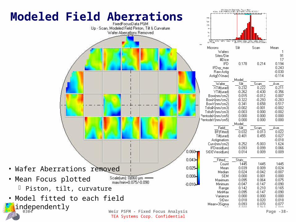
Modeled Field Aberrations
• Wafer Aberrations removed
• Mean Focus plotted Piston, tilt, curvature
• Model fitted to each field independently

0304 Weir PSFM - Fixed Focus Analysis Page -39-TEA Systems Corp. Confidential
Up-Scan, piston X-slit focus • Objective: View reticle flight-path in up-scan direction
• Wafer aberrations removed.
• Display modeled focus-piston (offset) of each row Illustrates the variation in scan uniformity
across wafer.
• Range of 50 nm seen in the plot of stable fields shown

0304 Weir PSFM - Fixed Focus Analysis Page -40-TEA Systems Corp. Confidential
Up-Scan, Y-scan focus Piston
• Modeled focus piston (offset) of each column.
• Objective: Observe slit-focus signature
• Graph is average of all fields Excluding top field at 12 o’clock

0304 Weir PSFM - Fixed Focus Analysis Page -41-TEA Systems Corp. Confidential
Up-Scan, Y-scan focus Piston
• Modeled focus piston (offset) of each column.
• Objective: Observe slit-focus signature
• Note retention of signature in all but the 4th column Reticle bowing?
• Variation is in the average offset of each field and tilt.
0304 Weir PSFM - Fixed Focus Analysis Page -42-TEA Systems Corp. Confidential
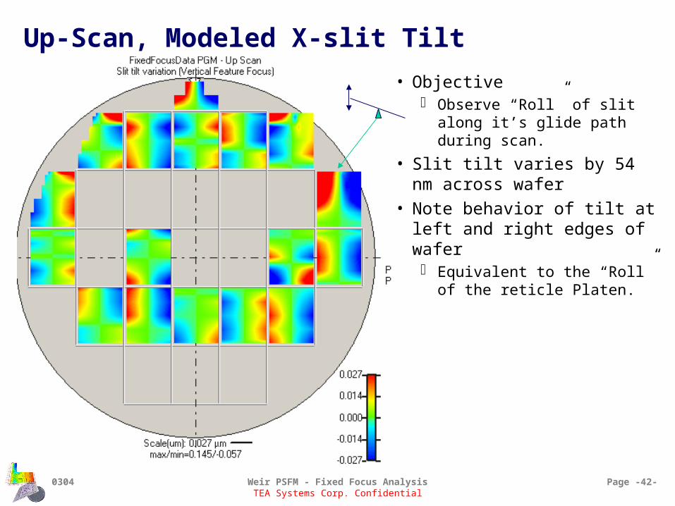
Up-Scan, Modeled X-slit Tilt
• Objective Observe “Roll” of slit along it’s
glide path during scan.
• Slit tilt varies by 54 nm across wafer
• Note behavior of tilt at left and right edges of wafer Equivalent to the “Roll” of the
reticle Platen.

0304 Weir PSFM - Fixed Focus Analysis Page -43-TEA Systems Corp. Confidential
Up-Scan, modeled Y-scan Tilt • Horizontal feature focus Wafer aberrations removed
• Pitch-variation of reticle scan-stage
• Objective: View pitch of scan stage as it
travels from bottom to top Tilt modeled for each column
of each field.
• Exhibits a 68 nm focus range contribution

0304 Weir PSFM - Fixed Focus Analysis Page -44-TEA Systems Corp. Confidential
Up-Scan, Y-scan flutter
• Y-scan curvature variation
• Objective: View 2nd order, curvature, stability
of each column Reticle is “flexed” as it scans
• This is a measure of the variation in focus at the start/stop of scan.
• Note the “flutter” at the extreme edges of the slit

0304 Weir PSFM - Fixed Focus Analysis Page -45-TEA Systems Corp. Confidential
Up-Scan – X slit curvature variation
• Variation in curvature of slit. Measured for each row of each
field.
• Objective: Direct measure of 2nd order
flexure of reticle during scan
• Caused by bowing of the reticle during the scan?

TEA Systems Corp. Confidential
Up-Scan, Residuals to modeled wafer & field
• Wafer aberrations are removed
• Field Piston, tilt and 2nd order systematics are removed
• Residuals contain: Higher-order focus variations (systematic errors) Random noise

0304 Weir PSFM - Fixed Focus Analysis Page -47-TEA Systems Corp. Confidential
Up-Scan, Mean - Focus
• Note presence of higher –order scan signature.
• High-point “bumps” in scan center Viewed earlier in slit profile box-plots

0304 Weir PSFM - Fixed Focus Analysis Page -48-TEA Systems Corp. Confidential
Up-Scan, Residuals to Wafer & Field
• Y-scan Sensitive to noise during scan-travel
• Bearing chatter, stage flex
• X-slit Sensitive to lens aberrations
Y-Scan Focus – Horizontal Features X-slit Focus – Vertical Features

0304 Weir PSFM - Fixed Focus Analysis Page -49-TEA Systems Corp. Confidential
X-Slit Focus, across slit residuals
• High-order focus errors due to lens aberrations.

TEA Systems Corp. Confidential
Down-scan focus behavior

0304 Weir PSFM - Fixed Focus Analysis Page -51-TEA Systems Corp. Confidential
Down-scan, raw data analysis

0304 Weir PSFM - Fixed Focus Analysis Page -52-TEA Systems Corp. Confidential
Down-Scan, Wafer aberrations

0304 Weir PSFM - Fixed Focus Analysis Page -53-TEA Systems Corp. Confidential
Down-scan, Wafer aberrations removed

0304 Weir PSFM - Fixed Focus Analysis Page -54-TEA Systems Corp. Confidential
Down-Scan, X-slit piston

0304 Weir PSFM - Fixed Focus Analysis Page -55-TEA Systems Corp. Confidential
X-Slit focus, Down-scan
• Objective: View reticle flight-path in up-scan
direction
• Field Piston plotted
• Analysis and up-scan comparison in next slide

0304 Weir PSFM - Fixed Focus Analysis Page -56-TEA Systems Corp. Confidential
Down-Scan, Scan flight path using X-slit piston
• Modeled X-focus piston (offset) for each row
• Graph (above) summarizes five (5) fields shown
• Illustrates average path of reticle-scan table

0304 Weir PSFM - Fixed Focus Analysis Page -57-TEA Systems Corp. Confidential
Down Scan, Slit Signature• Display of Modeled Y-scan piston
(offset) One model for each column of each field
• Results in focus signature across lens-slit
• Slit signature (below) is average of all fields Note large variation at field edges

0304 Weir PSFM - Fixed Focus Analysis Page -58-TEA Systems Corp. Confidential
Down-scan, Y-scan Focus piston

0304 Weir PSFM - Fixed Focus Analysis Page -59-TEA Systems Corp. Confidential
Down-Scan, X-slit focus tilt

0304 Weir PSFM - Fixed Focus Analysis Page -60-TEA Systems Corp. Confidential
Down-Scan, X-slit curvature
• Note that the lower-left corner of the wafer is typically the location of the greatest focus errors.

0304 Weir PSFM - Fixed Focus Analysis Page -61-TEA Systems Corp. Confidential
Down-scan, Y-scan focus Tilt

0304 Weir PSFM - Fixed Focus Analysis Page -62-TEA Systems Corp. Confidential
Down-scan, Y-scan focus Curvature

TEA Systems Corp. Confidential
Average Field Analysis
• Fields across the wafer are “collapsed” or overlaid.
• Metrology for each field-site is averaged
• Objective: View average performance of field Attempt to estimate the aberrations of the aerial scan image.

0304 Weir PSFM - Fixed Focus Analysis Page -64-TEA Systems Corp. Confidential
Mean Field & Scan DirectionUp-scan Scan glide-path
Lens-slit

0304 Weir PSFM - Fixed Focus Analysis Page -65-TEA Systems Corp. Confidential
Up-Scan – Wafer & Field tilt/piston removed
• Aberrations removed Wafer Field tilt Field piston
• Focus-error contributors Lens-aberrations Higher-order scan
errors

0304 Weir PSFM - Fixed Focus Analysis Page -66-TEA Systems Corp. Confidential
Up-Scan Direction piston
• View modeled piston variation Top: for each column
• Focus errors across lens-slit
Bottom: for each row• Focus errors caused by scan-
travel of slit
• Up-scan only
• Lens slit errors (top) Characteristic lens signature. On average, very level
• Bottom: scan glide-path Up-Scan exhibits a tilt Field-center point
• “bump” in scan
• This noise seen in earlier slides.
Scan glide-path
Lens-slit focus

0304 Weir PSFM - Fixed Focus Analysis Page -67-TEA Systems Corp. Confidential
Up-Scan – Field Tilt
• Vertical Features (left) X-slit tilt Note abrupt change in tilt at field center
Vertical Features Horizontal Features Mean Focus

TEA Systems Corp. Confidential
Astigmatic Behavior• (Y-X) Focus behavior
• The net-effect of this variation is to reduce the process window for this tool
• Objective: Analysis validation by comparison of values with those calculated
from an Artemis Zernike study.

0304 Weir PSFM - Fixed Focus Analysis Page -69-TEA Systems Corp. Confidential
Astigmatism – Up Scan
• Residuals to wafer aberrations• Y-scan focus
Primarily contributed by the scan-stage
• X-scan focus From lens focus errors From stage-scan errors

0304 Weir PSFM - Fixed Focus Analysis Page -70-TEA Systems Corp. Confidential
Astigmatism – Up ScanZ5
-10
-8
-6
-4
-2
0
2
4
6
8
10
-15 -10 -5 0 5 10 15
Artemis Astigmatic Zernike
Scan glide-path
Lens-slit
• Slit-astigmatic behavior (upper left) Tracks Z5 coefficient Note:
• Zernike analysis is from fy 2002 using a ring-aperture Estimated magnitude of Z5 errors is much less than
measured.
• Scan-glide path (bottom left) Scanning averages & removes errors Errors at scan-start and end
• Caused by scan-speed nonlinearity.– Acceleration errors in scan stabilize out in frist 7 to 12 mm– Acceleration errors at end of scan indicate start of turn-
around

0304 Weir PSFM - Fixed Focus Analysis Page -71-TEA Systems Corp. Confidential
Astigmatism – Up Scan
• No wafer removal Wafer aberrations previously also
included piston
• Otherwise, calculations same as in previous analysis.
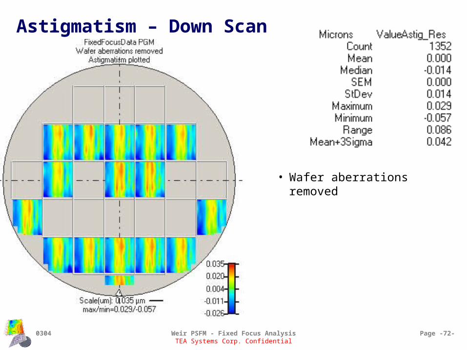
0304 Weir PSFM - Fixed Focus Analysis Page -72-TEA Systems Corp. Confidential
Astigmatism – Down Scan
• Wafer aberrations removed

0304 Weir PSFM - Fixed Focus Analysis Page -73-TEA Systems Corp. Confidential
Astigmatism – Down Scan
• Slit signature (top) Classic lens signature at center Wings vary from up-scan
• Scan signature (bottom) Fairly level Scan now moving down
• Right to left on graph
Non-flat performance suggest acceleration component present
After the 3rd row (from left), acceleration component again rises.

0304 Weir PSFM - Fixed Focus Analysis Page -74-TEA Systems Corp. Confidential
Astigmatism
• Wafer aberrations were removed
• Edge-field data culled
• Up and Down scans Up/down averaging removes signature resemblance to Z5 Zernike (upper right)
Z5
-10
-8
-6
-4
-2
0
2
4
6
8
10
-15 -10 -5 0 5 10 15