Embed Size (px)
Citation preview
Biological Journal of the Linnean Society
, 2002,
77
, 35–41. With 3 figures
© 2002 The Linnean Society of London,
Biological Journal of the Linnean Society,
2002,
77
, 35–41
35
Blackwell Science, LtdOxford, UKBIJBiological Journal of the Linnean Society0024-4066The Linnean Society of London, 2002XX 200277XX
Original Article
L.O. LUCIFORA and A. I. VASSALLOWALKING LOCOMOTION IN SKATES
*Correspondence: E-mail: [email protected]
Walking in skates (Chondrichthyes, Rajidae): anatomy, behaviour and analogies to tetrapod locomotion
LUIS O. LUCIFORA
1,
* and ALDO I. VASSALLO
2
1
Consejo Nacional de Investigaciones Científicas y Técnicas (CONICET) and Instituto Nacional de Investigación y Desarrollo Pesquero, Casilla de Correo 82, Correo Central, Mar del Plata 7600, Argentina
2
CONICET and Universidad Nacional de Mar del Plata, Departamento de Biología, Funes 3250, Mar del Plata 7600, Argentina
Received 10 December 2001; accepted for publication 15 April 2002
Pelvic fin walking in skates is common. However, the structure and function of pelvic fins have not been analysed.Pelvic fins of skates of the genus
Psammobatis
and
Rioraja agassizi
are externally divided into an anterior leg-likelobe and a posterior fin-like lobe. Internally, the anterior lobes are supported by a compound radial, a proximal radialand distal radials that resemble a thigh, a calf and a foot, respectively, and three associated radials arising from thepelvic girdle. A highly developed radial condyle on the pelvic girdle enables broad ‘limb’ movements. The musculararrangement of the anterior lobes is formed by protractor, retractor, flexor and extensor muscles, clearly departingfrom the generalized fin muscle arrangement of elasmobranchs. Walking is composed of propulsion and recoveryphases. A backward movement of the compound radial in the horizontal plane characterizes the propulsive phase.The proximal radial connects vertically the compound radial with the foot-like distal radials, which are anchored onthe bottom. During the recovery phase, the foot-like structure is lifted off the bottom and the entire limb-like anteriorlobe is moved forwards for starting a new cycle. Walking in skates resembles the ancestral tetrapod sprawling loco-motion seen in many salamanders and lizards. Pelvic fin anatomy and walking behaviour in skates and hemiscylliidsharks are compared. Ecological and evolutionary implications of walking locomotion in skates are alsodiscussed. © 2002 The Linnean Society of London,
Biological Journal of the Linnean Society
, 2002,
77
, 35–41.
ADDITIONAL KEYWORDS:
Elasmobranchii – biomechanics – vertebrate limbs – morphology – mosaic
evolution.
INTRODUCTION
The basic functional difference between aquatic andterrestrial paired appendages (fins and limbs) of ver-tebrates arises from the contrasting physical proper-ties of the media in which they act. The fins of fishes,aquatic turtles and penguins obliquely thrust againstthe water, the inertia of this dense medium causingit to push with equal force in the opposite direction(although these structures may also perform otherfunctions besides thrust generation; see Davenport &Clough, 1986). The forward component of this forcecauses the animal to be propelled. In contrast, thethrust of a foot against the ground exerted by a ter-restrial vertebrate, in the absence of the buoyancy of
water, should be shared between supportive (in oppo-sition to the pull of gravity) and propulsive force com-ponents (Hildebrand, 1988; Walker & Liem, 1994).However, aquatic vertebrates that walk along thebottom combine structural and functional features ofboth locomotion modes. Although benthic fishes areslightly denser than sea water (Hildebrand, 1988),they are much less denser relative to their mediumthan terrestrial vertebrates; so relatively light ‘limbs’can effectively propel them.
There are a number of ecological situations in whichswimming is not the selected option and other locomo-tive specializations appear in fishes. This is true forseveral lineages of benthic fishes that walk on thebottom by using different specialized structures(Helfman
et al.
, 1997). Common examples of walkingstructures in bony fishes are the free pectoral radialsof searobins (Scorpaeniformes, Triglidae) (Renous,
36
L. O. LUCIFORA and A. I. VASSALLO
© 2002 The Linnean Society of London,
Biological Journal of the Linnean Society,
2002,
77
, 35–41
Gasc, Bels & Davenport, 2000), the pelvic fins of flyinggurnards (Scorpaeniformes, Dactylopteridae), and thepaired fins of mudskippers (Perciformes, Gobiidae)(Moyle & Cech, 1982), and living Australian lung-fishes (
Neoceratodus forsteri
, Sarcopterygii) (Pough
etal.
, 1989).Chondrichthyans are less diverse than actinoptery-
gians but they still have many different lifestyles(Compagno, 1990). Many cartilaginous fishes arebenthic and some have locomotive specializations forwalking. These include epaulette sharks (
Hemiscyl-lium
spp., Hemiscyllidae, Orectolobiformes) (Michael,1993; Goto
et al.
, 1999), legged torpedos (
Typhlonarke
spp., Narkidae, Torpediniformes) (Compagno, 1999a)and legged-skates (Anacanthobatidae, Rajiformes)(Last, 1997; Compagno, 1999a,b). Many skate specieshave bilobed pelvic fins (Compagno, 1999b). There aresome anecdotal comments on the use of pelvic fins forwalking in skates (Berestovskiy, 1989; Compagno,1999b). The showy pelvic fin division into two lobes, adistinctive feature of ‘legged skates’ of the genera
Cru-riraja
and
Anacanthobatis
, leads to the hypothesisof sensorial/locomotor utilization of the anterior lobe(Bigelow & Schroeder, 1953; Last, 1997). However, theinternal anatomy and function of pelvic fins have notbeen analysed among skates. In this paper, the walk-ing behaviour of skates is documented and the anat-omy and function of skeleton components and musclesinvolved in walking are analysed.
MATERIAL AND METHODS
A
NATOMICAL
OBSERVATIONS
Frozen (
−
20
°
C) specimens of smallthorn sand skate,
Psammobatis rudis
, blotched sand skate,
P. bergi
,shortfin sand skate,
P. normani
(
n
=
3), smallnose fan-skate,
Sympterygia bonapartii
(
n
=
2), and Rio skate,
Rioraja agassizi
(
n
=
1) (Rajiformes, Rajidae) were dis-sected under a Wild Stereomicroscope (Heidelberg,Germany) to assess origin and insertion of pelvic finmuscles. Specimens were cleared and the cartilageswere stained with alcyan blue, using a techniquedescribed by Wassersug (1976), to survey internalstructure of pelvic girdle and fins. The pelvic girdleand fins of narrownose smoothhounds,
Mustelusschmitti
(Carcharhiniformes, Triakidae) were ob-served for comparative analysis. Skeletal terminologyfollows Compagno (1999a).
L
OCOMOTOR
BEHAVIOUR
Two individuals of
P. bergi
(12 cm total length) werekept in a tank of 145.2 L (i.e. 110
×
33
×
40 cm). Thefish were caught 2 days prior to the start of observa-tions with a dredge during a research cruise at
38
°
18
′
S
−
57
°
35
′
W and 38
°
22
′
S
−
57
°
31
′
W, 32 and 44 mdepth, respectively. Every observation session lastedat least 30 min. Skates were video-recorded duringobservations.
F
UNCTION
OF
MUSCLES
AND
SKELETON
DURING
WALKING
Function of each muscle and skeletal element wasinferred from analysing the correspondence betweenanatomical (i.e. origin and insertion of muscles, artic-ulations of pelvic fin skeletal elements) and behav-ioural observations (i.e. movements of every part ofthe anterior lobe of pelvic fins relative to the others).
RESULTS
A
NATOMICAL
OBSERVATIONS
Pelvic fins of
P. rudis
,
P. bergi
,
P. normani
and
R. agassizi
are composed of an anterior and a posteriorlobe (Fig. 1). The anterior lobe is functionally decou-pled from the posterior lobe due to the shortness ofradials 4 and 5. Pelvic fins of
Sympterygia bonapartii
were not as split as those from the former speciesbecause length of radials gently grade between ante-rior and posterior lobes.
The anterior lobe of pelvic fins is supported by acompound radial, a proximal radial, distal radials(Fig. 2A) and the three most anterior radials arisingfrom the pelvic girdle (Fig. 1). The base of the posteriorlobe is formed by a long basipterygium and the fin webis supported by basipterygial radials.
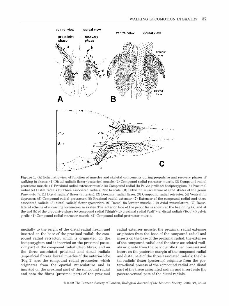
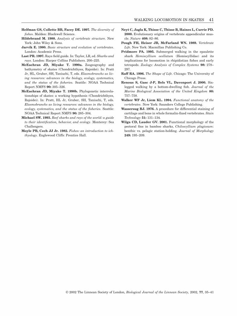
Pelvic fins are attached to the pelvic girdle by twojoints; an antero-lateral joint articulates the radialcondyle of the pelvic girdle with the compound radial(Fig. 2B). The semicircular radial condyle is greatlyenlarged providing a long articulation surface to thecompound radial (Fig. 2B). The proximal part of thecompound radial has a long fossa that accommodatesthe radial condyle (Fig. 2B). The distal end of the com-pound radial has a large terminal posterior processbehind the articulation face of the proximal radial(Fig. 2B). The articulation face of the compound radialis concave, thus accommodating the convex proximalpart of the proximal radial. A posterior joint articulatesthe basal condyle of the pelvic girdle with the basip-terygium, which supports the posterior lobe of the fin.
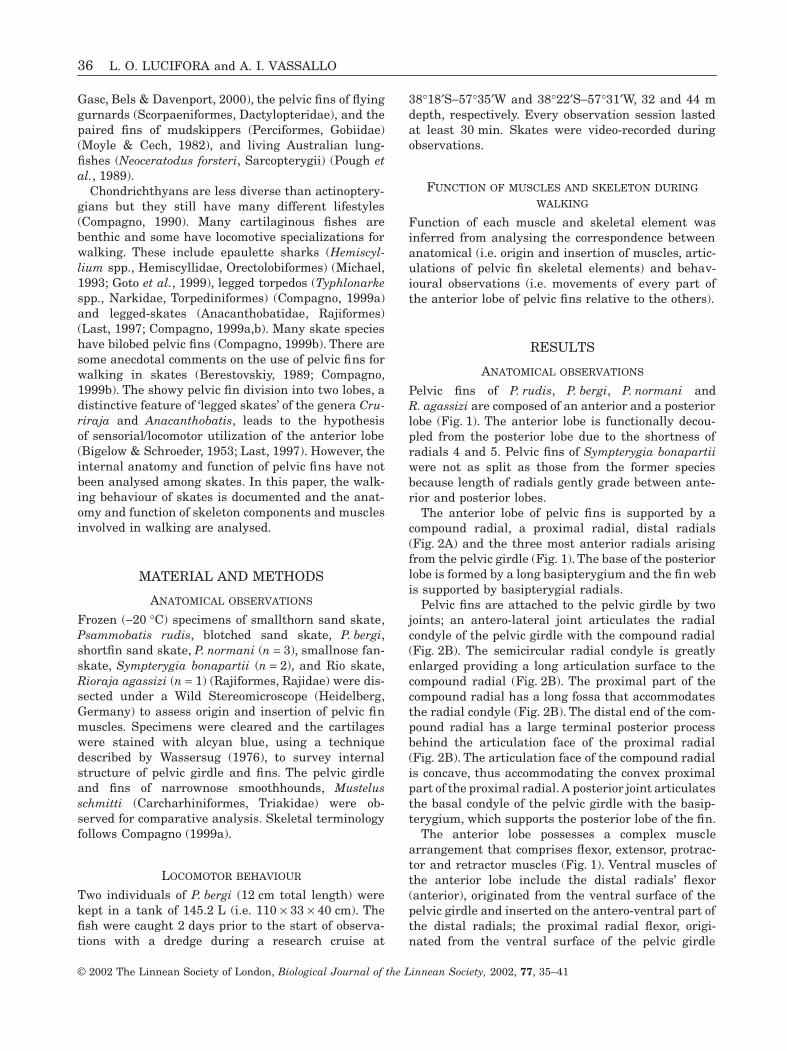
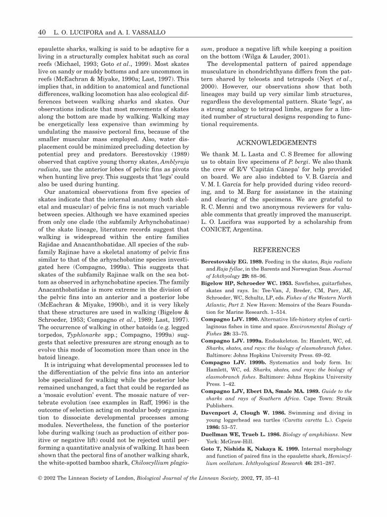
The anterior lobe possesses a complex musclearrangement that comprises flexor, extensor, protrac-tor and retractor muscles (Fig. 1). Ventral muscles ofthe anterior lobe include the distal radials’ flexor(anterior), originated from the ventral surface of thepelvic girdle and inserted on the antero-ventral part ofthe distal radials; the proximal radial flexor, origi-nated from the ventral surface of the pelvic girdle
WALKING LOCOMOTION IN SKATES
37
© 2002 The Linnean Society of London,
Biological Journal of the Linnean Society,
2002,
77
, 35–41
medially to the origin of the distal radial flexor, andinserted on the base of the proximal radial; the com-pound radial retractor, which is originated on thebasipterygium and is inserted on the proximal poste-rior part of the compound radial (deep fibres) and onthe three associated proximal and distal radials(superficial fibres). Dorsal muscles of the anterior lobe(Fig. 1) are: the compound radial protractor, whichoriginates from the epaxial musculature and isinserted on the proximal part of the compound radialand onto the fibres (proximal part) of the proximal
radial extensor muscle; the proximal radial extensororiginates from the base of the compound radial andinserts on the base of the proximal radial; the extensorof the compound radial and the three associated radi-als originate from the pelvic girdle (iliac process) andinsert on the posterior margin of the compound radialand distal part of the three associated radials; the dis-tal radials’ flexor (posterior) originate from the pos-tero-distal process of the compound radial and distalpart of the three associated radials and insert onto thepostero-ventral part of the distal radials.
Figure 1.
(A) Schematic view of function of muscles and skeletal components during propulsive and recovery phases ofwalking in skates. (1) Distal radial’s flexor (posterior) muscle. (2) Compound radial retractor muscle. (3) Compound radialprotractor muscle. (4) Proximal radial extensor muscle (a) Compound radial (b) Pelvic girdle (c) basipterygium (d) Proximalradial (e) Distal radials (f) Three associated radials. Not to scale. (B) Pelvic fin musculature of sand skates of the genus
Psammobatis
. (1) Distal radials’ flexor (anterior). (2) Droximal radial flexor. (3) Compound radial retractor. (4) Ventral findepressor. (5) Compound radial protractor. (6) Proximal radial extensor. (7) Extensor of the compound radial and threeassociated radials. (8) distal radials’ flexor (posterior). (9) Dorsal fin levator muscle. (10) Axial musculature. (C) Dorso-lateral scheme of sprawling locomotion in skates. The anterior lobe of the pelvic fin is shown at the beginning (a) and atthe end (b) of the propulsive phase (c) compound radial (‘thigh’) (d) proximal radial (‘calf ’) (e) distal radials (‘foot’) (f) pelvicgirdle. (1) Compound radial retractor muscle. (2) Compound radial protractor muscle.
38
L. O. LUCIFORA and A. I. VASSALLO
© 2002 The Linnean Society of London,
Biological Journal of the Linnean Society,
2002,
77
, 35–41
The musculature of the posterior lobe is fairly sim-ple (Fig. 1). It is composed by a dorsal fin levatormuscle originated from the epaxial musculature andinserted on the radials, and a ventral fin depressormuscle that originates on the basipterygium andinserts on the radials (Fig. 1).
No substantial differences were observed regardingmuscular and skeletal features of pelvic girdle andfins between the three species of
Psammobatis
and
R.agassizi
. Although pelvic fins of
S. bonapartii
are exter-nally not split into two lobes, no internal differencesregarding the musculature and skeleton of pelvic finswere found with respect to
Psammobatis
spp. and
R.agassizi
.Pelvic fin skeleton and musculature of
M. schmitti
are not different from those of a generalized elasmo-branch. That is, the anterior margin is supported by a
flat compound radial, and the base by the basiptery-gium. The articulation between the compound radialand the distal radials is not movable. The musculatureis fairly simple and is composed of a dorsal levator anda ventral depressor muscle.
L
OCOMOTOR
BEHAVIOUR
The analysis of video-recorded locomotion behaviourof aquarium-reared specimens of
Psammobatis bergi
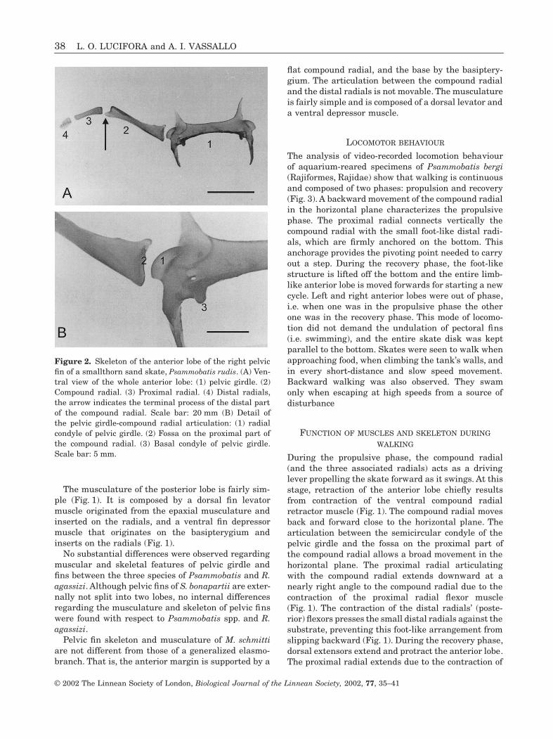
(Rajiformes, Rajidae) show that walking is continuousand composed of two phases: propulsion and recovery(Fig. 3). A backward movement of the compound radialin the horizontal plane characterizes the propulsivephase. The proximal radial connects vertically thecompound radial with the small foot-like distal radi-als, which are firmly anchored on the bottom. Thisanchorage provides the pivoting point needed to carryout a step. During the recovery phase, the foot-likestructure is lifted off the bottom and the entire limb-like anterior lobe is moved forwards for starting a newcycle. Left and right anterior lobes were out of phase,i.e. when one was in the propulsive phase the otherone was in the recovery phase. This mode of locomo-tion did not demand the undulation of pectoral fins(i.e. swimming), and the entire skate disk was keptparallel to the bottom. Skates were seen to walk whenapproaching food, when climbing the tank’s walls, andin every short-distance and slow speed movement.Backward walking was also observed. They swamonly when escaping at high speeds from a source ofdisturbance
F
UNCTION
OF
MUSCLES
AND
SKELETON
DURING
WALKING
During the propulsive phase, the compound radial(and the three associated radials) acts as a drivinglever propelling the skate forward as it swings. At thisstage, retraction of the anterior lobe chiefly resultsfrom contraction of the ventral compound radialretractor muscle (Fig. 1). The compound radial movesback and forward close to the horizontal plane. Thearticulation between the semicircular condyle of thepelvic girdle and the fossa on the proximal part ofthe compound radial allows a broad movement in thehorizontal plane. The proximal radial articulatingwith the compound radial extends downward at anearly right angle to the compound radial due to thecontraction of the proximal radial flexor muscle(Fig. 1). The contraction of the distal radials’ (poste-rior) flexors presses the small distal radials against thesubstrate, preventing this foot-like arrangement fromslipping backward (Fig. 1). During the recovery phase,dorsal extensors extend and protract the anterior lobe.The proximal radial extends due to the contraction of
Figure 2.
Skeleton of the anterior lobe of the right pelvicfin of a smallthorn sand skate,
Psammobatis rudis
. (A) Ven-tral view of the whole anterior lobe: (1) pelvic girdle. (2)Compound radial. (3) Proximal radial. (4) Distal radials,the arrow indicates the terminal process of the distal partof the compound radial. Scale bar: 20 mm (B) Detail ofthe pelvic girdle-compound radial articulation: (1) radialcondyle of pelvic girdle. (2) Fossa on the proximal part ofthe compound radial. (3) Basal condyle of pelvic girdle.Scale bar: 5 mm.
WALKING LOCOMOTION IN SKATES
39
© 2002 The Linnean Society of London,
Biological Journal of the Linnean Society,
2002,
77
, 35–41
the proximal radial extensor muscle. Coincident pro-traction of the compound radial is accomplished bycontraction of a short compound radial protractor mus-cle arising from the axial musculature (Fig. 1).
DISCUSSION
A walking terrestrial vertebrate advances by a succes-sion of steps in which one foot (or hand) is first placedon the ground to develop a thrust that accelerates thebody (propulsive phase of step), and then is removedfrom the ground and protracted (recovery or swingphase) (Walker & Liem, 1994). The structure and func-tion of the anterior pelvic fin lobe of skates resemblethis fundamental pattern of locomotion. In addition,because the proximal ‘limb’ segment (compoundradial) moves back and forth close to the horizontalplane, the two phases of walking resemble the ances-tral tetrapod sprawling locomotion seen in manysalamanders and lizards (Duellman & Trueb, 1986;Walker & Liem, 1994).
Crawling locomotion in epaulette sharks is per-formed by undulating the body and moving alter-nately the slightly modified paired fins (Pridmore,1995; Goto
et al
., 1999). Paired fins of
Hemiscyllium
spp. are not split into proximal, mesial and distalparts (i.e. thigh, calf and foot) (Goto
et al
., 1999) as inskates. As a consequence, the use and internal anat-omy of walking fins of epaulette sharks and skates iscompletely different. Skates do not bend the bodywhile walking: their vertebral column is kept straight
during walking contrasting with the bending reportedin hemiscyllid sharks. Most differences in the muscu-lature of both pectoral and pelvic fins between
Hemis-cyllium ocellatum
and generalized (i.e. swimming)forms were in the size of muscles rather than in thenumber and position of muscles (Goto
et al
., 1999).Goto
et al
. (1999) describe only one additional muscle,the levator pectoralis inferior, in relation to general-ized pectoral fins of other sharks. In contrast, our datashow that a very different and specialized muscula-ture occur in the pelvic fins of skates as compared tothe generalized pelvic fin of a generalized elasmo-branch or teleost fish (see Hildebrand, 1988).
Regarding skeletal features, one of the most strikingstructures of the anterior lobe is the highly developedradial condyle of the pelvic girdle and the deep com-pound radial fossa (Fig. 2), which greatly enable limbmovements in the horizontal plane during both pro-pulsive and swing phases. This condyle is absent orslightly developed in nonwalking elasmobranchs, butit is large and conspicuous in walking elasmobranchs(Goto
et al
., 1999; this study). As indicated by thestructure of the postcranial skeleton in osteolepiformfishes and basal labyrinthodonts (Jarvik, 1980), thearticulation between girdles and paired appendageswas one of the most important features in the evolu-tion of the appendicular skeleton of walking ancestralvertebrates. Hence the functional importance of thistrait in walking skates.
Walking has evolved many times among differentlineages of benthic fishes (Helfman
et al
., 1997). In
Figure 3.
Ventral view of a step succession in a blotched sand skate,
Psammobatis bergi
. In (A) the anterior lobe of theright pelvic fin is in the propulsive phase, while the left anterior lobe is in the recovery phase. The opposite is shown in(B). Scale bars: 30 mm.
40
L. O. LUCIFORA and A. I. VASSALLO
© 2002 The Linnean Society of London,
Biological Journal of the Linnean Society,
2002,
77
, 35–41
epaulette sharks, walking is said to be adaptive for aliving in a structurally complex habitat such as coralreefs (Michael, 1993; Goto et al., 1999). Most skateslive on sandy or muddy bottoms and are uncommon inreefs (McEachran & Miyake, 1990a; Last, 1997). Thisimplies that, in addition to anatomical and functionaldifferences, walking locomotion has also ecological dif-ferences between walking sharks and skates. Ourobservations indicate that most movements of skatesalong the bottom are made by walking. Walking maybe energetically less expensive than swimming byundulating the massive pectoral fins, because of thesmaller muscular mass employed. Also, water dis-placement could be minimized precluding detection bypotential prey and predators. Berestovskiy (1989)observed that captive young thorny skates, Amblyrajaradiata, use the anterior lobes of pelvic fins as pivotswhen hunting live prey. This suggests that ‘legs’ couldalso be used during hunting.
Our anatomical observations from five species ofskates indicate that the internal anatomy (both skel-etal and muscular) of pelvic fins is not much variablebetween species. Although we have examined speciesfrom only one clade (the subfamily Arhynchobatinae)of the skate lineage, literature records suggest thatwalking is widespread within the entire familiesRajidae and Anacanthobatidae. All species of the sub-family Rajinae have a skeletal anatomy of pelvic finssimilar to that of the arhynchobatine species investi-gated here (Compagno, 1999a). This suggests thatskates of the subfamily Rajinae walk on the sea bot-tom as observed in arhynchobatine species. The familyAnacanthobatidae is more extreme in the division ofthe pelvic fins into an anterior and a posterior lobe(McEachran & Miyake, 1990b), and it is very likelythat these structures are used in walking (Bigelow &Schroeder, 1953; Compagno et al., 1989; Last, 1997).The occurrence of walking in other batoids (e.g. leggedtorpedos, Typhlonarke spp.; Compagno, 1999a) sug-gests that selective pressures are strong enough as toevolve this mode of locomotion more than once in thebatoid lineage.
It is intriguing what developmental processes led tothe differentiation of the pelvic fins into an anteriorlobe specialized for walking while the posterior loberemained unchanged, a fact that could be regarded asa ‘mosaic evolution’ event. The mosaic nature of ver-tebrate evolution (see examples in Raff, 1996) is theoutcome of selection acting on modular body organiza-tion to dissociate developmental processes amongmodules. Nevertheless, the function of the posteriorlobe during walking (such as production of either pos-itive or negative lift) could not be rejected until per-forming a quantitative analysis of walking. It has beenshown that the pectoral fins of another walking shark,the white-spotted bamboo shark, Chiloscyllium plagio-
sum, produce a negative lift while keeping a positionon the bottom (Wilga & Lauder, 2001).
The developmental pattern of paired appendagemusculature in chondrichthyans differs from the pat-tern shared by teleosts and tetrapods (Neyt et al.,2000). However, our observations show that bothlineages may build up very similar limb structures,regardless the developmental pattern. Skate ‘legs’, asa strong analogy to tetrapod limbs, argues for a lim-ited number of structural designs responding to func-tional requirements.
ACKNOWLEDGEMENTS
We thank M. L. Lasta and C. S Bremec for allowingus to obtain live specimens of P. bergi. We also thankthe crew of R/V ‘Capitán Cánepa’ for help providedon board. We are also indebted to V. B. García andV. M. I. García for help provided during video record-ing, and to M. Barg for assistance in the stainingand clearing of the specimens. We are grateful toR. C. Menni and two anonymous reviewers for valu-able comments that greatly improved the manuscript.L. O. Lucifora was supported by a scholarship fromCONICET, Argentina.
REFERENCES
Berestovskiy EG. 1989. Feeding in the skates, Raja radiataand Raja fyllae, in the Barents and Norwegian Seas. Journalof Ichthyology 29: 88–96.
Bigelow HP, Schroeder WC. 1953. Sawfishes, guitarfishes,skates and rays. In: Tee-Van, J, Breder, CM, Parr, AE,Schroeder, WC, Schultz, LP, eds. Fishes of the Western NorthAtlantic, Part 2. New Haven: Memoirs of the Sears Founda-tion for Marine Research. 1–514.
Compagno LJV. 1990. Alternative life-history styles of carti-laginous fishes in time and space. Environmental Biology ofFishes 28: 33–75.
Compagno LJV. 1999a. Endoskeleton. In: Hamlett, WC, ed.Sharks, skates, and rays: the biology of elasmobranch fishes.Baltimore: Johns Hopkins University Press. 69–92.
Compagno LJV. 1999b. Systematics and body form. In:Hamlett, WC, ed. Sharks, skates, and rays: the biology ofelasmobranch fishes. Baltimore: Johns Hopkins UniversityPress. 1–42.
Compagno LJV, Ebert DA, Smale MA. 1989. Guide to thesharks and rays of Southern Africa. Cape Town: StruikPublishers.
Davenport J, Clough W. 1986. Swimming and diving inyoung loggerhead sea turtles (Caretta caretta L.). Copeia1986: 53–57.
Duellman WE, Trueb L. 1986. Biology of amphibians. NewYork: McGraw-Hill.
Goto T, Nishida K, Nakaya K. 1999. Internal morphologyand function of paired fins in the epaulette shark, Hemiscyl-lium ocellatum. Ichthyological Research 46: 281–287.
WALKING LOCOMOTION IN SKATES 41
© 2002 The Linnean Society of London, Biological Journal of the Linnean Society, 2002, 77, 35–41
Helfman GS, Collette BB, Facey DE. 1997. The diversity offishes. Malden: Blackwell Science.
Hildebrand M. 1988. Analysis of vertebrate structure. NewYork: John Wiley & Sons.
Jarvik E. 1980. Basic structure and evolution of vertebrates.London: Academic Press.
Last PR. 1997. Rays field guide. In: Taylor, LR, ed. Sharks andrays. London: Harper Collins Publishers. 200–225.
McEachran JD, Miyake T. 1990a. Zoogeography andbathymetry of skates (Chondrichthyes, Rajoidei). In: PrattJr, HL, Gruber, SH, Taniuchi, T, eds. Elasmobranchs as liv-ing resources: advances in the biology, ecology, systematics,and the status of the fisheries. Seattle: NOAA TechnicalReport NMFS 90: 305–326.
McEachran JD, Miyake T. 1990b. Phylogenetic interrela-tionships of skates: a working hypothesis (Chondrichthyes,Rajoidei). In: Pratt, HL Jr, Gruber, SH, Taniuchi, T, eds.Elasmobranchs as living resources: advances in the biology,ecology, systematics, and the status of the fisheries. Seattle:NOAA Technical Report NMFS 90: 285–304.
Michael SW. 1993. Reef sharks and rays of the world: a guideto their identification, behavior, and ecology. Monterey: SeaChallengers.
Moyle PB, Cech JJ Jr. 1982. Fishes: an introduction to ich-thyology. Englewood Cliffs: Prentice Hall.
Neyt C, Jagla K, Thisse C, Thisse B, Haines L, Currie PD.2000. Evolutionary origins of vertebrate appendicular mus-cle. Nature 408: 82–86.
Pough FH, Heiser JB, McFarland WN. 1989. VertebrateLife. New York: Macmillan Publishing Co.
Pridmore PA. 1995. Submerged walking in the epauletteshark Hemiscyllium ocellatum (Hemiscyllidae) and itsimplications for locomotion in rhipidistian fishes and earlytetrapods. Zoology: Analysis of Complex Systems 98: 278–297.
Raff RA. 1996. The Shape of Life. Chicago: The University ofChicago Press.
Renous S, Gasc J-P, Bels VL, Davenport J. 2000. Six-legged walking by a bottom-dwelling fish. Journal of theMarine Biological Association of the United Kingdom 80:757–758.
Walker WF Jr, Liem KL. 1994. Functional anatomy of thevertebrates. New York: Saunders College Publishing.
Wassersug RJ. 1976. A procedure for differential staining ofcartilage and bone in whole formalin-fixed vertebrates. StainTechnology 51: 131–134.
Wilga CD, Lauder GV. 2001. Functional morphology of thepectoral fins in bamboo sharks, Chiloscyllium plagiosum:benthic vs. pelagic station-holding. Journal of Morphology249: 195–209.