Embed Size (px)
Citation preview

Krithika Rangarajan, Chandan J Das, Atin Kumar, Arun Kumar Gupta
Krithika Rangarajan, Chandan J Das, Atin Kumar, Arun Ku-mar Gupta, Department of Radiodiagnosis, All India Institute of Medical Sciences, Ansari Nagar East, New Delhi 110029, IndiaAuthor contributions: Rangarajan K and Das CJ wrote most of the manuscript text as well as edited the images; Kumar A and Gupta AK provided valuable inputs, edited the manuscript and approved the content.Correspondence to: Chandan J Das, MD, Department of Radiodiagnosis, All India Institute of Medical Sciences, Ansari Nagar, Room number 60, New Delhi 110029, India. [email protected]: +91-11-26594868Received: January 9, 2014 Revised: July 13, 2014Accepted: July 27, 2014Published online: September 28, 2014
AbstractRecognition and characterization of central nervous system infections poses a formidable challenge to the neuro-radiologist. Imaging plays a vital role, the lesions typically being relatively inaccessible to tisue sampling. The results of an accurate diagnosis are endlessly re-warding, given the availability of excellent pharmaco-logical regimen. The availability of numerous magnetic resonance (MR) sequences which provide functional and molecular information is a powerful tool in the hands of the radiologist. However, the plethora of se-quences and the possibilities on each sequence is also intimidating, and often confusing as well as time con-suming. While a large number of reviews have already described in detail the possible imaging findings in each infection, we intend to classify infections based on their imaging characteristics. In this review we describe an algorithm for first classifying the imaging findings into patterns based on basic MR sequences (T1, T2 and enhancement pattern with Gadolinium), and then sub-classify them based on more advanced molecular and functional sequences (Diffusion, Perfusion, Susceptibili-ty imaging, MR Spectroscopy). This patterned approach
is intended as a guide to radiologists in-training and in-practice for quickly narrowing their list of differentials when faced with a clinical challenge. The entire content of the article has also been summarised in the form of flow-charts for the purpose of quick reference.
© 2014 Baishideng Publishing Group Inc. All rights reserved.
Key words: Central nervous system; Infection; Magnetic resonance imaging; Magnetic resonance spectroscopy; Perfusion weighted magnetic resonance imaging; Diffu-sion weighted magnetic resonance imaging
Core tip: The plethora of magnetic resonance sequenc-es available with the radiologist today provides a wealth of information about anatomical, pathological, physi-ological, functional and molecular aspects of the brain. While this provides an opportunity to transform patient management, the vast number of possibilities can be bewildering, particularly for the radiologist in-training. It is often easy to get lost in the details while forgetting the larger picture. In this article we first classify the in-fections into broad imaging patterns, and subsequently sub-classify them based on more advanced sequences (molecular and functional imaging). The flow-charts in the article are intended as a source of quick reference to the radiologist when faced with a clinical challenge.
Rangarajan K, Das CJ, Kumar A, Gupta AK. MRI in central nervous system infections: A simplified patterned approach. World J Radiol 2014; 6(9): 716-725 Available from: URL: http://www.wjgnet.com/1949-8470/full/v6/i9/716.htm DOI: http://dx.doi.org/10.4329/wjr.v6.i9.716
INTRODUCTIONCentral nervous system (CNS) infections are a significant
REVIEW
World Journal of RadiologyW J R
Submit a Manuscript: http://www.wjgnet.com/esps/Help Desk: http://www.wjgnet.com/esps/helpdesk.aspxDOI: 10.4329/wjr.v6.i9.716
World J Radiol 2014 September 28; 6(9): 716-725ISSN 1949-8470 (online)
© 2014 Baishideng Publishing Group Inc. All rights reserved.
716 September 28, 2014|Volume 6|Issue 9|WJR|www.wjgnet.com
MRI in central nervous system infections: A simplified patterned approach

cause of mortality and morbidity world-wide. This is par-ticularly true owing to its association with conditions of immunological compromise and the increasing incidence of human immunodeficiency virus (HIV) infection is fur-ther adding to the problem[1]. Today with the availability of excellent antimicrobials, many of these disorders are potentially treatable, making early recognition imperative. Like in other disorders of the CNS, non-invasive imaging based diagnosis is the key as possibility of a tissue diag-nosis by means of fine needle aspiration cytology (FNAC) or biopsy is difficult. Early diagnosis will also help to minimize long term complications related to the disease and its treatment.
The primary imaging modality, like in most CNS disor-ders is magnetic resonance imaging (MRI)[2]. Coming to an exact etiological agent on the basis of conventional MRI sequences with Gadolinium enhancement is always difficult due to overlapping imaging characteristics. With the possi-bility of molecular and functional imaging with newer MRI techniques however, the radiologist today is better equipped to handle this dilemma. Though the use of such multiple MRI sequences adds lots of information to narrow the differential possibilities, this vast information is difficult to recall when faced with a clinical problem.
The purpose of this review is to provide a rational MRI approach to narrow the list of differentials, to quickly classify and characterize CNS infections. The flow-charts presented in this review guides the radiolo-gist to first recognize the pattern of findings on routine MRI sequences and subsequently narrow the differential diagnosis based on the addition of other MR parameters such as diffusion weighted imaging (DWI) and MR spec-
troscopy (MRS).
CLASSIFICATIONMost infections in the CNS may be classified in one of the following categories based on their T1, T2 and con-trast enhancement characteristics (Figure 1) as follows (an image demonstrating a typical lesion in each category has been provided in Figures 2-5): Ring enhancing lesions (Figure 2), Basal ganglia space occupying lesions (Figure 3), Grey matter hyperintensities (Figure 4), White matter hyperintensities (Figure 5).
RING ENHANCING LESIONSPeripheral ring-like enhancement is a common finding in CNS imaging. Ring enhancing lesions on conventional MRI sequences have a long list of differentials ranging from infectious processes to high grade necrotic neo-plasm. Glioblastoma multiforme represents the most im-portant condition. Abscesses are usually associated with a thin smooth rim, in contrast to the nodular irregular rim seen in Glioblastoma multiforme. Satellite lesions are commonly seen in abscesses, unlike necrotic neoplasm[1]. Recent work by some investigators has suggested a role for susceptibility weighted imaging (SWI) in this differen-tiation. They found that a smooth, complete rim of sus-ceptibility is seen in abscesses in contrast to incomplete irregular rims seen in necrotic neoplasm[3].
All mature abscesses whether bacterial, fungal or pyogenic are hypointense on T1, hyperintense on T2 and show ring enhancement following intravenous Gado-
Rangarajan K et al . Patterned approach to CNS infections
717 September 28, 2014|Volume 6|Issue 9|WJR|www.wjgnet.com
Conventional MRI (T1, T2, T1 postcontrast)
Ring enhancing lesion
Figure 1 Classification of abnormalities on conventional magnetic resonance imaging sequences in suspected central nervous system infections.
Basal ganglia solr White matter T2 hyperintensity
Grey matter T2 hyperintensity
Figure 2 Ring enhancing lesions. T1 (A), T2 (B) and T1 (C) post gadolinium images from the magnetic resonance of an acutely ill child with fever. A ring enhancing lesion is seen in the right frontal lobe. This patient had a bacterial brain abscess. The differential diagnosis of this appearance would include a brain abscess of any etiology.
A B C
linium injection. Further differentiation of abscesses for possible etiological cause may be made as follows.
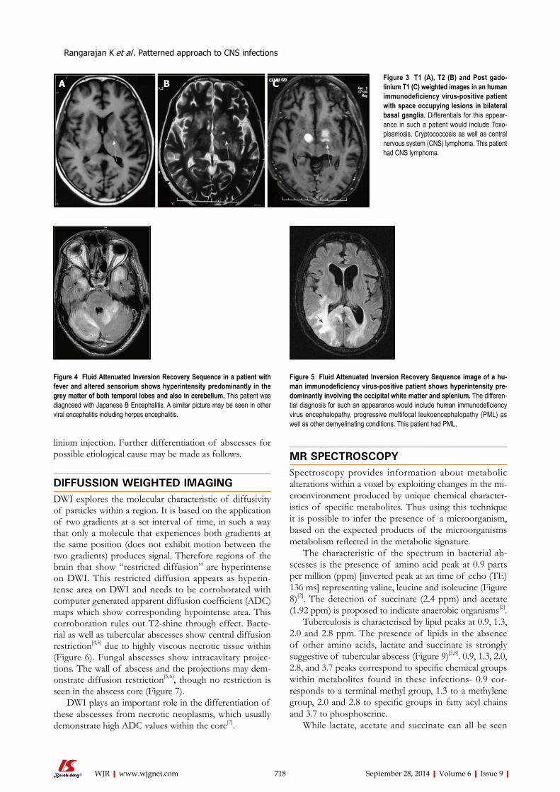
DIFFUSSION WEIGHTED IMAGINGDWI explores the molecular characteristic of diffusivity of particles within a region. It is based on the application of two gradients at a set interval of time, in such a way that only a molecule that experiences both gradients at the same position (does not exhibit motion between the two gradients) produces signal. Therefore regions of the brain that show “restricted diffusion” are hyperintense on DWI. This restricted diffusion appears as hyperin-tense area on DWI and needs to be corroborated with computer generated apparent diffusion coefficient (ADC) maps which show corresponding hypointense area. This corroboration rules out T2-shine through effect. Bacte-rial as well as tubercular abscesses show central diffusion restriction[4,5] due to highly viscous necrotic tissue within (Figure 6). Fungal abscesses show intracavitary projec-tions. The wall of abscess and the projections may dem-onstrate diffusion restriction[5,6], though no restriction is seen in the abscess core (Figure 7).
DWI plays an important role in the differentiation of these abscesses from necrotic neoplasms, which usually demonstrate high ADC values within the core[7].
MR SPECTROSCOPYSpectroscopy provides information about metabolic alterations within a voxel by exploiting changes in the mi-croenvironment produced by unique chemical character-istics of specific metabolites. Thus using this technique it is possible to infer the presence of a microorganism, based on the expected products of the microorganisms metabolism reflected in the metabolic signature.
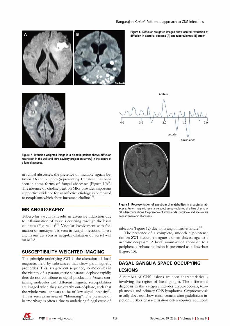
The characteristic of the spectrum in bacterial ab-scesses is the presence of amino acid peak at 0.9 parts per million (ppm) [inverted peak at an time of echo (TE) 136 ms] representing valine, leucine and isoleucine (Figure 8)[2]. The detection of succinate (2.4 ppm) and acetate (1.92 ppm) is proposed to indicate anaerobic organisms[2].
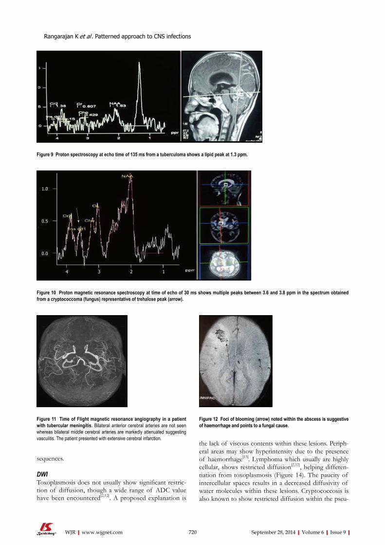
Tuberculosis is characterised by lipid peaks at 0.9, 1.3, 2.0 and 2.8 ppm. The presence of lipids in the absence of other amino acids, lactate and succinate is strongly suggestive of tubercular abscess (Figure 9)[5,8]. 0.9, 1.3, 2.0, 2.8, and 3.7 peaks correspond to specific chemical groups within metabolites found in these infections- 0.9 cor-responds to a terminal methyl group, 1.3 to a methylene group, 2.0 and 2.8 to specific groups in fatty acyl chains and 3.7 to phosphoserine.
While lactate, acetate and succinate can all be seen
718 September 28, 2014|Volume 6|Issue 9|WJR|www.wjgnet.com
A B CFigure 3 T1 (A), T2 (B) and Post gado-linium T1 (C) weighted images in an human immunodeficiency virus-positive patient with space occupying lesions in bilateral basal ganglia. Differentials for this appear-ance in such a patient would include Toxo-plasmosis, Cryptococcosis as well as central nervous system (CNS) lymphoma. This patient had CNS lymphoma.
Figure 4 Fluid Attenuated Inversion Recovery Sequence in a patient with fever and altered sensorium shows hyperintensity predominantly in the grey matter of both temporal lobes and also in cerebellum. This patient was diagnosed with Japanese B Encephalitis. A similar picture may be seen in other viral encephalitis including herpes encephalitis.
Figure 5 Fluid Attenuated Inversion Recovery Sequence image of a hu-man immunodeficiency virus-positive patient shows hyperintensity pre-dominantly involving the occipital white matter and splenium. The differen-tial diagnosis for such an appearance would include human immunodeficiency virus encephalopathy, progressive multifocal leukoencephalopathy (PML) as well as other demyelinating conditions. This patient had PML.
Rangarajan K et al . Patterned approach to CNS infections
infection (Figure 12) due to its angioinvasive nature [11].The presence of a complete, smooth hypointense
rim on SWI favours a diagnosis of an abscess against a necrotic neoplasm. A brief summary of approach to a peripherally enhancing lesion is presented as a flowchart (Figure 13).
BASAL GANGLIA SPACE OCCUPYING LESIONSA number of CNS lesions are seen characteristically involving the region of basal ganglia. The differential diagnosis in this category includes cryptococcosis, toxo-plasmosis and primary CNS lymphoma. Cryptococcosis usually does not show enhancement after gadolinium in-jection.Further characterisation often requires additional
in fungal abscesses, the presence of multiple signals be-tween 3.6 and 3.8 ppm (representing Trehalose) has been seen in some forms of fungal abscesses (Figure 10)[5]. The absence of choline peak on MRS provides important supportive evidence for an infective etiology as compared to neoplasms which show increased choline[7,9].
MR ANGIOGRAPHYTubercular vasculitis results in extensive infarction due to inflammation of vessels coursing through the basal exudates (Figure 11)[10]. Vascular involvement with for-mation of aneurysms is seen in fungal infections. These aneurysms are seen as irregular dilatation of vessel wall on MRA.
SUSCEPTIBILITY WEIGHTED IMAGING The principle underlying SWI is the alteration of local magnetic field by substances that show paramagnetic properties. This is a gradient sequence, so molecules in the vicinity of a paramagnetic substance dephase rapidly, thus do not contribute to signal production. Voxels con-taining molecules with different magnetic susceptibilities are imaged when they are exactly out-of-phase, such that the whole voxel appears to be of low signal intensity[2]. This is seen as an area of “blooming”. The presence of haemorrhage is often a clue to underlying fungal cause of
719 September 28, 2014|Volume 6|Issue 9|WJR|www.wjgnet.com
Figure 6 Diffusion weighted images show central restriction of diffusion in bacterial abscess (A) and tuberculomas (B) arrow.
Figure 7 Diffusion weighted image in a diabetic patient shows diffusion restriction in the wall and intra-cavitary projection (arrow) in the centre of a fungal abscess.
Acetate
4.0 3.0 2.0 1.0 0.0
Lactate
Amino acids
Figure 8 Representation of spectrum of metabolites in a bacterial ab-scess. Proton magnetic resonance spectroscopy obtained at a time of echo of 30 milliseconds shows the presence of amino acids. Succinate and acetate are seen in anaerobic abscesses.
Rangarajan K et al . Patterned approach to CNS infections
A B
sequences.
DWIToxoplasmosis does not usually show significant restric-tion of diffusion, though a wide range of ADC value have been encountered[2,12]. A proposed explanation is
the lack of viscous contents within these lesions. Periph-eral areas may show hyperintensity due to the presence of haemorrhage[13]. Lymphoma which usually are highly cellular, shows restricted diffusion[2,12], helping differen-tiation from toxoplasmosis (Figure 14). The paucity of intercellular spaces results in a decreased diffusivity of water molecules within these lesions. Cryptococcosis is also known to show restricted diffusion within the pseu-
720 September 28, 2014|Volume 6|Issue 9|WJR|www.wjgnet.com
Figure 9 Proton spectroscopy at echo time of 135 ms from a tuberculoma shows a lipid peak at 1.3 ppm.
1.0
0.5
0.0
4 3 2 1
Figure 10 Proton magnetic resonance spectroscopy at time of echo of 30 ms shows multiple peaks between 3.6 and 3.8 ppm in the spectrum obtained from a cryptococcoma (fungus) representative of trehalose peak (arrow).
Figure 11 Time of Flight magnetic resonance angiography in a patient with tubercular meningitis. Bilateral anterior cerebral arteries are not seen whereas bilateral middle cerebral arteries are markedly attenuated suggesting vasculitis. The patient presented with extensive cerebral infarction.
Figure 12 Foci of blooming (arrow) noted within the abscess is suggestive of haemorrhage and points to a fungal cause.
Rangarajan K et al . Patterned approach to CNS infections

docysts[2] owing to the viscosity of gelatinous material. Cryptococcomas have been shown to exhibit peripheral diffusion restriction akin to a necrotic brain tumour[14], though often they may not show any restricted diffu-sion[12] (Figure 15).
MRSPresence of peak between 3.6 to 3.8 ppm (trehalose) has been observed in cryptococcosis (which is a fungus)[5]
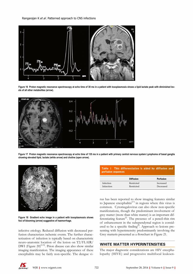
(Figure 10). Toxoplasma lesions (Figure 16) show mark-edly elevated lipid and lactate with diminished levels of all other metabolites[2]. Lymphoma (Figure 17) shows mild to moderate increase in lipid and lactate with mark-edly elevated choline peak[2].
MR perfusion Toxoplasmosis shows normal or decreased cerebral blood volume (CBV). Primary CNS lymphoma on the other
hand shows elevated CBV[12].
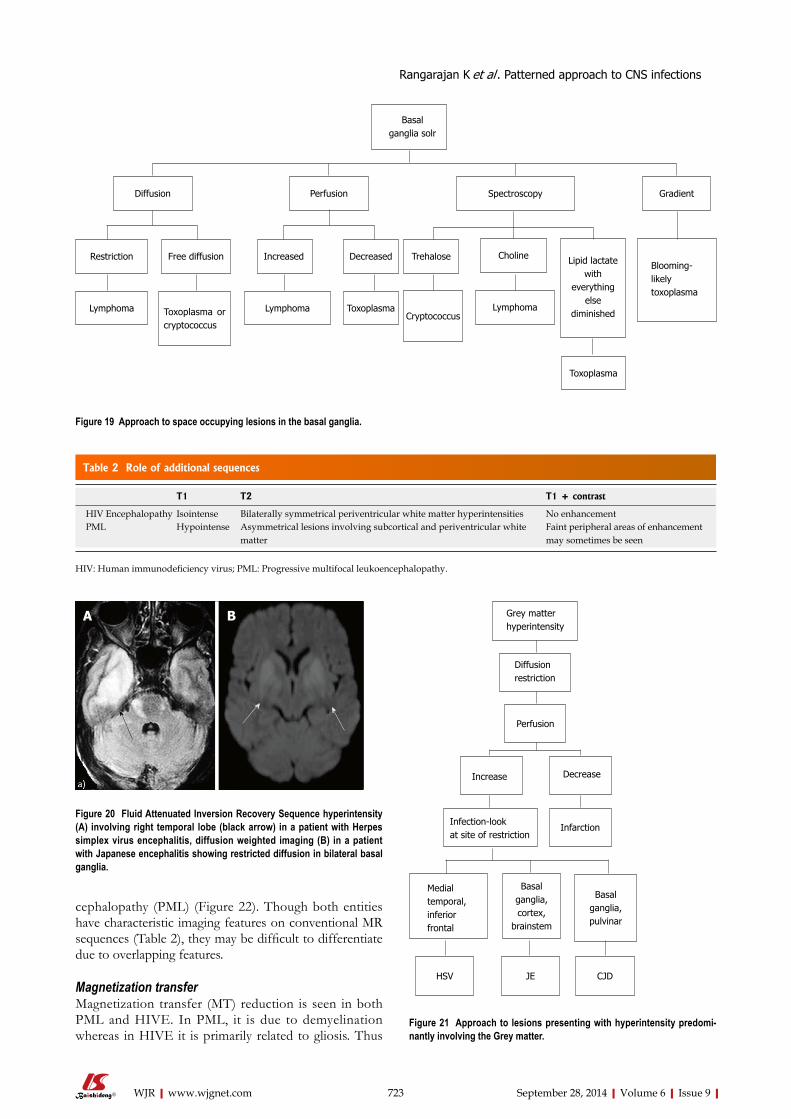
SWIPresence of hemorrhage (blooming on gradient echo se-quences) points towards toxoplasmosis, as lymphoma rarely show hemorrhage before treatment[12,15] (Figure 18). A brief summary of the approach to space occupying lesions in the basal ganglia is presented as a flowchart in Figure 19.
GREY MATTER HYPERINTENSITYT2/ FLAIR hyperintensity involving the grey matter may be seen in encephalitis as well as infarction. This differ-entiation is aided by diffusion and perfusion sequences (Table 1).
Diffusion and perfusionReduced diffusion with increased perfusion points to an
721 September 28, 2014|Volume 6|Issue 9|WJR|www.wjgnet.com
Ring enhancing
lesion
Blooming on gradient
Likelyfungaletiology
Diffusionrestriction Spectroscopy
Centralrestriction
Tubercularor bacterial Fungal Bacterial Tubercular Fungal
Aminoacid peaks
TrehaloseLipid peak
Rule out malignancy
Choline
Peripheral restriction, restriction in projections
Figure 13 Approach to ring enhancing lesions.
Figure 14 Diffusion weighted image from the Brain magnetic resonance im-aging of an human immunodeficiency virus-positive patient with lymphoma shows restriction in the right lentiform nucleus and the thalamus (arrow).
Figure 15 Apparent diffusion coefficient map showing free diffusion with-in a cryptococcoma (arrow) in a human immunodeficiency virus-positive patient.
Rangarajan K et al . Patterned approach to CNS infections
infective etiology. Reduced diffusion with decreased per-fusion characterizes ischemic events. The further charac-terization of infection is typically based on characteristic neuro-anatomic location of the lesion on T2/FLAIR/ DWI (Figure 20)[2,16]. Prion disease can also show similar imaging manifestation. The imaging appearance of these encephalitis may be fairly non-specific. The dengue vi-
rus has been reported to show imaging features similar to Japanese encephalitis[17] in regions where this virus is common. Cytomegalovirus can also show non-specific manifestations, though the predominant involvement of grey matter (more than white matter) is an important dif-ferentiating feature[1]. The presence of a pencil-thin rim of enhancement in the subependymal region is consid-ered to be a specific finding[1]. Approach to lesions pre-senting with hyperintensity predominantly involving the Grey matteris presented as a flowchart in Figure 21.
WHITE MATTER HYPERINTENSITIESThe major diagnostic considerations are HIV encepha-lopathy (HIVE) and progressive multifocal leukoen-
722 September 28, 2014|Volume 6|Issue 9|WJR|www.wjgnet.com
3
2
1
0
4 3 2 1
Figure 16 Proton magnetic resonance spectroscopy at echo time of 30 ms in a patient with toxoplasmosis shows a lipid lactate peak with diminished lev-els of all other metabolites (arrow).
0.4
0.2
0.0
-0.2
4 3 2 1
Figure 17 Proton magnetic resonance spectroscopy at echo time of 135 ms in a patient with primary central nervous system Lymphoma of basal ganglia showing elevated lipid, lactate (white arrow) and choline (open arrow).
Figure 18 Gradient echo image in a patient with toxoplasmosis shows foci of blooming (arrow) suggestive of haemorrhage.
Table 1 This differentiation is aided by diffusion and perfusion sequences
Diffusion Perfusion
Infection Restricted IncreasedInfarction Restricted Decreased
Rangarajan K et al . Patterned approach to CNS infections
Grey matter hyperintensity
Diffusionrestriction
Perfusion
Decrease
Infarction
Basal ganglia,pulvinar
Increase
Infection-lookat site of restriction
Medial temporal,inferiorfrontal
HSV JE CJD
Basal ganglia,cortex,
brainstem
Figure 21 Approach to lesions presenting with hyperintensity predomi-nantly involving the Grey matter.
cephalopathy (PML) (Figure 22). Though both entities have characteristic imaging features on conventional MR sequences (Table 2), they may be difficult to differentiate due to overlapping features.
Magnetization transferMagnetization transfer (MT) reduction is seen in both PML and HIVE. In PML, it is due to demyelination whereas in HIVE it is primarily related to gliosis. Thus
723 September 28, 2014|Volume 6|Issue 9|WJR|www.wjgnet.com
Diffusion
Restriction
Lymphoma
Free diffusion
Perfusion
Increased
LymphomaToxoplasma or cryptococcus
Decreased
Gradient
Toxoplasma
Spectroscopy
Trehalose Choline
CryptococcusLymphoma
Toxoplasma
Blooming-likelytoxoplasma
Lipid lactatewith
everything else
diminished
Basalganglia solr
Figure 19 Approach to space occupying lesions in the basal ganglia.
Figure 20 Fluid Attenuated Inversion Recovery Sequence hyperintensity (A) involving right temporal lobe (black arrow) in a patient with Herpes simplex virus encephalitis, diffusion weighted imaging (B) in a patient with Japanese encephalitis showing restricted diffusion in bilateral basal ganglia.
A B
Table 2 Role of additional sequences
T1 T2 T1 + contrast
HIV Encephalopathy Isointense Bilaterally symmetrical periventricular white matter hyperintensities No enhancementPML Hypointense Asymmetrical lesions involving subcortical and periventricular white
matterFaint peripheral areas of enhancement may sometimes be seen
HIV: Human immunodeficiency virus; PML: Progressive multifocal leukoencephalopathy.
Rangarajan K et al . Patterned approach to CNS infections

Figure 22 Fluid Attenuated Inversion Recovery Sequence image of a patient with (A) human immunodeficiency virus encephalopathy showing symmetrical periventricular white matter hyperintensity and (B) progres-sive multifocal leukoencephalopathy showing asymmetrical involvement of white matter, predominantly posterior subcortical white matter, with extension into the periventricular region.
A B
larger reduction in MT has been observed in PML as compared to HIVE[18]. The major role of MT sequence in this setting is in early detection of disease.
Diffusion and diffusion tensor imagingFractional anisotropy is seen to be reduced in HIVE and PML before the morphologic changes in conventional se-quences[19]. Reduced diffusion is seen in the periphery and free diffusion in the centre of PML lesions[12] (Figure 23).
MRSReduction of N-acetylaspartate is seen in HIVE even be-
fore the onset of symptoms. Raised choline and myoino-sitol is also seen in the spectra. A summary of suggested approach to these white matter lesions is presented as a flowchart in Figure 24.
CONCLUSIONImaging features of CNS infections constitute a complex myriad. Their classification based on conventional MRI sequences, may provide a quick guide to narrowing the differential diagnosis followed by further sub-differenti-ation into single etiology using advanced MRI sequences
724 September 28, 2014|Volume 6|Issue 9|WJR|www.wjgnet.com
Figure 23 Diffusion weighted image in a patient with progressive multifo-cal leukoencephalopathy showing peripheral diffusion restriction.
HIV PML
White matter hyperintensity
HIVPML
Magnetizationtransfer
Mild-moderate redictionSevere reduction
Location of T2/flair hyperintensity
Symmetrical periventricular,subcortical sparing
Assymetrical, posterior location, subcortical
involvement
Figure 24 Approach to lesions presenting with hyperintensity predominantly involving the white matter. PML: Progressive multifocal leukoencephalopathy; HIV: Human immunodeficiency virus.
Rangarajan K et al . Patterned approach to CNS infections
and techniques.
REFERENCES1 Aiken AH. Central nervous system infection. Neuroimaging
Clin N Am 2010; 20: 557-580 [PMID: 20974376 DOI: 10.1016/j.nic.2010.07.011]
2 Whiteman ML, Bowen BC, Post MJ, Bell MD. Intracranial infections. In Scott W Atlas, editor. Magnetic Resonance Im-aging of Brain and Spine. 3rd ed. Philadelphia: Lippincott Williams and Wilkins, 2002: 1099-177
3 Toh CH, Wei KC, Chang CN, Hsu PW, Wong HF, Ng SH, Castillo M, Lin CP. Differentiation of pyogenic brain abscesses from necrotic glioblastomas with use of suscep-tibility-weighted imaging. AJNR Am J Neuroradiol 2012; 33: 1534-1538 [PMID: 22422181 DOI: 10.3174/ajnr.A2986]
4 Desprechins B, Stadnik T, Koerts G, Shabana W, Breucq C, Osteaux M. Use of diffusion-weighted MR imaging in dif-ferential diagnosis between intracerebral necrotic tumors and cerebral abscesses. AJNR Am J Neuroradiol 1999; 20: 1252-1257 [PMID: 10472982]
5 Luthra G, Parihar A, Nath K, Jaiswal S, Prasad KN, Husain N, Husain M, Singh S, Behari S, Gupta RK. Comparative evaluation of fungal, tubercular, and pyogenic brain ab-scesses with conventional and diffusion MR imaging and proton MR spectroscopy. AJNR Am J Neuroradiol 2007; 28: 1332-1338 [PMID: 17698537 DOI: 10.3174/ajnr.A0548]
6 Gaviani P, Schwartz RB, Hedley-Whyte ET, Ligon KL, Robicsek A, Schaefer P, Henson JW. Diffusion-weighted imaging of fungal cerebral infection. AJNR Am J Neurora-diol 2005; 26: 1115-1121 [PMID: 15891169]
7 Lai PH, Ho JT, Chen WL, Hsu SS, Wang JS, Pan HB, Yang CF. Brain abscess and necrotic brain tumor: discrimina-tion with proton MR spectroscopy and diffusion-weighted imaging. AJNR Am J Neuroradiol 2002; 23: 1369-1377 [PMID: 12223380]
8 Gupta RK, Roy R, Dev R, Husain M, Poptani H, Pandey R, Kishore J, Bhaduri AP. Finger printing of Mycobacterium tuberculosis in patients with intracranial tuberculomas by using in vivo, ex vivo, and in vitro magnetic resonance spectroscopy. Magn Reson Med 1996; 36: 829-833 [PMID: 8946348]
9 Lai PH, Weng HH, Chen CY, Hsu SS, Ding S, Ko CW, Fu
JH, Liang HL, Chen KH. In vivo differentiation of aerobic brain abscesses and necrotic glioblastomas multiforme us-ing proton MR spectroscopic imaging. AJNR Am J Neurora-diol 2008; 29: 1511-1518 [PMID: 18499784 DOI: 10.3174/ajnr.A1130]
10 Trivedi R, Saksena S, Gupta RK. Magnetic resonance imaging in central nervous system tuberculosis. Indian J Radiol Imaging 2009; 19: 256-265 [PMID: 19881100 DOI: 10.4103/0971-3026.57205]
11 Jain KK, Mittal SK, Kumar S, Gupta RK. Imaging features of central nervous system fungal infections. Neurol In-dia 2007; 55: 241-250 [PMID: 17921653]
12 Smith AB, Smirniotopoulos JG, Rushing EJ. From the archives of the AFIP: central nervous system infections as-sociated with human immunodeficiency virus infection: radiologic-pathologic correlation. Radiographics 2008; 28: 2033-2058 [PMID: 19001657 DOI: 10.1148/rg.287085135]
13 Lee GT, Antelo F, Mlikotic AA. Best cases from the AFIP: cerebral toxoplasmosis. Radiographics 2009; 29: 1200-1205 [PMID: 19605667 DOI: 10.1148/rg.294085205]
14 Ho TL, Lee HJ, Lee KW, Chen WL. Diffusion-weighted and conventional magnetic resonance imaging in cerebral cryptococcoma. Acta Radiol 2005; 46: 411-414 [PMID: 16134319]
15 Trenkwalder P, Trenkwalder C, Feiden W, Vogl TJ, Einhäu-pl KM, Lydtin H. Toxoplasmosis with early intracerebral hemorrhage in a patient with the acquired immunodeficien-cy syndrome. Neurology 1992; 42: 436-438 [PMID: 1736179]
16 Ukisu R, Kushihashi T, Tanaka E, Baba M, Usui N, Fuji-sawa H, Takenaka H. Diffusion-weighted MR imaging of early-stage Creutzfeldt-Jakob disease: typical and atypical manifestations. Radiographics 2006; 26 Suppl 1: S191-S204 [PMID: 17050516 DOI: 10.1148/rg.26si065503]
17 Borawake K, Prayag P, Wagh A, Dole S. Dengue encephalitis. In-dian J Crit Care Med 2011; 15: 190-193 [PMID: 22013316 DOI: 10.4103/0972-5229.84896]
18 Ernst T, Chang L, Witt M, Walot I, Aronow H, Leonido-Yee M, Singer E. Progressive multifocal leukoencephalopathy and human immunodeficiency virus-associated white mat-ter lesions in AIDS: magnetization transfer MR imaging. Ra-diology 1999; 210: 539-543 [PMID: 10207441]
19 Pomara N, Crandall DT, Choi SJ, Johnson G, Lim KO. White matter abnormalities in HIV-1 infection: a diffusion ten-sor imaging study. Psychiatry Res 2001; 106: 15-24 [PMID: 11231096]
P- Reviewer: Asensi VC, Mueller WC, Radenovic L S- Editor: Wen LL L- Editor: A E- Editor: Lu YJ
725 September 28, 2014|Volume 6|Issue 9|WJR|www.wjgnet.com
Rangarajan K et al . Patterned approach to CNS infections

© 2014 Baishideng Publishing Group Inc. All rights reserved.
Published by Baishideng Publishing Group Inc8226 Regency Drive, Pleasanton, CA 94588, USA
Telephone: +1-925-223-8242Fax: +1-925-223-8243
E-mail: [email protected] Desk: http://www.wjgnet.com/esps/helpdesk.aspx
http://www.wjgnet.com