Embed Size (px)
Citation preview
1
VPM 403: INTRODUCTORY VETERINARY IMMUNOLOGY
LECTURE NOTES
DR M. A. OYEKUNLE (SP 1071)
DR. O. E. OJO
DR. M. AGBAJE
DEPARTMENT OF VETERINARY MICROBIOLOGY AND
PARASITOLOGY
COLLEGE OF VETERINARY MEDICINE
UNIVERSITY OF AGRICULTURE, ABEOKUTA
2
VPM 403: INTRODUCTORY VETERINARY IMMUNOLOGY COURSE OUTLINE Topics Lecturer History of Immunology M. A. Oyekunle Immunological concepts/terminologies M. A. Oyekunle Types of immunity Organs and cells involved in immune responses M. A. Oyekunle Iimmunoglobulin structures, classifications and functions M. A. Oyekunle The major histocompactibility complex M. A. Oyekunle Gene regulation of immune responses M. A. Oyekunle Antigen and requirements for antigenicity O. E. Ojo Adjuvants O. E. Ojo Antigen-antibody reaction O. E. Ojo Immune complexes O. E. Ojo Autoimmunity O. E. Ojo Autoimmune diseases O. E. Ojo Cytokines O. E. Ojo Antigenic variations O. E. Ojo Immune response to tumour O. E. Ojo Vaccine types and function O. E. Ojo Vaccine production and vaccination O. E. Ojo Complement systems M. Agbaje Immunological tolerance M. Agbaje Immunosuppression M. Agbaje Immune responses to bacterial M. Agbaje Immune responses to viral infections M. Agbaje Immune response to fungal and parasitic infections M. Agbaje Hypersensitivity reactions M. Agbaje
3
IMMUNOLOGY
DR. M. A. OYEKUNLE
Immunology is an area of science which helps in understanding the way by which animals gained protection from disease causing agents. It also includes the use of antibody-antigen reaction or other laboratory work i.e. serology and immunochemistry. History of Immunology The Nobel Prize in Physiology or medicine (1908) was awarded to llya llytic Metchni-Koff(1845-1916) with Paul Elich in recognition of their work in immunity. Late 18th century Jernner, Edward introduces cowpox vaccine for protection against smallpox (1798). Late 19th century
Pasteur; germ theory, attenuated & killed vaccines i.e. anthrax vaccine, developed rabies vaccine. Kock (1882) described tubercule bacillus and produced killed vaccine. Metchnikoff (1884) described phagocytosis. Pasteur (1885) developed rabies vaccine. VouBehring & Kitasato (1890) prepared killed vaccine. Bordet, Pfeiffer (1895) discovered complement activity. Ehrlich (1891) standardized diphtheria toxin so that its potency can be assessed and antitoxin measured
against it. Durhan- bacterial agglutination.
Mid 20th Century to date 1902 –Landsteiner discovered blood group. 1903 - Wright and others discovered antibody in the blood of immunized animals. 1903 –Antigenic determinant – Landsteiner ,Heidegerger, Murrack 1903 –Electrphoretic separation of gammaglobulin by Kabat & Tiselius 1903 – Antiglobulin test – Coobs, Mourant and Race 1903 – Recognition of immunity. 1955 –Clononal selection theory of immunity – Burnet & Jerae 1953 –Medabear – discovered immune tolerance 1962 – Porter – propose basic structure for immunoglobin G molecule
Transplant immunology, tumor immunology, Rhesus immunization, Deficiency states and role of thymus
Relationship between structure and biological activities of immunoglobin Molecules and genetic control mechanism -Determinant of immunogenicity of antigen molecule -Immunogenetic and evolution of immune system -Lymphocyte activation and cell cooperation. -Role of macrophages – antibacterial and cytotoxiceffects.
1975 – Monoclonal antibody production technique by Kholer & Milstein 1983-1984 –Mullis developed Polymerase Chain Reaction (PCR)
4
1986 -First vaccine (Hepatitis B vaccine) produced by genetic Engineering approved for human use. 1986 -Chickenpox vaccine approved for use in U. S IMMUNOLOGY CONCEPT Immunology is the study of host immune system from the moment o birth and sometimes even before that, the body exists in an environment filled with potentially harmful organisms and agents. Over the course of thousand of years of evolution, protective mechanism have developed in human – animal immune system reflects many aspect of this evolution ranging from the innate immunity afforded by the skin and mucous membranes to the highly complex specific response of T -cells and antibodies which recognizes invading pathogens if they are encountered again. TERMINOLOGIES Antibody (AB): A protein produced as a result of interaction with an antigen. The protein has the ability to combine with the antigen that stimulated its production. Antigen (Ag): A substance that can react with an antibody. Not all antigens can induce antibody production; those that can are also called immunology. B cell (also B lymphocyte): Strictly, a bursa–derived cell in avian species and, by analogy, a cell derived from the equivalent of the bursa in nonavian species. B cells are the precursors of plasma cells that produce antibody. Cell – mediated (cellular) immunity: Immunity in which the participation of lymphocytes and macrophages is predominant. Cell–mediated immunity is a term generally applied to the type IV hypersensitivity reaction (see below). Chemokines: low–molecular–weight protein that stimulate leukocyte movement. Chemotaxis: A process whereby phagocytic cells are attracted to the vicinity of invading pathogens. Complement: A set of plasma proteins that is the primary mediator of antigen-antibody reactions. Cytolysis: The lysis of bacteria or of cells such as tumor or red blood cells by insertion of the membrane attack complex derived from complement activation. Cytotoxic T cell: T cells that can kill other cells infected with intracellular pathogens. Endotoxins: Bacterial toxins released from damaged cells. Epitope: Site on an antigen recognized by an antibody. Also known as an antigenic determinant Hapten: A molecule that is not immunogenic by itself but can react with specific antibody. Histocompatible: Sharing transplantation antigens. Humoral immunity: Pertaining to immunity in a body fluid and used to denote immunity mediated by antibody and complement. Immune response: Development of resistance (immunity) to a foreign substance (e.g., infectious agent). It can be antibody-mediated (humoral) ,cell-mediated (cellular), or both. Innate immunity: Nonspecific resistance not acquired through contact with an antigen. It includes skin and mucous membrane barriers to infectious agent and a variety of non specific immunologic factors, and it may vary with age and hormoral or metabolic activity.
5
Adaptive immunity: Protection acquired by deliberate introduction of an antigen into a responsive host. Active immunity is specific and is mediated by either antibody or lymphoid cells (or both) Immunoglobulin: A glycoprotein, composed of H and L chain, that functions as antibody. All antibodies are immunoglobulin, but not all immunoglobulin have antibody function. Inflammation: Local accumulation of fluid and cells after injury or infection. Interferon: One of a heterogeneous group of low-molecular-weight proteins elaborated by infected host cells hat protect noninfected cells from viral infection. Interferons, which are cytokines, also have immunomodulating functions. Leukocyte: General term for a white cell. Lymphocyte: A molecule cell 7-12pm in diameter containing a nucleus with densely packed chromatin and a small rim of cytoplasma, lymphocytes include the T cells and B cells, which have primary roles in immunity. Macrophage: A phagocytic mononuclear cell derived from bone marrow monocyte and found in tissues and at the site of inflammation. Macrophages serve accessory roles in immunity, particularly as antigen presenting cells (APCs). Major histocompatibility complex(MHC): A cluster of genes located in close proximity eg, on human chromosomes 6, that encoded the histocompability antigens (MHC molecules) Membrane attack complex: The end product of activation of the complement cascade, which contains C5, C6, C7, and C8 (and C9). The membrane attack complex makes holes in the membrane of gram-negative bacteria killing them and, in red blood or other cells, resulting in lysis. Monoclonal antibodies: Each B lymphocyte produces antibody of a single specificity. However, normal B cells do not grow indefinitely. If B cells hybridization and fused cells that secret the desired antibody-producing cell line, known as a hybridoma, is contained, and these hybrid cells produce monoclonal antibodies. Monocyte: A circulating phagocytic blood cell that develops into tissue macrophages. Natural killer (NK) cells: Large lymphoid cells with no known antigen-specific receptors. They are able to recognize and kill certain abnormal cells, e g tumor cells. Opsonin: A substance capable of enhancing phagocytosis. Antibodies and complement are the two main opsonins Opsonization: The coatings of an antigen or particle (e.g., infectious agent) by substances, such as antibodies, complement components, fibronectin, and so forth, that facilitate uptake of the foreign partile into a phagocytic cell.
Immunology Immunology is the study of immunity or protein against infectious or other agents and conditions arising from the mechanisms involved in immunity. Immunity is the protection against infectious agents and other substance. There are two types of immunity,
1. Non adaptive immune response or Innate immunity. This is the immunity that is not affected by prior contact with the infectious agent or other material involved and is not mediated by lymphocytes.
2. Adaptive immune response/ specific immune response/Acquired immunity. This is the immune response that depends on the recognition and the elimination of antigens specific lymphocytes.
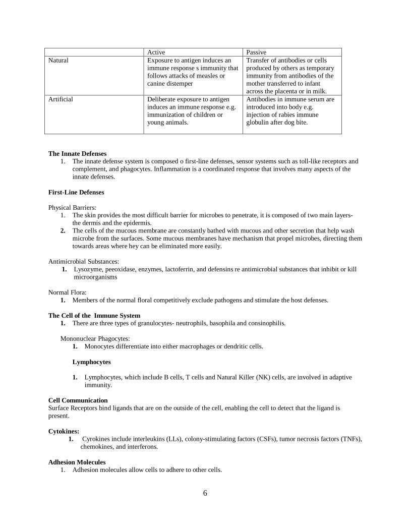
Adaptive/acquired Immunity can be natural or artificial, active or passive
6
Active Passive Natural Exposure to antigen induces an
immune response s immunity that follows attacks of measles or canine distemper
Transfer of antibodies or cells produced by others as temporary immunity from antibodies of the mother transferred to infant across the placenta or in milk.
Artificial Deliberate exposure to antigen induces an immune response e.g. immunization of children or young animals.
Antibodies in immune serum are introduced into body e.g. injection of rabies immune globulin after dog bite.
The Innate Defenses
1. The innate defense system is composed o first-line defenses, sensor systems such as toll-like receptors and complement, and phagocytes. Inflammation is a coordinated response that involves many aspects of the innate defenses.
First-Line Defenses Physical Barriers:
1. The skin provides the most difficult barrier for microbes to penetrate, it is composed of two main layers- the dermis and the epidermis.
2. The cells of the mucous membrane are constantly bathed with mucous and other secretion that help wash microbe from the surfaces. Some mucous membranes have mechanism that propel microbes, directing them towards areas where hey can be eliminated more easily.
Antimicrobial Substances:
1. Lysozyme, peeoxidase, enzymes, lactoferrin, and defensins re antimicrobial substances that inhibit or kill microorganisms
Normal Flora:
1. Members of the normal floral competitively exclude pathogens and stimulate the host defenses. The Cell of the Immune System
1. There are three types of granulocytes- neutrophils, basophila and consinophilis.
Mononuclear Phagocytes: 1. Monocytes differentiate into either macrophages or dendritic cells.
Lymphocytes 1. Lymphocytes, which include B cells, T cells and Natural Killer (NK) cells, are involved in adaptive
immunity. Cell Communication Surface Receptors bind ligands that are on the outside of the cell, enabling the cell to detect that the ligand is present. Cytokines:
1. Cyrokines include interleukins (LLs), colony-stimulating factors (CSFs), tumor necrosis factors (TNFs), chemokines, and interferons.
Adhesion Molecules
1. Adhesion molecules allow cells to adhere to other cells.
7
Sensor Systems Toll-Like Receptors
1. Toll-like receptor enables cells to detect molecules that signify the presence of microbe. The Complement System
1. Complement proteins circulate in the blood and the fluid that btches tissues, in response to certain stimui that indicate the presense of foreign material, they become activated.
2. The major protective outcomes of complement activation include opsonizatio,lyisof foreign cells, and initiation of inflammation.
Phagocytosis The Process of Phagocytosis
1. The step of phagocytosis includes chemotaxis, recognition and attachment, engultiment, destruction and digestion, and exocytosis.
Specialized Attributes of Macrophages:
1. Macrophages are always present in tissues to some extent, but are able to call in reinforcements when needed.
2. A macrophage can increase its killing power, becoming an activated macrophage. 3. Macrophage, giant cells, and T- helper cells form concentrated groups called granulomas that wall off and
retain organisms or other material that cannot be destroyd by mcrophages. Specialized Attributes of Neutrophils
1. Neutrophils play a critical role during the early stages of inflammation, being the first cell type recruited from the blood stream to the site of damage.
Inflammation-A Coordinated Response to Invasion or Damage
1. Swelling, redness, heat, and pain are the signs of inflammation, the attempt by the body to contain a site of damage, localized the response, and restore tissue function.
Factors that Initiate the Inflammatory Response:
1. Inflammation is initiated when pro inflammatory cytokines or other inflammatory mediators are released as a result of the engagement of tells like receptors or activation complement by invading microbes, or when tissue damage occurs.
The Inflammatory Process
1. The inflammatory process leads to a cascade that result in dilation of small blood vessels, leakage of fluids from those vessels, and the migration of lculocytes out of the bloodstream and into the tissues.
2. Acute inflammation is marked by a preponderance of neutrphils, chronic inflammation is characterized by the prevalence of macrophages, giant cells, and granulomas.
Outcomes of Inflammation
1. Inflammation can contain an infection, but the process itself can case damage, a system response can be life threatening.
Apoptosis –Controlled Cell Death that Circumvent the Inflammatory Process.
1. Apoptosis is a mechanism of eliminating self-cells without evoking an inflammatory response.
8
Interferons
1. One of the roles of interferons is to induce cells in the vicinity of a virally infected cell to prepare to cease protein synthesis in the event they become infected with a virus, double, stranded RNA signifies to the cell that it has been infected.
Fever
1. Fever occurs as a result of certain pro-inflammatory cytokines released by macrophages when their toll-like receptors bind microbial products.
2. Fever inhibits the growth of many pathogens and increases the rate of various body defenses. Strategy of the Adaptive Immune Response The Humoral Immunity Humoral immunity is mediateds by B.cells; in response to extracellular antigens, these maybe triggered to proliferate and then differentiate into plasma cells that function as antibody producing factories. Cellular Immunity Effector T- cytotoxic cells are able to induce apoptosis in ‘self” cells that present abnormal protein that signify danger. Effctor T-helper orchestrates the various response of cellular and humoral immunity. Anatomy of the lymphoid system Lymphatic Vessels. Lymph, which may contain antigens that have entered tissues, flows in the lymphatic vessels to the lymph nodes. Secondary lymphoid organs Secondary lymphoid organs are the sites at which lymphocytes gather to contact antigens; they facilitate the interactions and transfer of cytokines between the various cells of the immune system. Primary Lymphoid Organs
1. Primary lymphoid organs are the sites where B.cells and T.cells mature.
The Nature of Antigens
1. Antigens are molecules that react specifically with an antibody or lymphocyte, immunogen refers specifically to an antigen that elicits an immune response.
2. The immune response is directed to antigenic determinant, or epitopes, on the antigen. The Nature of Antibodies Structures and Properties of Antibodies
1. Antibodies monomers have a Y shape with an antigen-binding site at the end of each arm of the Y. The tail of the Y is the Fc region.
2. The antibody monomer is composed of two identical heavy chains and two identical light chains; each chain forms several domains. The variable region contains the antigen binding site; the constant region encompasses the entire Fc region as well as part of the Fab regions.
9
Protective Outcomes of Antibody-Antigens Binding
1. Antibody-antigens binding result in neutralization, immobilization and prevention of adherence, agglutination and precipitation, opsonization, complement activation, and antibody-dependent cytotoxicity.
Immunoglobulin Classes
1. There are five major antibody classes, IgM, IgG, IgA, IgD, and IgE, and each has distinct functions. Clonal Selection of Lymphocytes
1. When antigens enter a secondary lymphoid organ, only the lymphocytes that specifically recognize the antigen will respond; the antigen receptor they carry on their surface governs this recognition.
2. Lymphocytes may be immature, naïve, activated, effector, or memory cells. B.Lymphocytes and the Antibody Response The Response to T-Dependent Antigens
1. B.cells present antigen to effector T-helper cells for inspection. If an effector T-helper cell recognizes the antigen, it will deliver cytokines to the cell, initiating the process of clonal expansion, which ultimately forms plasma cells that produce antibody.
2. Under the direction of effector T-helper cells, the expanding B-cell population will undergo affinity maturation and class switching, and form memory cells.
3. In the primary response, a lag period occurs before antibodies can be detected; memory cells are responsible for the swift and effective secondary response, eliminating invaders before they cause noticeable harm.
The Response to T-Independent Antigens
1. T-independent antigens include polysaccharides that have multiple identical evenly spaced epitopes and LPS.
T Lymphocytes: Antigen Recognition and Response
1. The T-cell receptor recognizes antigen presented by major histocompatibility (MHC) molecules. 2. T-cytotoxic cells are referred to as CD8 T cell; T-helper are referred to as CD4 T-cells.
Functions of Effctor T-Cytotoxic (CD8) Cells
1. T-cytotoxic cells induce apoptosis in cell that produce proteins associated with danger, they also produce cytokines that allow neighboring cells to become more vigilant against intracellular invaders.
2. All nucleated cells present peptides from endogenous protein in the groove of MHC class molecules. Functions of Effector T-helper (CD4) Cells
1. T-helper cells respond to exogenous antigen, which are presented to MHC class II molecules. 2. ThI cells judge antigens presented by macrophages, a responding ThI cell activates that particular
macrophage and secrete cytokines that help orchestrate the immune response. 3. Th2 cells judge antigen presented by B.cells; a responding Th2 cell activates that particular B. cell and
supports actions that enhance its effectiveness Activation of T Cells
1. Naïve T-cells require signals to become activated, upon activation the cell stimulates ti own proliferation and ten gain its effector functions.
10
2. Dendritic cell sample material in tissues and the travel to the secondary lymphoid organs to present the antigen to naïve T-cells. Those that detect molecules associated with danger produce co stimulatory molecules and are able to activate both subcots of T-helper-cells
Natural Killer (NK) Cells
1. NK cells mediate antibody-dependent cellular-cytotoxicity (ADCC). 2. BK cells kill host that are not bearing MHC class I molecules on their surface.
Lymphocyte Development Generation of Diversity
1. Mechanisms used to generate the diversity of antigen specificity in lymphocytes include rearrangement of gene segments, imprecise joining of those segments, and combinatorial associations of heavy and light chains.
Negative Selection of Self-Reactions B-cells Negative selection occurs as B cells develop in the bone marrow, cells t which material binds to their B-cell receptor are induced to undergo apoptosis. References
1. Nester, EW, Anderson, D.Ce, Roberts (Jr), C. E Pearsal, N.N. Nester, M.T and Hurley. D (2004). Microbiology, A Human perspective 4th ed., published by McGraw Hill Higher Education
2. Brooks, G.F., Butel, J.S and Morse, S.A. (2004), Jawetz, Meinicte and Adelberg’s Medical Microbiology, 23rd ed. Published by McGrawHill Education
11
VPM 403: VETERINARY IMMUNOLOGY
LECTURE NOTES
DR O. E. OJO
DEPARTMENT OF VETERINARY MICROBIOLOGY AND
PARASITOLOGY
COLLEGE OF VETERINARY MEDICIN
UNIVERSITY OF AGRICULTURE, ABEOKUTA
12
ANTIGENS
Antigens are substances which are able to induce detectable immune responses when introduced
into an animal host. Immune responses could be cellular or humoral
Requirement for antigenicity
Molecular size: molecules with high molecular weight are capable of eliciting a better
immune response than those with low molecular weight.
Proteins>carbohydrates>lipids>nucleic acids. Molecules with molecular weight less than
10,000 dalton are weakly antigenic or non-antigenic.
Chemical complexity: molecules with high complexity are good antigens
Polymers are more antigenic than monomer
Genetic make-up of the animal host
The response of an animal to an antigen is regulated by genes
The ability to mount an immune response to a antigen varies with genetic
composition of the animal
Method of antigen administration
Immune response may differ according to the route of administration
Level of immune response is dose-dependent
Excessively high dose may induce a state of specific unresponsiveness
EPITOPES
Most foreign particles are composed of complex mixture of proteins, polysaccharides,
lipopolysaccharides, lipids and nucleoproteins
Such large molecules have specific regions responsible for antigenicity
Epitopes are regions on the surface of molecules that specifically trigger immune
reactions
Epitopes are also called antigenic determinants
An antigen mau possess more than one antigenic determinant
The antigenic determinants on an antigen vary in immunogenicity
Animal host respond better to an immunodominant epitope on an antigen
An antigen may possess similar epitopes to those present on the host’s self antigen
13
However, the cell of the immune system only recognize and respond to foreign epitopes
The number of epitopes on an antigen is related to its size
Usually about one epitope is present for each five kDa of protein
Immunopotency describes the capacity of a region of an antigen molecule to serve as an
antigenic determinant and induce the formation of specific antibody
Immunopotency is determined by:
Accesibility: exposure to the aqueous environment
Charge: electrical charges are dominant factor in specificity
Genetic factor: ability to induce immune response is under genetic control
HAPTENS
Small molecules (e.g. drugs, hormones), or chemical groups with molecular wight of less
than 1000Da which when bound to other larger molecules can function as epitopes
Haptens are too small to be appropriately processed and presented to the immune
system and are therefore not antigenic
When haptens are linked to a larger molecule, a new epitope is formed on the larger
molecule
When this is injected into an animal host, immune response develops with antibody
formation
The antibody can react with the hapten in the larger molecule
Haptens are non-immunogenic substances but can react with antibody in a specific
manner
Antigens are capable of inducing cellular immunity mediated by T-lymphocytes but
haptens are unable to do so.
The reactions of drugs which serve as haptens with body proteins may lead to allergies
Examples of haptens: dinitrophenols, penicillin
ADJUVANTS
Substances that enhance the immune response to an antigen when administered along
with that particular antigen
14
Mechanism of action:
Depot adjuvants: serve to protect antigen from rapid degradation and thereby
prolong immune responses
Particulate adjuvants: effectively deliver antigens to antigen presenting cells,
enhance cytokine production by antigen presenting cells, enhance T-helper cell
responses and enhance cell mediated immunity
Immunostimulatory adjuvants: enhance cytokines production, T-helper cell
response and enhance cell mediated immunity
Examples:
Depot adjuvants: Aluminium phosphate, Aluminium hydroxide, Treund’s
incomplete adjuvants (water-in-oil emulsion)
Particulate adjuvants: liposomes, microparticles, immunestimulatory complex
Immunostimulatory a djuvants: glucose, dextran sulphate, detergents, saponins,
lipopolysaccharides, anaerobic corynebacteria, bacillus calmette-Guerin (BCG m.
boris), Borditella pertussis etc.
Mixed adjuvant: treund’s complete adjuvant (water-in-oil emulsion plus
mycobacterium)
Tutorial Questions
Define the following terms
i. Antigen
ii. Autoimmunity
iii. Haptens
iv. Adjuvants
v. Epitopes (4 marks each = 20marks)
Tutorial Questions2 (10 marks)
i. Describe lupus erythematosus cells
ii. Give the examples of systemic autoimmune diseases
iii. Outline three features of lymphocytic thyroiditis
iv. In equine polyneuritis, what acts as autoantigen?
15
v. What is the distinct clinical feature of reproductive autoimmune diseases resulting from
the injection of terticular extract along with freund’s complex adjuvants in male
animals? (10 marks)
ANTIGEN-ANTIBODY REACTION
When an antibody comes in contact with its homologous antigen, it becomes attached to it by
one of its of its combining sites which reacts with a determinant area on the antigen. This
reaction leads into formation of an antigen-antibody complex
Ag + Ab --------------- Ag-Ab complex
The forces that hold these together are at their strongest under physiological conditions of ionic
strength and pH. If the pH is lowered, the antigen-antibody complex will dissociate.
Features of antigen-antibody reaction:
Close proximity: non-covalent binding forces are involved in antigen-antibody combination.
The shape of each of the combining site on an immunoglobulin molecule is an accurate
mould of the shape of the antigenic determinant and the two must be brought into very
close contact to fit into each other.
Specificity: the union of an antigen with its antibody is specific. The antigen react with its
corresponding antibody and with no other. Specificity is dictated by the presence of
determinant groups on the antigen and the type and pattern of amino acids present in the
antigen-binding region of immunoglobulins.
pH range: physiological range of pH (7.2-8.2) is required for a firm union. The optimal
temperature for an antigen-antibody reaction depends on the type of antibody. IgM reacts
best at 40C (cryoglobulin) while IgG reacts best at 370C.
Optimal proportion: there is an optimum concentration where antigen-antibody reaction
occurs. This optimum concentration is referred to as equivalence zone. The occurrence of
an antigen-antibody reaction can be detected by the presence of some secondary
phenomenon such as precipitation or agglutination complex. The presence of cisible
agglutination of precipitation reaction will be inhibited by an excess of antibody and this is
termed ‘prozone phenomenon’.
Forces Responsible for the Union of Antigen and Antibody
16
The forces of interaction responsible for antigen-antibody reaction are the same as those seen in
other proteins such as enzymes and transport proteins. The final strength of the bond is a
summation of the various binding or repelling forces present on both antigen and antibody
molecules. Covalent chemical bonding is not important and there is no obligatory requirement
for charged groups on antigens. However, there can be strong attraction or repulsion between
negatively charged ions and positively charged ions on these molecules at physiologic pH. The
forces invoved in antigen-antibody union include the followings:
1. Electrostatic forces
2. Hydrogen bonding
3. Hydrophobic attraction
4. Van der waal forces
Electrostatic forces: these are due to the attraction between oppositely charged ionic groups on
proteins side chains. An example is the interaction between an ionized amino group (-NH3+) on a
lysine of one protein and an ionized carboxyl group (-coo--) on a glutamate of another protein.
Hydrogen bonding: if molecules carrying hydrophilic groups such as –OH, -NH2 and –COOH
approach closely, they form hydrogen bridges which are relatively weak and reversible. The
interaction between threonine and tyrosine is an example of hydrogen boding.
Hydrophobic attraction: non-polar hydrophobic groups such as those of the side chains of valine,
leucine and phenylalanine tend to associate in an aquepus environment, just like oil droplets in
water merge to form a single large drop. It has been estimated that hydrophobic forces may
contribute up to 50% of the total strength of the antigen-antibody bond.
Van der waals forces: these are very weak forces which depend on interaction between the
external “electron cloud” of molecules. Complimentary electron cloud shapes on the combining
site of an antibody and on the surface determinant of an antigen fit the two molecules strongly
together like a lock and key.
Antibody Affinity and Avidity
The antibodies that are first produced by the body after it has been stimulated with an antigen do
not mate with so large an area of the antigenic seterminant as do those which are synthesized
later and especially those which appear after repeated immunization have been carried out. Thus,
antibodies produced soon after a first stimulation are very specific and have high affinity for a
17
particular area of the antigenic determinant. They are termed non-avid (i.e. the complexes they
formed with the antigen are easily broken down). The strength of the interaction of an antibody
with a monovalent hapten or a single antigenic determinant is referred to as affinity. Antibodies
produced later or after repeated immunization are avid. The strength of the interaction of an
antiserum with a fall antigen with its multiple determinants is termed avidity. The force binding
two determinant groups by antibody is usually many fold greater that the arithmetic sum of the
forces binding each separate antigenic determinant. Avidity makes for stronger bonds with the
antigen and often able to cross-react with other related antigens.
a. Early non-avid antibody molecules only combine with a small area of the antigenic
determinant
b. Later antibody, and antibody produced after repeated restimulation is very avid. It
combines strongly with a larger portion of the antigenic determinant than does non-avid
antibody.
c. Avid antibody is also able to combine with related antigenic determinants. The fit
however is not very close and the binding is weak.
Mechanism of Protection by Antigen-Antibody Reaction
Antibody can protect the body from infection or its effect by neutralizing soluble toxins, coating
organisms and thus promote phagocytosis, by direct lysis of organisms in the presence of the
compliment proteins and by preventing the spread of intracellular organisms.
Consequences of Antigen-Antibody Reactions in-vitro
Following the primary union of antigen to antibody in the laboratory, a number of events occur
which produce visible effects. This primary interaction gives rise to a number of secondary
phenomena such as precipitation, agglutination, flocculation, phagocytosis, cytolysis and
neutralization. These secondary reactions are the basis of a number of standard immunological
techniques. The primary reaction can simply be viewed as the specific recognition and
combination of the antigenic determinant with the binding site of its corresponding antibody.
Generally, primary tests are more sensitive than secondary tests. The quantitative tests that
employ the primary reaction include immunoflourescence, radioimmunoassay and
immunoenzymatic assays.
Harmful Effects of Antigen-Antibody Reaction in the Body
18
Antibody-antigen reactions in the body are not only helpful but can equally be harmful. In some
situations the immune attack on the invading organisms also damage host tissues. Autoimmune
reactions and hypersensitivity reaction and graft rejection are examples of harmful reactions.
AUTOIMMUNITY
The body produces self-antigens
Lymphocytes capable of binding and responding to self antigens in the body are
suppressed
Self-antigens to which the immune system is exposed during foetal life are recognized as
self and the body develop tolerance to them
Autoimmunity is a state in which the natural unresponsiveness of the lymphocytes
(tolerance) to self antigens is lost
In autoimmunity, autoantibodies are produced which react with self components. This
may lead to disease condition and tissue damage
Not all autoimmune responses are harmful. Infact, some are beneficial and crucial to
survival. Some autoantibodies serve physiological functions e.g. destruction of
senescent red blood cells
The exact cause and mechanisms of autoimmunity are not well understood
Autoimmunity could be mediated by either B cells or T cells (auto antibodies or T cells)
Mechanism of autoimmune diseases
Normal immune response to an unusual or abnormal antigen
Abnormal immune response to a normal antigen: a situation in which regulations
preventing development of self-responsive T-cells fails
Aberrant response to a single specific antigen
General defect in the regulation of B- or T- cells functions
Normal immune response
Normal immune response to a previously hidden antigen
Cross reactivity between an infectious agent and a normal body component
Abnormal antigen processing
Abnormal immune response
19
Sustained immune response to hidden epitopes
Lymphoid tumour cells producing autoantibody
Defective destruction of self-reactive lymphocytes
Virus-induced autoimmunity
Vaccine-induced autoimmunity: vaccines with adjuvants, especially excessive use
Example:
Endocrine diseases like lymphocytic thyroiditis, hyperthyroidism,
Neurological diseases: equine polyneuritis, canine polyneuritis,
degenerative myelopathy
Eye diseases: equine recurrent ureitis
Muscle diseases: myasthenia gravis, canine cardiomyopathy, polymyositis
Skin diseases: perphigus complex, epidermolysis bullosa
AUTOIMMUNE DISEASES
Systemic autoimmune diseases
Associated with the presence of circulating immune complexes and complement in
tissues
The deposition of immune complexes lead to chronic inflammation
The initiating antigens are unknown but may well be infectious agents
There is genetic predisposition linked with MHC
Examples:
Systemic Lupus Erythematosus
There is impaired clearance of apoptotic cells by macrophage phagocytosis
Apoptotic cells accumulate in the tissue
Nuclear fragments of apoptotic cells are processed by dendritic cells (antigen-presenting
cells)
There is formation of autoantibodies (antinuclear antibodies, ANA)
This leads to formation and deposition of immune complex and tissue damage
There is dermatitis (skin lesions), polyarthritis, heamolytic anaemia, thrombocytopaenia,
proteinuria, positive ANA test, and positive LE cell test
20
LE cells: cells that have phagocytosed opsonised nuclei oftern present in the bone
marrow of SLE patients
Seen in humans, other primates, dogs, rats, horses, mice
Sjogrens Syndrome: (Horses, dogs)
Characterized by keratoconjuctivitis sicca (conjuctival dryness), xerostomia (mouth
dyness) and rheumatoid factors
Autoimmunity against salivary and lacrimal glands
There is gingivitis, dental caries, excessive thirst, corneal dyness and abrasion leading to
kretitis and conjunctivitis as well as other ocular lesions\there is also rheumatoid
arthritis and polimuositis
Autoimmune polyarthritis
Deposition of immunoglobulins and immune complex within joints leading to joint
diseases
Could be erosive polyarthritis (e.g reumathoid arthritis) or non-erosive (e.g. equine and
canine polyarthritis)
Organ-specific/Tissue-specific Autoimmune Diseases
Endocrine:
Lymphocytic thyroditis
Lymphocytic parathyroditis
Insulin-dependent diabetes mellitus
Atropic lymphocityx pancreatitis
Sutoimmune immune adrenatitis
Hyperthyroidism
Neurological
Degenerative neuropathy
Cerebellar degeneration
Equine polyneuritis
Steroid meningitis-arteritis
21
Canine polyneuritis
Eye diseases
Equine recurrent ureitis
Ureodermatological syndrome
Reproductive
Skin diseases
The pemphigus complex
Skin basement membrane disease
Alopecia areata
Relapsing poluchondritis
Nephritis
Autoimmune immune nephritis
Autoimmune haemolytic anaemia
Autoimmune immune thrombocytopenia
Muscle
Myasthenia Gravis
Polymyositis
Autoimmune masticatoryy myopathy
Canine cardinmyopathy
Organ-Specific Autoimmune Diseases
Autoimmune diseases that affect a single organ or tissue
Arises as a result of abnormal reponse to a small number of self- or foreign antigen but
not necessarily a major loss of control of the entire immune system
Examples:
A Autoimmune endocrine diseases
I. Lymphocytic thyroditis
- Described in human, dogs and chicken
22
- Production of autoantibody against throglobulin which may also react with
triialothyronine (T3) or thyroxine (T4)
- There is dull, dry, coarse coat, scaling, hypotrichosis, hyperpigmentation, pyoderma. Affected
animals are fat sluggish and have area of in the skin
II. Lymphocytic parathyroiditis
i. Affects dogs and cats
ii. History of neurological or neuromuscular disorder like seizures
iii. There is marked lymphocalcaemia and low level of serum parathormones
iv. At histology, the normal parathyroid tissue is replaced by infiltrating
lymphocutes and some plasma cells
III. Insulin-dependent diabetes mellitus
i. There is development of autoantibodies against islet cells enzyme called
glutamic acid carboxylase
ii. There is atrophy of pancreatic islet and loss of cells. Lymphocytes
infiltrate the islets.
B. Autoimmune neurological diseases e.g. development of autoantibody to brain tissue
following administration of rabies vaccines prepared in brain tissue
I. Equine polyneuritis
II. Peripheral myelin protein P2 acts as autoantigen stimulating the formation of
autoantibodies: There is a chronic granulomatous inflammation in the region of the
extradural nerve roots. The nerves affected are thickened and discoloured. There is loss
of myelinated axon, macrophage, lymphocyte, giant cells and plasma cells and plasma
cells infiltration and deposition of fibrous material in the perineurium
C. Autoimmune reproductive diseases
Damage to the testes may release hidden antigens and consequently
autoimmunity
Injection of testicular extract in Treund’s complete adjuvant may produce
autoimmune orchitis in male animals
23
The presence of sperm antigens in the circulation stimulates the production of
IgE or IgA autoantibodies
The autoantibodies can agglutinate and immobilize sperm cells leading to
infertility
Autoimmune dermatitis may occur in intact female dogs as a result of
hypersensitivity to endogenous progesterone or oestrogen
This autoimmune dermatitis may coincide with oestrus or pseudopregnancy and
it is characterized by bilateral erythema and popular eruption with intense
pruritus
D. Autoimmune Muscle Diseases
a. Myasthenia Gravis
i. Seen in humans, dogs and cats
ii. Disease of skeletal muscle characterized by abnormal fatique and
weakness following mild exercise
iii. There is degradation of acetylcholine receptors by IgG autoantibodies
iv. Autoantibodies also block acetylcholine binding sites and trigger
compliment-mediated damage
v. The deficiency of acetylcholine receptor: this leads to failure of
transmission of nerve impulses across the motor end-plate of striated
muscle
E. Autoimmune Haemolytic Anaemia
i. Destruction of red blood cells mediated by autoantibodies to red blood
cells antigens
ii. Red blood cells destruction could be intravascular haemolysis mediated
by complement or phagocytosis of antibody coated RBC in spleen and
liver by macrophages (extravascular)
iii. Autoimmune haemolytic anaemia has been attributed to alteration in red
blood cell surface antigen induce by drugs or viruses
24
iv. The condition is characterized by anaemia, weakness, lethargy, fever,
uterus and hapto- splebomegaly. There could be tarchycardia, anorexia,
vomiting or diarrhea
v. It has been described in human, dogs, horses, cats, mice, cattle and
rabbits
CYTOKINES
Proteins secreted by the cells of the immune system that regulate the immune
response by communicating among cells
Characteristics:
Cell rarely secrete only one cytokine at a time e.g. macrophages secrete at least
five: IL-1, IL-6, IL-12, IL-18, and TNF-
They affect a wide variety of cells and organs
Many different cytokines may have similar effect (redundancy) e.g. IL-1, TNF-,
TNF-, IL-6, all have pyrogenic effect
Types and Groups:
1. Interleukins: cytokines that regulate the interaction between lymphocytes and
other leukocytes. They are numbered sequentially in order of their discovery, IL-
1 – IL-30
2. Interferons
Antiviral cytokines produced in response to immune stimulation and virus
infection
Interferes with viral RNA and protein synthesis
There are 2types: type I and type II
Type I: interferon alpha (IFN-) and interferon beta (IFN-) (antiviral)
Type II: interferon gamma (IFN-) (immune activation)
Some interferon are important in maintenance of pregnancy (e.g. type I
IFN-)
3. Tumor Necrotic Factors (TNFs)
Derived from macrophages and T-cells
25
They destroy tumor cells
They are important in acute inflammatory reactions especially TNF-
They play dominant role in immune regulation and inflammation
4. Growth Factors
Colony stimulating factors
Control leukocyte production by regulating stem cell growth
Make immune cells available for body defence
5. Chemokines
Regulates leukocyte circulation and migration (chemotaxis) during
inflammation
They also activate leukocytes
Example: Interleukin-8 (CXCL-8)
Functions of Cytokines
i. Cytokines are produced by antigenic stimuli acting through the T-cell and B-cell
receptors
ii. Antigen-antibody complex acting through Fc receptors
iii. Super antigens acting through the T-cell receptors
iv. Pathogen-associated molecules such as lipopolysaccharides acting through toll-like
receptors
Pattern of Cytokine activities
Autocrine: they bind to receptors on the cell that produced them.
Paracrine: they bind only to receptors on cells in close proximity to the cell of origin
Endocrine: they spread throughout the body thereby affecting cells in distant location from the
source of production
Functions
when bound to target cells, cytokines may induce the target cell to divide or
differentiate
They may stimulate the production of new proteins by the target cell
26
They may inhibit cell division and differentiation
They may inhibit the process of protein synthesis in the target cell
Most cytokines act on different target cell types and initiate different responses in
each. This phenomenon in termed PLEIOTROPY
Many different cytokines may act on a single target cell. This is termed
REDUNDANCY e.g. IL-3, IL-4, IL-5, IL-6, all affect B-cell function
Some cytokines work optimally only when in association with other cytokines. This is
called SYNERGY e.g.IL-4 combines with IL-5 to stimulate B-cell switching to IgE
synthesis
Some cytokines may prevent/inhibit the action of others. This is called
ANTAGONISM e.g. IL-4 and IFN- are mutual antagonists.
IMMUNE RESPONSE TO TUMOUR
Events leading to the development of tumour are pooly understood
Tumour arises as a result of:
1. Infection with a tumourgenic virus e.g. herpes virus, papilloma virus
2. Mutation in gene controlling cell growth
3. Expression of pre-existing oncogenes (tumour genes)
4. Disturbance in normal growth control mechanisms so that a genetically
normal cell no longer displays normal differentiation
Tumour antigens
1. Antigens expressed on chemically or physically induced tumours
2. Antigens expressed on virally induced tumours
3. Antigens associated with oncodevelopmental products
4. Antigens of spontaneous tumour
Types of Tumour Antigens
i. Antigens of chemically induced tumours
27
ii. Antigens of virally induced tumours
iii. Onco-developmental tumour antigens
iv. Antigen of spontaneous tumours
The major difference between a normal cell and a tumour cell is a loss of regulated
cell growth as a result of multiple mutation
Mutation may make the tumour cells express abnormal proteins on their surfaces
The abnormal proteins may be recognized by the body’s defence mechanism as
being foreign
This recognition will induce immunological attack
Antigenic Features of Tumour Cells
Changes on the cell surface of tumour cells that make them different from the normal cells
- loss or gain of histocompatibility antigen
- loss of blood group carbohydrates
- appearance of virus-associated antigen (tumour associated viral antigen TAVA)
- tumour-associated transplantation antigens common for the tumour of the same
hytologic type (TATA)
- Tumour-specific transplantation antigen present on only one tumour type (TSTA)
- Antigen detected only by serologic reaction unique for a given tumour (Tumour-
associated serologic defined antigens TASA)
- Tumour-associated developmental antigens (TADA): markers shared by embryonic
or developing tumours and established tumours
Tumour-associated antigens
- Tumour cells may produce new proteins
- Tumour cells may produce excessive amounts of normal proteins
1. Some tumour cells may express the products of developmental genes that are turned
off in adult cells and are normally only expressed early in an individual’s development.
These proteins are called onco foetal antigens e.g. carcinoembryonic antigen (CEA,
CD66e) is a glycoprotein produced by tumour cells of the gastrointestinal tract which
28
should normally be found only in fetal intestine; -fetoprotein produced by lepatoma
cells is an onco-foetal antigen normally found only in the foetal liver
Onco-fetal antigens are poor immunogens and do not provoke protective
immunology
Measurement of their level in blood may be useful in diagnosis and in
monitoring the progress of tumour
2. Antigens to spontaneous tumour
Rarely demonstrate tumour-specific antigens/new antigens
Normal antigens are expressed in unusual quantities
There may be abnormal proteins associated with cell division e.g. glycosylation
of proteins
3. Antigens due to oncogenic viruses
Tumour cells gained new antigenic character of inducing virus
Antigens are coded in viral genome but not part of the virion
4. chemically induce tumour
chemical may induce mutation
tumour cells therefore expressed mutated surface antigens
carcinogenic chemicals may produce different mutation
Tumour induced by a particular chemical may be antigenically different
Resistant to one chemically induced tumour does not prevent the growth of
another tumour induced by same chemical
The ability of tumour cells to elicit immune reaction depends on their ability to
cause/induce inflammation
A tumour cell that does not invade the lymphoid organs may not elicit immune
reaction
Tumour cells that invade the lymphoid organs may elicit either a strong or a weak
immune reaction
Tumour cells that are processed by dendritic cells elicit a strong T-cell response
29
Tumour cells that are walled off may not be processed enough and thus only a weak
immune response
Tumour cells that produce inflammation in tissue also trigger dendritic cell activation
and processing
Effector Mechanism in Tumour Immunity
Tumour cells express different antigens from normal cells
However, tumour cells are not always recognized as foreign
The normal molecules on tumour cells are not appropriately presented to the immune
cells especially cytotoxic T-cells
However, tumour cells may be attacked by natural killer cells, cytotoxic T-cells, activated
macrophages and antibodies
Natural killer cells are the most important in immunity to tumour
Humoral response
Antibodies can be demonstrated in the body against tumour
The presence of antibodies does not induce resistance to tumour
Antibody detection are important in serological characterization and isolation of
tumour-associated antigen
Therefore, antibodies can mediate anti-tumour activities
o Compliment-mediated lysis
o Opsonization and phagocytosis
o Loss of cell adhesion
Cell-mediated responses
Direct lysis by T-lymohocytes
o Immune T-lymphocytes can specifically recognize and kill target cells that share
the same antigens as the immunizing tumour cells
o Able to destroy solid tissue as well as dispersed tumours
Antibody-dependent cell-mediated cytotoxicity (ADCC)
o Tumour target cells coated with IgG can be destroyed by effector celss such as
granulocytes, macrophages and killer cells
30
Killing by activated macrophages
o Activated macrophages have tumouricidal capabilities
Lysis by natural killer cells
o They can discriminate between normal and abnormal cells
Evasion of Immune Mechanism by Tumour Cells
Tumour in privilege sites
o Tumour in the central nervous system and eyes
o Effector cells can not reach them
Antigenic modulation
o Loss of antigenicity or change in antigenic marker
o Tumour cells avoid immunologic destruction
Enhancement and blocking factors
o Humoral factors enhance tumour survival by interfering with the cellular assault
against tumour
o Early production of antibodies may result in absorption to tumour surface and
most tumour antigen
o This prevent induction of T-killer cell-mediated immunity
Immune capacity versus tumour mass
o If tumour challenge is sufficiently larger, the animal may succumb to the growth
of lethal cancer
Suppressor of T-lymphocytes
o Tumour-specific suppressor T-cels have been demonstrated in tumour-bearing
mice and may play a role in the apparent ineffectiveness of the response in
tumour-bearing mice
Suppression mediated by the tumour
o Some tumour synthesize various materials such as prostagladins which affect the
activity of immune response
Immunodiagnosis
Based on:
31
1. Detection of tumour markers e.g. alpha fetoproteins, carcinoembryonic antigen (CEA),
prostate-specific antigens (PSA)
2. detection of tumour-specific immunity using the presence of humoral or cellular
antibodies autoimmune immunity for diagnosis
Immunotherapy
Active immunotherapy
Stimulate the immune system non-specifically e.g. use of attenuated strain of
glycobaterium bovis BCG which activate macrophages and stimulates cytokines
release thereby promoting T-cells activity
Use of tumour cells/antigens to stimulate immune response X-irradiated,
neuraminidase or glutaraldehyde-treated cells can be used in tumour vaccines
Passive immunotherapy
Cytokine therapy: IFN-, TFN-, IL-2
Activated cytotoxic cell therapy: NK and NK-like cells activated
Antibody therapy: use of monoclonal antibodies
VACCINES AND VACCINATION
The term vaccine eas coined from vacca (cow)
Edward Jenner was the first to discover the use of vaccine to prevent infectious disease
Jenner used vaccinia virus of cow to protect against smallpox in human in 1798
Vaccines can be directed against infectious agents or its toxin
History of vaccination
Ancient time practices of vaccination fir disease protection:
- King Mithridates of Pontus protected himself from poison by drinking the blood of duck
given the poison
- Pliny the Elder in Rome ate liver of ‘mad dogs’ to protect against rabies
- Edward Jenner inoculated James Philip on the arm with material from a typical cowpox
on the hand of a milk maid
32
- Pasteur produced different vaccines against livestock diseases: Fowl cholera (using dead
bacteria to protect chicken in 1880). Anthrax vaccine for cattle and sheep in 1881 by
growing B. anthracis at 420C. Rabbis vaccine in 1885
Types of vaccines
Homologous vaccines
Developed from the pathogen or from its virulent mutant e.g. Salmonella typhi
vaccine for the protection of typhoid in human, E. Dublin vaccine to protect
animals from virulent strains.
Heterologous vaccines
Developed from different organisms to protect against another sharing close
antigenic properties e.g. rinderpest vaccine (TCRV) used for the protection of
goats from PPR
Autogenous vaccine
Vaccine developed from organism recovered/isolated form an infected animal
and the vaccine administered to the same animal for protection. Used in case of
chronic diarrhea of animals
VACCINATION
Active immunization
Artificially acquired
Long lasting protection against infectious agents
Advantages:
Better and cheaper than chemotherapy; some diseases can only be prevented
Prevention is better than cure; prevention of zoonotic disease
Decreases morbidity
Decreases mortality
Duration of protection is influenced by:
Age
Immune complexes
33
Nutritional status
Nature of the antigen
Presence of adjuvants
Presence of maternal antibodies
Modified Live vaccine confers more prolonged immunity than killed, inactivated
vaccines
Routes of administration
Aphthization
A crude method produced by Fulani herdsmen
In an outbreak of foot-and-mouth disease, cattle rearer obtained saliva from
clinically-ill cattle and rub it on the tongue of healthy cattle in the flock.
Infection is in the head and recovery is synchronized
Mucus membranes
Newcastle disease vaccines given intravenously to day-old chicks
Infectious laryngotracheotis (ILT) vaccines rubbed into the mucus membranes of
cloaca
Subcutaneous
B. pertusis vaccine, Brucella S19, T1 vaccine of CBPP, typhoid vaccine (TAB)
Intramuscular
Yellow fever vaccine, tetanus toxoid
Intradermal
Pox vaccines, tuberculosis (BCG) vaccine
Oral
E. coli vaccine
Poliomyelitis vaccine
Time of vaccination
Depends on the disease to be prevented
Influence by government policies
34
Age susceptibility of host
Examples: BCG, polio, PPR, cumboro, rabies
Pregnant animals may be vaccinated for passive protection of offspring
C. perfrigens type B and type D infection in lamb prevented by vaccinating
pregnant ewes 4 weeks and 2 weeks before lambing
Brucella vaccine given to calves 4-8 months old. M. paratuberculosis given to 30-
day old calves
Advantage of vaccination over chemotherapy
Some diseases can not be treated but can only be prevented e.g. viral diseases
Vaccination is cheaper than chemotherapy
Production of organic meat
Danger of vaccination
Accidental self-innoculation
Precipitation of the disease to be prevented
Vaccine failure
Hypersensitivity
Contamination of vaccine by extraneous organism
Vaccine production
Capital intensive
Require skill personnels
Process of vaccine preparation
Killed viral or bacterial vaccine
Inactivated toxin or toxoids
Line attenuated vaccines
Recombinant vaccines
Killed vaccines
35
Chemical killing e.g. formalin, beta-propiolactone
Heat killing, high temperature
Radiation killing e.g. UV light, ultrasonic wave, x-rays
Viability may be destroyed i.e. decreased immunogenicity
Beta-propiolactone destroys nucleic acid and preserve antigenicity
Toxoids
Detoxified toxin
Use formalin or glutenaldehyde for detoxification
Antigenicity increased by adsorption on mineral carrier
Live attenuated vaccines
Passages/several subculturing in monolayer tissue culture e.g. viral vaccine
Cultivation at abnormal temperature e.g. B. anthracis at 420C for anthrax vaccine
Culture on unusual media e.g. B. abortus S19 on potato medium or ox bile
medium for BCG
Use of avirulent strain of poor growth e.g. streptomycin-dependent mutants of
blingis spp
Biochemically-deficient S. typhimurium
Recombinant vaccine
Mutant hybrids
Safe and effective
Genetic modification
Live attenuated:
a number of route of administration because they have relevant antigens for
protective immunity
high level of cell-mediated and humoral and mucosal surface protection
no need for adjuvants; they can replicate in the recipients
booster dose can be spaced widely. Spaced interval if needed because of good
immunological memory
36
Live attenuated vaccines can produce adverse reactions such as
immunosupression
Inactivated:
Can induce high level of antibodies but less cell-mediated and mucosal immunity.
Inactivated vaccines often contain many irrelevant antigenic substances some with
undesirable biological activity
Advantages of Live vaccines
Good antigen with good antibody production
Excretion of vaccine strains may protect those infected with the strain
Back mutation extremely rare. When present, it is due to deletion rather than
spontaneous mutation
Early non-specific protection is initiated within 1-2 days of administration in
cases of viral
Disadvantages of Live vaccine
Residual virulence may produce clinical signs e.g. S19 in bulls may produce
orchitis
Cannot withstand rough handling; storage condition is very strigent
Limited shelf-life or danger of contamination with other organism found on
tissue culture
Mutation of vaccine organism
Immunosuppression especially in young
Advantages of Killed Vaccine
Can withstand rough handling and ambient temperature
No overt diseases produced
Long shelf-life
Disadvantages of Killed Vaccine
Killing destroys essential antigens
37
Poor immunogens, therefore requires several inoculation
Adjuvants may be required with possible adverse reaction
Repeated vaccination may lead to hypersensitivity
Note: many disease agents still don’t have vaccines for their prevention
Recombinant Vaccine/Biotechnology: subunit or genetically engineered live vaccines
Increased efficacy
Increases safety
RECOMBINANT VACCINES
There are three categories:
1. Type 1 recombinant vaccine: composed of antigens produced by genetic engineering
2. Type II recombinant vaccine: genetically attenuated microorganism
3. Type III recombinant vaccine: composed of modified live viruses or bacteria into which
DNA encoding a particular antigen is introduced
Type I: subunit proteins produced by recombinant bacteria or other microorganisms. DNA
encoding the required antigen is isolated and introduced into a suitable bacterium or yeast in
which the recombinant gene/antigen is expressed. There is need for adjuvants to enhance their
immunogenicity. Have been used for FMD, feline leukemia and Lyne diseases (Borrlia
burgdoferi)
Type II: virulent microorganisms are rendered less virulent by gene deletion or site directed
mutagenesis. The genome of large DNA viruses (e.g. ) contains many genes not required for in
vitro replication. With DNA technology, a pseudorabies vaccine lacking the gene for thymidine
kinase has been produced. Thymidine kinase is required by this herpes virus to replicate in non-
dividing cells such as neurons. The vaccine virus with deleted gene can infect neurons but unable
to replicate in their cells. The deleted mutants induce a protective immune response in pigs.
Deletion of the gene encoding for the glycoprotein gI on the pseudorabies virus prevent
differentiation of infected pigs which permit differentiation of infected pigs which produce
antibodies against gI from vaccinated pigs which lack the antibodies. Thus vaccination can be
done in countries where the disease is being eradicated without interfering with serological
recognition and removal of the infected pigs.
38
Type III: Necessitated because vaccine failure often result from delivery system.
Type III: modified live organism called vectors into which a gene is inserted and this organism
also serves as a delivery system in the recipient. Vector must not pose any threat to the host.
A vaccinia virus vector carrying the rabine G glycoprotein gene has been successfully used as an
oral vaccine administered to wild carnivores in baits.
39
VETERINARY VIROLOGY (VPM 402) LECTURE NOTES DEPT. OF VETERINARY MICROBIOLOGY AND PARASITOLOGY
COLLEGE OF VETERINARY MEDICINE UNIVERSITY OF AGRICULTURE ABEOKUTA, OGUN STATE,
NIGERIA. Dr. MICHAEL AGBAJE
THE COMPLEMENT SYSTEM
Introduction
Complement is a collective noun that described a series of approximately 30 plasma proteins.
When activated they interact sequentially, forming a self-assembling enzymatic cascade and
generating biologically active molecules mediating a range of end processes that are significant
in the immune and inflammatory response. The basic principle of this cascade system is depicted
in general terms; this is similar to the coagulation pathways involved in secondary haemostasis.
Intrinsic in such pathways is the presence of a regulatory system that can switch off the cascade
when no longer required in order to avoid inappropriate damage to normal tissue.
There are four complement pathways, known as the classical, lectin, alternative and terminal
pathways. The first three of these pathways share a common end point, which in turn is the start
of the shared terminal pathways. Complement components generated by the activations of these
pathways will be described below.
Complements recognized in all animal species and the constituent components are relatively
conserved. These components are mostly described using the abbreviation “C” (for
complement), with a number to indicate the specific component (e.g. C4) and a lower case letter
to indicate a subfraction of that component (e.g C4a and C4b). The numbering of the
components does not always follow a logical sequence, as each component was numbered in the
order in which it was discovered. Rather than by the position it holds in the hierarchy of the
system. Some components and regulatory proteins do not conform to the “C’ nomenclature. To
add further confusion, there is traditionally some minor difference in nomenclature used in North
America and Europe (for example the classical pathway C3 convertase is C4bC2b in Europe and
C4bC2a in North America). Readers should be aware of this, as t is the European system that is
presented here.
40
The Classical and Lectin Pathways
The classical pathway, so called because it was the first discovered, is triggered by the
binding of the first component of complement (C1) to the surface of an antigen (e.g a pathogen).
In the case of some bacteria, this binding might be directly to a cell wall structural component
(e.g lipteichoic acid of the wall of gram-positive bacteria), or C1 may bind C-reactive protein
that is in turn attached to a bacteria polysaccharide. Most often, C1 attached to an immune
complex of antigen and antibody (IgG or IgM) by binding to a specific area of the
immunoglobulin component of the complex (e.g to the CH2 domain of IgG). It is also possible
for C1 to bind to an aggregate of antibody molecules in the absence of antigen. The antigen in
these situations is generally relative large (e.g a cell or microbe) and provides a surface area onto
which the complement molecules deposit as they are activated. C1 is comprised of three
enzymatic subunits, C1q, C1r and C1s, which are activated in that sequence following
attachment to the immunoglobulin molecule.
C1s then acts on the next component in the sequence, which is C4, and splits this into two
subunits, C4a and C4b. the C4b fraction attaches to the surface of the antigen and binds C2,
which is also cleaved by C1s to C2a and C2b. The C2b fragment remains associated with the
C4b fraction and this complex has now become the classical pathway ‘C3 convertase’. As this
name suggests, the next stage of the sequence is that C4bC2b (C3 convertase) acts on C3 to split
this molecule into C3a and C3b. C3a has a major biological role, but it does not affix to the
surface of the antigen. In contrast, C3b deposit adjacent to the C4bC2b complex to form a new
complex of C4bC2bC3b, which become a ‘C5 convertase,. The generation of this C5 convertase
is the final stage in the classical pathway.
As for all complement pathways, a system of regulatory control is built into the classical
pathway to inactivate the system when no longer required. The first means of control is relatively
simple and relates to the fact that these complement components have a short half-life when
generated. Moreover, complement molecules are highly susceptible to heat and, in vitro, the
molecules may be inactivated by heating a sample of serum (containing the molecules) to 56 oC
for a short period of time (a process known as ‘heat-inactivation’ of complement). Additional
means of controlling the classical pathway relate to the presence of a series of specific inhibitory
factors that act at different points of the pathway. The C1 inhibitor cleaves C1r from C1s,
thereby disrupting the activity of this complex. The C4 binding protein displaces C2b from C4b
41
and work in combination with factor 1, which subsequently cleaves C4b into two active
subfractions, C4c and C4d. factor 1 is also able to cleave C3b into the inactive subdractions C3c
and C3d.
As these complement molecules have high biological potency, there must be a means of
protecting potency, there must be a means of protecting normal cells that lie in the immediate
vicinity of the activated classical pathway and may be potentially susceptible to complement
fragments that diffuse away from the site of activation. In fact, normal cells have an in-built
protective mechanism, as their cell membrane includes a series of constitutively expressed
proteins that are able to disrupt the C3 convertase should it form on their surface. These proteins
include the decay accelerating factor (DAF), complement receptor 1 (CR1) and membrane co-
factor protein (MCP).
The lectin pathway of complement was the most recently discovered and shares elements
of the classical pathway. Essentially, the serum mannan-binding lectin (MBL) initially associates
with a bacterial surface carbohydrate and is then able to activate the MBL-associated serine
protease-2 (MASP-2). Activated MASP-2 replicates the effects of C1 by activating C4 and C2 to
form the C3 convertase complex.
The Alternative Pathway
The alternative pathway of the complement system is older in evolutionary terms then the
classical pathway and as it does not require the presence of antibody to be activated, it may be
considered part of the innate immune system. The alternative pathway has two distinct phases.
The first of these continually cycles at low level in clinically normal animals and is often referred
to as the ‘tick over’ phase. The key feature of the tick over phase is that it occurs within the
extracellular fluid space and is not associated with the surface of cells. The second phase reflects
full activation of the system and require the presence of an appropriate trigger factor, which in
the case of the alternative pathway is the presence of a ‘trigger surface’ that permits deposition of
molecules of the enzymatic cascade. Such trigger surfaces are provided by microbes (particularly
bacteria or yeast), by abnormal tissue cells (e.g virally infected or neoplastic cells), by aggregates
of immunoglobulin or by foreign material (e.g asbestos).
The tick over phase is initiated by C3 in the extracellular fluid, which undergoes
spontanbeous hydrolysis to form C3i. In the presence of Mg2+, some C3i is bound by Factor B
to form a complex of C3iB. Bound Factor B is acted upon by Factor D, which fragments the
42
molecule to Bb and Ba. Bb remains associated with C3i to form a C3 convertase, which in turn
splits further C3 molecules in the fluid phase to C3a and C3b. in the tick over pathway, the
majority of the C3b that is generated undergoes spontaneous hydrolysis and inactivation. Should
any C3b deposit onto the surface of adjacent normal cells, it will be inactivated by the
combination of MCP, DAF, CR 1, Factor H and Factor I.
The Terminal Pathway
In contrast to the pathways described above, the terminal pathway is relatively
straightforward and benefits from the fact that the constituent molecules are named in correct
numerical sequence. Accordingly, the pathway is initiated by C5 convertase splitting C5 into the
subfractions C5a and C5b, C5a has potent biological activity similar to that of C3a, but has its
effect distant to the surface of the cell that is the target of complement activation.
Activated C5b recruits C6 and C7 to form a complex of C5bC6C7, which associates with
the membrane of the target cell. This complex in turn binds C8 that penetrates the cell membrane
and recruits a number of C9 molecules that insert into the membrane to form a ‘donut-like’
transmembrane pore. This is known as the membrane attack complex (MAC). The formation of
the MAC represents the end point of the terminal pathway. Biological consequences of Complement activation
Cytolysis
Once the terminal complement pathway is activated in reality not one, but thousands of MACs
are generated within the membrane of the cell that is the target of complement activation. The
surface of this target cell becomes riddled with holes and an osmotic imbalance between the cell
cytoplasm and extracellular fluid is established such that there is a net influx of water into the
cell. The cell swells and subsequently bursts in a phenomenon known as ‘osmotic lysis’. If that
target cell is a bacterium or an abnormal tissue cell, then clearly this mechanism of cellular lysis
(cytolysis) is beneficial to the host and is a valuable part of the protective immune response.
Antibody-mediated (classical pathway) complement cytolysis is the basis for the type II
hypersensitivity reaction.
Generation of Bioactive Substances
43
The second major consequence of complement activation is the generation of those
fragments of complement that do not associate with the membrane of target cells. The two most
important of these are C3a and C5a. C5a is more potent than C3a, but much larger quantities of
C3a are generated. These molecules are sometimes known as anaphylatoxins or chemoattractants
after the fundamental effects that they mediate.
C3a and C5a have key roles in the tissues inflammatory response via a number of
mechanisms. These molecules mediate vasodilation of small vessels within the tissue in which
they are generated. Dilated vessels with leaky endothelial cell junctions permit the egress of
fluid, protein and cells from the circulation into the tissue. If the generation of C3a and C5a has
resulted from infection or damage within that tissue, this process has clear benefit to the tissue.
Vascular fluid loss leads to tissue oedema, which may be important in diluting locally produced
toxins. Leakage of blood proteins (immunoglobulins and complement molecules) may be
beneficial if these molecules participate in a local immune response. Migration of leucocytes,
particularly phagocytic cells such as neutrophils and macrophages, is likely to be of benefit in the
removal of infectious agents or tissue debris. C3a and C5a may additionally have direct
activating effects on neutrophils in order to enhance their phagocytic function and release of
further inflammatory mediators. The chemotactic role of C3a and C5a may further enhance the
value of local tissue recruitment of leucocytes. After migrating from the circulation into tissue,
phagocytic cells may be directed towards the location of pathogens or damaged cells by moving
up a ‘chamotactic gradient’. This gradient is formed by complement molecules, which are at
their highest concentration at the point that they were generated and at progressively lower
concentration the further away from that source they are within that tissue.
In some circumstances the action of C3a and C5a may cross the border between s useful
role in local inflammation and induction of a pathological inflammatory response. Both
molecules may act on tissue mast cells, causing them to degranulate and further expand the tissue
inflammatory response. In the respiratory tract this may result in contraction of bronechiolar
smooth muscle (bronchoconstriction).
Removal of Particulate Antigen or Immune Complexes
The third biological consequence of complement activation is in enhancing the removal
and destruction of particulate antigen or complexes of antigen and antibody by phagocytic cell
44
such as neutrophils and macrophages. A phagocytes might encounter such antigen within the
tissue space by random encounter instance the phagocytic cell might internalize and destroy the
particle and this process might involve the interaction of receptor molecules expressed by the
phagocyte with ligands found on the target. Particulate antigen would be more likely to be
phagocytosed were it aggregated together by antibody (particularly IgM antbody), making it a
‘larger target’ for the phagocytic cell. The interaction between target antigen and phagocyte can
also be enhanced by the complement system by the processes of opsonization and immune
adherence.
The phenomenon of opsonization arises because phagocytic cells express on their
membrane receptors for IgG (the Fcy Receptor) and receptors for the complement molecule C3b
(C3bR). Simply put, if a particulate antigen is already coated with IgG, it is more likely to be
phagocytosed due to the enhanced contact with that cell mediated thought the IgG-FeR
interaction. Even better is if the antigen is also coated with C3b, so that there is dual interaction
involving IgG-FcR and C3b-C3bR binding. ‘opsonization’ comes from the Greek ‘to make tasty
for the table’ and the analogy is often used that coating a target antigen with IgG and C3b is akin
to covering dinner with gravy!.
A related event is that of immune adherence, a process that is of great importance in
clearing particulate antigen from the bloodstream. Immune adherence arises from the fact that
erythrocytes express a C3bR (but not an FcR) and this permits circulating antigen that is coated
with C3b (with or without antibody) to attach to the surface membrane of red cells. As these
antigen-laden red cells pass through the hepatic sinusoids and spleen, they encounter phagocytic
cells resident in those tissues that bind the antigen via their own C3b receptors and remove the
antigen from the red cells for internalization and destruction.
Interaction with other Inflammatory Pathways
As discussed above, the complement pathways have key roles in the inflammatory and
immune response. During inflammation a series of biochemical pathways is activated in parallel
within the same tissue, so it is not surprising that there may be molecular interactions between
the mediators within the different pathways. Complement molecules may have a range of effects
on the kinin, coagulation and fibrinolytic pathways, the fine details of which are beyond the
scope of this discussion.
Tests of Complement Function
45
The complement pathways have been defined in most domestic animals species and,
although rarely performed, it is possible to test for complement function. Complement molecules
are very heat-labile (and may be destroyed by heating a serum sample to 56oC for 30 minutes),
so such assays require freshly collected serum frozen rapidly to – 70oC to preserve the
components. The total haemolytic complement assay (CH50) tests for the overall function of the
classical and terminal pathways. Essentially, this test involves incubating patient serum with an
indicator system of antibody-coated red blood cells. Complement present within the serum will
cause lysis of these erythrocytes. A titration of the serum sample is performed and the CH50
value for the sample is defined as the reciprocal of the serum dilution that gives 50% haemolysis.
An alternative to the tube-based CH50 assay is to perform the test by adding serum to wells cut
in an agarose gel that contains the sensitized erythrocytes. After appropriate incubations,
haemolysis in such plates is indicated by the formation of a clear zone around the well. The
diameter of this cleared ring is proportional to the log sample. Finally, it is also possible to
measure the using single radial immunodiffusion (SRID) or a haemolytic assay in which an
excess of all complement components, except the one being tested, is provided, such that the
missing component derives from the test sample.
HYPERSENSITIVITY Occasionally, the immune system responds inappropriately to the presence of antigen. These responses are referred to as hypersensitivities. There are four different types of hypersensitivities that result from different alterations of the immune system. These types are classified as: Type I: Immediate Hypersensitivity Type II: Cytotoxic Hypersensitivity Type III: Immune Complex Hypersensitivity Type IV: Delayed Hypersensitivity This section describes the four types of hypersensitivity, giving examples of diseases that may result. TYPE I HYPERSENSITIVITY Type I or Immediate Hypersensitivity can be illustrated by considering the following experiment: 1. First, a guinea pig is injected intravenously with an antigen. For this example, bovine
serum albumin (BSA, a protein) will be used. After two weeks, the same antigen will be reinjected into the same animal. Within a few minutes, the animal begins to suffocate and dies by a process called anaphylactic shock.
46
2. Instead of reinjecting the immunized guinea pig, serum is transfered from this pig to a "naive" (unimmunized) pig. When this second guinea pig is now injected with BSA, it also dies of anaphylactic shock. However, if the second pig is injected with a different antigen (e.g. egg white albumin), the pig shows no reaction.
3. If immune cells (T-cells and macrophages instead of serum) are transfered from the immunized pig to a second pig, the result is very different; injection of the second pig with BSA has no effect.
These results tell us that: The reaction elicited by antigen occurs very rapidly (hence the name "immediate
hypersensitivity"). The hypersensitivity is mediated via serum-derived components (i.e. antibody). The hypersensitivity is antigen-specific (as one might expect for an antibody-mediated
reaction). The details of this reaction can be summarized as follows : 1. Initial introduction of antigen produces an antibody response. More specifically, the type
of antigen and the way in which it is administered induce the synthesis of IgE antibody in particular.
2. Immunoglobulin IgE binds very specifically to receptors on the surface of mast cells, which remain circulating.
3. Reintroduced antigen interacts with IgE on mast cells causing the cells to degranulate and release large amounts of histamine, lipid mediators and chemotactic factors that cause smooth muscle contraction, vasodilation, increased vascular permeability, broncoconstriction and edema. These reactions occur very suddenly, causing death.
Examples of Type I hypersensitivities include allergies to penicillin, insect bites, molds, etc. A person's sensitivity to these allergens can be tested by a cutaneous reaction. If the specific antigen in question is injected intradermally and the patient is sensitive, a specific reaction known as wheal and flare can be observed within 15 minutes. Individuals who are hypersensitive to such allergens must avoid contact with large inocula to prevent anaphylactic shock.
TYPE II HYPERSENSITIVITY Type II or Cytotoxic Hypersensitivity also involves antibody-mediated reactions. However, the immunoglobulin class (isotype) is generally IgG. In addition, this process involves K-cells rather than mast cells. K-cells are, of course, involved in antibody-dependent cell-mediated cytotoxicity (ADCC). Type II hypersensitivity may also involve complement that binds to cell-bound antibody. The difference here is that the antibodies are specific for (or able to cross-react with) "self" antigens. When these circulating antibodies react with a host cell surface, tissue damage may result. There are many examples of Type II hypersensitivity. These include:
47
Pemphigus: IgG antibodies that react with the intracellular substance found between
epidermal cells. Autoimmune hemolytic anemia (AHA): This disease is generally inspired by a drug
such as penicillin that becomes attached to the surface of red blood cells (RBC) and acts as hapten for the production of antibody which then binds the RBC surface leading to lysis of RBCs.
Goodpasture's syndrome: Generally manifested as a glomerulonephritis, IgG antibodies that react against glomerular basement membrane surfaces can lead to kidney destruction.
TYPE III HYPERSENSITIVITY Type III or Immune Complex hypersensitivity involves circulating antibody that reacts with free antigen. These circulating complexes can then become deposited on tissues. Tissue deposition may lead to reaction with complement, causing tissue damage. this type of hypersensitivity develops as a result of systematic exposure to an antigen and is dependent on i) the type of antigen and antibody and ii) the size of the resulting complex. More specifically, complexes that are too small remain in circulation; complexes too large are removed by the glomerulus; intermediate comlexes may become lodged in the glomerulus leading to kidney damage. One example of a Type III hypersensitivity is serum sickness, a condition that may develop when a patient is injected with a large amount of e.g. antitoxin that was produced in an animal. After about 10 days, anti-antitoxin antibodies react with the antitoxin forming immune complexes that deposit in tissues. Type III hypersensitivities can be ascertained by intradermal injection of the antigen, followed by the observance of an "Arthus" reaction (swelling and redness at site of injection) after a few hours. TYPE IV HYPERSENSITIVITY Type IV or Delayed Hypersensitivity can be illustrated by considering the following experiment: 1. First, a guinea pig is injected with a sub-lethal dose of Mycobacterium tuberculosis (MT).
Following recovery of the animal, injection of a lethal dose of MT under the skin produces only erythema (redness) and induration (hard spot) at the site of injection 1-2 days later.
2. Instead of reinjecting the immunized guinea pig, serum is transfered from this pig to a "naive" (unimmunized) pig. When this second guinea pig is now injected with MT, it dies of the infection.
3. If immune cells (T-cells and macrophages instead of serum) are transfered from the immunized pig to a second pig, the result is very different; injection of the second pig with MT causes only erythema and induration at the site of injection 1-2 days later.
4. In a separate experiment, if the immunized guinea pig is injected with a lethal dose of Listeria monocytogenes (LM) instead of MT, it dies of the infection. However, if the pig is simultaenously injected with both LM and MT, it survives.
These results tell us that:
48
The reaction elicited by antigen occurs relatively slowly (hence the name "delayed
hypersensitivity"). The hypersensitivity is mediated via T-cells and macrophages. The hypersensitivity illustrates both antigen-specific (T-cell) and antigen non-specific
(macrophage) characteristics. The details of this reaction can be summarized as follows (click the image to animate): 1. Initial introduction of antigen produces a cell-mediated response. Mycobacterium
tuberculosis is an intracellular pathogen and recovery requires induction of specific T-cell clones with subsequent activation of macrophages.
2. Memory T-cells respond upon secondary injection of the specific (i.e. MT) antigen, but not the non-specific (i.e. LM) antigen.
3. Induction of the memory T-cells causes activation of macrophages and destruction of both specific (MT) and non-specific (LM) microorganisms.
Immune responses to infectious agents
The immune Response to Viral Infection
Viruses are a highly successful class of pathogens responsible for very significant morbidity and
mortality amongst both animal and human populations. This in part relates to the ability of these
organisms to evolve a range of strategies to evade or inhibit the host immune response. Some
viruses (e.g retroviruses such as feline leukaemia virus, Fel V). have been able to integrate their
genetic material into the host genome, others are able to alter their antigenic appearance to
produce repeated epidemics or pandemics of disease (e.g human and animal influenza viruse)
and yet other viruses have been able to capture host genes and express host-related proteins that
interfere with development of the protective immune response (e.g the capture of the human IL-
10 gene by Epstein-Barr virus).
As an example of a model immune response to virus infection, we shall consider how the
immune system might deal with a viral infection of the enterocyte lining of the intestinal tract, as
might occur with, for example, an intestinal rotavirus of domestic livestock. When infectious
virus particles arrive at their target surface, they will encounter an array of innate immune
defences relevant to that surface. In the intestinal mucosa these will include the enterocyte
barrier, the secretions that coat the luminal surface of that barrier (including mucus,
antimicrobial enzymes and defensins and polyreactive immunoglobulins) and the range of innate
49
immune cells that normally populate the epithelial compartment (e.g the TCR T cells) and the
underlying lamina propria (e.g macrophages, dendritic cells and NK cells).
Virus particles generally infect host cells by binding to a receptor molecule expressed on
the surface of the target cell. This is usually a normal surface protein that the virus uses as a
receptor or co-receptor to gain entry to the target cell. In the present example, the virus interacts
with receptor on the enterocyte surface to gain access to the host cell. Once within the cell, the
aim of the virus is to replicate in order to produce new virions that might then leave that cell (and
in the process destroy the cell) to infect new targets. The means by which this is achieved
depends on the nature of the virus and its genomic materials. Fortunately, most virus-infected
cells begin to secrete the antiviral cytokines IFN-α and IFN-β. These antiviral interferon’s bind
receptors on adjacent non-infected tissue cells and stimulate the uninfected cell to produce an
array of other proteins that confer a measure of resistance to certain viral infections. The virus-
infected cell may also display viral antigen on its surface membrane and if that expression is
concurrent with down-regulation of MHC class I molecules, the infected cell becomes a target
for innate NK cells in the vicinity. The antiviral interferons may also positively influence local
NK cells. Alternatively, the infected cell may process and present virus antigen in the context to
MHC class I and II molecules but this may only be of significance later.
At this stage of the viral infection it would be hoped that lamina propria dendritic cells
may be able to sample virus antigen or even become infected by virus particles, allowing classic
processing and presentation by these APCs. The interaction between virus and APCs involves
viral Pathogen Associated Molecular PatternsPAMPs (often of nucleic acid origin) and dendritic
cell PRRs (pattern recognition receptor) that are predominatly cytoplasmic. These interaction
presumptively lead to selective gene activation in the APC. In addition, the dendritic cell should
enter lypmhatics and migrate to the regional mesenteric lymph nodes. It is also possible that
virus antigen broaches the intestinal barrier through the Peyer’s patch lymphoid tissue.
Once the antigen-laden APC has entered the paracortex of the mesenteric lymph mode, it
will aim to locate and activate recirculating antigen-specific naïve peptides. The interaction
between Th0 cell and APC will be guided by the range of co-stimulatory surface molecules and
cytokines that have been activated within the APC following PRR-PAMP interaction. As the
most ‘relevant’ type of adaptive immune response for this type of infection would be a Thl-
regulated cytotoxic effector response, it would be hoped that the APC will activate clones of ThI
50
CD4- T cells and CD8 cytotoxic T cells. Recalling that Thl cells may provide restricted help for
those B cells committed to producing the subclass of IgG antibody able to opsonize or take part
in ADCC, it would also be appropriate for virus-specific B cells to be activated and class switch
to that IgG subclass.
The range of Thl, Tc and B cells generated must then leave the mesenteric lymph node in
efferent lymph in order eventually to enter the bloodstream and ‘home’ to the anatomical site to
viral infection (the intestinal mucosa). This will involve the interaction of homing receptors such
as the α4-β7 integrin with the vascular addressin MAdCAM (Mucosaladdressin cell adhesion
molecule). Once these adaptive immune cells arrive in the mucosa, the full effector phase of
adaptive immunity will come into play. Thl-derived IFN-y will amplify the effects of NK cells
and Tc cells, and such cell-mediated cytotoxicity is the major effector mechanism in the antiviral
immune response. The Thl cell will also stimulate B-Cell transformation to plasma cells
secreting IgG subclass that contributes to the cytotoxic process. Antibody bound to infected cells
may also mediate their lysis via activation of the classical pathway of complement. Although in a
protective anti-viral immune response the Thl arm of adaptive immunity is important, it is also
likely that some Th2 effectors are generated within the mesenteric lymph node and also home
back to the site of infection. These cells may have a relevant role in stimulating the local
production of anti-viral IgA that could be secreted across the mucosal barrier to bind virus
particles and block their interaction with receptors. Locally secreted IgG may act in a similar
fashion. If this adaptive immune response is successful in containing the infection, then late stage
immunosuppression (induced T-Cell receptors TCRs) and the development of T-and B-cell
memory will occurs.
The Immune Response to Bacterial Infection
To remain with the intestinal model, we will next consider the nature of the immune response
that might be generated in response to an enteric bacterial pathogen such a Escherichia coli or
Salmonella spp. On arrival in the intestinal tract these organisms will also immediately encounter
the range of innate immune defences outlined above. However, of particular importance in this
context would be the presence of the endogenous intestinal bacterial microflora, which will
compete with the pathogen for space and nutrients, making colonization more challenging.
Another element of innate immunity that may have greater relevance to this class of infection is
51
the ᵧᵟT cell within the enterocyte layer. These cells are well situated for early interaction with
bacterial pathogens and are thought to be primarily activated in response to this type of
organism.
As for viruses, bacteria often require an initial receptor-mediated interaction with target host
cells. For example, the K88 and K99 pili of E.coli permit attachment to receptors at the
enterocyte interface between these bacteria and host tissue. Enteric pathogens, such as E.coli or
S. Typhimurium, may utilize a variety of different mechanisms to induce disease, dependent on
the genetic strain of the bacterium. Some may produce locally active enterotoxin that bind toxin
receptors and lead to osmotic imbalance and metabolic diarrhea. Others may attach to and
disrupt the epithelial surface or invade the intestinal mucosa and regional lymph nodes, leading
to a local pyogranulomatous inflammatory response. Such gram-nagative rods, are also
characterized by the ability to produce severe generalized disease (endotoxaemia).
Once the mucosal surface is colonized, the adaptive immune response will be required to
help resolve the infection. Again, mucosal dendritic cells should sample bacterial antigen and
this process involves the interaction of PRRs with a range of bacterial PAMPs. Particulate
bacterial antigen may be more likely to gain access to the lamina propria immune compartment
via M cells within the dome epithelium overlying the Peyer’s patch. The activated dendritic cells
will migrate to the regional mesenteric lymph nodes in orders in order to activate paracortical T
cells and, in turn, follicular B cells. The desired effector immune response in this situation would
be one dominated by the production of antigen-specific immunoglobulin, so APC signalling of
the Th0 cell would generate Th2 effector. These, together with antigen-specific B cells, would
then exit the mesenteric lymph node to home back to the mucosal surface.
The most beneficial effector immune response in this context will involve the synthesis of
specific IgA and IgG antibodies. For those organisms mediating pathology via toxin production,
IgG neutralization of toxin will be important, IgG antibodies may also opsonize invasive
organisms for phagocytosis or permit the complement-mediated lysis of the bacteria. Bacterium-
specific IgA antibodies will be secreted to the luminal surface, where they may interfere with the
interaction of organism with receptor molecules. Again, in a successful immune response, final
down-regulation of the effector populations will be required together with the generation of
immunological memory. The Immune response to fungal infection
52
As for nematodes, many fungal pathogens provide a challenging target for the immune system
because of the relative size of the colonies of organisms. In this final example we shall consider
the immune response of the dog to colonization of the nasal sinuses and nasal cavity by the
organism Aspergilus fumigatus. This fungus form a large colony over the mucosa of these nasal
tissues, but the organisms tend not to infiltrate into the lamina propria. The colonies comprise a
tangled mass of fungal hyphae, with intermittent conidia that represent the sites of spore
formation.
In order for such a colony to establish, the organism must broach the normal innate
immune barriers of the upper respiratory tract, including the antimicrobial substances found
within nasal secretions. Although innate phagocytic cells (neutrophils and macrophages) are
capable of phagocytosing fungal spores, they appear unable effectively to kill these elements.
The fungal hypae are simply too large a target for effective phagocytosis.
The adaptive immune response to this infection has recently been well characterized. It is
likely that APCs carrying fungal antigen stimulate this response in regional lymphoid tissue such
as the nasopharyngeal tonsil or retropharygeal lymph nodes. The effector phase of the nucosal
immune response involves infiltration by CD4+ Thl and probably Thl7 cells, as determined by
up-regulation of gene expression for IFN-y and IL-23 in inflamed tissue. There is also a marked
pyogranulomatous element to the reaction and infiltration of IgG-bearing plasma cells. Thl-
derived IFN-y most likely provides stimulation to macrophages to permit these cells to destroy
any phagocytosed fungal spores. Antibody and complement molecules are likely to coat the
hyphal elements and form a bridge to FcR-bearing granulocytes. Similar to helminth infection,
these cells may degranulate locally and induce focal damage to the hyphae. Infected dogs
generally mount a strong serum IgG antibody response to the organism. The inflammatory
response itself it likely responsible for the extensive tissue and bone destruction that may occur
in this disease. Similar to observations in leishmaniosis, there is an additional regulatory element
to the response, as there is concurrent up-regulation of lL-10 gene expression. Again, this is
interpreted as an attempt by the adaptive immune of systemic sequelae, but at the same time
allows persistence of the infection and the development of chronic sinonasal disease.