Embed Size (px)
Citation preview
Chapman UniversityChapman University Digital Commons
Pharmacy Faculty Articles and Research School of Pharmacy
2012
Vorinostat: A Potent Agent to Prevent and TreatLaser-Induced Corneal HazeAshish TandonHarry S. Truman Memorial Veterans’ Hospital
Jonathan C. K. ToveyHarry S. Truman Memorial Veterans' Hospital
Michael R. WaggonerHarry S. Truman Memorial Veterans' Hospital
Ajay SharmaChapman University, [email protected]
John W. CowdenUniversity of Missouri, Columbia
See next page for additional authors
Follow this and additional works at: https://digitalcommons.chapman.edu/pharmacy_articles
Part of the Animal Experimentation and Research Commons, Animals Commons,Musculoskeletal, Neural, and Ocular Physiology Commons, Ophthalmology Commons, and theOther Pharmacy and Pharmaceutical Sciences Commons
This Article is brought to you for free and open access by the School of Pharmacy at Chapman University Digital Commons. It has been accepted forinclusion in Pharmacy Faculty Articles and Research by an authorized administrator of Chapman University Digital Commons. For more information,please contact [email protected].
Recommended CitationTandon A, Tovey JCK, Waggoner MR, et al. Vorinostat: A Potent Agent to Prevent and Treat Laser-induced Corneal Haze. J RefractSurg. 2012;28(4):285-290. doi:10.3928/1081597X-20120210-01.
CORE Metadata, citation and similar papers at core.ac.uk
Provided by Chapman University Digital Commons

Vorinostat: A Potent Agent to Prevent and Treat Laser-Induced CornealHaze
CommentsThis is a pre-copy-editing, author-produced PDF of an article accepted for publication in Journal of RefractiveSurgery, volume 28, issue 4, in 2012 following peer review. The definitive publisher-authenticated version isavailable online at DOI: 10.3928/1081597X-20120210-01.
CopyrightSlack
AuthorsAshish Tandon, Jonathan C. K. Tovey, Michael R. Waggoner, Ajay Sharma, John W. Cowden, Daniel J.Gibson, Yuanjing Liu, Gregory S. Schultz, and Rajiv R. Mohan
This article is available at Chapman University Digital Commons: https://digitalcommons.chapman.edu/pharmacy_articles/541
Vorinostat: A Potent Agent to Prevent and Treat Laser-induced Corneal Haze
Ashish Tandon, PhD, Jonathan C.K. Tovey, MD, Michael R. Waggoner, MD, Ajay Sharma, PhD, John W. Cowden, MD, Daniel J. Gibson, Yuanjing Liu, PhD, Gregory S. Schultz, PhD, and Rajiv R. Mohan, PhDHarry S. Truman Memorial Veterans’ Hospital (Tandon, Tovey, Waggoner, Sharma, Mohan), Mason Eye Institute, School of Medicine, (Tandon, Tovey, Waggoner, Sharma, Cowden, Mohan), and College of Veterinary Medicine, University of Missouri (Mohan), Columbia, Missouri; and the Department of Ob/Gyn, University of Florida, Gainesville, Florida (Gibson, Liu, Schultz)
Abstract
PURPOSE—This study investigated the efficacy and safety of vorinostat, a deacetylase (HDAC)
inhibitor, in the treatment of laser-induced corneal haze following photorefractive keratectomy
(PRK) in rabbits in vivo and transforming growth factor beta 1 (TGFβ1) -induced corneal fibrosis
in vitro.
METHODS—Corneal haze in rabbits was produced with −9.00 diopters (D) PRK. Fibrosis in
cultured human and rabbit corneal fibroblasts was activated with TGFβ1. Vorinostat (25 μm) was
topically applied once for 5 minutes on rabbit cornea immediately after PRK for in vivo studies.
Vorinostat (0 to 25 μm) was given to human/rabbit corneal fibroblasts for 5 minutes or 48 hours
for in vitro studies. Slit-lamp microscopy, TUNEL assay, and trypan blue were used to determined
vorinostat toxicity, whereas real-time polymerase chain reaction, immunocytochemistry, and
immunoblotting were used to measure its efficacy.
RESULTS—Single 5-minute vorinostat (25 μm) topical application on the cornea following PRK
significantly reduced corneal haze (P<.008) and fibrotic marker proteins (α-smooth muscle actin
and f-actin; P<.001) without showing redness, swelling, or inflammation in rabbit eyes in vivo
screened 4 weeks after PRK. Vorinostat reduced TGFβ1-induced fibrosis in human and rabbit
corneas in vitro in a dose-dependent manner without altering cellular viability, phenotype, or
proliferation.
CONCLUSIONS—Vorinostat is non-cytotoxic and safe for the eye and has potential to prevent
laser-induced corneal haze in patients undergoing PRK for high myopia.
Correspondence: Rajiv R. Mohan, PhD, Mason Eye Institute, School of Medicine University of Missouri, 1 Hospital Dr, Columbia, MO 65212. Tel: 573.884.1449; Fax: 573.884.4100; [email protected].
The authors have no financial or proprietary interests in the materials presented herein.
AUTHOR CONTRIBUTIONSStudy concept and design (D.J.G., R.R.M.); data collection (A.T., J.C.K.T., M.R.W., A.S., J.W.C., D.J.G., Y.L., G.S.S., R.R.M.); analysis and interpretation of data (R.R.M.); drafting of the manuscript (A.T., J.C.K.T., M.R.W., A.S., R.R.M.); critical revision of the manuscript (J.W.C., D.J.G., Y.L., G.S.S.); obtained funding (G.S.S., R.R.M.); supervision (G.S.S., R.R.M.)
HHS Public AccessAuthor manuscriptJ Refract Surg. Author manuscript; available in PMC 2015 July 20.
Published in final edited form as:J Refract Surg. 2012 April ; 28(4): 285–290. doi:10.3928/1081597X-20120210-01.
Author M
anuscriptA
uthor Manuscript
Author M
anuscriptA
uthor Manuscript
Approximately 80% of Americans older than 12 years have refractive errors.1 Laser eye
surgeries such as photorefractive keratectomy (PRK), LASIK, and laser epithelial
keratomileusis are frequently used to correct refractive errors and reduce dependency on
spectacles or contact lenses.1–3 Photorefractive keratectomy is considered safest among
refractive surgeries but is often associated with postoperative corneal haze in some cases.2,3
Extensive research revealed that excessive cytokine and growth factor activity in the stroma
following PRK induces abnormal corneal wound healing, extracellular matrix deposition,
keratocyte transformation to myofibroblasts, and haze formation in the cornea.4–10 Among
many cytokines, transforming growth factor beta 1 (TGFβ1) has been identified to play a
major role in haze development, triggering transformation of quiescent keratocytes into
corneal fibroblasts and myofibroblasts.6–10 Selective modulation of TGFβ1 has emerged as
an effective strategy to control laser-induced corneal haze.7–10
Histone acetyltransferase and histone deacetylase (HDAC) are enzymes involved in
epigenetic regulation of DNA transcriptional activity via acetylation-deacetylation of histone
proteins including TGFβ1.11–14 Histone deacetylase inhibitors are shown to reduce TGFβ1-
induced collagen synthesis, myofibroblast formation, and fibrosis in many tissues including
the cornea.12–14 In line with our hypothesis that epigenetic modulation is a novel and
effective approach to treat corneal haze, we found significant inhibition of TGFβ1-mediated
human corneal fibroblast transformation to myofibroblasts in vitro and PRK-induced corneal
haze in rabbits in vivo by a potent HDAC inhibitor, trichostatin-A.14 Unfortunately, it is not
approved for human use; however, in 2006 an analog of trichostatin-A, vorinostat
(suberoylanilide hydroxamic acid) was approved by the United States Food and Drug
Administration for medical use. Currently, vorinostat is used clinically to treat cancer in
human patients. The purpose of this study was to evaluate the usefulness of vorinostat in
preventing postoperative PRK corneal haze by testing its efficacy and toxicity using in vivo
PRK corneal haze rabbit and in vitro TGFβ1-induced corneal fibrosis models.
MATERIALS AND METHODS
In Vitro Studies
Culture Conditions and Viability Assay—Donor human and rabbit corneas were used
to generate primary corneal fibroblasts using minimal essential medium (MEM)
supplemented with 10% serum. Corneal fibroblasts grown in the presence of TGFβ1 (1
ng/mL) under serum-free conditions produced myofibroblasts. Short- and long-term
vorinostat toxicity was examined by incubating cultures for 5 minutes and 48 hours,
respectively. Cultures were seeded at 3×10⁴ cells/well in 48 well culture plate in MEM 10%
serum medium. When cells reached approximately 75% to 80% confluence, medium was
switched to serum-free medium, and cells were incubated with/without vorinostat (0 to 25
μm) for 5 minutes or 48 hours, allowed to reach ~90% confluence, trypsinized, and stained
with 0.4% trypan blue solution. Toxicity was determined by counting blue and white cells
following manufacturer instructions.
Quantitative Real-time Polymerase Chain Reaction—Total ribonucleic acid (RNA)
and complementary deoxyribonucleic acid (cDNA) were prepared as described
Tandon et al. Page 2
J Refract Surg. Author manuscript; available in PMC 2015 July 20.
Author M
anuscriptA
uthor Manuscript
Author M
anuscriptA
uthor Manuscript
previously.10,14 Real-time polymerase chain reaction (PCR) containing SYBR green, cDNA,
forward/reverse primers for smooth muscle action (SMA)/fibronectin, and β-actin as
housekeeping gene was performed as reported previously.10,14
Immunoblotting—Protein lysates were prepared and quantified by Bradford assay as
reported previously.10,14 Samples were resolved on 4% to 12% sodium dodecyl sulfate
polyacrylamide gel, transferred onto polyvinylidene fluoride membrane, incubated with
SMA and β-actin or β-tubulin antibodies followed by alkaline phosphatase-conjugated anti-
mouse secondary antibodies and Nitro-blue tetrazolium chloride and 5-Bromo-4-chloro-3′-
indolyphosphate p-toluidine (NBT-BCIP) developing reagents.
Quantification and Statistical Analyses—Smooth muscle actin-positive cells in six
randomly selected areas in corneal sections were counted and standard error means were
calculated as reported previously.10,14 Statistical analysis was performed with two-way
analysis of variance (ANOVA) and Bonferroni multiple comparisons for real-time PCR,
one-way ANOVA followed by Tukey multiple comparisons for cellular viability, and one-
way ANOVA with Wilcoxon rank sum test for corneal haze. The value P<.05 was
considered significant.
In Vivo Studies
Haze Generation, Vorinostat Treatment, Microscopy, and Tissue Collection—The Institutional Animal Care and Use Committee approved the study and animals were
treated in accordance with the Association for Research and Vision in Ophthalmology
Statement for the use of animals in ophthalmic and vision research.
Twelve female New Zealand white rabbits were anesthetized with intramuscular ketamine
(50 mg/kg) and xylazine (10 mg/kg) and one drop of 0.5% proparacaine was topically
instilled in the cornea. To induce haze, −9.00-diopter (D) PRK with a 6-mm ablation zone
was performed on the central corneal stroma with an excimer laser.10,14 Vorinostat (25 μm)
or balanced salt solution was topically applied on the stroma for 5 minutes immediately after
PRK. Contralateral eyes served as controls. Corneal health and haze levels in rabbits were
gauged with slit-lamp and stereo-microscopy in a masked manner as reported
previously.10,14 Rabbits were euthanized 4 weeks after PRK (pentobarbitone, 150 mg/kg)
and corneas were subsequently excised and snap frozen in optical coherence tomography
compound. Tissue sections (7 μm) were cut and maintained at −80°C.
Immunofluorescence Studies to Quantify Fibrosis and Toxicity—Immunostaining and terminal deoxynucleotidyl transferase-mediated dUTP nick end
labeling (TUNEL assay [ApopTag kit, Chemicon; Millipore Corp, Billerica,
Massachusetts]) were performed by incubating rabbit corneal sections with α-SMA antibody
or Alexa594-conjugated phalloidin or TdT Enzyme followed by secondary antibody or anti-
digoxigenin conjugate, and mounting in DAPI-containing medium following previously
reported protocols.10,14
Tandon et al. Page 3
J Refract Surg. Author manuscript; available in PMC 2015 July 20.
Author M
anuscriptA
uthor Manuscript
Author M
anuscriptA
uthor Manuscript
RESULTS
Effect of Vorinostat on Corneal Fibroblast Viability and TGFβ1-induced Myofibroblast formation in vitro
Figure 1 shows vorinostat did not alter cellular viability of corneal fibroblasts significantly
in a dose-dependent manner after 5 minutes of treatment. Rabbit corneal fibroblasts on 48-
hour vorinostat incubation under similar conditions showed similar results (data not shown).
Figure 2 shows the effect of single 5-minute vorinostat treatment on mRNA levels of SMA
and fibronectin. Transforming growth factor beta 1-treated human corneal fibroblast
produced a 9-fold increase in SMA (P<.001) and 3-fold increase in fibronectin mRNA
levels (P<.01). Vorinostat treatment of 10 and 25 μm reduced TGFβ1-induced mRNA levels
of SMA 80% (P<.001) and 86% (P<.001) and fibronectin mRNA 54% (P<.001) and 64%
(P<.001), respectively.
Figure 3A shows anti-fibrotic effects of single 5-minute vorinostat treatment on corneal
fibroblasts measured with western blot analysis. Transforming growth factor beta 1
treatment significantly increased SMA protein levels and 5-minute 10- or 25-μm vorinostat
dose decreased SMA protein by 45% or 50%, respectively (Fig 3B).
Figure 4 shows anti-fibrotic response of long-term (48 hours) vorinostat exposure in human
corneas in vitro. The anti-fibrotic effects of vorinostat were dose-dependent as incremental
decrease in TGFβ1-induced SMA expression was noted at increasing vorinostat doses with a
complete absence of SMA at 10 and 25 μm (data not shown).
Microscopy and Immunohistochemical Measurements of the Effect of Vorinostat on Corneal Haze In Vivo
Figure 5 shows results of single topical application of 25 μm vorinostat on haze reduction in
rabbit corneas 4 weeks after PRK evaluated with slit-lamp microscopy. Photorefractive
keratectomy-treated rabbit corneas showed a mean haze score of 3±0.7 whereas vorinostat
treatment demonstrated significant haze reduction (1.1±0.5; P<.008).
Figure 6 shows inhibitory effects of vorinostat on myofibroblast development measured with
SMA and f-actin immunohistochemistry. High SMA and f-actin expression in PRK-treated
rabbit corneas confirmed myofibroblast and haze formation (Figs 6A and 6C). Smooth
muscle actin and f-actin staining was significantly decreased in rabbit corneas (Figs 6B and
6D) that received single 5-minute vorinostat (25 μm) topically immediately after PRK,
suggesting that vorinostat is an efficient anti-fibrotic agent for preventing corneal haze.
Quantification of SMA in these corneas revealed that vorinostat inhibited SMA by 42% (P<.
001) in rabbit corneas in vivo (Fig 7).
Vorinostat Toxicity In Vivo
No detection of inflammation, redness, swelling, or discharge in the rabbit eye with slit-
lamp microscopy suggests that vorinostat is non-toxic to the rabbit eye. The effects of
vorinostat on keratocyte death were analyzed with TUNEL assay. Figure 8 shows TUNEL
staining observed in rabbit corneas collected 6 hours or 4 weeks after PRK with/without
Tandon et al. Page 4
J Refract Surg. Author manuscript; available in PMC 2015 July 20.
Author M
anuscriptA
uthor Manuscript
Author M
anuscriptA
uthor Manuscript
vorinostat (25 μm) application. Quantification of TUNEL+ cells in rabbit corneas with/
without vorinostat collected 6 hours or 4 weeks after PRK detected no significant
differences in TUNEL+ cells, suggesting that topical vorinostat application is safe for the
rabbit cornea in vivo. Detection of many TUNEL+ cells in the anterior stroma 6 hours after
PRK or in corneal epithelium 4 weeks after PRK is not surprising and is consistent with
earlier reports.5,13
DISCUSSION
Maintenance of corneal transparency is imperative for normal vision. Infection or injury to
the cornea can initiate wound healing, resulting in scarring and vision loss.4–6 Corneal repair
is orchestrated primarily by TGFβ1-mediated excess deposition of extracellular matrix along
with the transformation of keratocytes to myofibroblasts, which causes corneal fibrosis and
reduces optical clarity.6–10 The present study provides further support to our central
hypothesis that epigenetic modulation in the cornea offers an effective approach to prevent
corneal fibrosis in vivo.
The break in the epithelial barrier following refractive laser surgery exposes the stroma to
many cytokines and growth factors released from corneal epithelium, tears, and transient
inflammatory cell population often leading to excessive corneal healing and haze
complication.6–9 Our recent RNA interference experiments suggest that TGFβ1 induces
fibrosis in the cornea via Smad signaling (Mohan et al, unpublished data, September 2011)
and histone acetylation is reported to regulate Smad-mediated gene expression.15 Thus, we
tested the anti-fibrotic effect of vorinostat in a rabbit model of PRK-induced corneal haze.
Our data demonstrate that a single topical prophylactic vorinostat treatment significantly
decreased postoperative PRK corneal haze in vivo in rabbits as detected with slit-lamp
microscopy and histological examinations. These results suggest that epigenetic modulation
by HDAC inhibition can effectively serve as a means of preventing PRK-induced corneal
haze.
At present, steroids and mitomycin C are used for the clinical management of corneal
haze.3,5 The beneficial effects of steroids for inhibiting haze are inconclusive. Mitomycin C
topical use after PRK is highly effective and generally provides excellent outcomes;
however, it has been associated with multiple severe adverse effects including limbal and
scleral necrosis, corneal endothelial damage, and loss of keratocytes.3,16–19 Our in vitro
human cornea and in vivo rabbit cornea toxicity data suggest that vorinostat is safe for
corneal application. Our results indicate that a single topical application of this agent has
therapeutic potential for treatment of corneal fibrosis in vivo with no major side effects.
This study demonstrates that vorinostat can effectively prevent corneal haze by interrupting
the biological effects of TGFβ1 and may have potential clinical applications for preventing
corneal haze in patients undergoing PRK for high myopia.
Tandon et al. Page 5
J Refract Surg. Author manuscript; available in PMC 2015 July 20.
Author M
anuscriptA
uthor Manuscript
Author M
anuscriptA
uthor Manuscript
Acknowledgments
Supported in part by grants RO1EY17294 (R.R.M.), R01EY005587 (G.S.S.) from National Eye Institute, Bethesda, Maryland; 1I01BX00035701 (R.R.M.) from Veteran Health Affairs, Washington, DC; and an unrestricted grant from Research to Prevent Blindness, New York, New York.
The authors thank Heartland Eye Bank, St Louis, Missouri, for providing donor human corneas; Vanessa Lopez, MD and Dr Frank G. Rieger III from the Harry S. Truman VA Hospital, and Chuck W. Hamm, Mason Eye Institute, University of Missouri-Columbia, for their help in slit-lamp microscopy.
References
1. Vitale S, Elwein L, Cotch MF, Ferris FL, Sperduto R. Prevalence of refractive error in the United States, 1999–2004. Arch Ophthalmol. 2008; 126(8):1111–1119. [PubMed: 18695106]
2. Taneri S, Weisberg M, Azar DT. Surface ablation techniques. J Cataract Refract Surg. 2011; 37(2):392–408. [PubMed: 21241926]
3. Reynolds A, Moore JE, Naroo SA, Moore CB, Shah S. Excimer laser surface ablation - a review. Clin Experiment Ophthalmol. 2010; 38(2):168–182. [PubMed: 20398106]
4. Wilson SE, Mohan RR, Mohan RR, Ambrósio R Jr, Hong J, Lee J. The corneal wound healing response: cytokine-mediated interaction of the epithelium, stroma, and inflammatory cells. Prog Retin Eye Res. 2001; 20(5):625–637. [PubMed: 11470453]
5. Salomao MQ, Wilson SE. Corneal molecular and cellular biology update for the refractive surgeon. J Refract Surg. 2009; 25(5):459–466. [PubMed: 19507799]
6. Tandon A, Tovey JC, Sharma A, Gupta R, Mohan RR. Role of transforming growth factor beta in corneal function, biology and pathology. Curr Mol Med. 2010; 10(6):565–578. [PubMed: 20642439]
7. Bühren J, Nagy L, Swanton JN, et al. Optical effects of anti-TGF-beta treatment after photorefractive keratectomy in a cat model. Invest Ophthalmol Vis Sci. 2009; 50(2):634–643. [PubMed: 18952913]
8. Jester JV, Barry-Lane PA, Petroll WM, Olsen DR, Cavanagh HD. Inhibition of corneal fibrosis by topical application of blocking antibodies to TGF beta in the rabbit. Cornea. 1997; 16(2):177–187. [PubMed: 9071531]
9. Møller-Pedersen T, Cavanagh HD, Petroll WM, Jester JV. Neutralizing antibody to TGFbeta modulates stromal fibrosis but not regression of photoablative effect following PRK. Curr Eye Res. 1998; 17(7):736–747. [PubMed: 9678420]
10. Mohan RR, Tandon A, Sharma A, Cowden JW, Tovey JC. Significant inhibition of corneal scarring in vivo with tissue-selective, targeted AAV5 decorin gene therapy. Invest Ophthalmol Vis Sci. 2011; 52(7):4833–4841. [PubMed: 21551414]
11. Pang M, Zhuang S. Histone deacetylase: a potential therapeutic target for fibrotic disorders. J Pharmacol Exp Ther. 2010; 335(2):266–272. [PubMed: 20719940]
12. Guo W, Shan B, Klingsberg RC, Qin X, Lasky JA. Abrogation of TGF-beta1-induced fibroblast-myofibroblast differentiation by histone deacetylase inhibition. Am J Physiol Lung Cell Mol Physiol. 2009; 297(5):L864–L870. [PubMed: 19700647]
13. Kitano A, Okada Y, Yamanka O, Shirai K, Mohan RR, Saika S. Therapeutic potential of trichostatin A to control inflammatory and fibrogenic disorders of the ocular surface. Mol Vis. 2010; 16:2964–2973. [PubMed: 21203344]
14. Sharma A, Mehan MM, Sinha S, Cowden JW, Mohan RR. Trichostatin a inhibits corneal haze in vitro and in vivo. Invest Ophthalmol Vis Sci. 2009; 50(6):2695–2701. [PubMed: 19168895]
15. Struhl K. Histone acetylation and transcriptional regulatory mechanisms. Genes Dev. 1998; 12(5):599–606. [PubMed: 9499396]
16. Chen S, Feng Y, Stojanovic A, Wang Q. Meta-analysis of clinical outcomes comparing surface ablation for correction of myopia with and without 0.02% mitomycin C. J Refract Surg. 2011; 27(7):530–541. [PubMed: 21243972]
Tandon et al. Page 6
J Refract Surg. Author manuscript; available in PMC 2015 July 20.
Author M
anuscriptA
uthor Manuscript
Author M
anuscriptA
uthor Manuscript
17. Einollahi B, Baradaran-Rafii A, Rezaei-Kanavi M, et al. Mechanical versus alcohal-assisted epithelial debridement during photorefractive keratectomy: a confocal microscopic clinical trial. J Refract Surg. 2011; 27(12):887–893. [PubMed: 21877678]
18. Netto MV, Mohan RR, Sinha S, Sharma A, Gupta PC, Wilson SE. Effect of prophylactic and therapeutic mitomycin C on corneal apoptosis, cellular proliferation, haze, and long-term keratocyte density in rabbits. J Refract Surg. 2006; 22(6):562–574. [PubMed: 16805119]
19. Roh DS, Funderburgh JL. Impact on the corneal endothelium of mitomycin C during photorefractive keratectomy. J Refract Surg. 2009; 25(10):894–897. [PubMed: 19835330]
Tandon et al. Page 7
J Refract Surg. Author manuscript; available in PMC 2015 July 20.
Author M
anuscriptA
uthor Manuscript
Author M
anuscriptA
uthor Manuscript
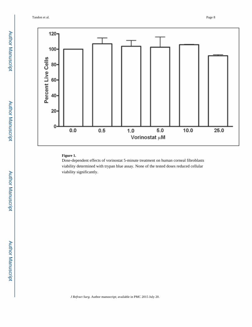
Figure 1. Dose-dependent effects of vorinostat 5-minute treatment on human corneal fibroblasts
viability determined with trypan blue assay. None of the tested doses reduced cellular
viability significantly.
Tandon et al. Page 8
J Refract Surg. Author manuscript; available in PMC 2015 July 20.
Author M
anuscriptA
uthor Manuscript
Author M
anuscriptA
uthor Manuscript
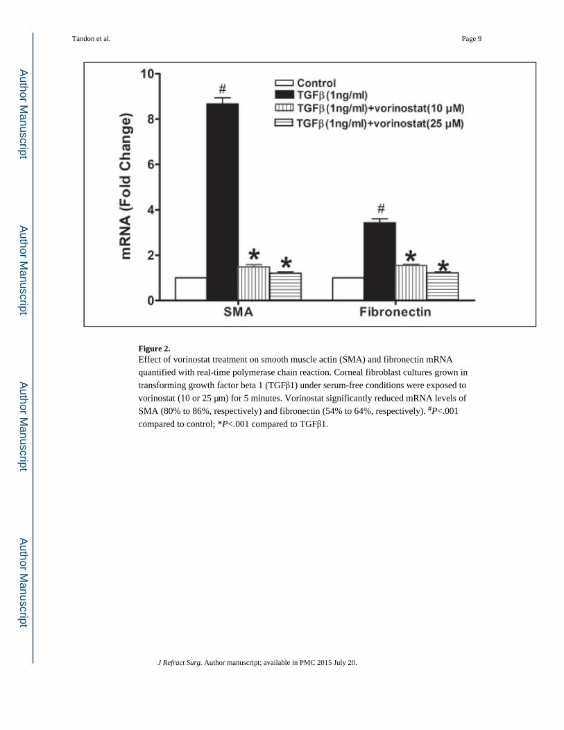
Figure 2. Effect of vorinostat treatment on smooth muscle actin (SMA) and fibronectin mRNA
quantified with real-time polymerase chain reaction. Corneal fibroblast cultures grown in
transforming growth factor beta 1 (TGFβ1) under serum-free conditions were exposed to
vorinostat (10 or 25 μm) for 5 minutes. Vorinostat significantly reduced mRNA levels of
SMA (80% to 86%, respectively) and fibronectin (54% to 64%, respectively). #P<.001
compared to control; *P<.001 compared to TGFβ1.
Tandon et al. Page 9
J Refract Surg. Author manuscript; available in PMC 2015 July 20.
Author M
anuscriptA
uthor Manuscript
Author M
anuscriptA
uthor Manuscript
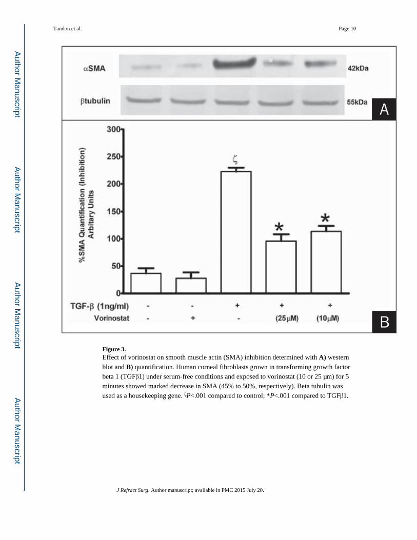
Figure 3. Effect of vorinostat on smooth muscle actin (SMA) inhibition determined with A) western
blot and B) quantification. Human corneal fibroblasts grown in transforming growth factor
beta 1 (TGFβ1) under serum-free conditions and exposed to vorinostat (10 or 25 μm) for 5
minutes showed marked decrease in SMA (45% to 50%, respectively). Beta tubulin was
used as a housekeeping gene. ζP<.001 compared to control; *P<.001 compared to TGFβ1.
Tandon et al. Page 10
J Refract Surg. Author manuscript; available in PMC 2015 July 20.
Author M
anuscriptA
uthor Manuscript
Author M
anuscriptA
uthor Manuscript
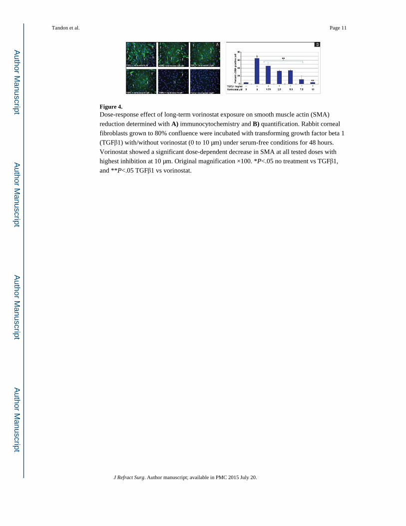
Figure 4. Dose-response effect of long-term vorinostat exposure on smooth muscle actin (SMA)
reduction determined with A) immunocytochemistry and B) quantification. Rabbit corneal
fibroblasts grown to 80% confluence were incubated with transforming growth factor beta 1
(TGFβ1) with/without vorinostat (0 to 10 μm) under serum-free conditions for 48 hours.
Vorinostat showed a significant dose-dependent decrease in SMA at all tested doses with
highest inhibition at 10 μm. Original magnification ×100. *P<.05 no treatment vs TGFβ1,
and **P<.05 TGFβ1 vs vorinostat.
Tandon et al. Page 11
J Refract Surg. Author manuscript; available in PMC 2015 July 20.
Author M
anuscriptA
uthor Manuscript
Author M
anuscriptA
uthor Manuscript
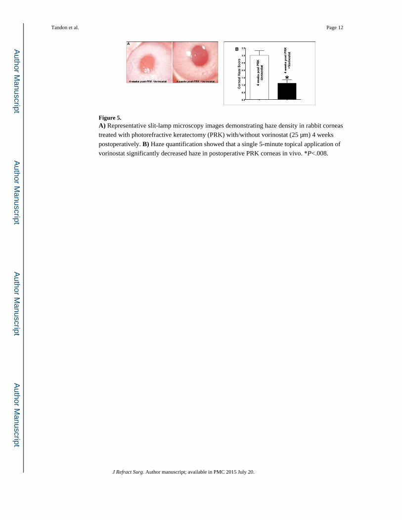
Figure 5. A) Representative slit-lamp microscopy images demonstrating haze density in rabbit corneas
treated with photorefractive keratectomy (PRK) with/without vorinostat (25 μm) 4 weeks
postoperatively. B) Haze quantification showed that a single 5-minute topical application of
vorinostat significantly decreased haze in postoperative PRK corneas in vivo. *P<.008.
Tandon et al. Page 12
J Refract Surg. Author manuscript; available in PMC 2015 July 20.
Author M
anuscriptA
uthor Manuscript
Author M
anuscriptA
uthor Manuscript
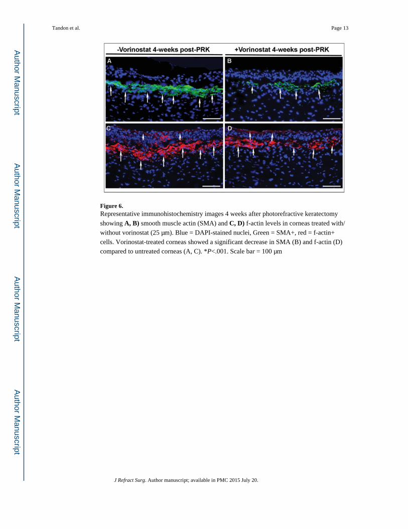
Figure 6. Representative immunohistochemistry images 4 weeks after photorefractive keratectomy
showing A, B) smooth muscle actin (SMA) and C, D) f-actin levels in corneas treated with/
without vorinostat (25 μm). Blue = DAPI-stained nuclei, Green = SMA+, red = f-actin+
cells. Vorinostat-treated corneas showed a significant decrease in SMA (B) and f-actin (D)
compared to untreated corneas (A, C). *P<.001. Scale bar = 100 μm
Tandon et al. Page 13
J Refract Surg. Author manuscript; available in PMC 2015 July 20.
Author M
anuscriptA
uthor Manuscript
Author M
anuscriptA
uthor Manuscript

Figure 7. Quantification of smooth muscle actin (SMA)+ cells 4 weeks after photorefractive
keratectomy (PRK) in corneas treated with/without vorinostat (25 μm). Vorinostat treatment
significantly decreased SMA+ cells (*P<.001).
Tandon et al. Page 14
J Refract Surg. Author manuscript; available in PMC 2015 July 20.
Author M
anuscriptA
uthor Manuscript
Author M
anuscriptA
uthor Manuscript
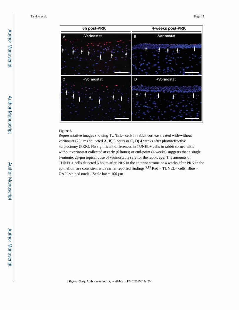
Figure 8. Representative images showing TUNEL+ cells in rabbit corneas treated with/without
vorinostat (25 μm) collected A, B) 6 hours or C, D) 4 weeks after photorefractive
keratectomy (PRK). No significant differences in TUNEL+ cells in rabbit cornea with/
without vorinostat collected at early (6 hours) or end-point (4 weeks) suggests that a single
5-minute, 25-μm topical dose of vorinostat is safe for the rabbit eye. The amounts of
TUNEL+ cells detected 6 hours after PRK in the anterior stroma or 4 weeks after PRK in the
epithelium are consistent with earlier reported findings.5,13 Red = TUNEL+ cells, Blue =
DAPI-stained nuclei. Scale bar = 100 μm
Tandon et al. Page 15
J Refract Surg. Author manuscript; available in PMC 2015 July 20.
Author M
anuscriptA
uthor Manuscript
Author M
anuscriptA
uthor Manuscript
![Pentobra: A Potent Antibiotic with Multiple Layers of ...wonglab.seas.ucla.edu/pdf/2015 JID [Schmidt, Wong] Pentobra A Potent... · Pentobra: A Potent Antibiotic with Multiple Layers](https://img.pdfslide.us/doc/110x75/5e79535b6eb666031e579d24/pentobra-a-potent-antibiotic-with-multiple-layers-of-jid-schmidt-wong-pentobra.jpg)