Embed Size (px)
Citation preview

DentalAnthropology
A Publication of the Dental Anthropology Association
Volume 17, Number 1, 2004 ISSN 1096-9411

1Dental AnthropologyVolume 17, Number 1, 2004
Dental Anthropology is the Official Publication of the Dental Anthropology Association.
Editor: Edward F. HarrisBook Review Editor: Greg C. Nelson
Address for Manuscripts
Dr. Edward F. HarrisCollege of Dentistry, University of Tennessee
870 Union Avenue, Memphis, TN 38163 U.S.A.e-mail address: [email protected]
Address for Book Reviews
Dr. Greg C. NelsonDepartment of Anthropology, University of Oregon
Condon Hall, Eugene, Oregon 97403 U.S.A.e-mail address: [email protected]
Published at
Craniofacial Biology Laboratory, Department of OrthodonticsCollege of Dentistry, The Health Science Center
University of Tennessee, Memphis, TN 38163 U.S.A.
Editorial Board
Kurt W. Alt (2004-2009) Jules A. Kieser (2004-2009) Tseunehiko Hanihara (2004-2009) Richard T. Koritzer (1999-2004) A. M. Haeussler (2004-2009) Helen Liversidge (2004-2009) Simon W. Hillson (1999-2004) John T. Mayhall (1999-2004) Kenneth A. R. Kennedy (1999-2004) Phillip W. Walker (1999-2004)
Officers of the Dental Anthropology Association
Debbie Guatelli-Steinberg (Ohio State University, OH) President (2004-2006)Simon W. Hillson (University College London, UK) President-Elect (2004-2006)
Heather H. Edgar (Maxwell Museum of Anthropology, NM) Secretary-Treasurer (2003-2005)Joel D. Irish (University of Alaska, Fairbanks, AK) Executive Board Member 2004-2006

1
The Genetics of Odontogenesis: Implications in Dental Anthropology and Palæo-Odontology
Geoffrey H. Sperber*
Department of Dentistry, University of Alberta, Edmonton, Alberta, Canada
*Address for correspondence: G. H. Sperber,Department of Dentistry, Faculty of Medicine and Dentistry, University of Alberta, Edmonton, Alberta T6G 2N8, CanadaE-mail: [email protected]
Editor’s note: The Editor solicited Professor Sperber to write this review article for Dental Anthropology.
EVOLUTIONARY GENETICS
“The crown of the human tooth even in its minute details represents little that is fortuitous. It is the resultant of inherited ancestral conditions, modifying further by evolution and involution.”
A. Hrdlička, 1924
Dental characters predominate in the identification of most species and genera, both of fossil and extant varieties. In this respect, teeth are unique among organs in enabling direct comparisons to be made between fresh specimens formed a few months previously and fossils excavated from sediments formed millions of years ago. Teeth depict their genetically inherited patterns, and thus their evolutionary history, more accurately than all other organs. This precision of genetic expression is due to their highly protected developmental environment, ensconced as they are in their submerged dental follicles until their full morphological maturity, before emerging into the potentially damaging environment.
By casting their primeval and delicate genotypic templates into the enduringly fossilized form of highly mineralized phenotypic morphology, teeth are the ultimate and amongst the most perfect extrinsic
ABSTRACT Palaeoanthropology and forensic odontology rely significantly upon detailed dental morphology that is ultimately the phenotypic expression of the underlying genotype and developmental phenomena. Odontogenesis is the consequence of a complex series of molecular interactions controlled by epigenetic signals acting on embryonic epithelial-mesenchymal tissues of ectodermal, neural crest and mesodermal origin. Of the estimated 24,847 genes of the human genome (Pearson, 2003) some 200 or more genes have been directly or indirectly involved in tooth development (http://bite-it.helsinki.fi). The loci of these genes on the 22 pairs of autosomes and the pair of sex chromosomes are being identified by their mutations that give rise to phenotypic dental abnormalities. The sequential cascades of stages from initiation through
the bud, cap, bell, mineralization, root formation and eruption of teeth are all under genetic control but subject to environmental influences. Identification of specific genes with clinical phenotypes provides invaluable clues to familial, racial and evolutionary affinities, all of jurisprudential, heredity and evolutionary significance to odontologists. Combining the genetics of odontogenesis with forensic evidence and palaeoanthropological fossil data provides an unparalled source of information on heredity, environmental and evolutionary events through teeth, the most durable of all biological structures after death. It is paradoxical that teeth are most susceptible to decay during life, but postmortem are the last structures to disintegrate. Teeth truly tell tales of the living and the dead. Dental Anthropology 2004;17(1):1-7.
expressors of the intrinsic units of evolutionary change, the mutations of genes.
The intricate morphology of the crowns of human teeth reflects both a long and complex phylogenetic archival record and a brief but extraordinarily elaborate ontogenetic formulation. This combination of long hereditary and short embryologic developments lies within the genes determining tooth shapes. The influence of phylogenetic factors upon the ontogeny of teeth is responsible for many of the factors peculiar to odontogenesis, making the study of dental development at the forefront of “evo-devo” exploration. The divergence of taxa heretofore based exclusively on fossil remnants may now be pursued by studying the selective action of genes during developmental processes (McCollum and Sharpe, 2001). New pathways of palaeoanthropological research are now being revealed by the genetic revolution.
The genetics underlying phenotypic dental characteristics that are directly observable has enabled rates and degrees of gene flow to be calculated and genetic drift to be estimated in divergent populations. Mutations may be traced in this manner, and the

2 3
selective advantages of particular dental conformations might account for dental micro-evolution. The development of cusps, ridges and fissures that enhance the predatory and masticatory capability of teeth are evolutionary advancements that correlate with different diets and environmental niches.
DEVELOPMENTAL GENETICS
The complexity of contributions of over 200 genes to odontogenesis makes the elucidation of each genes’ individual responsibility for each stage of development a daunting task. Most of these genes encode signals as well as their receptors, both in the cytoplasm and in transcription factors regulating gene expression in the nucleus (Thesleff, 2000). It is the mutation or deletion of these genes, by phenotypically expressing dental malformations or anodontia, or by experimental “knock outs” of specific genes, that some of the responsibilities of each gene is revealed. The intricacies of RNA editing, complex regulatory networks and criss-crossing molecular pathways makes meaningless the exact identification of genetic units. Moreover, the overlapping and redundancy of genetic expression patterns during development make the unravelling of the skein of influences particularly difficult.
Teeth initially developed in primitive fishes from the adaption of placoid scales overlying their jaws to form dermal denticles (Smith and Johanson, 2003). With the pending identification of genes responsible for the development of ectodermally-derived hard tissues, the revelation of the evolution of teeth becomes a possibility in the newly emerging discipline of phylogenomics (Eisen and Fraser, 2003). The synteny of conserved genes across species will account for the identification of “dental” genes in human odontogenesis having initially evolved in piscine species. This phylogenetic dermal origin of teeth is reflected in the embryonic development of human teeth, which although they develop submerged beneath the oral gingival epithelium, originate in part from ectodermal tissue. Teeth are derived from two of the primary germ layers, ectoderm and mesoderm, with a neural crest contribution. The enamel of teeth is derived from oral ectoderm, and neural crest mesenchyme provides material for the dentine, pulp and cementum. The periodontium is of both neural crest and mesodermal origin.
The morphogenesis of the maxillary and mandibular teeth is under the control of two different genetic programs, accounting for variation between upper and lower dentitions that provide for taxonomic distinctions. Combinations of different sized teeth within individuals reflect mosaic evolutionary derivations (McCollum and Sharpe, 2001).
An early signally event in tooth development at 6 weeks postconception is the induction of odontogenic mesenchyme by bone morphogenetic proteins (BMPs)
and fibroblast growth factors (FGFs) from the oral ectoderm. These initial odontogenic epithelial signals induce in the mesenchyme the expression of reciprocal signal molecules to the epithelium that results in the formation of the dental placode. The placodal signals, expressed as Sonic hedgehog (SHH), Wingless (Wnt) and tumor necrosis factor (TNF) molecules regulate the budding of the epithelium and condensation of the mesenchyme, effectively creating tooth buds (Thesleff and Mikkola, 2002). The number of tooth buds developing in each jaw is genetically determined, with an initial identicality that is later altered by their location. The differential odontogenic patterning creating a variety of tooth shapes (incisors, canines, molars) is organized by a homeodomain code of transcription factors expressed in restricted regions during development (Sharpe, 1995; Tucker and Sharpe, 1999; Cobourne and Sharpe, 2003). These factors include the Msx genes, Dlx family members, Pax 9, Lhx genes and Barx1 (Francis-West et al., 1998, Maas and Bei, 1997; Jung et al., 2003). The precise role of many of these signaling molecules during early budding is still under investigation. Barx1 expression is restricted to the presumptive proximal (posterior) region of the first pharyngeal arch, influencing the tooth buds to a molarization pattern (Tucker et al., 1999). The LIM homeodomain protein Islet 1 (ISL1) that is exclusively expressed in the presumptive incisor epithelium coincides with expression of Bmp4 that induces MSX1 expression in the underlying mesenchyme (Mitsiadis et al., 2003). The mesenchyme of the presumptive distal (anterior) region of the first arch expresses both Msx1 and Alx3 homeobox genes that determine incisiform shapes to the developing tooth buds (ten Berge et al., 1993). The region of overlap between Msx and Dlx genes codes for canines and premolars (Fig. 1).
The transcription factor Runx 2 and the signal Fgf 3 regulate epithelial morphogenesis from bud to cap stages. A primary enamel knot forms at the tip of the tooth bud, consequent to BMP 4 induction. The exit of enamel knot cells marks the onset of development of the tooth crown to form a cap-like structure that surrounds the underlying mesenchyme, referred to as the dental papilla. A SHH signal from the enamel knot is required for the growth of the epithelial cervical loops flanking the enamel knots and encompassing the dental papilla (Thesleff, 2003). Primary enamel knots initiate secondary enamel knots, thereby regulating the patterning of the tooth crown.. The arrangements and intercuspal dimensions of molar teeth are determined by the enamel knots (Townsend et al., 2003). Enamel knots are transient signaling centers that disappear by apoptosis (Vaahtokari et al., 1996). The consequent epithelial sheet folds in an exact sequence to produce undulating peaks and valleys, adumbrating cusps and fissures in the future crowns. This folding must involve
G. H. SPERBER
2 3ANTHROPALAEO-ODONTOLOGICAL GENETICS
Fig
1. S
chem
atic
dep
ictio
n of
fact
ors
oper
atin
g in
odo
ntog
enes
is.

4 5
differential mitotic activity by inhibition and activation determined by gene expression patterns to produce different tooth shapes (Salazar-Ciudad and Jernvall, 2002).
ENAMEL FORMATION
The secretion of the proteins unique to the enamel matrix, ameloblastin, amelogenin, enamelin and tuftelin by ameloblasts precedes the most intense mineralization of any tissue in the body (Dong et al., 2000). The ameloblast, the heralder of the hardest of human tissues, lays down a matrix that by mineralization becomes petrified, providing fossilized, immortal remains within the living jaws. Enamelin, the largest enamel extracellular matrix protein is a uniquely ameloblastic secretion, and is involved in the nucleation of apatite crystals (Gibson, 1999). Enamelin persists in mature enamel, whereas ameloblastin and amelogenin occur only temporarily in immature enamel (Robinson et al., 1989; Deutsch 1989). Moreover, there is an evolutionary sequence to the appearance of these proteins, with enamelins appearing earlier in phylogenetic history than amelogenins, and differing in their distribution among species (Herold et al., 1989), emphasizing the relationship of molecular biology to phylogeny. The tuftelin gene (TUFT1) has been mapped to chromosome 1q (Deutsch et al., 1994). The gene for the ameloblastin protein, AMBN, is located on chromosome 4q, and is a single copy gene containing 13 exons (Toyosawa et al., 2000).
ENAMEL THICKNESS
The speed and direction of migration of the ameloblasts in laying down enamel matrix, again under genetic control, determines the ultimate thickness of the enamel cap of the dental crown. The limited life of postmitotic ameloblasts, determined by their programmed early cell death, varies in different locations on the dental crown surface. This accounts for the varying ultimate thickness of enamel, from minimal along the cervical margins and in fissure depth, to maximal over the cusp peaks. This variation of enamel thickness not only reflects the longevity of ameloblasts, but also the speed of their migration. This combination of ameloblastic activities varies phylogenetically, accounting for the different maximal thicknesses of enamel found among hominoids and hominins (Beynon and Wood, 1986; Grine and Martin, 1989). The thin enamel of the gorilla, chimpanzee and orangutan contrasts strongly with the thick enamel of Homo sapiens and the australopithecines. The folivorous diet of the great apes, relatively free of abrasive grit, is not as wearing on dental enamel as the gritty omnivorous diet of hominins. Enamel thickness is correlated with longevity, as hominins long outlive pongids. The periodicity of incremental deposition of the enamel
matrix leading to the striae of Retzius, allows for age assessment at the time of death or exfoliation of extant and fossil teeth (Boyde 1963; FitzGerald 1996; Shellis 1998). During the year or two that a tooth develops and erupts, it accumulates isotopes of carbon and oxygen. Variations in the ratios of C13
to C12 and O18 to O16 provide evidence of the ambient diets of fossilized teeth. This isotopic evidence, in turn, may provide information on the provenance of recovered remains, even to the extent of tracing habitats and migrations during a lifetime, as revealed by the peregrinations of the Alpine Iceman (Müller et al., 2003).
Ameloblasts are extremely sensitive to metabolic, dietary and drug influences during enamel matrix deposition. The mechanisms of mineralized tissue deposition during amelogenesis provide a kymographic record of the state of metabolism and nutrition of the individual that is permanently entombed in the hard dental tissues.
Accordingly, illnesses and drug therapy during amelogenesis may be recorded as hypoplasias, hypomineralization or distinctive marks in matured enamel. Such examples as tetracyline staining or the neonatal line reflecting the change from intrauterine to extrauterine nutrition are ineradicably imprinted on enamel.
Incremental enamel apposition produces surface perikymata that allows determination of variations in their spacing, reflecting chronological deposition rates (Guatelli-Steinberg 2003). These rates have been determined to differ between apes, hominids and hominins (Dean et al., 2001). Amelogenesis can provide insights into cladistic relationships of the different species of hominoids, and their different rates of body maturation (Beynon and Dean, 1998; Smith, Martin and Leakey, 2003). The rapid growth of the Neanderthals has been based upon incremental dental data (Rozzi and de Castro, 2004).
The direct association of the sex chromosome genes that influences enamel development with the thickness of this tissue and with taurodontism indicates the ontogenetic link of dental morphology with evolutionary changes and phylogenetic influences. The aneuploid presence of extra sex chromosomes (47, XXX females, 47, XYY males) manifest thicker than normal enamel (Alvesalo et al., 1985; Alvesalo et al. 1987). Taurodontism, a trait carrying strong Neandertaloid associations is linked with aberrant sex-chromosome syndromes (Gage, 1978; Varrela et al., 1990).
ODONTOGENESIS
Each tooth germ consists of an enamel organ and a dental papilla surrounded by a dental follicle or sac. The dental papilla, of neural crest origin, and dental follicle of mesodermal origin, are the anlagen of the dental pulp and part of the periodontal apparatus
G. H. SPERBER

4 5
respectively.Each enamel organ during its development changes
from its initial small bud shape, enlarging by rapid mitosis of the basal cells into a cap shape, and later cupping into a large bell shape, by which shapes the three stages of enamel organ development are designated. Concomitant with these morphological alterations, histodifferentiation occurs within the enamel organ. Its external layer forms the outer enamel epithelium, a layer of cuboidal cells subjacent to the developing follicle. The stellate reticulum, composed of stellate cells set in a fluid matrix, constitutes the central bulk of the early enamel organ. The indented inner layer, lining the dental papilla, forms the inner enamel epithelium, part of which differentiates into the transient secretory columnar ameloblasts that form enamel. Lining a portion of the stellate reticular surface of the inner enamel epithelium is a squamous cellular condensation, the stratum intermedium, that probably assists the ameloblasts in forming enamel. The inner and outer enamel epithelia form the cervical loop, elongating into Hertwig’s epithelial root sheath, that, by enclosing more and more of the dental papilla, outlines the root(s) of the tooth. The number of roots of a tooth is determined by the subdivision, or lack thereof, of the root sheath into one, two or three compartments. The regulation of root development is dependent upon genes encoding nuclear factor I (NFI) transcription-replication proteins (Steele-Perkins et al., 2003). Aneuploid variation of the X chromosome’s “dental genes” appears to influence the mitotic activity of odontoblasts to produce taurodontic teeth (Varrela and Alvesalo, 1988; Varrela et al., 1990).
The inner enamel epithelium interacts with the ectomesenchymal cells of the dental papilla, whose peripheral cells differentiate into odontoblasts. The formation of dentine by the odontoblasts precedes, and is necessary for, the induction of premeloblasts into ameloblasts to produce enamel. The inner enamel epithelium of the root sheath induces odontoblast differentiation but, lacking a stratum intermedium, fails to differentiate itself into enamel-forming ameloblasts, accounting for the absence of enamel from the roots. Cementum forms on dentine adjacent to the sites of disintegration of the outer enamel epithelium of the root sheath. The fragmentation of the root sheath, due to programmed cell death (apoptosis) leaves clusters of cells, the epithelial rests of Malassez, in the periodontal ligament. These rests are the source of potential periodontal cysts. The fibers in the initial cementum derive solely from fibers of the pre-existing dental follicle that form the first principal fibers of the periodontal ligament.
The ameloblasts of the inner enamel epithelium and the adjacent odontoblasts together form a bilaminar membrane, which spreads by mitosis under genetic
control and varies among the tooth germs in different areas as previously described. The ameloblasts secrete a protein matrix of amelogenins and enamelins that later mineralize as enamel rods or prisms as they retreat from the membrane. Concomitantly, the odontoblasts secrete the collagen matrix of predentine, which later calcifies to dentine. Dentine deposition is a continuous process throughout life. The dental papilla differentiates into the dental pulp, the peripheral cells into odontoblasts, and the remaining cells into fibroblasts. Enamel formation is restricted to the pre-eruptive phase of odontogenesis and ends with the deposition of an organic layer, the enamel cuticle. The enamel organ collapses after deposition of this cuticle. The inner and outer enamel epithelia together with the remains of the stratum intermedium form the reduced enamel epithelium, which later fuses with the overlying oral mucous membrane to initiate the pathway for eruption.
The tissues of the dental pulp, the only unmineralized dental tissues, are confined within the enclosed pulp chamber, protected by the surrounding mineralized tissues. This protection provides the possibility of preservation of pulp tissues beyond death, enabling both forensic and palaeo-odontological investigations to be performed on tissues that may reveal DNA formulations (Komuro et al., 1998). Moreover, dental pulp tissues may contain stem cells of highly proliferative clonogenic capability, with the potentiality to differentiate into a variety of cell types (Gronthos et al., 2002; Miura et al., 2003). The possibility of clinical application of this stem cell source for therapies and tissue engineering remains to be explored, but the cloning of a whole individual from a dental pulp cell is still a fictional absurdity. Nonetheless, dental pulp cells have been shown to provide neurotrophic support for dopaminergic neurons as a treatment modality for Parkinson’s disease (Nosrat et al., 2004). Moreover, the cultivation of stem cells to produce teeth has been successfully achieved in experiments with mice, and portends the future therapeutic replacement of teeth in humans (Ohazama et al., 2004).
CONCLUSIONS
Odontogenesis and phylogenesis are inextric-ably interlinked through genetics in a combination that accounts for the complex functional morphology of the total dentition and its individual units, the teeth. The dental components-the crowns and their cusps, the roots, the pulp chambers and their tissues and the periodontal apparatus-are moulded by the twin forces of evolution and embryonic development. Thus, a synthesis of the features of comparative anatomy and developmental biology with the systematics of evolution is necessary for an understanding of the morphologic diversity and intricate structure of the dentition.
ANTHROPALAEO-ODONTOLOGICAL GENETICS

6 7
LITERATURE CITED
Alvesalo L, Tammisalo E, Hakola P. 1985. Enamel thickness in 47, XYY males’ permanent teeth. Ann Hum Biol 2:421-427.
Alvesalo L, Tammisalo E, Therman E. 1987. 47, XXX females, sex chromosomes, and tooth crown structure. Hum Genet 77:345-348.
Beynon AD, Dean MC. 1988. Distinct development patterns in early fossil hominids. Nature 335:509-514.
Beynon AD, Wood BA. 1986. Variations in enamel thickness and structure in East African hominids. Am J Phys Anthrop 70:177-193.
Beynon AD, Wood BA. 1987. Patterns and rates of enamel growth in the molar teeth of early hominids. Nature 326:493-496.
Boyde A. 1963. Estimation of age at death of young human skeletal remains from incremental lines in the dental enamel. Excerpta medica Int Cong Ser 80:36.
Cobourne MT, Sharpe PT. 2003. Tooth and jaw: molecular mechanisms of patterning in the first branchial arch. Arch Oral Biol 48:1-14.
Dean MC, Leakey MG, Reid D, Schrenk F, Schwartz GT, Stringer C, Walker A. 2001. Growth processes in teeth distinguish modern humans from Homo erectus and earlier hominins. Nature 414:628-631.
Deutsch D. 1989. Structure and function of enamel gene products. Anat Rec 224:189-210.
Deutsch D, Palmon A, Young MF, Selig S, Kearns WG, Fisher LW. 1994. Mapping of the human tuftelin gene (TUFT1) to chromosome 1 by fluorescence in situ hybridisation. Mamm Genome 5:461-462.
Dong J, Gu TT, Simmons D, MacDougall M. 2000. Enamelin maps to human chromosome 4q21 within the autosomal dominant amelogenesis imperfecta locus. Eur J Oral Sci 108:353-358.
Eisen JA, Fraser CM. 2003. Phylogenomics: Intersection of evolution and genomics. Science 300:1706-1712.
FitzGerald CM. 1996. Tooth crown formation and the variation of enamel microstructural growth markers in modern humans. Ph.D. dissertation, University of Cambridge.
Francis-West P et al. 1998. Signalling interactions during facial development. Mech Dev 75:3-28.
Gage JP. 1978. Taurodontism and enamel hypomaturation associated with X-linked abnormalities. Clin Genet 14:159-164.
Gibson CW. 1999. Regulation of amelogenin gene expression. Crit Rev Eukaryote Gene Expr 9:45-57.
Grine FE, Martin LB. 1989. Enamel thickness and development in Australopithecus and Paranthropus. In: Grine FE, ed. Evolutionary History of the ‘Robust’ Australopithecines. New York: Aldine de Gruyter, p 3-42.
Gronthos S, Brahim J, Li W, Fisher LW, Cherman N, Boyde A, Den Bestern P, Robey G, Shi S. 2002. Stem cell properties of human dental pulp stem cells. J Dent Res 81:531-535.
Guatelli-Steinberg D. 2003. Macroscopic and microscopic analyses of linear enamel hypoplasia in plio-pleistocene South African hominins with respect to aspects of enamel development. Am J Phys Anthrop 120:309-322.
Herold R, Rosenbloom J, Granovsky M. 1989. Phylogenetic distribution of enamel proteins: evolutionary appearance of enamelins prior to amelogenins. Calc Tiss Int 45:88-94
Hrdlička A. 1924. New data on the teeth of early man and certain fossil European apes. Am J Phys Anthrop 7:109-132.
Hu JC-C, Sun X, Zhang C, Simmer JP. 2001. A comparison of enamelin and amelogenin expression in developing mouse molars. Eur J Oral Sci 109:125-132.
Jung HS, Hitoshi Y, Kim HJ. 2003. Study on tooth development, past, present, and future. Microsc Res Tech 60:480-482
Komuro T, Nakamura M, Tsutsumi H, Mukoyama R. 1998. Gender determination from dental pulp by using capillary gel electrophoresis of amelogenin locus. J Forensic Odontosomatol 16:23-26.
Maas R, Bei M. 1997. The genetic control of early tooth development. Crit Rev Oral Biol Med 8:4-39.
McCollum MA, Sharpe PT. 2001. Developmental genetics and early hominid craniodental evolution. Bioessays 23:481-493.
Miletich I, Sharpe PT. 2003. Normal and abnormal dental development. Hum Mol Genet 12:R69-R73.
Mitsiadis TA, Angeli I, James C, Lendahl U, Sharpe PT. 2003. Role of Islet1 in the patterning of murine dentition. Development 130:4451-4460.
Miura M, Gronthos S, Zhao M, Lu B, Fisher LW, Robey PG, Shi S. 2003. SHED: Stem cells from human exfoliated deciduous teeth. Proc Nat Acad Sci 100:5807-5812.
Müller W, Fricke H, Halliday AN, McCulloch WJ-A. 2003. Origin and migration of the Alpine Iceman. Science 302:862-866.
Nosrat IV, Smith CA, Mullally P, Olson L, Nosrat CA. 2004. Dental pulp cells provide neurotrophic support for dopaminergic neurons and differentiate into neurons in vitro: implications for tissue engineering and repair in the nervous system. Eur J Neurosci 19:2388-2398.
Ohazama A, Modino SAC, Miletich I, Sharpe PT. 2004. Stem cell-based tissue engineering of murine teeth. J Dent Res [In press].
Pearson H. 2003. Geneticists play the numbers game in vain. Nature 423:576.
Robinson C, Weatherall JA, Hobling HJ. 1983.
G. H. SPERBER

6 7ANTHROPALAEO-ODONTOLOGICAL GENETICS
Formation and mineralization of dental enamel. Trends Biochem Sc 8:284-287.
Rozzi FVR, de Castro JMB. 2004. Surprisingly rapid growth in Neanderthals. Nature 428:936-938.
Salazar-Ciudad I, Jernvall J. 2002. A gene network model accounting for development and evolution of mammalian teeth. Proc Nat Acad Sci USA 99:8116-8120.
Sharpe PT. 1995. Homeobox genes and orofacial development. Conn Tiss Res 1995; 32:17-25.
Shellis RP. 1998. Utilization of periodic markings in enamel to obtain information on tooth growth. J Hum Evol 35:387-400.
Smith MM, Johanson Z. 2003. Separate evolutionary origins of teeth from evidence in fossil jawed vertebrates. Science 299:1235-1236.
Smith TM, Martin LB, Leakey GM. 2003. Enamel thickness, microstructure and development in Afropithecus turkanensis. J Hum Evol 44:283-306.
Steele-Perkins G, Butz KG, Lyons GE, Zeichner-David M, Kim H-J, Cho M-I, Richard M. 2003. Essential role for NFI-C/CTF transcription-replication factor in tooth root development. Mol Cell Biol 23:1075-1084.
ten Berge D, Brouwer A, el Bahi S, Guenet JL, Robert B, Meijlink F. 1998. Mouse Alx3: an aristaless-like homeobox gene expressed during embryogenesis in ectomesenchyme and lateral plate mesoderm. Dev Biol 199:11-25.
Thesleff I. 2000. Genetic basis of tooth development and dental defects. Acta Odont Scand 58:191-194.
Thesleff I. 2003. Epithelial-mesenchymal signalling regulating tooth morphogenesis. J Cell Sci 116:1647-
1648.Thesleff I, Mikkola M. 2002. The role of growth factors
in tooth development. Int Rev Cytol 217:93-134.Townsend G, Richards L, Hughes T. 2003. Molar
intercuspal dimensions. J Dent Res 82:350-355. Townsend GC, Alvesalo L. 1985. Tooth size in 47
X 44 males: evidence for a direct effect of the Y chromosome on growth. Aust Dent J 30:268-272.
Toyosawa S, Fujiwara T, Oeshima T, Shintani T, Sato A, Ogawa Y, Sobue S, Ijuhin N 2000. Cloning and characterization of the human ameloblastin gene. Gene 256:1-11.
Tucker AS, Matthews KL, Sharpe PT. 1998. Transformation of tooth type induced by inhibition of BMP signaling. Science 282:1136-1138.
Tucker AS, Sharpe PT. 1999. Molecular genetics of tooth morphogenesis and patterning: the right shape in the right place. J Dent Res 78:826-834.
Vaahtokari A, Åberg T, Thesleff I. 1996. Apoptosis in the developing tooth: association with an embryonic signaling center and suppression by EGF and TGF-4. Development 122:121-129.
Varrela J, Alvesalo L. 1988. Taurodontism in 47,XXY males: an effect of the extra X chromosome on root development. J Dent Res 67:501-502.
Varrela J, Alvesalo L, Mayhall J. 1990. Taurodontism in 45,X females. J Dent Res 69:494-495.
WEBSITE
http://bite-it.helsinki.fi

8 9
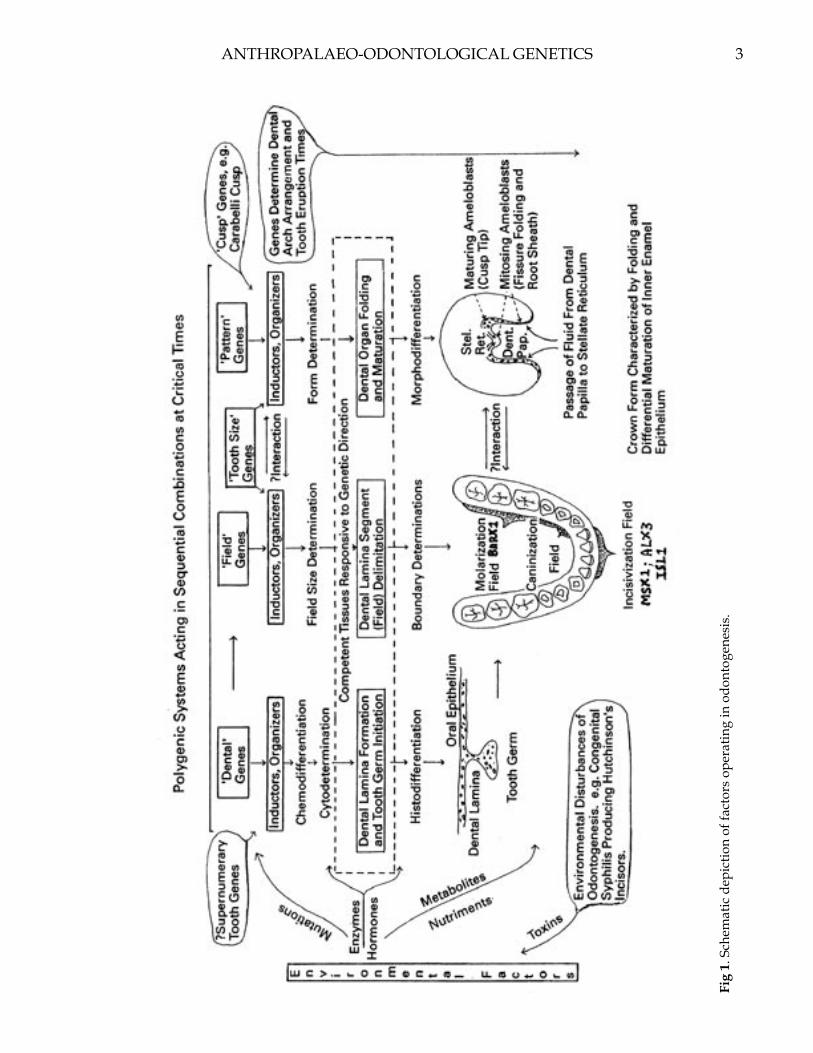
The Canary Islands are located in the Atlantic Ocean off the northwest coast of Africa (Fig. 1). Seven small islands comprise the archipelago: La Palma, Gomera, Hierro, Tenerife, Grand Canaria, Fuerteventura, and Lanzarote. Of the seven islands, Fuerteventura is nearest the continent, approximately 100 km west of Cape Juby, Morocco. The Canary Islands have been a part of Spain since the late 15th century. However, prior to that time they were occupied by the Guanche—the aboriginal inhabitants of the archipelago. These early people were primarily cereal agriculturalists who practiced a Neolithic lifestyle (Cavalli-Sforza et al., 1994). They possessed domesticated goats and pigs, and supplemented their diet with shellfish, fish, and various wild plants (Mercer, 1980).
An Odontometric Investigation of Canary Islander Origins
Address for correspondence: Joel D. Irish, Department of Anthropology, P.O. Box 757720, University of Alaska Fairbanks, Fairbanks, AK 99775-7720, U.S.A.E-mail: [email protected]
Joel D. Irish* and Brian E. Hemphill**
*Department of Anthropology, University of Alaska, Fairbanks, AK 99775.**Department of Sociology & Anthropology, California State University, Bakersfield, CA 93311
ABSTRACT Attempts by anthropologists to account for the peopling of the Canary Islands have led to theories that call for one, two, and even four immigration events. However, most agree the Canary Island Guanche are biologically closest to Berbers from Morocco and Algeria. Genetic contributions from Arabs, Romans, and Carthaginians have also been proposed. An earlier study by Irish using Penrose analysis of odontometric data in samples of Guanche, Shawia and Kabyle Berbers, and Bedouin Arabs supports many of these proposed genetic relationships.
The present investigation expands upon this earlier work by adding samples of Carthaginians, Egyptians, and Nubians, and by using tooth size apportionment analysis, a more robust statistical approach for
assessing inter-sample differences in the distribution, or allocation, of tooth size in the maxillary and mandibular dental arcades. The analysis yielded three components that account for >80% of the total variance. Cluster analysis and three-dimensional ordination of group component scores provide additional insight into Canary Island/North African relationships. Except for one early Nubian sample, the Guanche exhibit some measure of affinity to all others. However, they are most like Berbers and Carthaginians. These results suggest that Canary Islanders belong to a greater North African gene pool, yet show the closest affinities to Northwest Africans—which corroborates earlier dental and non-dental findings. Dental Anthropology 2004;17:8-17.
Authors’ note: A preliminary version of this paper was included in the 2001 volume La Paléo-Odontologie: Analyses et Méthodes d’Étude, Paris: Éditions Artcom, edited by Djillali Hadjouis and Bertrand Mafart. That article (Irish and Hemphill, 2001) was published in French, is generally not available outside of western Europe, and contained several publisher errors in the tables and figures. As such, we decided to provide a modified and expanded English translation to facilitate dissemination of our findings to a wider audience of dental and Canary Island researchers.
Over the past 100 years, numerous researchers have attempted to determine the origins and biological affinities of the Guanche (e.g., Verneau 1887, 1891; Hooton 1916, 1925; Falkenburger 1939; Fusté 1959, 1965; Schwidetzky 1963; Roberts et al., 1966; Vallois 1969; Mercer 1980; Gonzalez and Tejera, 1981; Onrubia Pintado, 1987; Bermudez de Castro, 1989). As a result, the original Guanche homeland has alternately been identified as Africa, Europe, and/or the eastern Mediterranean area. The purpose of the present investigation is to reexamine four of these origins hypotheses using evidence from principal components analysis of odontometric data in Canary Island, North African, and West Asian-derived samples. Although other theories exist (see Vallois 1969 for an overview), the four examined here afford a representative sampling of those envisioned by all researchers. Components obtained from statistical analyses yield information on overall crown size, as well as the allocation of size across dimensions and tooth types in both jaws among samples. This approach, termed tooth size apportionment analysis (see Harris and Bailit, 1988; Harris and Rathbun, 1991; Lukacs and Hemphill, 1993),
8 9
African mainland during the Neolithic. These two groups consisted of “Cro-Magnoid” and “Mediterranean-like” cranial types, asserted to be evident in prehistoric Guanche remains. These same findings are echoed by Fusté (1959, 1965), Vallois (1969), and others. The former cranial type is said to be characterized by a wide low face with robust features, whereas the latter is more gracile with a narrow, high face.
Roberts and coworkers (1966) proposed that the Guanche were the product of an ancient colonization from Europe (which reprises Verneau’s thesis to some extent (see Vallois 1969)). They based their conclusions on perceived osteological affinities of ancient Guanche skeletons (per Hooton, 1925; Hiernaux, 1975) and serological and dermatoglyphic affinities of living Canary Islanders (Mourant, 1954; Roberts et al., 1966) to Northwest Europeans.
Lastly, Mercer (1980) described an immigration of Northwest African Berbers during the Roman era, based on 15th-17th century ethnographic accounts of Guanche oral traditions and paleo-serological analyses of Guanche mummies. He suggested that Berber malcontents from the Atlas Mountains of northern Morocco and Algeria were exiled to the islands as punishment for resistance to Roman rule. Mercer also sees a lack of definite radiocarbon dates prior to the first century AD in the archipelago as supportive of this
CANARY ISLANDER ODONTOMETRICS
is intended to provide new insight into the old problem of understanding Guanche ancestry.
PREVIOUS PEOPLING HYPOTHESES
Hooton (1916, 1925) was one of several early anthropological researchers to investigate the origins and population history of Canary Islanders (see also Verneau 1887, 1891, and among others, Quatrefages and Hamy 1874, Shrubsall 1896, von Luschan 1896, von Behr 1908 (as presented in Vallois 1969)). He hypothesized that four migrations to the islands from North Africa took place during the Neolithic and Bronze Age. Based on the analyses of craniometric and ethnographic data, Hooton maintained that the Guanche were comprised of different stocks of people largely exhibiting Mediterranean and Alpine Caucasoid components, supplemented perhaps, by sub-Saharan and other elements. He further proposed that they originated from populations inhabiting southern Morocco, the Atlas Mountains of northern Morocco and Algeria, and the eastern Mediterranean (Fig. 1). Subsequent intermixture among these four groups, along with later Arab, Berber, and Carthaginian gene flow, was thought to have resulted in the pre-European Contact peoples of the Canary Islands.
Based on cranial morphometric data, Schwidetzky (1963) envisioned two migrations from the adjacent
Fig. 1. Regional map showing Canary Islands, North Africa, and the Mediterranean area.
10 11
late-arrival model. In addition, his hypothesis provides an explanation for sea transportation to the islands—an ability the Guanche apparently did not possess at the time of European Contact. However, others maintain (e.g., Cavalli-Sforza et al., 1994) that the Guanche originally sailed to the islands of their own accord, and subsequently lost the skill to make adequate sea-going vessels. Like Hooton, Mercer suggests later contact by Carthaginians and Arabs may have provided an additional genetic contribution to the Canary Island gene pool.
Despite these widely varying scenarios all workers agree that, at the time of European Contact, the native Guanche comprised a lightly-pigmented population (Murdock, 1959; Vallois, 1969) reminiscent of peoples living throughout Europe, the Mediterranean area, and parts of North Africa. This contention is based on 15th century French and Spanish accounts, in addition to the aforementioned ethnographic, serological, skeletal, and other data. Further, excluding Roberts et al. (1966), most researchers believe the Guanche were closely related to Northwest African Berbers (see Hooton, 1916; Schwidetzky, 1963; Gonzalez and Tejera, 1981; Onrubia Pintado, 1987; Bermudez de Castro, 1989); perhaps those from the Atlas Mountains region of northern Morocco and Algeria (Mercer, 1980). Support for this relationship is bolstered by recent genetic analyses (Cavalli-Sforza et al., 1994), as well as long-standing linguistic evidence that Guanche, the Canary Islander’s extinct language (Bynon, 1970), shows a close affinity to the Afroasiatic Berber language (Hooton, 1916, 1925; Greenberg, 1966; Mercer, 1980). The Berber language may in turn be derived from the Late Paleolithic North African Mechta and Capsian cultures (Hiernaux, 1975; Mercer, 1980; Onrubia Pintado, 1987). However, as Hooton (1925) and Mercer (1980) note, the islands’ population may have also been influenced by Arab, Roman, and Carthaginian contact prior to the 15th century Spanish occupation.
ODONTOMETRIC ANALYSES
In a preliminary study (Irish, 1993a), aspects of the four hypotheses were tested via Penrose shape analysis of tooth crown diameters in samples of pre-European Contact Canary Islanders (n=163), and historic Northwest African Shawia Berbers (n=26), Kabyle Berbers (n=32), and Bedouin Arabs (n=49). Although metric data are employed, the Penrose shape component is analogous to morphological analysis because it emphasizes differences in the form of a structure (crown form) rather than size (Penrose, 1954; Rahman, 1962; Corruccini, 1973). The results tentatively support a Canary Island/Northwest Africa link. The Guanche comparison to the Shawia and Kabyle Berbers yielded low, insignificant shape values (0.09 and 0.10, respectively), indicating a close phenetic similarity that would be expected if Berbers colonized the Islands. The magnitude of the Guanche/ Arab value is twice that of the other comparisons (0.18) and is significant
(Rahman, 1962), suggesting a more distant affinity.The present investigation expands upon this previous
odontometric study. Besides the Guanche, Berbers, and Arabs, samples of West Asian-derived Carthaginians and Northeast African Egyptians and Nubians are added. In total, 12 prehistoric through historic Northwest and Northeast African samples, comprising 669 dentitions, are analyzed and compared. Moreover, in place of Penrose, tooth size apportionment analysis (Harris and Bailit, 1988; Harris and Rathbun, 1991; Lukacs and Hemphill, 1993) is used on the odontometric data. This technique provides a more robust statistical approach that uses principal components analysis for assessing inter-sample differences in allocation of tooth size.
MATERIALS AND METHODS
The samples
The Canary Islands sample used in both the previous and present odontometric studies consists of 163 skeletal dentitions (male=70, female=52, indeterminate=41). Eight crania are from the island of La Palma, 25 from Gomera, 54 from Tenerife, 56 from Gran Canaria, 11 from Fuerteventura, and nine from unidentified locations in the archipelago. Most specimens are curated at the Musée de l’Homme, Paris, although 13 are located at the American Museum of Natural History, New York, and two are at the National Museum of Natural History in Washington, D.C. The exact date(s) of the series is unknown, but radiocarbon dating of grottoes, caves, and tumuli similar to those from which the present materials were removed range from 20 BC to AD 1690+70, with a median range of AD 400-900 (Mercer, 1980; Bermudez de Castro, 1989).
The Shawia Berber sample consists of 26 historic individuals who originally lived just south of Constantine, Algeria (see Fig. 1). The sample consists of dentitions from 16 males, seven females, and three individuals of unknown sex, all from the Musée de l’Homme. Greenberg (1966) characterizes Berbers as speaking one of several dialects (e.g., Shawia) of the Berber language, which belongs to the Berber language family in the Afroasiatic superfamily. Their language also reflects influence from Phoenician, Latin, and Arabic sources (Bynon, 1970). Such heterogeneity is consistent with the fact that Berber populations, especially those from the less-mountainous regions of Algeria and Morocco, show evidence of admixture with Arabs and other intrusive peoples (i.e., Carthaginian, Greek, Roman, Spanish, Turkish, French) (Wysner, 1945).
The Kabyle Berber sample is made up of 32 historic crania (male=21, female=7, indeterminate=4) from the Algiers and Oran region of the Djurdjura Mountains in northern Algeria (Wysner, 1945). They are all curated at the Musée de l’Homme. Unlike many Berbers, the Kabyle remained isolated from the many outsiders who successively conquered lands throughout northern Africa
J.D. IRISH AND B.E. HEMPHILL
10 11
beginning in 750 BC. As such, they experienced relatively little genetic admixture (Wysner, 1945). The Berbers may be indigenous to North Africa, being descended from earlier Capsian and perhaps Mechta peoples (Hiernaux, 1975; Irish, 1998a,b, 1999, 2000).
The Bedouin Arab sample (n=49) is composed of a heterogeneous mix of historic crania (male=18, female=24, indeterminate=7). Thirty-six individuals were recovered from the coast of Morocco between Rabat and Mogador, ten are from Algeria between Oran and Algiers, two are from Tunis, Tunisia, and one is from the Sahel region of Libya. The latter specimen was recorded at the University of Minnesota; the rest are at the Musée de l’Homme. Arabs first entered Africa along the Suez isthmus in the 7th century, conquering Byzantine lands in Egypt and to the west. A second wave of Arabs arrived in the 11th century, when entire tribes of Bedouin immigrated from the Syrian desert (Julien, 1970; Hiernaux, 1975). These nomadic peoples are similar in physical appearance to the Berbers with whom they are heavily admixed (Julien, 1970; Hiernaux, 1975).
The Carthaginian sample is made up of 28 individuals (male=16, female=8, indeterminate=4) from the site of Carthage, north of Tunis, Tunisia. Twenty-four crania were recovered from Punic period levels (751?-146 BC) (Charles-Picard and Picard, 1968). The four remaining skulls may be from the Punic period, or are perhaps from early Roman times (146 BC-AD 435) (Wysner, 1945). All of the material is curated at the Musée de l’Homme. Carthage was founded in ca. 751 BC by the Phoenicians, a West Asiatic people from the area now comprising Lebanon (Charles-Picard and Picard, 1968). In 146 BC, Carthage was conquered by the Romans, who remained in control until AD 435. Both the Carthaginians and Romans are thought to have had extensive contact with local Berber populations (Wysner, 1945).
The remaining seven samples, from Northeast Africa, are included in the dental analysis to help delineate Guanche affinities on a broader, geographically-oriented scale. Three samples comprise 12th Dynasty through Byzantine Egyptians (1991 BC-AD 600) (Elliot Smith and Wood-Jones, 1910; Baines and Malek, 1982) from Lisht (n=61), El Hesa (n=72), and Kharga Oasis (n=26) in Egypt. The specimens are located at the American Museum of Natural History and National Museum of Natural History. There are several hypotheses concerning Egyptian origins; they may be non-African (i.e., West Asian or southern European) (Angel, 1972; Curto, 1972; Hiernaux, 1975; Mourant, 1983), an admixed people, with African and non-African roots (e.g., Hamid Zayed, 1981), or indigenous (White, 1970; Davidson, 1974; Trigger, 1976; July, 1992; Phillipson, 1994; Newman, 1995; Williams, 1997). Whichever the case, by the Dynastic period they were likely a heterogeneous people from the combining of many ethnic elements (Curto, 1972; Davidson, 1974). The other four Northeast samples are from Nubia, in
northern Sudan. One sample consists of 18th Dynasty Pharonic Nubians (1575-1380 BC) (Trigger, 1976) from Soleb (n=32); the others are Meroitic (n=91), X-Group (n=39), and Christian (n=18) Nubians (100 BC-AD 1400) from Semna (Zabkar and Zabkar, 1982) (see Irish, 1993b, 1998b for a more complete description of all samples). The Pharonic sample was recorded at the Musée de l’Homme; the others are curated at Arizona State University, Tempe. The origin of the Nubians is unclear; they may be locals that possess a sub-Saharan component (e.g., Greene, 1967, 1972; Carlson and Van Gerven, 1977, 1979), or are heavily-admixed migrants from elsewhere in North Africa (Irish and Turner, 1990; Turner and Markowitz, 1990).
Methods employed
Mesiodistal and buccolingual dental crown measurements were taken by Irish on each individual’s maxillary and mandibular permanent teeth (I1-M3), following the method of Moorrees (1957), with Boley gauge vernier calipers accurate to 0.1 mm. Excessively worn or carious teeth, as well as those antimere pairs exhibiting obvious size asymmetry (most often M3s), were not measured. The degree of intra-observer measurement error was assessed by comparing replicate measurements of the left side of 25 Meroitic dentitions. The mean measurement error between sessions one month apart is 0.2 mm; this figure is within the range noted by Wolpoff (1971). Moreover, none of the measurements are significantly different based on paired-sample t-tests.
Dimensions of teeth on the left side in each sample were used for statistical analysis because, based on paired-sample t-tests, no significant differences occurred between antimeres for any dimensions (per Hemphill, 1991; Hemphill et al., 1992; Lukacs and Hemphill, 1993). If a significant difference (p < 0.05) would have existed, the average of the dimensions from the antimere pairs would have been used per individual to compute the sample average. In cases where a tooth on the left side was missing in an individual, the right antimere (if present) was measured to maximize sample size. The resulting 32 or fewer mesiodistal and buccolingual dental crown measurements per individual were then used to calculate mean crown diameters for use in the assessment of odontometric affinity among samples.
Tooth size apportionment analysis was conducted according to the procedures of Harris and Bailit (1988) and Harris and Rathbun (1991), as modified by Hemphill (1991). The covariance matrix of mean crown diameters for each of the 12 samples was submitted to principal components analysis to obtain component loadings. Crown diameters for each sample were multiplied by the loadings for each tooth diameter, and this product was summed across all 32 crown diameters. This methodology yielded three component scores per sample (see Lukacs and Hemphill, 1993).
The mean total crown area (MD X BL) for all 16 teeth,
CANARY ISLANDER ODONTOMETRICS

12 13
per sample, was used to assess differences in overall tooth size. If samples differed significantly in total crown area (>5%), residual component scores were calculated for those components significantly correlated with overall tooth size. Group component scores were then submitted to cluster analysis and three-dimensional ordination. A minimum spanning tree (Hartigan, 1975) was imposed on the array of component scores for ease of interpretation of association among the individual samples. All statistical analyses were performed with SYSTAT statistical software (Wilkinson, 1990).
Ideally, odontometric research should involve separate analyses by sex. However, out of necessity, the
sexes were pooled by sample in this study. This approach follows the lead of Harris and Rathbun (1991), and Lukacs and Hemphill (1991), who report that any dental size variation between the sexes was not great enough to justify the markedly smaller sample sizes. Moreover, Hemphill et al. (1992) and Lukacs and Hemphill (1993) found that while males and females within an ethnic group differ in absolute tooth size, apportionment of tooth size is unaffected by sex dimorphism.
RESULTS
Tooth size apportionment analysis of the 12 samples’ crown measurements yielded the component loadings in Table 1; component eigenvalues and percentage of the variance explained are also tabulated. The dental crown measurements themselves will be presented in a separate publication on African odontometric variation, and thus are not listed. Although six principal components possess eigenvalues greater than 1.0, the first three alone account for 80.4% of the total variance.
Component one is dominated by a general size factor, which is illustrated by the strong positive loadings for most variables (see top of Fig. 2). Nevertheless, a second factor involving relative dimensions of the teeth is also evident, as reflected by much lower loadings for buccolingual dimensions of the maxillary and, particularly, mandibular anterior teeth. In other words, high scorers along this component are characterized by generally large dentitions, with anterior teeth that exhibit long mesiodistal relative to narrow buccolingual diameters.
The second component separates samples on the basis of two criteria (see middle of Fig. 2). The first is similar to the secondary factor of component one. Anterior teeth (I1, I2, C) feature dimensional segregation, with buccolingual breadths receiving higher loadings than mesiodistal lengths; this is true for both maxillary and, especially, mandibular teeth. The second distinction involves the distal molars (M2, M3). Mandibular mesiodistal and buccolingual diameters receive fewer negative loadings than their maxillary counterparts. This difference is slightly greater for the mesiodistal than buccolingual dimensions. Thus, high scorers along component two exhibit broad buccolingual diameters among anterior maxillary and, especially, mandibular teeth relative to mesiodistal dimensions, as well as relatively large mandibular distal molars compared to their maxillary isomeres.
The loadings for component three are, at first glance, confusing. However, there appears to be a distinction in buccolingual dimensions by isomere; that is, with the exception of P4 and M2, maxillary breadths receive higher loadings than their mandibular counterparts (see bottom of Fig. 2). This is especially true for I1 and C. Thus, high scorers for component three possess maxillary teeth that are broader in their buccolingual
J.D. IRISH AND B.E. HEMPHILL
Components Variable 1 2 3
UI1MD 0.837 0.078 0.151UI1BL 0.377 0.631 0.606UI2MD 0.960 -0.102 0.090UI2BL 0.724 0.263 0.043UCMD 0.563 0.570 0.332UCBL 0.491 0.642 0.280UP3MD 0.952 0.021 0.076UP3BL 0.911 -0.056 0.232UP4MD 0.730 -0.057 -0.424UP4BL 0.923 -0.089 0.081UM1MD 0.774 -0.066 -0.425UMIBL 0.909 0.044 0.198UM2MD 0.777 -0.371 -0.312UM2BL 0.770 -0.325 -0.312UM3MD 0.499 -0.661 0.428UM3BL 0.802 -0.485 0.008LI1MD 0.737 0.175 0.235LI1BL 0.177 0.497 -0.511LI2MD 0.833 0.216 0.149LI2BL 0.177 0.850 -0.161LCMD 0.807 0.252 -0.340LCBL 0.347 0.765 -0.347LP3MD 0.817 -0.343 -0.010LP3BL 0.817 -0.129 0.358LP4MD 0.847 0.051 -0.257LP4BL 0.933 0.040 -0.123LMIMD 0.844 -0.034 0.207LM1131 0.927 0.100 -0.023LM2MD 0.917 0.055 -0.150LM2BL 0.895 -0.035 -0.254LM3MD 0.781 -0.222 0.207LM3BL 0.837 0.275 -0.094
Eigenvalue 19.147 4.133 2.462Variance (%) 59.834 12.916 7.695Total Variance 80.445
TABLE 1. Component loadings, eigenvalues, and variance explained for the 12 dental samples.

12 13
dimensions than the corresponding mandibular isomeres.
Once component loadings were obtained, total crown areas by sample were regressed on component scores to determine if overall tooth size represents a significant contributing factor behind group scores. As is often the case, component one scores are highly associated with size (see Table 2)—in this case overall tooth size (F=1537.84, p=0.00). However, components two and three do not show a significant association. To compensate for the effect of overall tooth size, the regression formula was used to obtain expected component one scores. Expected scores were subtracted from the observed to calculate group departures (residuals) from expected results from general tooth size.
The next step in analysis requires the use of some technique to illustrate the patterning of biological distances delineated by the residual component one, component two, and component three scores (Table 2). In the present investigation four methods of cluster analysis—complete linkage, single linkage, average linkage, and Ward’s minimum variance, as well as three-dimensional ordination were employed.
The complete linkage dendrogram is presented in Figure 3. Results obtained with other associating algorithms produced analogous results. The Guanche sample is phenetically most similar to Northwest African Shawia Berbers, a relationship revealed by the previous Penrose analysis (Irish, 1993a). The Guanche also show a close affinity to the Carthaginian and Kabyle samples. Members of this four-group aggregate share anterior teeth of intermediate buccolingual size, and maxillary and mandibular isomeres of proportionate dimensions.
The Guanche are next most-like the aggregate at the center of the dendrogram that contains Christian, X-Group, and Meroitic Nubians, Lisht, El Hesa, and Kharga Egyptians, and Bedouin Arabs. The earlier Penrose analysis (Irish, 1993a) also showed the Arab sample
Fig. 2. Loadings among the 12 dental samples for com-ponents one, two, and three.
CANARY ISLANDER ODONTOMETRICS
Sample TCA COMP1 RCMP1 COMP2 COMP3
Guanche 1098.09 -0.399 -0.185 0.150 -0.377Shawia 1100.64 -0.164 0.002 0.820 -0.112Kabyle 1117.97 0.125 -0.038 2.001 -0.654Bedouin 1084.59 -0.457 0.014 -0.489 0.659Carthage 1058.07 -1.058 -0.084 0.931 -1.394Lisht 1050.73 -1.191 -0.077 -0.730 0.110El Hesa 1051.15 -1.130 -0.024 -0.701 0.865Kharga 1086.70 -0.624 -0.194 -0.508 0.983Soleb 1193.56 1.566 -0.043 1.176 2.012Meroitic 1145.27 0.750 0.068 -0.746 0.009X-Group 1191.73 1.431 -0.134 -1.239 -0.883Christian 1177.20 1.162 -0.127 -0.664 -1.218
TABLE 2. Total crown area (TCA), component scores (COMP), and residuals (RCMP) for the 12 dental samples.

14 15
to be slightly divergent from the Guanche. Moreover, except for the West Asian-derived Arabs who, as noted comprise a mix of individuals from throughout North Africa, this seven-group aggregate is composed entirely of Northeast Africans. For the most part, these samples exhibit a tendency toward broad maxillary teeth relative to the corresponding mandibular isomeres. This pattern is particularly evident in the Christian and X-Group Nubian samples; they also possess relatively large teeth (see TCA in Table 2).
Lastly, the Guanche, as well as all other samples, are most divergent from Pharonic Nubians from Soleb. The Soleb sample is characterized by the largest teeth of all samples, as well as broad buccolingual anterior tooth diameters and large mandibular molars relative to the maxillary counterparts.
Similar dental relationships are illustrated by ordination of the three principal component scores (Figure 4). Axes X, Y, and Z correspond to the sample scores for residual component one (RCMP1), component two (COMP2), and component three (COMP3). The Guanche (CAN), located on the far left of the figure, link most closely with Northwest Africans; that is,
Fig. 4. Three-dimensional ordination with minimum spanning tree of principal component scores among the 12 samples. See text for explanation of abbreviations.
Fig. 3. Complete linkage cluster analysis dendrogram of principal component scores among the 12 samples.
J.D. IRISH AND B.E. HEMPHILL

14 15
with Carthaginians (CAR), Shawia Berbers (ALG), and Kabyle Berbers (KAB). However, they also exhibit some affinities to Northeast Africans. This affinity is evident by the Guanche connection to the Meroitic sample (MER) from Semna. Meroitic Nubians are in turn linked to X-Group (XGR) and Christian (CHR) Nubians, and to Lisht (LIS), the Bedouin Arabs (BED), El Hesa (HES), Kharga (KHA), and the Soleb (SOL) outlier, respectively.
DISCUSSION AND CONCLUSIONS
Although the timing and circumstances under which the immigration event(s) occurred have not been addressed by these odontometric results, tooth size apportionment analysis has revealed two important findings that pertain to other aspects of the four peopling hypotheses. First, the Canary Island Guanche show closest dental affinities to Northwest Africans, relative to other samples of various ages. Second, the pattern of phenetic affinities possessed by the Guanche suggest that some degree of biological relatedness extends beyond the adjacent mainland to Nubians and Egyptians in Northeast Africa.
The Guanche share a very similar pattern of tooth size apportionment with the Shawia and, to a lesser extent, Kabyle Berbers. This similarity corroborates results of a preliminary odontometric study (Irish, 1993a), and supports those aspects of Hooton’s (1916, 1925), Schwidetzky’s (1963), and other’s (e.g., Fusté 1959, 1965; Vallois 1969) models that suggest at least some Guanche originated in Northwest Africa; it specifically sustains Mercer’s (1980) and other’s (e.g., Gonzalez and Tejera, 1981; Onrubia Pintado, 1987; Bermudez de Castro, 1989; Cavalli-Sforza et al., 1994) claims for a sole Berber ancestry from populations living in northern Morocco and Algeria.
Conversely, this finding cannot completely rule out Hooton’s (1925), Schwidetzky’s (1963), and other’s (e.g., Fusté 1959, 1965; Vallois 1969, etc.) evidence for some eastern Mediterranean input, considering the Guanche affinity to most Northeast Africans. Moreover, Guanche similarity to West Asian-derived Carthaginians could be interpreted as support for this contention. However, such an affinity may simply identify evidence for Berber/Carthaginian admixture, or could imply genetic relatedness via the latter’s proposed direct contact (Hooton, 1916, 1925; Mercer, 1980) with the Guanche; a similar situation might explain the slightly more distant Guanche affinity to West Asian-derived Bedouin Arabs. In addition, Hooton’s (1925) suggestion for a sub-Saharan genetic component has not been directly tested here, although data from dental morphological studies (see Irish, 1993b, 1997, 1998a,b, 2000) do not support such a relationship. Whatever the case, the concordance of skeletal, ethnographic, linguistic, genetic, and now dental data, should put to rest any notion of a non-
African (i.e., European) origin for aboriginal Canary Islanders (as per Roberts et al., 1966).
The evidence for a lesser Guanche affinity to Egyptian and three of four Nubian samples implies aboriginal Canary Islanders belong to a greater North African gene pool. Some level of diachronic dental homogeneity apparently exists throughout North Africa—from the Canary Islands to Egypt and northern Sudan. Indeed, this east-west similarity suggests that a clinal relationship in tooth size apportionment existed, considering the separation of Northwest and Northeast African samples. These conclusions support previous findings based on dental morphological analyses published elsewhere (Irish, 1993b, 1997, 1998a,b; Guatelli-Steinberg et al., 2001).
ACKNOWLEDGMENTS
We thank the individuals at the institutions from which the Guanche and comparative data were collected, including: Christy G. Turner II, Charles Merbs, and Donald Morris from Arizona State University; Guy Gibbon and the late Elden Johnson from the University of Minnesota; Douglas Ubelaker and David Hunt from the National Museum of Natural History; Ian Tattersall, Jaymie Brauer, and Gary Sawyer from the American Museum of Natural History; Andre Langaney, Frances Roville-Sausse, and Miya Awazu Periera da Silva from the Musée de l’Homme; Fred Wendorf and Sue Linder-Linsley from Southern Methodist University, and Henry de Lumley and Dominique Grimaud-Herve from the Institut de Paléontologie Humaine. Research was supported by grants from the National Science Foundation (BNS-9013942), the Arizona State University Research Development Program, and the American Museum of Natural History.
LITERATURE CITED
Angel JL. 1972. Biological relations of Egyptian and east-ern Mediterranean populations during pre-dynastic and dynastic times. J Hum Evol 1:307-313.
Baines J, Malek J. 1982. Atlas of ancient Egypt. New York: Facts On File Publications.
Bermudez de Castro JM. 1989. The Carabelli trait in hu-man prehistoric populations of the Canary Islands. Hum Biol 61:117-131.
Bynon J. 1970. The contribution of linguistics to history in the field of Berber studies. In: Dalby D, editor. Language and history in Africa. New York: Africana Publishing Corporation, p 64-77.
Carlson DS, Van Gerven DP. 1977. Masticatory function and post-Pleistocene evolution in Nubia. Am J Phys Anthropol 46:495-506.
Carlson DS, Van Gerven DP. 1979. Diffusion, biological determinism, and biocultural adaptation in the Nu-bian corridor. Amer Anthrop 81:561-580.
Cavalli-Sforza LL, Menozzi P, Piazza A. 1994. The his-
CANARY ISLANDER ODONTOMETRICS

16 17
tory and geography of human genes. Princeton: Princeton University Press.
Charles-Picard G, Picard C. 1968. The life and death of Carthage. New York: Taplinger Publishing Co.
Corruccini RS. 1973. Size and shape in similarity coef-ficients based on metric characters. Am J Phys An-thropol 38:743-754.
Curto S. 1972. Archaeological outline of Egypt from the Paleolithic to the modern Arab state. J Hum Evol 1:141-146.
Davidson B. 1974. Africa in history. New York: MacMil-lan Publishing Company.
Elliot Smith G, Wood-Jones F. 1910. The archaeological survey of Nubia: Report for 1907-1908, Volume II, Report on Human Remains. Cairo: National Print-ing Department.
Falkenburger F. 1939. Essai d’une nouvelle classifica-tion craniologique des anciens habitants des Canar-ies. L’Anthropologie 49:330-364, 523-541.
Fusté M. 1959. Contribution à l’anthropologie de la Grande-Canarie. L’Anthropologie 63:295-318.
Fusté M. 1965. Physical anthropology of the Canary Islands. Am J Phys Anthropol 23:285-292.
Gonzalez R, Tejera A. 1981. Los aborigenes Canarios. La Laguna, Tenerife: Universidad de La Laguna.
Greenberg JH. 1966. The languages of Africa. Bloom-ington: Indiana University.
Greene DL. 1967. Dentition of Meroitic, X-Group, and Christian populations from Wadi Halfa, Sudan. Anthropol. Paper 85, Nubian Ser. 1. Salt Lake City: University of Utah Press.
Greene DL. 1972. Dental anthropology of early Egypt and Nubia. J Hum Evol 1:315-324.
Guatelli-Steinberg D, Irish JD, Lukacs J. 2001. Canary Island-North African population affinities: Mea-sures of divergence based on dental morphology. Homo 52:173-188.
Hamid Zayed A. 1981. Egypt’s relations with the rest of Africa. In: Mokhtar G, editor. General history of Africa II: Ancient civilizations of Africa. Berkeley: University of California Press, p 136-154.
Harris EF, Bailit HL. 1988. A principal components analysis of human odontometrics. Am J Phys An-thropol 75:87-99.
Harris EF, Rathbun TA. 1991. Ethnic differences in the apportionment of tooth sizes. In: Kelley MA, Larsen CS, editors. Advances in dental anthropology. New York: Wiley-Liss, p 121-142.
Hartigan JH. 1975. Clustering algorithms. New York: Wiley.
Hemphill BE. 1991. Tooth size apportionment among contemporary Indians: An analysis of caste, lan-guage, and geography. Ph.D. dissertation, Univer-sity of Oregon, Eugene.
Hemphill BE, Lukacs JR, Rami Reddy V. 1992. Tooth size apportionment among contemporary Indians:
Factors of caste, language, and geography. J Hum Ecol 2:231-253.
Hiernaux J. 1975. The people of Africa. New York: Charles Scribner’s Sons.
Hooton EA. 1916. Preliminary remarks on the arche-ology and physical anthropology of Tenerife. Am Anthropol 18:358-365.
Hooton EA. 1925. The ancient inhabitants of the Canary Islands. Cambridge: Peabody Museum of Harvard University.
Irish JD. 1993a. Dental morphometric affinity of Canary Islanders with North African Maghreb populations. Am J Phys Anthropol Supplement 16:114.
Irish JD. 1993b. Biological affinities of late Pleistocene through modern African aboriginal populations: The dental evidence. Ph.D. Dissertation, Arizona State University, Tempe. Ann Arbor: University Microfilms.
Irish JD. 1997. Characteristic high- and low-frequency dental traits in Sub-Saharan African populations. Am J Phys Anthropol 102:455-467.
Irish JD. 1998a. Diachronic and synchronic dental trait affinities of Late and post-Pleistocene peoples from North Africa. Homo 49:138-155.
Irish JD. 1998b. Dental morphological affinities of Late Pleistocene through recent sub-Saharan and North African peoples. Bull Mem Societé d’Anthropol Paris. Nouvelle serie 10:237-272.
Irish JD. 1999. Chi erano gli Iberomaurusiani? Affinità biologiche tra popolazioni Nord-Africane del Pleis-tocene Superiore e più recenti. Attualità dell’ Antro-pologia Ricerca e Insegnamento nel XXI secolo, p 113-114.
Irish JD. 2000. The Iberomaurusian enigma: North African progenitor or dead end? J Hum Evol 39:393-410.
Irish JD, Hemphill BE. 2001. Les Canaries ont-elles été colonisées par les Berbčres d’Afrique du Nord? La contribution de l’analyse odontométrique. In: Had-jouis D, Mafart B, editors. La paléo-odontologie: Analyses et méthodes d’étude. Collection paléo-anthropologie et paléopathologie osseuse. Paris: Artcom, p 122-137.
Irish JD, Turner CG II. 1990. West African dental affinity of late Pleistocene Nubians: Peopling of the Eurafri-can-South Asian triangle II. Homo 41:42-53.
Julien C. 1970. History of North Africa. London: Rout-ledge and Kegan Paul.
July RW. 1992. A history of the African people, 4th edi-tion. Prospect Heights, Ill: Waveland Press. Lukacs JR, Hemphill BE. 1991. The dental anthropology of prehistoric Baluchistan: A morphometric approach to the peopling of South Asia. In: Kelley MA, Larsen CS, editors. Advances in dental anthropology. New York: Wiley-Liss, p 77-119.
Lukacs JR, Hemphill BE. 1993. Odontometry and
J.D. IRISH AND B.E. HEMPHILL

16 17
biological affinity in South Asia: Analysis of three ethnic groups from Northwest India. Hum Biol 65:279-325.
Mercer J. 1980. The Canary Islanders: Their prehistory, conquest and survival. London: Rex Collings.
Moorrees CFA. 1957. The Aleut dentition: A correla-tive study of dental characteristics in an Eskimoid people. Cambridge: Harvard University Press.
Mourant AE. 1954. The distribution of the human blood groups. Springfield, IL: Charles C. Thomas, Pub-lisher.
Mourant AE. 1983. Blood relations: Blood groups and anthropology. Oxford: Oxford University Press.
Murdock GP. 1959. Africa: Its peoples and their culture history. New York: McGraw-Hill.
Newman JL (1995) The peopling of Africa: A geographic interpretation. New Haven: Yale University Press.
Onrubia Pintado J. 1987. Les cultures préhistoriques des Îles Canaries état de la questions. L’Anthropologie 91:653-678.
Penrose LS. 1954. Distance, size and shape. Ann Eugen-ics 18:337-343.
Phillipson DW. 1994. African archaeology, 2nd ed. Cambridge: Cambridge University Press.
Rahman NA. 1962. On the sampling distribution of the studentized Penrose measure of distance. Ann. Hum. Genetics 26:97-106.
Roberts DF, Evans M, Ikin EW, Mourant AE. 1966. Blood groups and the affinities of the Canary Island-ers. Man 1:512-525.
Trigger BG. 1976. Nubia under the pharaohs. Boulder, CO: Westview Press.
Turner CG II, Markowitz M. 1990. Dental discontinu-
ity between late Pleistocene and recent Nubians. I. Peopling of the Eurafrican-South Asian Triangle. Homo 41:42-53.
Schwidetzky I. 1963. La población Prehispánica de las Islas Canarias. Santa Cruz de Tenerife: Museo Ar-queologico de Tenerife.
Vallois HV. 1969. Les hommes de Cro-Magnon et les Guanches: Les faits acquis et les hypothèses.
Simposio de Cro-Magnon. Anuario de Estudias Atlanti-cos. Madrid 15:97-119.
Verneau R. 1887. Rapport sur une mission scientifique dans l’archipel Canarien. Archives des Missions sci-entifieques et littéraires, 3e série 13:569-817.
Verneau R. 1891. Cinq années de séjour aux îles Canar-ies. Paris.
White JEM. 1970. Ancient Egypt: Its culture and history. New York: Dover Publications, Inc.
Williams B. 1997. Egypt and Sub-Saharan Africa: Their interaction. In: Vogel J, editor. Encyclopedia of precolonial Africa: Archaeology, history, languages, cultures, and environments. Walnut Creek: Alta Mira Press, p 465-472.
Wilkinson L. 1990. SYSTAT: The system for statistics. Evanston, IL: Systat, Inc.
Wolpoff MH. 1971. Metric trends in hominid dental evolution. Studies in Anthropology, no. 2. Cleve-land: Case Western Reserve University Press.
Wysner GM. 1945. The Kabyle people. New York: Pri-vately Printed.
Zabkar LV, Zabkar J. 1982. Semna South. A preliminary report of the 1966-68 excavation of the University of Chicago Oriental Institute expedition to Sudanese Nubia. J Am Res Center Egypt 19:21-28.
CANARY ISLANDER ODONTOMETRICS
18 19
This case highlights the fact that asymmetries in dental crown form, whether they be fluctuating or directional, need to be viewed as resulting from a con-tinuum of developmental disturbances that may range from minor to severe. As our knowledge of the mo-lecular basis of dental development continues to grow, we should eventually be able to explain in cellular and molecular terms the specific causes of the whole range of asymmetrical expressions in dental crown form that we observe within the human dentition.
The phenotypic appearance of newly-emerged den-tal crowns results from an interplay between an individ-ual’s genotype and environmental influences operating during the period of odontogenesis. Environmental fac-tors may also alter crown appearance after teeth emerge into the oral cavity, for example due to trauma, caries or wear. However, careful examination of teeth intra-oral-ly or indirectly via dental models will normally enable the examiner to distinguish between those crown varia-tions that have occurred during development compared with those that have resulted after emergence.
It is generally assumed that the genetic influences operating on antimeric tooth pairs are identical so, in the absence of post-emergence effects, differences in crown morphology between corresponding teeth on opposite sides of the dental arch can be considered to reflect the influence of developmental disturbances during odontogenesis. These disturbances may vary in their timing, duration and severity.
Asymmetry in dental crown size is referred to as being directional if there is a tendency for dimensions on one side to be consistently larger than those of their corresponding antimeres. There is some evidence of
directionality in deciduous and permanent crown size in relatively large human samples that exclude indi-viduals with major developmental disorders (Harris, 1992; Townsend et al., 1999). However, whether these findings reflect real underlying biological influences or represent chance effects remains unclear.
There also are various pathological conditions that may lead to directional asymmetries in dental crown size and shape. For example, in hemifacial microso-mia—a developmental abnormality affecting the first and second branchial arches—the posterior teeth are smaller than normal, with the reduction in size being most marked on the affected side (Seow et al., 1998). This is an example of directional asymmetry where the affected teeth are smaller on the affected side.
Fluctuating dental asymmetry refers to the small random differences in crown size or morphology com-monly observed between antimeric tooth pairs. These differences may be due, for example, to differences in blood supply or space availability between sides. More severe space constraints leading to distortion of devel-oping tooth germs may result in compression of a tooth or teeth on one side producing more marked asymme-try in size and/or shape.
The magnitude of fluctuating dental asymmetry is increased in laboratory animals exposed to external stressors during development (Siegel et al., 1977) and in certain human chromosomal disorders, for example Down syndrome, where the aneuploidy is thought to disrupt homeostasis, leading to increased developmen-tal instability (Townsend, 1983). A similar explanation has been put forward to account for increased fluctuat-ing asymmetry in crown size noted in individuals with
Localized Asymmetry in Human Dental Crown Form—an Interesting CaseJohn Wetherell, Tracey Winning and Grant Townsend*
Dental School, The University of Adelaide, South Australia 5005
ABSTRACT. A case of a 20-year-old female is described in which the premolars and molars on the right side of the arch display altered crown proportions and altered occlusal morphology. There is no evidence of an orofacial congenital disorder or history of trauma. It is argued that the asymmetrical expression of crown form does not fall within the normal range of variation but has resulted from a localized disruption
in cellular function within the developing tooth germs, probably upsetting the folding of the internal enamel epithelia. This has produced crowns that have rounded cuspal outlines and reduced intercuspal distances. Superimposed space constraints in the mandible may have also led to compression of the lower molar crowns mesiodistally and affected their root formation. Dental Anthropology 2004;17(1):18-23.
Editor’s note: It is hoped that this case report will stimulate some productive discussion in the Journal. Please submit comments to the Editor.
*Address for correspondence: Grant Townsend, Dental School, The University of Adelaide, South Australia 5005Email: [email protected]

18 19
sex chromosomal aneuploidies (Townsend et al., 1986) and in individuals with cleft lip and palate (Narayanan et al., 1999). Of considerable interest is the report of increased directional asymmetry in the occlusal mor-phology of permanent first molars in 45,X/46,X mosa-ics (Pirttiniemi et al., 1998). This study indicates that different cell lines regulated by different genes may be responsible for differences in crown form on opposite sides of the dental arches.
In this paper we report on an interesting example of dental asymmetry that is evident in the maxillary and mandibular posterior segments of the permanent dentition of a young woman who has no history or signs of orofacial trauma or a congenital disorder. This case provides a good opportunity to ponder on how factors that have presumably operated unilaterally on the developing dental arches can lead to marked asymmetries in final crown forms in an otherwise healthy person.
CASE REPORT
Figures 1 and 2 show occlusal views of the maxillary and mandibular dental arches of a 20-year-old female of European ancestry who presented at the Adelaide Dental School in 2001 for a routine dental check-up. The woman had no history of any major medical problems, nor was there any history of her mother suffering ill-health during pregnancy. She had chicken pox as an 8-9 year-old but did not take any medication at that time. There was also no history or evidence of visible facial asymmetry.
In both arches, the first premolars had been extracted previously for orthodontic reasons, and the third molars had not emerged in the maxilla. The woman had also worn an upper removable orthodontic appliance for seven months in 1995. A supernumerary tooth had been extracted from the maxillary right molar region distal to the first molar prior to the commencement of orthodon-tic treatment.
The maxillary right third molar was not present and the woman confirmed that it had not been extracted. The mandibular left third molar was partly erupted. The maxillary left first and second molars and man-dibular left first molar had fissure sealants placed on their surfaces in 2001 and the occlusal surface of the maxillary right first molar had been restored in amal-gam in 1994, then the amalgam had been replaced with composite resin in 2001.
The crowns of the maxillary right second premolar and the first and second molars were markedly differ-ent in form to those on the left. There was also some minor variation in crown form of the maxillary right canine. The mandibular right second premolar and first, second and third molars all showed different and unusual crown form compared with those on the left. The affected maxillary and mandibular premolar and molar teeth showed similar features, with altered crown shapes and rounded forms with small intercus-pal distances. The maxillary right canine crown showed increased labial convexity compared with its antimere, but this variation was less marked than those of the pre-molars and molars. Intraoral examination did not dis-close any hypoplasia or hypocalcification of the enamel of affected teeth.
Examination of a panoramic radiograph obtained at 20 years 5 months of age showed that the maxillary right third molar was congenitally missing (Fig. 3). This film disclosed some differences in the root morphology of the mandibular right first and second molars com-pared with the corresponding teeth on the left. The roots of the mandibular right first molar appeared to be more slender than those of the mandibular left first molar. The roots of the mandibular right second molar were more curved (like plier handles) than those of its antimere. The buccal roots of the maxillary right molars also appeared to converge more than the corresponding molar roots on the left that displayed a distal curve. The roots of all teeth were fully formed, except for the man-dibular left third molar that was distally impacted.
Fig. 2. Occlusal view of the mandibular dentition of the woman.
Fig. 1. Occlusal view of the maxillary dentition of the woman.
LOCALIZED ASYMMETRY IN THE PERMANENT DENTITION

20 21
Bitewing radiographs were also available that en-abled an assessment of enamel and dentine thickness and pulp cavity anatomy. The altered contours of the proximal surfaces of affected teeth made it difficult to locate homologous points on the mesial and distal sur-faces of antimeric teeth. However, using the methods described by Stroud et al. (1994) and comparing the woman’s data with the standards provided by Stroud and colleagues, enamel and dentine thickness fell within the normal ranges and there were only minor differences between the sides.
The sizes of the dental crowns were compared be-tween sides and with normal data published for indi-viduals of European ancestry (Townsend et al., 1986). Maximum mesiodistal and buccolingual crown diam-eters were recorded according to the definitions of Sei-pel (1946) and expressed as z-scores against standards for girls. All of the z-scores except two were positive, indicating that the woman’s dental crown size was gen-erally larger than normal. In particular, the z-score for the buccolingual crown diameter of the maxillary right second premolar was 3.5 (compared with 2.8 on the left) and the z-score for the buccolingual crown diameter of the mandibular right first molar was 3.4 (compared with 1.9 on the left). In contrast, the z-score for the mesiodis-tal crown diameter of the mandibular right first molar was -0.3 (compared with 1.7 on the left) and the z-score for the mesiodistal crown diameter of the mandibular right second molar was 0.1 (compared with 1.0 on the
left). Therefore, the mandibular right molars showed markedly reduced mesiodistal crown diameters but increased buccolingual diameters compared with their antimeres.
Intercuspal distances were also recorded for the woman’s first molars and maxillary second premolars, then comparisons were made between sides and with unpublished normal values that had been computed previously in our laboratory for a sample of females of European ancestry. The woman’s intercuspal distances were expressed as z-scores and all of these values were positive on the left side, consistent with the fact that overall crown size of these teeth was also larger than average. However, the values of z-scores for intercuspal dimensions of the right first molars and the maxillary right second premolar were all negative. They ranged from -0.8 for the distance between the mesiobuccal and distobuccal cusps of the mandibular first molar, to -2.0 for the distance between the mesiobuccal and mesiolin-gual cusps of the maxillary first molar. These measure-ments confirmed the visual impression that the cusp tips were closer together on the posterior teeth on the right compared with the left.
The mandibular right first molar was a four-cusped tooth compared with its antimere that displayed the typical five-cusped appearance. There was also altered expression of Carabelli trait between the maxillary right and left first molars, the former displaying a groove form of the feature whereas the latter showed a cuspal
Fig. 3. Panoramic radiograph of the woman.
J. WETHERELL ET AL.

20 21LOCALIZED ASYMMETRY IN THE PERMANENT DENTITION
form.When the dental casts were examined from the buc-
cal view with the teeth in maximum intercuspation, the premolars and first molars on the right did not occlude whereas there was contact between opposing teeth on the left (Figs. 4 and 5). The maxillary right canine was in a crossbite relationship with the mandibular right canine and lateral incisor. The central incisors displayed a normal overbite and overjet relationship (Fig. 6), al-though the mandibular incisors were retroclined and the mandibular arch midline was displaced 2-3 mm to the right. Given that orthodontic treatment had been carried out, including extraction of first premolars, we did not attempt to develop a common hypothesis to explain the altered crown form of the premolars and molars, and the posterior open bite, on the right side.
DISCUSSION
Although some of the woman’s teeth showed asymmetry in overall crown size, especially the mandibular first molars, the most striking feature was the asymmetrical expression of crown shape of both maxillary and mandibular posterior teeth. The posterior teeth on the right showed more rounded cuspal outlines with smaller intercuspal distances than their antimeres on the left. The alteration in crown form was localized mainly to the maxillary and mandibular posterior segments, specifically the premolars and molars, although there was some minor variation in the labial convexity of the maxillary right canine. The first premolars had been extracted for orthodontic reasons so it was not possible to examine them. Nor was it possible to examine any of the woman’s primary teeth.
Given that there was no indication that the enamel on the affected teeth was hypoplastic or hypocalcified, it would seem that some disturbance must have affected the morphogenesis of the developing premolar and molar tooth germs on the right side only. The location of the cusp tips on premolars and molars is associated with the development of enamel knots in the enamel organ, that is those regions of the internal enamel epithelium that cease mitosis, leading to the buckling of its surface (Thesleff et al., 2001). The final shape of the cusps depends on the subsequent deposition of enamel
by ameloblasts. As the woman’s enamel was apparently normal both qualitatively and quantitatively, the most likely site of the disruption is the internal enamel epithelium.
We have reported that heritability estimates for intercuspal distances of molar teeth derived from a large sample of twins are only moderate in magnitude compared with those for overall crown dimensions (Townsend et al., 2003). Intercuspal distances were also associated with higher coefficients of variation than overall crown measures, confirming that they display relatively greater phenotypic variation than maximum mesiodistal and buccolingual diameters. These results are consistent with the findings of molecular studies (e.g., Tucker and Sharpe, 1999; Thesleff et al., 2001), in-dicating that epigenetic influences related to the release of specific signalling molecules from the regions of the enamel knots are important in determining how the internal enamel epithelium folds during odontogenesis. It is possible, therefore, that the localized alteration of crown form in this case has resulted from a disruption to the development of enamel knots on one side of the arch. This may have been triggered by traumatic event.
It is difficult to say what the cellular or molecular basis of such a disturbance could be, but it is tempting to suggest that an upset to neural crest cell migration, or to the reciprocal interaction between the ectomesenchymal cells of the dental papilla and the epithelial cells of the internal enamel epithelium, might underlie the problem. It is very unlikely that a genetic mutation has caused the morphological asymmetry, as this would be most likely to affect teeth on both sides of both dentitions. A pos-
Fig. 5. Left buccal view of the woman’s dentition, with models occluded in intercuspal occlusion.
Fig. 4. Right buccal view of the woman’s dentition, with models occluded in intercuspal occlusion.
Fig. 6. Labial view of the woman’s dentition, with models occluded in intercuspal occlusion.

22 23
sible exception could be mosaicism, with different cell lines regulated by discrete genes producing morpho-logical asymmetry. Although the woman reported here showed no other signs of physical abnormality, we were unable to test for mosaicism.
The observed pattern of morphological variation within the human dentition usually follows Butler’s field theory (Dahlberg, 1945; Butler, 2001), with the more distal tooth in each class showing greater varia-tion than more mesially positioned teeth. For example, third molars generally show considerable variation in morphology, much more than first molars. In this case, however, the “key” molar tooth seemed to be affected to the same degree as the more distal members of the class. This tends to confirm that the localized variation in crown form resulted from a distinct, though relatively minor, developmental disturbance and does not merely represent an extreme example of the normal range of development.
Another model that may prove useful in trying to decipher the underlying basis of variation within the human dentition is the so-called “facial homeobox code” described by Sharpe (1995). The homeobox genes in the developing face are restricted to specific domains, with incisor, canine and molar fields being described. As Sharpe (1995) points out, it is possible that neural crest cells are pre-patterned with homeobox genes prior to or during their migration. Subsequent reciprocal interac-tions between those neural crest cells contributing to the ectomesenchyme of the dental papilla with the epithe-lial cells of the internal enamel organ would then define tooth type and shape. It is possible that some localized upset to expression of the molar homeobox code has produced the unilateral variation in dental morphology that we have observed in this case.
The timing of onset and duration of crown forma-tion of the affected teeth provide further insights into the possible nature of the disturbance. The crowns of the permanent first molars begin to calcify at around birth, so the period of folding of the internal enamel epi-thelium is mainly a pre-natal event, although distortion could still occur post-natally until the cusp tips have been united by the spread of calcification. The second premolar crowns commence their calcification at around 2.0 to 2.5 years and the second molars around 2.5 to 3.0 years, so folding and potential distortion of the internal enamel epithelia of these teeth persists into the post-na-tal period. The third molars may not commence crown calcification until 7-10 years, so there are several years after birth during which disturbances may affect their crown form (Hillson, 1996:123).
Given that all of the affected teeth in this case show similar alterations in their crown form, it would seem that some ongoing localized disturbance in the func-tion of one or more cell lines in the developing teeth is the most likely etiological factor. It is possible that
there could have also been superimposed local space constraints that led to the alterations in overall crown shape of the mandibular molars, compressing them me-siodistally but allowing them to grow buccolingually. For example, Taylor (1978:257) has described in detail the appearance of compressed teeth and suggested that their appearance may have resulted from crowding of tooth buds prior to calcification. Space constraints may also account for the apparent differences in molar root form between the sides.
Several researchers have reported on asymmetrical expression of so-called non-metric crown variants, such as Carabelli trait (e.g., Saunders and Mayhall, 1982; Pinkerton et al., 1999). This normal variation may take the form of a large cusp on one side and a smaller cusp on the other, or there may be different expressions of grooves on each side. However, it is rare to find a cuspal form of Carabelli trait on one side but no expression or a small groove on the other. Again, then, the observed expression of Carabelli trait in this case suggests that a specific disturbance has occurred and that the variation in expression does not fall within the so-called normal range of variation.
This case highlights the fact that asymmetries in dental crown form, whether they be fluctuating or directional, need to be viewed as resulting from a con-tinuum of developmental disturbances that may range from minor to severe. As our knowledge of the mo-lecular basis of dental development continues to grow, we should eventually be able to explain in cellular and molecular terms the specific causes of the whole range of asymmetrical expressions in dental crown form that we observe within the human dentition.
LITERATURE CITED
Butler PM. 2001. What happened to the field theory. In: Brook A, editor. Dental morphology 2001. Sheffield: Sheffield Academic Press, p 3-12.
Dahlberg AA. 1945. The changing dentition of man. Am J Dent Assoc 32:676-690.
Harris EF. 1992. Laterality in human odontometrics: analysis of a contemporary American White series. In: Lukacs JR, editor. Culture, ecology and dental anthropology. Delhi, Kamla-Raj, p 157-170.
Hillson S. 1996. Dental Anthropology. Cambridge: Cam-bridge University Press, p 123.
Moorrees CFA, Thomsen SO, Jensen E, Yen PKJ. 1957. Mesiodistal crown diameters of human permanent teeth in individuals. J Dent Res 36:39-47.
Narayanan A, Smith S, Townsend G. 1999. Dental crown size in individuals with cleft lip and palate. Perspect Hum Biol 4:61-70.
Pinkerton S, Townsend G, Richards L, Schwerdt W, Dempsey P. 1999. Expression of Carabelli trait in both dentitions of Australian twins. Perspec Hum Biol 4:19-28.
J. WETHERELL ET AL.

22 23LOCALIZED ASYMMETRY IN THE PERMANENT DENTITION
Pirttiniemi P, Alvesalo L, Silven O, Heikkila J, Julku J, Karjalahti P. 1998. Asymmetry in the occlusal mor-phology of first permanent molars in 45,X/46,XX mosaics. Archs Oral Biol 43:25-32.
Saunders SR, Mayhall J. 1982. Developmental patterns of human dental morphological traits. Archs Oral Biol 27:45-49.
Siegel MI, Doyle WJ, Kelley C. 1977. Heat stress, fluctu-ating asymmetry and prenatal selection in the labora-tory rat. Am J Phys Anthropol 46:121-126
Seipel CM. 1946. Variation of tooth position. Sven Tand-lak Tidskr 39: Suppl.
Seow WK, Urban S, Vafaie N, Shusterman S. 1998. Mor-phometric analysis of the primary and permanent dentitions in hemifacial microsomia: a controlled study. J Dent Res 77:27-38.
Sharpe PT. 1995. Homeobox genes and orofacial devel-opment. Connect Tiss Res 32:17-25.
Stroud JL, Buschang PH, Goaz PW. 1994. Sexual dimor-phism in mesiodistal dentin and enamel thickness. Dentomaxillofac Radiol 23:169-171.
Taylor RMS. 1978. Variation in the morphology of teeth. Springfield, IL: Charles C Thomas, p 257.
Thesleff I, Keranen S, Jernvall J. 2001. Enamel knots as
Decoding Your SubscriptionWant to know when your subscription to
Dental Anthropology expires? Membership in the Association and, thus, your subscription to Dental Anthropology is on an annual basis coinciding with the calendar year. Have a look at the mailing label on the evelope that this issue arrived in, and you will see the year for which your dues have been paid. The year is located in parentheses to the right of your name. So, if the mailing label says “(2004)” you are paid to the end of this calendar year.
In order to extend your membership, fill-out the relevant portions of the enclosed form (or e-mail the Secretary)—remember to include appropriate pay-ment—and mail it to the Secretary-Treasurer of the Association:
Dr. Heather H. EdgarMaxwell Museum of AnthropologyMSC01 10501 University of New MexicoAlbuquerque, New Mexico 87131-0001 USA
telephone: (505) 277-4415e-mail: [email protected]
signalling centers linking tooth morphogenesis and odontoblast differentiation. Adv Dent Res 15:14-18.
Townsend GC. 1983. Fluctuating dental asymmetry in Down’s syndrome. Aust Dent J 28:39-44.
Townsend GC and Brown T. 1981. The Carabelli trait in Australian Aboriginal dentition. Archs Oral Biol 26:809-814.
Townsend G, Dempsey P, Richards L. 1999. Asymmetry in the deciduous dentition: fluctuating and direc-tional components. Perspec Hum Biol 4:45-52.
Townsend G, Richards L, Hughes T. 2003. Molar in-tercuspal dimensions: genetic input to phenotypic variation. J Dent Res 82:350-355.
Townsend GC, Alvesalo L, Jensen B, Kari M. 1986. Pat-terns of tooth size in human chromosomal aneuploi-dies. In: Russell DE, Santoro JP, Sigogneau-Russell D, editors. Teeth revisited: Proceedings of the VIIth International Symposium on Dental Morphol-ogy, Mem Mus Natn Hist Nat Paris (series C), Paris, France 53:25-45.
Tucker AS, Sharpe PT. 1999. Molecular genetics of tooth morphogenesis and patterning: the right shape in the right place. J Dent Res 78:826-834.

24 25
During data collection for a larger study (Edgar, 2002), it was noted that some mandibular premolars were more rectangular than ‘normal’ premolars. The affected teeth appear to be either compressed in the buccolingual dimension or longer in the mesiodistal dimension. The affected premolar is thus more rectangular or elliptical than hexagonal or rounded, like most mandibular premolars. Additionally, the cusps of the affected premolars appear closer together, and somewhat angled toward each other in some cases. Although affected posterior mandibular premolars were noted, more anterior premolars were seen with this condition.
MATERIALS
A total of 458 individual dentitions were examined for the presence of affected premolars. A list of the samples from which these individuals were drawn, their ancestry, and the frequency of trait presence is listed in Table 1. In these dentitions 19 elongated mandibular premolars were noted, 14 anterior and five posterior.
METHODS
Measurements were made of 14 affected and 22 non-affected anterior premolars. All individuals are represented by the following three measurements:
Mesiodistal diameter (MD): This measure was the greatest length of the tooth in the mesiodistal plane. Adjustments were made if the tooth was rotated out of ‘normal’ occlusion (Goose, 1963; Hillson, 1986, 1996; Moorrees, 1957; Moorrees et al., 1957). Teeth with excessive wear were excluded from the study (Keiser, 1990).
Buccolingual diameter (BL): the maximum diameter of the tooth crown perpendicular to the MD measurement (Goose, 63; Hillson, 1986, 1996; Moorrees, 1957; Moorrees et al. , 1957).
Cusp Distance (CD): This is a measure of the distance between apices of the two main cusps of the premolar, one buccal and one lingual. If more than one lingual cusp was present, the largest cusp was the one included in the measurement. If there was no lingual cusp, the measure was taken from the apex of the buccal cusp to the center of the lingual marginal ridge.
In order to investigate the nature of the size and shape differences between the affected and non-affected premolars, we performed principal components analysis (PCA) on the logarithmically transformed BL, MD, and CD dimensions. We used this approach as it allows the analysis of variation in the premolars to include multiple measures, while accounting for the interaction among the measures. Noting the pattern of these relationships often provides insights into the relationships between the size and shape of features as well as how size and shape interact in the development of the feature (Jolicoeur, 1963).
PCA was performed with SAS software (SAS Statistical Institute, 2003). The calculation of the principal components of a covariance matrix yields the direction cosines and lengths of the principal axes of
Elongated Mandibular Premolar: A New Morphological Variant
Heather J. H. Edgar* and Paul W. Sciulli**
*Maxwell Museum of Anthropology, University of New Mexico, Albuquerque, New Mexico**Department of Anthropology, The Ohio State University, Columbus, Ohio
ABSTRACT A previously unreported morphological variant, elongated premolar, is described and analyzed. The elongated premolar is mandibular and may affect the anterior or posterior premolar in the field. It appears phenotypically to be more rectangular (with the long axis mesiodistal) than ‘normal’ premolars. Dentitions of European Americans, African Americans, and an admixed group of Native African/European Americans were examined for the presence of this characteristic. Elongated premolars were found in 19 teeth in the 458 individuals included in the study. Mesiodistal diameter,
buccolingual diameter, and cusp distance were measured for 14 affected and 22 unaffected anterior premolars. Principal components analysis shows that elongated and non-elongated premolars differ primarily in shape and not size, with elongated premolars attaining their overall shape due primarily to an increase in the mesiodistal dimension. Thus, the suggested description of this feature is elongated premolar (referring to the mesiodistal dimension), rather than compressed premolar (referring to the buccolingual dimension). Dental Anthropology 2004;17(1):24-27.
*Address for correspondence: Heather H. J. Edgar,Maxwell Museum of Anthropology, MSC01 1050, 1 University of New Mexico, Albuquerque, New Mexico 87131-0001E-mail: [email protected]

24 25
the multidimensional scatter of points. This will show which combination of features is most variable and uncorrelated with each other. The eigenvector matrix contains the direction cosines of the principal axes. If all of the direction cosines of an eigenvector are of the same sign, this axis represents a simultaneous increase (or decrease) in all of the features, and is usually considered to represent size variation. If, on the other hand, the signs of the direction cosines differ, then some features increase while others decrease. Axes of this sort are often considered to reflect shape variation, as this represents an increase in some dimensions and a simultaneous decrease in others.
In addition to differences in sign, the direction cosines may differ in magnitude. For example, if a feature increased in such a way that the proportions of all dimensions remained constant, then the relations among the dimensions would be isometric. This situation will be reflected in the direction cosines of an eigenvector when they all have the same sign and magnitude equal to 1/√p, where p is the number of variables.
RESULTS
Figures 1 and 2 are affected anterior and posterior premolars, respectively. If every dentition examined in this study contained a full complement of mandibular premolars, 1,832 teeth would have been examined, and the trait frequency of expression would be 1.09%. However, a minority of observed dentitions was incomplete, so the actual frequency of the trait expression is somewhat higher. Out of the 458 dentitions observed, 14 had at least one affected tooth,
giving a sample frequency of 3.06%. Of these 14, seven individuals showed unilateral expression in an anterior premolar, four showed unilateral expression in a posterior premolar, two showed bilateral expression in the anterior premolars, and one showed expression in both one anterior and one posterior premolars.
Table 2 contains the results of the PCA of the affected and unaffected premolar samples. In the unaffected sample the direction cosines of the first axis are all the same sign, indicating this axis represents size variation. This observed first eigenvector differs from hypothetical isometry (0.5774 0.5774 0.5774) by only 45°, indicating that the proportions of all dimensions are constant. The second and third axes have direction cosines of differing signs indicating that they represent shape variation. The second axis shows direction cosines for the BL and MD dimensions of the same sign and approximate magnitude, while the direction cosine corresponding to the CD dimension is about 2.5 times smaller. The third axis from the PCA of the unaffected sample represents a contrast between the BL and MD dimensions, with the former increasing while the latter decreases.
In the affected premolar sample, the direction cosines of the first axis are all of the same sign. This axis, like the first axis of the unaffected premolar sample, represents size variation. However, unlike the first axis of the unaffected premolar sample, the first axis of the affected premolar sample does not appear to represent isometry. The direction cosine of the MD diameter is about 1.5 times smaller than those of the BL and CD dimensions, and the eigenvector differs from hypothetical isometry by 14.6°. The second and third axes of the affected premolar sample, like the sample of unaffected premolars, contain direction cosines with different signs, indicating these two axes represent
Fig 1. Elongated anterior premolars. Bilaterally, the anterior premolars exhibit a marked reduction in size of the buccolingual dimension, but no loss of mesiodistal length.
Fig 2. Elongated posterior premolars. The two posterior premolars, but notably the tooth on the right, is compressed buccolingually, but no loss of mesiodistal length.
ELONGATED PREMOLARS

26 27
shape variation. Both the second and third axes show a large direction cosine for the MD dimension in relation to the BL dimension. Compared to the sample of unaffected premolars the MD dimension on the second and third axes of the affected premolars represents a much larger relative increase. The direction cosine for the CD dimension on the second and third axes have the same sign as in the sample of unaffected premolars, but with a smaller magnitude in the second axis and a greater magnitude in the third axis.
CONCLUSIONS
In the PCA results for both unaffected and affected premolars, the first axis represents size variation. The measures of the unaffected premolars are isometric on the first axis, but the affected premolars deviate from
Unaffected N=22
Eigenvector matrix Means Means 1 2 3 Loge Raw
BL 0.6020 0.3915 0.6959 2.0374 4.67 MD 0.6142 0.3300 -0.7169 2.0156 7.51 CD 0.5103 -0.8590 0.0419 1.5613 4.77
λi 75.2% 18.9% 5.9%
Affected N=44
Eigenvector matrix Means Means 1 2 3 Loge Raw BL 0.6016 0.3234 -0.7304 1.9882 7.30 MD 0.3926 0.6767 0.6229 2.0600 7.8 CD 0.6957 -0.6615 0.2801 1.5555 4.73
λi 76.2% 17.3% 6.5%
TABLE 2. Eigenvector matrices, percent of total variation accounted for by the eigenvalues, and mean vectors from PCA of logrithmically transformed BL, MD, and CD dimensions.
isometry, with the BL dimension being larger that the MD. This result is not expected, as the affected premolars appear to be narrower in the BL direction. However, the first axis of the affected premolar may simply reflect the general size component of premolars in which increase in the BL dimension is fairly constant.
The second and third axes represent shape variation in both premolar samples, and it is in these axes that the overall phenotypic differences are manifest. For the unaffected premolar sample, in the second axis, the BL and MD direction cosines are approximately equal and the CD direction cosine is larger in magnitude but of opposite sign. In the third axis the direction cosine of the BL dimension is greater than that of the MD dimension, and the CD dimension direction cosine is negligible. These results describe a ‘normal’ premolar that is generally oval in outline, with the BL dimension slightly
Affected Group Ancestry Reference N Anterior Posterior
New York Twins European American Osborne et al., 1958 102 4 1U T Memphis European American Lease and Harris, 2001; Edgar, 2002 101 0 0 African American 100 1 3Gullah African American Menegaz-Bock, 1968; Edgar, 2002 55 9 1Hollister-Haliwa Native American African American European American Menegaz-Bock, 1968 100 0 0
Totals 458 14 5
TABLE 1. Sample materials.
H.J.H. EDGAR AND P.W. SCIULLI

26 27ELONGATED PREMOLARS
larger than the MD.For the affected premolar sample, the direction cosine
for the MD dimension is somewhat larger than the BL direction cosine in the second axis, and much larger than the BL direction cosine in the third axis. The direction cosine of the CD dimension is not as small in the second axis as it is in the unaffected sample, and it is larger in the third axis than in the ‘normal’ sample. This pattern of interaction describes a tooth that is primarily elongated in the MD direction, more rectangular than ‘normal’ premolars, and with cusps somewhat closer together.
It should be noted that elongated premolars are detectable as a morphological variant. It is not necessary to perform metric analysis to include an observation of its presence or absence in a morphological analysis of a dentition. The metric analysis described here was performed simply to determine if the final phenotype was the result of a predominant reduction in BL growth or and extension of MD growth. We are interested to hear from other researchers who may have noted this characteristic and/or have data concerning its expression and frequency.
LITERATURE CITED
Edgar HJH. 2002. Biological Distance and the African American Dentition. Ph.D. dissertation, Ohio State University.
Goose DH. 1963. Dental measurement: an assessment of its value in anthropological studies. In: Brothwell DR, editor. Dental anthropology. London: Pergamon Press, p 263-270.
Hillson SW. 1986. Teeth. Cambridge: Cambridge University Press.
Hillson SW. 1996. Dental anthropology. Cambridge: Cambridge University Press.
Jolicoeur P. 1963. The multivariate generalization of the allometry equation. Biometrics 19:497-499.
Keiser JA. 1990. Human adult odontometrics. Cambridge: Cambridge University Press.
Lease LR, Harris EF. 2001. Absence of association between body size and deciduous tooth size in American Black children. Dental Anthropology 15: 7-12.
Menegaz-Bock RM. 1968. An Investigation of the Genetic Basis for Structural Relationships in the Anterior Dentition. Ph.D. dissertation, University of Chicago.
Moorrees CFA. 1957. The Aleut dentition. Cambridge: Harvard University Press.
Moorrees CFA, Thomsen SO, Jensen E, Yen PKJ. 1957. Mesiodistal crown diameters of deciduous and permanent teeth. J Dent Res 36:39-47.
Osborne RH, Horowitz SL, de George FV. 1958. Genetic variation of tooth dimensions: a twin study of the permanent anterior teeth. Am J Hum Gen 10:350-356.
SAS Statistical Institute Inc., 2003.
13th InternationalSymposium on Dental
MorphologyThe Organizing Committee of the 13th Inter-
national Symposium on Dental Morphology has announced the dates and location of the next Symposium on Dental Morphology. The Committee is headed by Dr. Elzbieta Zadzinska, and the Symposium dates are from Wednesday 24th to Saturday 27th, August, 2005. The venue will be in Lodz, Poland.
The Committee announces that the deadline for preregistration is 10 June 2004, so the Committee can get a sense of the potentional number of participants.
Organizing Committee Address:Chair of AnthropologyUniversity of LodzBanacha 12/1690-237 LodzPolande-mail: [email protected]: +48 42 635-44-13
More information will be supplied as it becomes available from the Organizing Committee
The Editor
The Cathedral in Lodz

28 29
In January and February of 2003, members of the Combined Prehistoric Expedition, under the direction of Romuald Schild, Polish Academy of Sciences, continued an ongoing investigation of Neolithic archaeological sites in Upper Egypt’s Western Desert. One goal of the field season was to complete the excavation of a cemetery near Gebel Ramlah, a promontory located approximately 30 km northwest of Nabta Playa (see Wendorf and Schild, 2001), and some 250 km southwest of Aswan (Fig. 1).
Previous fieldwork at the cemetery, which was originally discovered in 2000, revealed three concentrations of human skeletons and grave goods. During 2001, the easternmost concentration was excavated, and 30 sets of remains were recovered. Typological analyses of associated pottery and other artifacts originally suggested that they date to the Late Neolithic period. However, recent radiocarbon dating (Kobusiewicz et al., n.d.) places them in the first half of the fifth millennium BC, which corresponds (see Wendorf et al., 1984; Wendorf and Schild, 1999, 2001) to an early Final Neolithic assignation. Of the many finds at the cemetery, one of the more interesting entails evidence of extreme care that was given to those remains disturbed by later inhumations. Beyond collecting all of the bones and grave goods for secondary burial, the Neolithic gravediggers also made an effort to recover dental remains that had fallen from their jaws during handling. In two cases, teeth had been reinserted into the alveoli; this action was discerned because several were found to have been placed in incorrect anatomical positions (Irish et al., 2003). Additional details concerning this treatment, as well information on the positioning of remains, the grave goods, and the site in general, among
*Address for correspondence: Joel D. Irish, Department of Anthropology, P.O. Box 757720, University of Alaska Fairbanks, Fairbanks, AK 99775-7720, U.S.A.E-Mail: [email protected]
An Artificial Human Tooth from the Neolithic Cemetery at Gebel Ramlah, Egypt
Joel D. Irish*, Przemyslaw Bobrowski**, Michal Kobusiewicz**, Jacek Kabaciski**, and Romuald Schild***
*Department of Anthropology, University of Alaska Fairbanks, Fairbanks, AK 99775**Institute of Archaeology and Ethnology, Polish Academy of Sciences, 60-814 Poznan, Poland***Institute of Archaeology and Ethnology, Polish Academy of Sciences, 00-140 Warsaw, Poland
ABSTRACT Excavations at the Gebel Ramlah cemetery, in Upper Egypt’s Western Desert, have provided numerous data concerning mortuary practices of the local Final Neolithic period populace. Previous articles have chronicled treatment of disturbed inhumations, in which great care had been taken to recover and rebury all grave goods and skeletal elements including, most notably, dental remains. In several cases, the Neolithic
gravediggers apparently went so far as to reinsert, or to in other ways reincorporate, teeth that had fallen from their alveoli during handling. This report describes and interprets a new find, i.e., an anatomically accurate, life-size shell carving of a human incisor, that provides additional insight into the apparent importance of teeth to these desert people. Dental Anthropology 2004;17(1):28-31.
others (paleodemography, geologic information, etc.), are presented in Irish et al. (2003), Schild et al. (2002), and Kobusiewicz et al. (n.d.).
In 2003 the remaining burial concentrations were excavated. Both also date to the Final Neolithic. During the course of fieldwork, two finds were made that provide additional evidence concerning the apparent importance of teeth to the local inhabitants. One, again, involves deliberate dental repositioning during the mortuary process; it is detailed elsewhere (Irish et
Fig. 1. Location of Gebel Ramlah.

28 29
al., n.d.). The other find, a shell fragment carved in the shape of a human tooth, is discussed here.
THE CARVED TOOTH
During archaeological reconnaissance of the Gebel Ramlah cemetery, the second author discovered a small, purposefully-carved object located approximately equidistant to the three burial concentrations. This object was recovered from the desert surface, so its original, exact provenience is unknown. However, because it was found near bone fragments and artifacts analogous to those within the recovered Final Neolithic burials, it is likely contemporary. Because of deflation, only those skeletons and grave goods that had been deeply buried remain in situ. A Neolithic date is also inferred because no remains affiliated with other cultural/temporal periods were observed at the cemetery.
The material from which the object was carved is shell. Although the species is unknown, it appears to have been a large mollusk—which is more indicative of a salt-, rather than freshwater origin. Thus, it may
be derived from the Red Sea; several identifiable shells from this water source were found within intact burials. Although other interpretations are possible (e.g., mini-phallus?), it is almost certainly a life size rendition of a human maxillary incisor—specifically, a left central or perhaps lateral tooth. As evident in Figures 2 through 5, the object’s “morphology” closely corresponds to this determination. Everything from an incisor’s large, pointed single root, to its constricted neck and straight incisal edge are skillfully rendered. Moreover, an indication of slight shoveling is detectable on what would be the object’s lingual surface (Fig. 2). The lingual and labial (Fig. 4) aspects of the “crown” are similar in appearance to that of a left central incisor. However, the occlusal view (Fig. 5) is suggestive of a more asymmetrical left lateral incisor. Indeed, mesiodistal and buccolingual measurements (using the method of Moorrees, 1957) taken of the object’s “crown,” 7.7 and
Fig. 2. Lingual view of the carved shell tooth. Note indication of slight shoveling. Tick marks on the margins are 1 millimeter intervals.
Fig. 3. Mesial view of the carved shell tooth. Tick marks on the margins are 1 millimeter intervals.
Fig. 4. Labial view of the carved shell tooth. Tick marks on the margins are 1 millimeter intervals.
Fig. 5. Occlusal view of the carved shell tooth. Tick marks on the margins are 1 millimeter intervals.
ARTIFICIAL NEOLITHIC TOOTH

30 31
7.1 mm respectively, are close to that of mean diameters obtained from actual Gebel Ramlah male lateral incisors. These and other values (i.e., males, females, sexes pooled) for both maxillary central and lateral incisors are presented in Table 1 for comparison. Although not measured in the actual Gebel Ramlah teeth, the object’s “crown” height (7.2 mm), “root” length (15.3 mm), and overall length (22.5 mm) are well within the normal range of variation for human maxillary incisors (e.g., Lavelle, 1968; personal observations by first author).
Because the carved tooth was not recovered in situ, its intended purpose is difficult to ascertain. Still, it is plausible that it might fit into one of two broad functional categories, i.e., decorative object or surrogate human incisor.
With reference to the first category, the tooth may have been an objet d’art, and/or was meant to be worn as jewelry. Shell (as well as ivory, bone, and stone) bracelets, rings, and bead necklaces were found within the intact burials. Yet, if it was part of a necklace, or in some other way intended to be worn, it would probably exhibit a hole to facilitate attachment—in the same manner that all of the aforementioned beads were delicately drilled. Another possibility is that the tooth was an amulet. As described by Bonner (1950) and reviewed by Becker (1999), such amulets were often found in more recent Egyptian burials. It is reported that these “votive objects and other types of charms . . . were commonly placed [fittingly] in the mouths of Egyptian mummies” (per Jonckheere, 1958, as quoted in Becker, 1999:22). The carved tooth may represent a Neolithic example of this later, more widespread practice.
Because the tooth is, more or less, anatomically accurate in size and appearance, there may be another possibility regarding its function. Perhaps it was intended as a replacement for an actual human incisor. The Egyptians are documented to have practiced basic dentistry by at least 2900 BC (Perine, 1883, as quoted in Becker, 1999; Ring, 1985). More to the point, there are reports that they may have employed false teeth or prostheses (Puech, 1995; Ring, 1985); later Mediterranean area populations (e.g., Phoenicians, Etruscans) certainly did (Ring, 1985; Becker, 1994a, 1996; Teschler-Nicola et al., 1998). However, as demonstrated by Becker (1999:20) (but see Puech, 1995 for another view), there is “no evidence that dental prostheses were made before 630 BC or that they were fashioned in Egypt or even present there until after 400 BC.” To further dissuade any idea that the tooth was a prosthesis, it has been shown that all examples of ancient false teeth are limited to the crowns only. In order for the present shell tooth (which includes the root) to be employed, it would have had to be implanted into an alveolus. Dental implantation is a relatively recent invention, having first been clinically introduced in 1918 (Ring, 1985); to date, no irrefutable ancient examples have been documented (see Becker,
1994b, 1999).Although it seems unlikely that the shell incisor
served in a functional masticatory capacity, it may still have been intended to take the place of an actual human tooth. That is, perhaps it was inserted into the alveolus of an incisor lost postmortem. As noted above and elsewhere (Irish et al., 2003, n.d.), extreme care was taken by the Neolithic inhabitants during reburial of disturbed remains. Such care included collection and, in some cases, reinsertion of loose teeth. Perhaps the shell tooth was fashioned to replace the misplaced incisor of an individual disturbed by a later burial. After all, it does seem that the intent at Gebel Ramlah was to “… return these [disturbed] individuals to the soil in as complete of a state as possible” (Irish et al., 2003:281). In what may be deemed analogous treatment, two sets of more recent, Old Kingdom (ca. 2500 BC) remains from Giza and El Qatta in Lower Egypt, exhibit apparent post-mortem insertion of several teeth during the mummification process; however, in these cases actual human teeth, bound together with gold wire, were employed (see Junker, 1914; Harris et al., 1975; Ring, 1985; Puech, 1995). As stated by Ring (1985:36), in accordance with Junker’s (1914) own observations (and similar to that noted above), this treatment was
TABLE 1. Measurements of carved shell tooth compared to mean crown diameters of maxillary incisors in the Gebel Ramlah (GR) skeletal sample.
Mesiodistal BuccolingualSpecimen or Sample Dimension Dimension
Shell Tooth 7.7 mm 7.1 mm
GR Maxillary Central Incisors
Sexes Pooled1 8.91 7.36 (n=22) (n=23)
Males Only 9.13 7.80 (n=6) (n=6)
Females Only 8.78 7.20 (n=12) (n=13)GR Maxillary Lateral Incisors
Sexes Pooled 6.97 6.74 (n=20) (n=21)
Males Only 7.60 7.06 (n=6) (n=5)
Females Only 6.72 6.70 (n=11) (n=12)
1Gebel Ramlah is coded GR. Sexes pooled samples include individuals of indeterminate sex.
J.D. IRISH ET AL.

30 31ARTIFICIAL NEOLITHIC TOOTH
apparently done to “… inter a corpse in as complete a state as possible, for they [the Egyptians] firmly believed that the body must be kept intact to house the soul in the afterworld.” The only other documented pre-modern example that may serve as a corollary for the Neolithic tooth’s postmortem functional interpretation comes from Honduras. An AD 600 Mayan mandible from the Ulúa Valley contains three artificial teeth, also carved from shell, that were inserted into the incisor alveoli (Ring, 1985).
CONCLUSION
The actual purpose of the carved shell tooth is, of course, conjectural and will likely never be conclusively determined. Yet, whether decorative or functional, the fact that the time was taken to carve such an anatomically accurate rendering suggests that teeth may have played a relatively important role in everyday life, or death. Moreover, although small, it and other better documented finds (Irish et al., 2003, n.d.) continue to provide insight into Egyptian Neolithic mortuary practices, and help add a measure of humanness to these desert folk beyond that ordinarily encountered in an archaeological setting.
ACKNOWLEDGMENTS
Our gratitude is extended to the Geological Survey of Egypt and the Supreme Council of Antiquities. We also thank Dr. Fred Wendorf, Southern Methodist University, and Ms. Heba Baset, Antiquities Inspector, Abu Simbel Office, Egypt. Fieldwork was financed by the State Committee of Scientific Research through the Institute of Archaeology and Ethnology, Polish Academy of Sciences. A portion of the travel funding for the first author was made possible through a National Science Foundation grant (BCS-0119754) awarded to Dr. Jerome Rose, University of Arkansas.
LITERATURE CITED
Becker MJ. 1994a. Etruscan gold dental appliances: Origins and functions as indicated by an example form Orvieto in the Danish National Museum. Dental Anthropology Newsletter 8:2-8.
Becker MJ. 1994b. Spurious “examples” of ancient dental implants or appliances. Dental Anthropology Newsletter 9:5-10.
Becker MJ. 1996. An unusual Etruscan gold dental appliance from Poggio Gaiella, Italy. Dental Anthropology Newsletter 10:10-16.
Becker MJ. 1999. Ancient “dental implants”: A recently proposed example from France evaluated with other spurious examples. Int J Oral Maxillofacial Implants 14:19-29.
Bonner C. 1950. Studies in magical amulets, chiefly Graeco-Egyptian. Ann Arbor: University of Michigan Press.
Harris JE, Iskander Z, Farid S. 1975. Restorative dentistry in ancient Egypt: an archaeological fact. J Michigan Dent Assoc 57:401-404.
Irish JD, Kobusiewicz M, Schild R, Wendorf F. 2003. Neolithic tooth replacement in two secondary burials from Southern Egypt. J Archaeol Sci 30:281-285.
Irish JD, Kobusiewicz M, Kabaciski J, Schild R. n.d. Two additional Egyptian Neolithic burials exhibiting unusual mortuary treatment of teeth. J Int Osteoarch (Under Review).
Junker H. 1914. Expedition der Weiner Akademie 1914 auf dem Friedhof von Gizeh. Vorbericht.
Kobusiewicz M, Kabaciski J, Schild R, Irish J, Wendorf F. n.d. A Late Neolithic Cemetery at Gebel Ramlah Playa. Archaeology (In Press).
Lavelle CLB. 1968. Anglo-Saxon and modern British teeth. J Dent Res 47:811-815.
Moorrees CFA. 1957. The Aleut dentition: A correlative study of dental characteristics in an Eskimoid people. Cambridge: Harvard University Press.
Puech PF. 1995. Dentistry in ancient Egypt: Junker’s teeth. Dental Anthropology Newsletter 10:5-7.
Ring ME. 1985. Dentistry: an illustrated history. New York: Harry N. Abrams, Inc., Publishers.
Schild R, Kobusiewicz M, Wendorf F, Irish JD, KabaciΩski J, Królik H. 2002. Gebel Ramlah Playa. In: Lenssen-Erz T, Tegtmeier U, Kröpelin S, editors. Tides of the desert: contributions to the archaeology and environmental history of Africa in honour of Rudolph Kuper. 14 Africa Praehistorica, monographs on African archaeology and environment. Cologne: Heinrich-Barth Institut, University of Cologne, p 117-123.
Teschler-Nicola M, Kneissel M, Brandstätter, Prossinger H. 1998. A recently discovered Etruscan dental bridgework. In: Alt KW, Rösing FW, Teschler-Nicola M, editors. Dental anthropology: fundamentals, limits, and prospects. New York: Springer, p 57-68.
Wendorf F, Schild R. 1999. Introduction. In: Nelson K, editor. Archaeology manual: Archaeological techniques in Saharan archaeology. Prepared for the Institute of International Education, Subcontract No. 99-048, p 1-14.
Wendorf F, Schild R. 2001. Introduction. In: Wendorf F, Schild R, editors. Holocene settlements in the Egyptian Sahara. Vol. 1, The archaeology of Nabta Playa. New York: Kluwer Academic/Plenum Press, p 1-10.
Wendorf F, Schild R (assemblers), Close AE, editor. 1984. Cattle keepers of the Eastern Sahara: The Neolithic of Bir Kiseiba. Dallas: Southern Methodist University.

32 33
Constitution and By-Laws: Dental Anthropology Association
ARTICLE I: NameThe name of this organization shall be Dental Anthropology Association (DAA).
ARTICLE II: ObjectivesThe general nature, object, and purpose of this Association shall be for any and all of the following purposes:(a) For the exchange of educational, scientific and scholarly knowledge in the field of dental anthropology.(b) To stimulate interest in the field of dental anthropology.(c) To publish a journal, DENTAL ANTHROPOLOGY, the Official Publication of the Dental Anthropology Association.
ARTICLE III: MembershipSection 1. Membership in this organization shall be of two classes: (a) Regular and (b) Student.Section 2. Those eligible for membership in this organization shall be persons who have an
academic, research, and/or clinical interest in dental anthropology.
ARTICLE IV: Board of DirectorsSection 1. The business of the Association shall be under the management of the Board
of Directors, composed of the following elected officers: President, President-Elect, Secretary-Treasurer, Editor of DENTAL ANTHROPOLOGY, and one Executive Board Member.
Section 2. The Board of Directors shall meet annually, exceptions to be determined by the President.
Section 3. Special meetings may be called by the President.Section 4. A quorum will consist of those members present.Section 5. The elected officers of the Association shall constitute the Executive Committee,
which may meet to consider any important matters which may arise between meetings of the Association. Every member of the Executive Committee having been notified of meeting, those present shall constitute a quorum.
Section 6. Members of the Association may attend the Board of Directors Meetings and may vote. They may have the privilege of the floor by consent of the presiding officer.
ARTICLE V. Officers and ElectionsSection 1. Designation of officers. The elected officers of this organization shall be the
President, President-Elect, Secretary-Treasurer, Editor of DENTAL ANTHROPOLOGY , and one Executive Board Member. The President, president-Elect, and Secretary-Treasurer shall serve for a period of two years, the Executive Board member for a period of three years, and the Editor of DENTAL ANTHROPOLOGY for a period of
Editor’s note: The Association’s Constitution and By-Laws are published on a periodic basis so they will be available to all members.
32 33
four years.Section 2. The slate of incoming officers shall be presented by the Nominations-Elections
officer to the general membership before the Annual Meeting.Section 3. Nominations may be made from the general membership at the Annual Meeting.Section 4. If there is more than one nominee for an office, election shall be secret ballot counted
by the Secretary-Treasurer. In case of a tie the President shall cast the deciding vote. If only one nominee is presented for an office, that office may be filled by instruction from the floor to have the Secretary-Treasurer cast a unanimous vote for such nominee.
Section 5. Vacancies among officers may be filled by vote of the remaining members of the Board of Directors.
ARTICLE VI: Duties of officersSection 1: President:
(a) Shall preside at all general membership Annual Meetings and Board Meetings.(b) Shall be an ex-officio member of all standing and special committees.(c) Shall appoint the chairs of all standing and special committees.(d) Shall serve as a liaison officer between the Association and other professional organizations.
Section 2: President-Elect:(a) Shall assume the office of President following the term of President.(b) Shall stand in and assume the duties for the President in the event that the President is not able to perform his or her duties.
Section 3: Secretary-Treasurer:(a) Shall assist the President in the discharge of his or her duties.(b) Shall keep the minutes of meetings of the Board of Directors and general membership Annual Meetings and submit them for approval. A copy of such minutes shall be sent to the President within ten days of the meeting. (c) Shall keep an accurate roll call of each Board Meeting.(d) All reports of officers and committees shall be filed with the Secretary-Treasurer for record.(e) Shall conduct the official correspondence of the Association under the direction of the President.(f) Shall be the custodian of all funds of the Association which he or she shall disburse only on order of the Board of Directors. All bills must be accompanied by an itemized statement or receipt when reimbursement is in order.(g) Shall send dues statements to all eligible members.(h) Shall submit a regular written report at each Board Meeting, and at the general membership Annual Meeting shall present a full and written report of the finances of the Association.(i) Shall file all appropriate federal, state, and local forms according to law.
Section 4. Editor of DENTAL ANTHROPOLOGY :(a) Shall publish DENTAL ANTHROPOLOGY .
Section 5. Executive Board Member:(a) Shall serve as Nominations-Elections Officer, Program Chair, and Meeting Facilitator.
DAA CONSTITUTION AND BY-LAWS

34 35
ARTICLE VII: CommitteesSection 1. Standing committees may be established at the discretion of the President.
Section 2. Special committees may be created by the Board of Directors to perform the special function for which they are so created. The Chair of such committees shall be appointed by the President.
ARTICLE VIII: MeetingsSection 1. Unless otherwise ordered by the Association or the Board of Directors, regular
meetings shall be held annually.Section 2. Special meetings may be called by the President with the consent of the Board of
Directors, with adequate notification of the membership. Section 3. The annual meeting shall be designated as the Annual Meeting of the Dental
Anthropology Association, held in conjunction with the American Association of Physical Anthropologists.
ARTICLE IX: Dues and FinanceSection 1. Dues:
(a) To be included in the membership of the Association and receive a subscription to DENTAL ANTHROPOLOGY, dues must be paid by January 31 of the current fiscal year.(b) Dues of this organization shall be set by the Board of Directors with the approval of the general membership. The membership shall be notified of the proposed change at the Annual Meeting.
Section 2. Finance:(a) The Finance Committee shall consists of the Board of Directors.(b) The Finance Committee shall present a proposed budget to the membership for approval at the Annual Meeting.(c) The disbursement of monies not provided for in the budget shall be voted upon at the Annual Meeting.(d) The signature of the President and the Secretary-Treasurer shall be on record at the depository and either signature is valid for all banking transactions.
Section 3. The fiscal year shall be from June 1 of one year through May 31 of the following year.
ARTICLE X. Amendments and Rules of OrderSection 1. The By-Laws may be revised or amended at any meeting of the general membership
by a two-thirds vote of those present and eligible to vote, the proposed amendments or revisions having been mailed to the general membership thirty (30) days prior to the date the vote is to be taken.
Section 2. Robert’s Rules of Order, Newly Revised, shall be the parliamentary authority for all matters of procedures not specially covered by the By-Laws of this organization.
ARTICLE XI: Dissolution of the Dental Anthropology AssociationNo persons shall possess any property right in or the property or assets of the Association. Upon dissolution of the corporation, and after all obligations are satisfied, all assets shall be distributed exclusively to the American Association of Physical Anthropologists.
DAA CONSTITUTION AND BY-LAWS
34 35
DENTAL MORPHOLOGY 2001: PROCEEDINGS OF THE 11TH INTERNATIONAL SYMPOSIUM ON DENTAL MORPHOLOGY. Edited by Alan Brook. Sheffield: Sheffield Academic Press Ltd. (hardback), 2001. 350 pp. ISBN 1-84127-289-2.
A symposium provides an arena for scientists to forge new syntheses based on their most recent research findings and methodologies. Unfortunately, the reality of most published symposium proceedings is that the information is no longer cutting edge once the papers are published one (to two) years later. The organizers of the 12th International Symposium on Dental Morphology attempted to address this problem by publishing the peer-reviewed papers in Dental Morphology 2001 at the time of the symposium. However, because only those papers submitted well in advance of the conference could be included in Dental Morphology 2001, many of the papers presented at the symposium were left out of the volume. Consequently, the resulting volume is timely, but incomplete.
As with the past symposium publications, the volume is divided into six sections consisting of: Dental Anthropology, Dental Evolution, Ontogeny, Technology, Morphological Integration within the Dental and Craniofacial Complex, and Dental Genetics. In comparison to the previous volume, Dental Anthropology 1998, there are substantially fewer papers included in each section. In the present volume, seven papers comprise the Dental Evolution section. The Dental Anthropology portion of the book is the second largest with six papers. Three subjects, Ontogeny, Technology, and Dental Genetics, have four articles each. Finally, the Morphological Integration within the Dental and Craniofacial Complex segment has five papers. By contrast, in the previous volume, Dental Anthropology had 23 papers, Dental Evolution consisted of 11, Ontogeny was comprised of seven papers, Technology had four, and Morphological Integration within the Dental and Craniofacial Complex had seven papers. The Dental Genetics section was the only section to increase in number, from two to four papers, from the earlier volume to the present one.
The scope and depth of the papers also seem to be diminished in comparison to the previous volumes of the series, and again, this may be the result of the early publication deadline. Several papers seem to be overviews of the research performed or pilot studies for further research. On the other hand, while the volume does not have the complete proceedings of the symposium, several articles are of note.
P. M. Butler’s “What happened to the field theory” provides a unique opportunity to appreciate the background and logic behind one of the earliest and most significant theories in Dental Anthropology. In this article, Butler traces the influence of experimental work in the induction of limbs in urodele larvae on his speculations
regarding the development of the dentition evolving as part of an integrated system across the mammalian order (p.4). The field theory has influenced numerous research endeavors, such as AA Dahlberg’s study dental morphological variation in human populations, experimental histology work on the enamel organs, and Osborne’s clone theory. Butler notes that while there have been great advances in the field of Dental Anthropology over the last 60 years, fundamental questions, such as what determines the distribution of cusps between teeth and among species, remain unanswered.
Other articles of note include EF Harris’s contribution, “Deciduous tooth size distributions in recent humans: A world-wide survey.” Previous comparative studies conducted in this manner have been performed primarily on the permanent dentition. This overview is one of the first comparative studies performed strictly on the deciduous dentition in recent human populations.
Besides providing a forum for research and comparative studies, the symposium also introduced various methodology papers. Brook et al.’s article, “The development of a new index to measure enamel defects” proposed the basis for a new Enamel Defects Index for common usage. The pilot study of the index showed promising results for a quick and straightforward method for data collection, and is being further developed.
Kelley and co-researchers expanded the information regarding the reconstruction of primate species’ life histories in “Molar growth in the late Miocene hominoid, Dryopithecus laietanus.” In this paper, the authors discuss the life history of a Miocene hominoid using comparative data on first molar development in other extinct and extant primates.
The physical format of the book was a bit awkward. Unlike the previous volume, there are no color images in the 2001 volume and the majority of the tables, graphs, and images follow the papers. As a result, it is some times necessary to hunt for a particular figure or image, which can be distracting.
Accompanying the volume is a CD of the papers in PDF format. While the papers themselves and their associated graphs and photos are clear, the format of the file is cumbersome to use. Each page is bookmarked, rather than each paper, within a section. In order to find particular papers out of order, the reader must scroll back to the Table of Contents constantly. On computers with slower speeds, this can be a frustrating exercise.
Overall, while the organizers of the symposium tackle the issue of data relevancy by the early publishing of the proceedings, the result, unfortunately, is an unfinished record of the presented papers. Regrettably, this volume is not on a par with the previous editions of the series, such as the 1978 volume Development, Function and Evolution of Teeth and the 1982 volume Teeth: Form,
BOOK REVIEW
Continued on page 36. col 2.

36
Function and Evolution. While the reviewer can appreciate the need for timeliness, the volume could have been improved through the inclusion of the additional papers presented and by employing a more user-friendly CD format. As problems associated with the early publication of the volume are smoothed out, future volumes of the Dental Anthropology series will return to the high standards of the past.
LOREN R. LEASESOUTHWEST FOUNDATION FOR BIOMEDICAL RESEARCH
Changing of the Guard
The American Journal of Physical Anthropology has about a score of Associate Editors who facilitate the anonymous peer review process. These people are listed on the inside of the front cover of the AJPA. Each handles the papers submitted to the Journal specific to his or her topical area of expertise. Terms run for four years, and the outgoing Associate Editor for matters dental, Edward Harris, has recently been replaced by Dr. John R. Lukacs (photo above) at the University of Oregon. John took over from Edward this April at the meeting of the editorial group at the AAPA meetings in Tampa. John is a founding member of the Dental Anthropology Associaton. Dr. Paul R. Sciulli (The Ohio State University), also a founding member of the DAA, served in this editorial role before Edward.
Among the Associate Editor’s duties are to suggest appropriate reviewers to the Editor-in-Chief, Clark Spencer Larsen (The Ohio State University), collect these reviews and provide Clark with a summary review and suggested disposition for the article. Normally, the whole review process occurs electronically using a web-based program supplied by Wiley-Liss. The work load varies with the seasons (and when authors get to do some writing during the academic year), but there always is plenty to do.
The DAA wishes John the best as he takes on these numerous duties in addition to his normal work load.
The Editor

36
NOTICE TO CONTRIBUTORS
Dental Anthropology publishes research articles, book reviews, announcements and notes and comments relevant to the membership of the Dental Anthropology Association. Editorials, opinion articles, and research questions are invited for the purpose of stimulating discussion and the transfer of information. Address correspondence to the Editor, Dr. Edward F. Harris, Department of Orthodontics, University of Tennessee, Memphis, TN 38163 USA (e-mail: [email protected]).
Research Articles. The manuscript should be in a uniform style (one font style, with the same 10- to 12-point font size throughout) and should consist of seven sections in this order: Title page Tables Abstract Figure Legends Text Figures Literature CitedThe manuscript should be double-spaced on one side of 8.5 x 11’’ paper (or the approximate local equivalent) with adequate margins. All pages should be numbered consecutively, beginning with the title page. Submit three (3) copies—the original and two copies—to the Editor at the address above. Be certain to include the full address of the corresponding author, including an e-mail address. All research articles are peer reviewed; the author may be asked to revise the paper to the satisfaction of the reviewers and the Editor. All communications appear in English.
Title Page. This page contains (a) title of the paper, (b) authors’ names as they are to appear in publication, (c) full institutional affiliation of each author, (d) number of manuscript pages (including text, references, tables, and figures), and (3) an abbreviated title for the header.
Abstract. The abstract does not contain subheadings, but should include succinct comments relating to these five areas: introduction, materials, methods, principal results, and conclusion. The abstract should not exceed 200 words. Use full sentences. The abstract has to stand alone without reference to the paper; avoid citations to the literature in the abstract. Figures. One set of the original figures must be provided with the manuscript in publication-ready format. Drawings and graphics should be of high quality in black-and-white with strong contrast. Graphics on heavy-bodied paper or mounted on cardboard are encouraged; label each on the back with the author’s name, figure number, and orientation. Generally it is preferable to also send graphs and figures as computer files that can be printed at high resolution (600 dpi or higher). Most common file formats (Windows or Macintosh) are acceptable; check with the Editor if there is a question. The journal does not support color illustrations. Print each table on a separate page. Each table consists of (a) a table legend (at top) explaining as briefly as possible the contents of the table, (b) the table proper, and (c) any footnotes (at the bottom) needed to clarify contents of the table. Whenever possible, provide the disk-version of each table as a tab-delimited document; do not use the “make table” feature available with most word-processing programs. Use as few horizontal lines as possible and do not use vertical lines in a table.
Literature Cited. Dental Anthropology adheres strictly to the current citation format of the American Journal of Physical Anthropology. Refer to a current issue of the AJPA or to that association’s web-site since the “current” style is periodically updated. As of this writing, the most recent guidelines have been published in the January, 2002, issue of the AJPA (2002;117:97-101). Dental Anthropology adheres to the in-text citation style used by the AJPA consisting of the author’s last name followed by the year of publication. References are enclosed in parentheses, separated by a semicolon, and there is a comma before the date. Examples are (Black, 2000; Black and White, 2001; White et al., 2002). The list of authors is truncated and the Latin abbreviation “et al.” is substituted when there are three or more authors (Brown et al., 2000). However, all authors of a reference are listed in the Literature Cited section at the end of the manuscript.
Diskette Submission. Electronic submission in addition to sending hard copies of articles is strongly encouraged. For articles that undergo peer review, the editor will request submission of the final revision of a manuscript in electronic format, not interim versions. Files can be submitted on a 3.5” diskette or a 100-megabyte Iomega Zip disk, either in Windows or Macintosh format. Files can also be sent as e-mail attachments. Microsoft Word documents are preferred, but most common formats are suitable. Submit text and each table and figure as a separate file. Illustrations should be sent in EPS format (with preview), or check with the Editor before submitting other file types. Be certain to label the disk with your name, file format, and file names.

Published atCraniofacial Biology Laboratory, Department of Orthodontics
College of Dentistry, The Health Science CenterUniversity of Tennessee, Memphis, TN 38163 U.S.A.
Dental AnthropologyVolume 17, Number 1, 2004
Original Articles
Geoffrey H. SperberThe Genetics of Odontogenesis: Implications inDental Anthropology and Palæo-Odontology . . . . . . . . . . . . . . . . . . . . . . . . . . . . . 1
Joel D. Irish and Brian E. HemphillAn Odontometric Investigation of Canary Islander Origins . . . . . . . . . . . . . . . . 8
John Wetherell, Tracey Winning and Grant TownsendLocalized Asymmetry inHuman Dental Crown Form—An Interesting Case . . . . . . . . . . . . . . . . . . . . . . . 18
Heather J. H. Edgar and Paul W. SciulliElongated premolar: A new morphological variant . . . . . . . . . . . . . . . . . . . . . . . 24
Joel D. Irish, Przemyslaw Bobrowski, Michal Kobusiewicz, Jacek Kabaciski, and Romuald Schild
An Artificial Human Tooth from the Neolithic Cemeteryat Gebel Ramlah, Egypt . . . . . . . . . . . . . . . . . . . . . . . . . . . . . . . . . . . . . . . . . . . . . . . 28
Book Review
Loren R. LeaseDental Morphology 2001 . . . . . . . . . . . . . . . . . . . . . . . . . . . . . . . . . . . . . . . . . . . . . . 35
Dental Anthropology Association News and Events
Constitution and By-Laws: Dental Anthropological Association . . . . . . . . . . . 32