Embed Size (px)
Citation preview
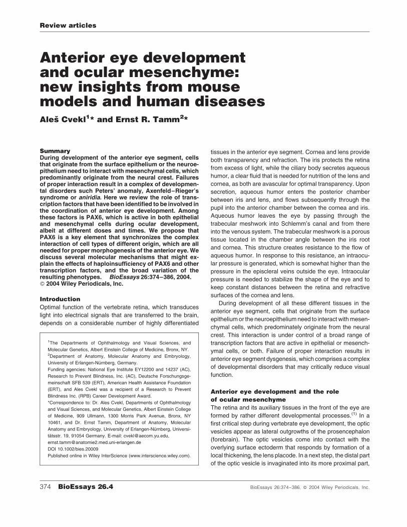
Anterior eye developmentand ocular mesenchyme:new insights from mousemodels and human diseasesAles Cvekl1* and Ernst R. Tamm2*
SummaryDuring development of the anterior eye segment, cellsthat originate from the surface epithelium or the neuroe-pithelium need to interact withmesenchymal cells, whichpredominantly originate from the neural crest. Failuresof proper interaction result in a complex of developmen-tal disorders such Peters’ anomaly, Axenfeld–Rieger’ssyndrome or aniridia. Here we review the role of trans-cription factors that have been identified to be involved inthe coordination of anterior eye development. Amongthese factors is PAX6, which is active in both epithelialand mesenchymal cells during ocular development,albeit at different doses and times. We propose thatPAX6 is a key element that synchronizes the complexinteraction of cell types of different origin, which are allneeded for propermorphogenesis of the anterior eye.Wediscuss several molecular mechanisms that might ex-plain the effects of haploinsufficiency of PAX6 and othertranscription factors, and the broad variation of theresulting phenotypes. BioEssays 26:374–386, 2004.� 2004 Wiley Periodicals, Inc.
Introduction
Optimal function of the vertebrate retina, which transduces
light into electrical signals that are transferred to the brain,
depends on a considerable number of highly differentiated
tissues in the anterior eye segment. Cornea and lens provide
both transparency and refraction. The iris protects the retina
from excess of light, while the ciliary body secretes aqueous
humor, a clear fluid that is needed for nutrition of the lens and
cornea, as both are avascular for optimal transparency. Upon
secretion, aqueous humor enters the posterior chamber
between iris and lens, and flows subsequently through the
pupil into the anterior chamber between the cornea and iris.
Aqueous humor leaves the eye by passing through the
trabecular meshwork into Schlemm’s canal and from there
into the venous system. The trabecular meshwork is a porous
tissue located in the chamber angle between the iris root
and cornea. This structure creates resistance to the flow of
aqueous humor. In response to this resistance, an intraocu-
lar pressure is generated, which is somewhat higher than the
pressure in the episcleral veins outside the eye. Intraocular
pressure is needed to stabilize the shape of the eye and to
keep constant distances between the retina and refractive
surfaces of the cornea and lens.
During development of all these different tissues in the
anterior eye segment, cells that originate from the surface
epitheliumor the neuroepitheliumneed to interact withmesen-
chymal cells, which predominately originate from the neural
crest. This interaction is under control of a broad range of
transcription factors that are active in epithelial or mesench-
ymal cells, or both. Failure of proper interaction results in
anterior eye segment dysgenesis, which comprises a complex
of developmental disorders that may critically reduce visual
function.
Anterior eye development and the role
of ocular mesenchyme
The retina and its auxiliary tissues in the front of the eye are
formed by rather different developmental processes.(1) In a
first critical step during vertebrate eye development, the optic
vesicles appear as lateral outgrowths of the prosencephalon
(forebrain). The optic vesicles come into contact with the
overlying surface ectoderm that responds by formation of a
local thickening, the lens placode. In a next step, the distal part
of the optic vesicle is invaginated into its more proximal part,
374 BioEssays 26.4 BioEssays 26:374–386, � 2004 Wiley Periodicals, Inc.
1The Departments of Ophthalmology and Visual Sciences, and
Molecular Genetics, Albert Einstein College of Medicine, Bronx, NY.2Department of Anatomy, Molecular Anatomy and Embryology,
University of Erlangen-Nurnberg, Germany.
Funding agencies: National Eye Institute EY12200 and 14237 (AC),
Research to Prevent Blindness, Inc. (AC), Deutsche Forschungsge-
meinschaft SFB 539 (ERT), American Health Assistance Foundation
(ERT), and Ales Cvekl was a recipient of a Research to Prevent
Blindness Inc. (RPB) Career Development Award.
*Correspondence to: Dr. Ales Cvekl, Departments of Ophthalmology
and Visual Sciences, and Molecular Genetics, Albert Einstein College
of Medicine, 909 Ullmann, 1300 Morris Park Avenue, Bronx, NY
10461, and Dr. Ernst Tamm, Department of Anatomy, Molecular
Anatomy and Embryology, University of Erlangen-Nurnberg, Universi-
tatsstr. 19, 91054 Germany. E-mail: [email protected],
DOI 10.1002/bies.20009
Published online in Wiley InterScience (www.interscience.wiley.com).
Review articles

thereby converting into a double-layered optic cup. The inner
layer of the optic cup will form the neural retina; the outer layer
will differentiate into the retinal pigmented epithelium. In
parallel to the development of the optic cup, the lens placode
enlarges and sinks below the level of the surrounding
ectoderm to form the lens pit. Subsequently, it forms the lens
vesicle, which at first remains connected to the surface
ectoderm by the lens stalk. Finally, the lens vesicle detaches
from the surface ectoderm and invaginates into the optic cup.
The optic cup is incomplete inferiorly at the so-called
embryonic (choroidal) fissure, which is used by the hyaloid
artery to pass into the optic cup. This artery supplies nutrients
to the inner layer of the cup and the lens vesicle during ocular
development.
Shortly after the lens vesicle has become detached from
the surface ectoderm, mesenchymal cells start to migrate into
the space between the anterior epithelium of the lens vesicle
and the surface ectoderm (Fig. 1A). In the mouse eye, four to
seven layers of mesenchymal cells are present at embryonic
day (E) 12. The cells form long cytoplasmic processes and
have a stellate, star-shaped phenotype.(2) As the number of
cells between the lens and surface ectoderm continuously
increases, the cells condense more and more to form several
layers of flat mesenchymal cells that are separated from each
other by a loose fibrillar extracellular matrix (Fig. 1B). In
parallel, the cavity of the lens vesicle becomes completely
closed as it is filled by the primary lens fibers. During the next
days (E14.5–E15.5) of mouse anterior eye development, the
posterior mesenchyme cells closest to the lens flatten and
extend to form apicolateral contacts with adjacent cells.(3–7)
Finally, the cells become connected to each other through
continuous bands of junctional complexes and an endothelial
monolayer is formed (Fig. 2A). At the end of this process, all
layers of the future cornea have been defined (Fig. 2A). The
endothelial monolayer that has been formed from posterior
mesenchyme cells will become the corneal endothelium,
the surface ectoderm that covers the anterior side of the
mesenchyme will become the corneal epithelium. Mesench-
yme cells between the corneal epithelium and endothe-
lium start to differentiate into corneal stroma fibroblasts or
Figure 1. Schematic diagram of ocular mesenchyme
development in the mouse eye between embryonic days
(E) 12.5–14.5. A: At E 12.5–13.5, the lens vesicle (LV)
has detached from the surface epithelium (SE) and has
become invaginated into the optic cup. Mesenchymal
cells (ME) start to migrate into the space between the
anterior epithelium of the lens vesicle and the sur-
face ectoderm. The inner layer of the optic cup forms
the neural retina (Re), the outer layer the retinal pigment-
ed epithelium (PE). The optic cup is incompletely inferior
at the so-called embryonic (choroidal) fissure (EF), where
the hyaloid artery (HA) enters the optic cup.B:AtE 13.5–
14.5, themesenchyme cells condense to form several flat
layers that are separated from each other by a loose
fibrillar extracellular matrix. In the lens (Le), the primary
lens fibers elongate to close the lumen of the lens vesicle.
Review articles
BioEssays 26.4 375
keratocytes,which is responsible for the synthesis of the highly
specialized extracellular matrix of the corneal stroma. As a
result, the cornea finally becomes transparent.
While the early development of themammalian cornea has
been primarily studied in the mouse eye, the available data on
corneal development in other mammalian species (cat, cow,
pig, hamster) suggest essentially similar mechanisms.(4) In
humanembryos,migratingmesenchymecells are seenduring
the 6th week of development, which form several layers of
loosely aggregated cells filling the space between surface
epithelium and lens epithelium;(1,8) this is similar to the
situation in the mouse eye at E12. The next step appears to
differ somewhat from mouse development, as most of the
mesenchyme cells condense to a dense layer that will give rise
to the future corneal endothelium.(1,4,8) Some cells remain in
the stromal space between surface ectoderm and future
corneal endothelium, which thickens again while mesench-
yme cells proliferate and/or continue to migrate to the future
cornea. A comparable scenario has been observed for corneal
development in monkey eyes.(9)
It is of interest to note that corneal development in the
mammalian eye appears to differ from that of the avian eye.(10)
In birds, an acellular primary corneal stroma consisting of
about 30 orthogonally arranged strata of collagen fibrils is
deposited between the surface epithelium and the lens.
Mesenchymal cells migrate along the inner surface of the
primary stroma to form the corneal endothelium. Subse-
quently, the primary corneal stroma swells and is invaded by
secondary mesenchymal cells destined to become kerato-
cytes. A primary corneal stroma comparable to that in birds is
not formed during mammalian eye development.(1,3,4)
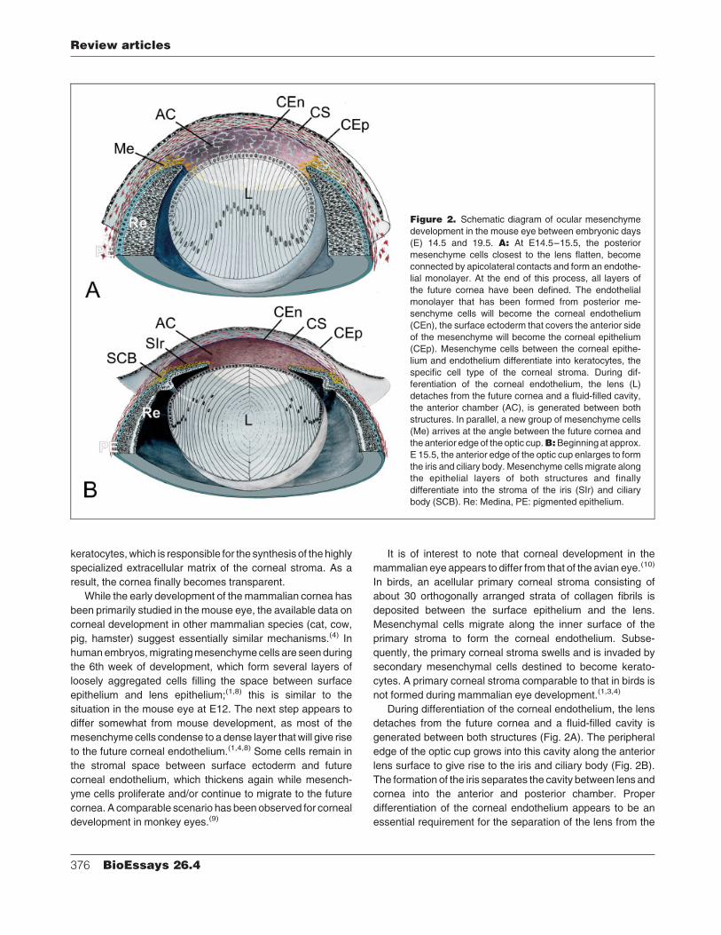
During differentiation of the corneal endothelium, the lens
detaches from the future cornea and a fluid-filled cavity is
generated between both structures (Fig. 2A). The peripheral
edge of the optic cup grows into this cavity along the anterior
lens surface to give rise to the iris and ciliary body (Fig. 2B).
The formation of the iris separates the cavity between lens and
cornea into the anterior and posterior chamber. Proper
differentiation of the corneal endothelium appears to be an
essential requirement for the separation of the lens from the
Figure 2. Schematic diagram of ocular mesenchyme
development in the mouse eye between embryonic days
(E) 14.5 and 19.5. A: At E14.5–15.5, the posterior
mesenchyme cells closest to the lens flatten, become
connected by apicolateral contacts and form an endothe-
lial monolayer. At the end of this process, all layers of
the future cornea have been defined. The endothelial
monolayer that has been formed from posterior me-
senchyme cells will become the corneal endothelium
(CEn), the surface ectoderm that covers the anterior side
of the mesenchyme will become the corneal epithelium
(CEp). Mesenchyme cells between the corneal epithe-
lium and endothelium differentiate into keratocytes, the
specific cell type of the corneal stroma. During dif-
ferentiation of the corneal endothelium, the lens (L)
detaches from the future cornea and a fluid-filled cavity,
the anterior chamber (AC), is generated between both
structures. In parallel, a new group of mesenchyme cells
(Me) arrives at the angle between the future cornea and
the anterior edge of the optic cup.B:Beginning at approx.E 15.5, the anterior edge of the optic cup enlarges to form
the iris and ciliary body. Mesenchyme cells migrate along
the epithelial layers of both structures and finally
differentiate into the stroma of the iris (SIr) and ciliary
body (SCB). Re: Medina, PE: pigmented epithelium.
Review articles
376 BioEssays 26.4
cornea, and subsequent anterior chamber formation. In
several strains of mutated mice, the corneal endothelium fails
to develop and the lens remains attached to the posterior side
of the cornea.(5–7)
While the corneal endothelium differentiates, and the
lens and future cornea become separated, a new group of
mesenchyme cells migrates to the anterior eye in a second
larger wave of migration. These cells appear first at the angle
between the future cornea and the anterior edge of the optic
cup (Fig. 2A). When the anterior edges of the optic cup ex-
tend to form the iris and ciliary body, the mesenchyme cells
migrate along the epithelial layers of both structures and
finally differentiate into the stroma of the iris and ciliary body
(Fig. 2B).
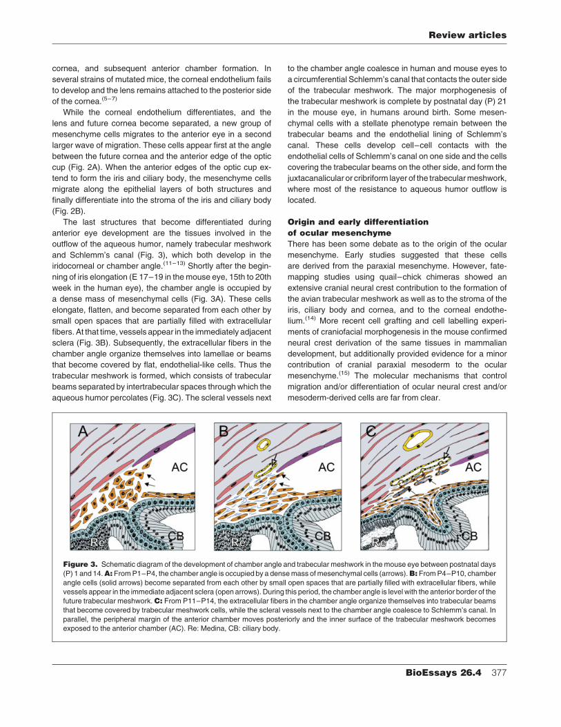
The last structures that become differentiated during
anterior eye development are the tissues involved in the
outflow of the aqueous humor, namely trabecular meshwork
and Schlemm’s canal (Fig. 3), which both develop in the
iridocorneal or chamber angle.(11–13) Shortly after the begin-
ning of iris elongation (E 17–19 in the mouse eye, 15th to 20th
week in the human eye), the chamber angle is occupied by
a dense mass of mesenchymal cells (Fig. 3A). These cells
elongate, flatten, and become separated from each other by
small open spaces that are partially filled with extracellular
fibers. At that time, vessels appear in the immediately adjacent
sclera (Fig. 3B). Subsequently, the extracellular fibers in the
chamber angle organize themselves into lamellae or beams
that become covered by flat, endothelial-like cells. Thus the
trabecular meshwork is formed, which consists of trabecular
beams separated by intertrabecular spaces through which the
aqueous humor percolates (Fig. 3C). The scleral vessels next
to the chamber angle coalesce in human and mouse eyes to
a circumferential Schlemm’s canal that contacts the outer side
of the trabecular meshwork. The major morphogenesis of
the trabecular meshwork is complete by postnatal day (P) 21
in the mouse eye, in humans around birth. Some mesen-
chymal cells with a stellate phenotype remain between the
trabecular beams and the endothelial lining of Schlemm’s
canal. These cells develop cell–cell contacts with the
endothelial cells of Schlemm’s canal on one side and the cells
covering the trabecular beams on the other side, and form the
juxtacanalicular or cribriform layer of the trabecularmeshwork,
where most of the resistance to aqueous humor outflow is
located.
Origin and early differentiation
of ocular mesenchyme
There has been some debate as to the origin of the ocular
mesenchyme. Early studies suggested that these cells
are derived from the paraxial mesenchyme. However, fate-
mapping studies using quail–chick chimeras showed an
extensive cranial neural crest contribution to the formation of
the avian trabecular meshwork as well as to the stroma of the
iris, ciliary body and cornea, and to the corneal endothe-
lium.(14) More recent cell grafting and cell labelling experi-
ments of craniofacial morphogenesis in the mouse confirmed
neural crest derivation of the same tissues in mammalian
development, but additionally provided evidence for a minor
contribution of cranial paraxial mesoderm to the ocular
mesenchyme.(15) The molecular mechanisms that control
migration and/or differentiation of ocular neural crest and/or
mesoderm-derived cells are far from clear.
Figure 3. Schematic diagram of the development of chamber angle and trabecular meshwork in the mouse eye between postnatal days
(P) 1 and 14.A:FromP1–P4, the chamber angle is occupied by a densemass ofmesenchymal cells (arrows).B:FromP4–P10, chamber
angle cells (solid arrows) become separated from each other by small open spaces that are partially filled with extracellular fibers, while
vessels appear in the immediate adjacent sclera (open arrows). During this period, the chamber angle is level with the anterior border of the
future trabecular meshwork.C: From P11–P14, the extracellular fibers in the chamber angle organize themselves into trabecular beams
that become covered by trabecular meshwork cells, while the scleral vessels next to the chamber angle coalesce to Schlemm’s canal. In
parallel, the peripheral margin of the anterior chamber moves posteriorly and the inner surface of the trabecular meshwork becomes
exposed to the anterior chamber (AC). Re: Medina, CB: ciliary body.
Review articles
BioEssays 26.4 377
Data from classical transplantation experiments in avian
embryos suggest that the differentiation of mesenchymal cells
in the cornea and the formation of an anterior chamber
depends on inductive signals from the lens.(16,17) Apparently,
such inductive lenticular signals are also important during
mammalian eye development, as primary defects in lens
development are usually associated with malformation of
mesenchyme-derived anterior eye segment tissues. Good
examples for this are the phenotypes that result from muta-
tions in the transcription factors MAF, FOXE3 and PITX3
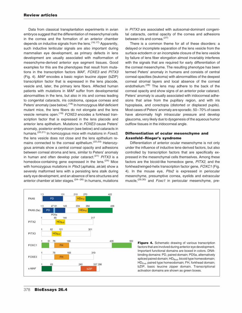
(Fig. 4). MAF encodes a basic region leucine zipper (bZIP)
transcription factor that is expressed in the lens placode,
vesicle and, later, the primary lens fibers. Affected human
patients with mutations in MAF suffer from developmental
abnormalities in the lens, but also in iris and cornea leading
to congenital cataracts, iris coloboma, opaque corneas and
Peters’ anomaly (see below).(18) In homozygousMaf-deficient
mutant mice, the lens fibers do not elongate and the lens
vesicle remains open.(19) FOXE3 encodes a forkhead tran-
scription factor that is expressed in the lens placode and
anterior lens epithelium. Mutations in FOXE3 cause Peters’
anomaly, posterior embryotoxon (see below) and cataracts in
humans.(20,21) In homozygous mice with mutations in Foxe3,
the lens vesicle does not close and the lens epithelium re-
mains connected to the corneal epithelium.(22,23) Heterozy-
gous animals show a central corneal opacity and adhesions
between corneal stroma and lens, similar to Peters’ anomaly
in human and often develop polar cataract.(21) PITX3 is a
homeobox-containing gene expressed in the lens.(24) Mice
with homozygous mutations in Pitx3 (aphakia, ak/ak) show a
severely malformed lens with a persisting lens stalk during
early eye development, and an absence of lens structures and
anterior chamber at later stages.(24–26) In humans, mutations
in PITX3 are associated with autosomal-dominant congeni-
tal cataracts, central opacity of the cornea and adhesions
between iris and cornea.(27)
There is a common theme for all of these disorders: a
delayed or incomplete separation of the lens vesicle from the
surface ectoderm or an incomplete closure of the lens vesicle
by failure of lens fiber elongation almost invariably interferes
with the signals that are required for early differentiation of
the corneal mesenchyme. The resulting phenotype has been
termed Peters’ anomaly in humans and consists of central
corneal opacities (leukoma) with abnormalities of the deepest
corneal stromal layers and local absence of the corneal
endothelium.(28) The lens may adhere to the back of the
corneal opacity and show signs of an anterior polar cataract.
Peters’ anomaly is usually associated with iridocorneal adhe-
sions that arise from the pupillary region, and with iris
hypoplasia, and corectopia (distorted or displaced pupils).
Most casesofPeters’ anomaly are sporadic. 50–70%of cases
have abnormally high intraocular pressure and develop
glaucoma, very likely due to dysgenesis of the aqueous humor
outflow tissues in the iridocorneal angle.
Differentiation of ocular mesenchyme and
Axenfeld–Rieger’s syndrome
Differentiation of anterior ocular mesenchyme is not only
under the influence of inductive lens-derived factors, but also
controlled by transcription factors that are specifically ex-
pressed in the mesenchymal cells themselves. Among these
factors are the bicoid-like homeobox gene, PITX2, and the
forkhead/winged-helix transcription factor gene, FOXC1 (Fig.
4). In the mouse eye, Pitx2 is expressed in periocular
mesenchyme, presumptive cornea, eyelids and extraocular
muscle,(29,30) and Foxc1 in periocular mesenchyme, pre-
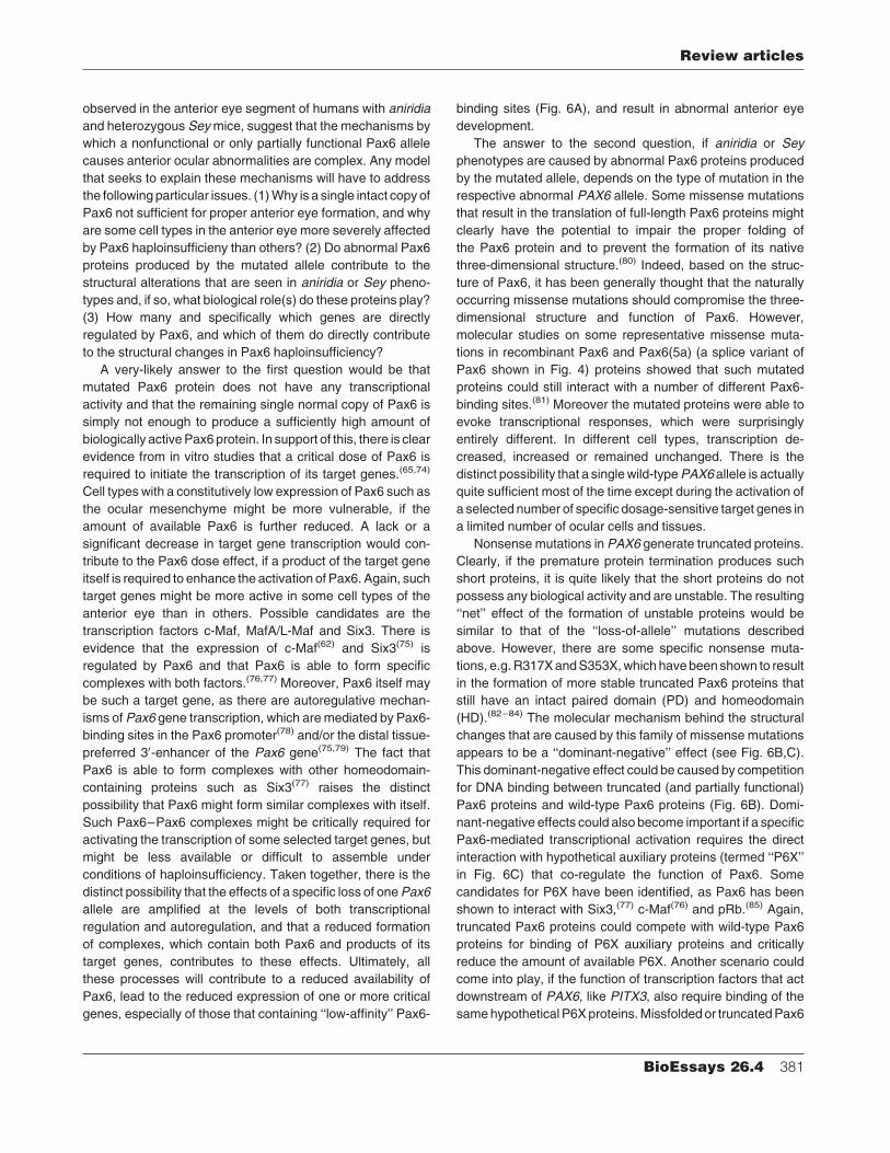
Figure 4. Schematic drawing of various transcription
factors that are involvedduring anterior eyedevelopment.
Important functional domains are boxed in colors. DNA-
binding domains: PD, paired domain; PD5a, alternatively
spliced paired domain; HDBcd, bicoid type homeodomain;
HDPrd, paired type homeodomain; FH, forkhead domain;
bZIP, basic leucine zipper domain. Transcriptional
activation domains are shown as green boxes.
Review articles
378 BioEssays 26.4
sumptive cornea and trabecular meshwork.(5,31,32) Neither of
the factors is expressed in retina nor lens. In humans,
mutations in PITX2 or FOXC1 result in a broad spectrum of
abnormalities during anterior eye development with different
specific clinical phenotypes.(32–37)
Most of these phenotypes belong to the broad spectrum
of clinical disorders, which are part of Axenfeld–Rieger’s
syndrome.(38) Subtypes of Axenfeld–Rieger’s syndrome
include Rieger’s anomaly or syndrome, Axenfeld’s anomaly
and iridogoniodysgenesis, all of which are commonly inherited
in an autosomal-dominant fashion.(39) In Rieger’s anomaly,
midpheripheral adhesions from the iris to cornea are seen. In
addition, there is marked iris hypoplasia and structural defects
such as polycoria (extra holes in the iris) and corectopia.When
the ocular findings of Rieger’s anomaly are associated with
characteristic systemic developmental defects such as dental
or facial abnormalities, the term Rieger’s syndrome is used.
Axenfeld’s anomaly is characterized by iris strands that attach
to a structure called posterior embryotoxon, which is a ring
of collagenous fibers at the peripheral end of Descemet’s
membrane, the basement membrane of the corneal endothe-
lium. It is clinically recognized as a ring-shaped opacity in the
peripheral cornea. Patientswith iridogoniodysgenesis havean
iris with hypoplastic stroma, abnormal chamber angle tissue,
and glaucoma. In general, patients with Axenfeld–Rieger’s
syndrome develop glaucoma in about 50% of cases.
Some patients with mutations in PITX2 or FOXC1 have
been reported that exhibit the phenotype of Peters’ anom-
aly.(33,34,40) The reasons for this wide spectrum of phenotypes
caused by mutations in PITX2 and FOXC1 are not clear and
have been discussed recently.(41) Mutant proteins may retain
partial functions resulting in milder phenotypes. However,
individuals with the same mutation may have different
phenotypes, even within the same family.(33,40) There is the
distinct possibility that different phenotypes result from modi-
fying genes that interact with mutant genes. Indeed, Foxc1þ/�
mice exhibit phenotypes comparable to those of human
patients with mutations in FOXC1 depending on strain, and
therefore genetic background.(42) Still, even between mice
from the same inbred strain (with essentially the same
background), or between right and left eye of the same animal,
the severity of the phenotype varies. It has been suggested
that stochastic developmental events and/or the local envir-
onment during development influence the outcome for each
individual eye.(41) Such events may lead to the presence of a
more or less active gene product at a given critical point during
development. Data from mouse mutants suggest that dosage
is an important factor. Heterozygous mutants show pheno-
types that resemble those in humans with Axenfeld–Rieger’s
syndrome,(42) whereas homozygous mutant mice, which die
shortly after birth, show corneolenticular adhesions and failure
of anterior chamber development similar to the condition in
Peters’ anomaly.(5) Homozygous Pitx2�/� deficient mice are
also not viable after birth, and embryonic data suggest a
phenotype comparable to that in homozygous Foxc1�/�
mutant mice with a persistence of corneolenticular adhesions
and lens stalk.(43)
Ocular mesenchyme and Pax6
Another gene that is critically required for the morphogenesis
of mesenchyme-derived tissues in the anterior eye is PAX6,
which codes for a paired domain and paired-like home-
odomain transcription factor (Fig. 4). Pax6 is a key regulator
of eye development that is both essential for eye formation
in different organisms as well as capable of inducing ecto-
pic eyes in flies and frogs upon misexpression.(44–46)
Humans with heterozygote mutations in PAX6 exhibit the
phenotype aniridia, a panocular disease that is associated
with iris hypoplasia, corneal opacification, cataract and
foveal dysplasia.(47–49) About 50–75%of patientswith aniridia
develop glaucoma, because of abnormal differentiation of the
trabecular meshwork and/or complete absence of Schlemm’s
canal.(50,51) Mutations in PAX6 have also been found in
patients with Peters’ anomaly, autosomal dominant keratitis
and isolated foveal hypoplasia.(52) Heterozygous Small eye
(Sey) mice or heterozygous Pax6 lacZ/þ mutant mice, which
both have null alleles ofPax6, showa reduction in eye size and
cataracts.(53,54) The iris of the animals remains hypoplastic
and corneal abnormalities are present, which include an
irregular lamellar alignment, cellular infiltrates and vascular-
ization of the corneal stroma.(53,55) Defects of the corneal
epithelium contribute to the corneal abnormalities.(55,56) In a
third of Pax6 lacZ/þ mutant mice, the separation of the cornea
from the lens is incomplete, the epithelial layers of lens and
cornea are continuous and iridocorneal adhesions are
present, all hallmarks of Peters’ anomaly.(53) Persistence of
the lens stalk has also been observed in Small eye (Sey)
mice.(55) In addition, the trabecular meshwork of Pax6 lacZ/þ
mutant mice remains undifferentiated and Schlemm’s canal
is absent.(53)
Overall, the phenotype of Pax6 haploinsufficiency in mice
and humans indicates that Pax6 is critically required for the
differentiation of those tissues of the anterior eye segment that
are of mesenchymal origin. Pax6 could indirectly act on the
morphogenesis of ocular mesenchyme as a strong Pax6
expression has been described in cells that derive from the
neuroectoderm of the optic cup or from the anterior surface
ectoderm.(57–60) This strong expression is seen during ocular
development and ismaintained in adulthood. To clarify, if Pax6
acts also directly on mesenchymal differentiation in the devel-
oping eye, we recently studied Pax6 expression by b-galactosidase staining of Pax6 lacZ/þ heterozygous mice, and
by immunostaining of wild-type littermates and cultured
murine trabecular meshwork cells.(53) Positive signals were
observed in cells of mesenchymal origin, but the intensity was
weaker than in cells of surface ectodermal or neuroepithelial
Review articles
BioEssays 26.4 379
origin and only observed in mid-fetal stages, but not in adult
animals. A similar staining pattern was observed by Collinson
and coworkers using different antibodies.(61) In addition, these
authors analysed gene expression and distribution of Pax6�/�
cells in Pax6þ/þ$Pax6�/� chimeras to find that Pax6 is
autonomously required for cells to contribute to corneal stroma
and corneal endothelium.(61)
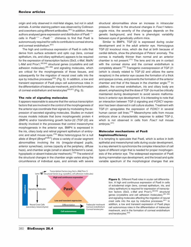
The high and continuous expression of Pax6 in cells that
derive from surface ectoderm and optic cup (lens, corneal
epithelium, iris, and ciliary epithelium) appears to be required
for the expression of transcription factors (Six3, c-Maf, MafA/
L-Maf and Prox1,(62,64) structural genes (crystallins and cell
adhesion molecules),(65–69) and signaling molecules, which
are critical for the morphogenesis of those tissues, and
subsequently for the migration of neural crest cells into the
eye by inductive processes(53) (Fig. 5). In addition, a low and
transient expression of Pax6 plays cell autonomous roles in
thedifferentiation of trabecularmeshwork, and in the formation
of corneal endothelium and keratocytes(53,61) (Fig. 5).
The role of signaling molecules
It appears reasonable to assume that the various transcription
factors that are involved in the control of themorphogenesis of
the anterior eye coordinate their signals by modulating the ex-
pression of secreted signaling molecules. Studies on mutant
mouse models indicate that bone morphogenetic protein 4
(BMP4) and/or transforming growth factor-b2 (TGF-b2) aredirectly involved in the processes that control mesenchyme
morphogenesis in the anterior eye. BMP4 is expressed in
the iris, ciliary body and retinal pigment epithelium of embry-
onic and adult mouse eyes.(70) Mice heterozygous for a null
allele of Bmp4 (Bmp4tmBLh) show a variety of ocular segment
abnormalities involving the iris (irregular-shaped pupils,
anterior synechiae), cornea (opacity at the periphery, diffuse
haze), and chamber angle (small or absent Schlemm’s canal,
hypoplastic or absent trabecular meshwork).(70) The extend of
the structural changes in the chamber angle varies along the
circumference of individual eyes, and animals with severe
structural abnormalities show an increase in intraocular
pressure. Similar to the structural changes in Foxc1 hetero-
zygote mice, the severity of the changes depends on the
genetic background, and there is phenotypic variability
between eyes of genetically uniform mice.
Similar to BMP4, TGF-b2 is expressed during ocular
development and in the adult anterior eye. Homozygous
TGF-b2 knockout mice, which die that at birth because of
cardial defects, show the phenotype of Peters’ anomaly. The
cornea is markedly thinner than normal and an anterior
chamber is not present.(71) The lens and iris are in contact
with the corneal stoma and the corneal endothelium is
completely absent.(72) Transgenic overexpression of TGF-b1(which is closely related to TGF-b2 and utilizes the same
receptors) in the anterior eye causes the formation of a thick
and opaque cornea, and prevents the formation of the anterior
chamber and that of the tissues in the chamber angle.(7) In
addition, the corneal endothelium, iris and ciliary body are
absent, emphasizing that the dose of TGF-bsmust be critically
maintained during development to avoid severe malforma-
tions in anterior eye development. It is interesting to note that
an interaction between TGF-b signaling and FOXC1 expres-
sion has been observed in cell culture studies. Treatment with
TGF-b1 upregulates the expression of FOXC1 in several
human cancer cell lines.(73) Mesenchyme cells from mouse
embryos show a characteristic response to added TGF-b,which is not observed in cells from Foxc1 null mouse
embryos.(31)
Molecular mechanisms of Pax6
haploinsufficiency
It is tempting to speculate that Pax6, which is active in both
epithelial and mesenchymal cells during ocular development,
is a key element to synchronize the complex interaction of cell
types of different origin that is needed for proper morphogen-
esis of the anterior eye. The widespread expression of Pax6
during mammalian eye development, and the broad and quite
variable spectrum of the morphological changes that are
Figure 5. Different Pax6 roles in ocular cell differentia-
tion. A high and continuous expression of Pax6 in cells
of ectodermal origin (lens, corneal epithelium, iris, and
ciliary epithelium) is required for expression of transcrip-
tion factors (Six3, c-Maf and Prox1),(62,63) structural
genes (crystallins and cell adhesion molecules),(65–69)
and signaling molecules affecting the migration of neural
crest cells into the eye by inductive processes.(61) In
addition, a low and transient expression of Pax6 plays
cell autonomous roles in the differentiation of trabecular
meshwork, and in the formation of corneal endothelium
and keratocytes.(53)
Review articles
380 BioEssays 26.4
observed in the anterior eye segment of humans with aniridia
and heterozygous Seymice, suggest that the mechanisms by
which a nonfunctional or only partially functional Pax6 allele
causes anterior ocular abnormalities are complex. Any model
that seeks to explain these mechanisms will have to address
the following particular issues. (1)Why is a single intact copyof
Pax6 not sufficient for proper anterior eye formation, and why
are some cell types in the anterior eye more severely affected
by Pax6 haploinsufficieny than others? (2) Do abnormal Pax6
proteins produced by the mutated allele contribute to the
structural alterations that are seen in aniridia or Sey pheno-
types and, if so, what biological role(s) do these proteins play?
(3) How many and specifically which genes are directly
regulated by Pax6, and which of them do directly contribute
to the structural changes in Pax6 haploinsufficiency?
A very-likely answer to the first question would be that
mutated Pax6 protein does not have any transcriptional
activity and that the remaining single normal copy of Pax6 is
simply not enough to produce a sufficiently high amount of
biologically activePax6 protein. In support of this, there is clear
evidence from in vitro studies that a critical dose of Pax6 is
required to initiate the transcription of its target genes.(65,74)
Cell types with a constitutively low expression of Pax6 such as
the ocular mesenchyme might be more vulnerable, if the
amount of available Pax6 is further reduced. A lack or a
significant decrease in target gene transcription would con-
tribute to the Pax6 dose effect, if a product of the target gene
itself is required to enhance the activation of Pax6. Again, such
target genes might be more active in some cell types of the
anterior eye than in others. Possible candidates are the
transcription factors c-Maf, MafA/L-Maf and Six3. There is
evidence that the expression of c-Maf(62) and Six3(75) is
regulated by Pax6 and that Pax6 is able to form specific
complexes with both factors.(76,77) Moreover, Pax6 itself may
be such a target gene, as there are autoregulative mechan-
isms of Pax6 gene transcription, which are mediated by Pax6-
binding sites in the Pax6 promoter(78) and/or the distal tissue-
preferred 30-enhancer of the Pax6 gene(75,79) The fact that
Pax6 is able to form complexes with other homeodomain-
containing proteins such as Six3(77) raises the distinct
possibility that Pax6 might form similar complexes with itself.
Such Pax6–Pax6 complexes might be critically required for
activating the transcription of some selected target genes, but
might be less available or difficult to assemble under
conditions of haploinsufficiency. Taken together, there is the
distinct possibility that the effects of a specific loss of onePax6
allele are amplified at the levels of both transcriptional
regulation and autoregulation, and that a reduced formation
of complexes, which contain both Pax6 and products of its
target genes, contributes to these effects. Ultimately, all
these processes will contribute to a reduced availability of
Pax6, lead to the reduced expression of one or more critical
genes, especially of those that containing ‘‘low-affinity’’ Pax6-
binding sites (Fig. 6A), and result in abnormal anterior eye
development.
The answer to the second question, if aniridia or Sey
phenotypes are caused by abnormal Pax6 proteins produced
by the mutated allele, depends on the type of mutation in the
respective abnormal PAX6 allele. Some missense mutations
that result in the translation of full-length Pax6 proteins might
clearly have the potential to impair the proper folding of
the Pax6 protein and to prevent the formation of its native
three-dimensional structure.(80) Indeed, based on the struc-
ture of Pax6, it has been generally thought that the naturally
occurring missense mutations should compromise the three-
dimensional structure and function of Pax6. However,
molecular studies on some representative missense muta-
tions in recombinant Pax6 and Pax6(5a) (a splice variant of
Pax6 shown in Fig. 4) proteins showed that such mutated
proteins could still interact with a number of different Pax6-
binding sites.(81) Moreover the mutated proteins were able to
evoke transcriptional responses, which were surprisingly
entirely different. In different cell types, transcription de-
creased, increased or remained unchanged. There is the
distinct possibility that a singlewild-typePAX6 allele is actually
quite sufficient most of the time except during the activation of
a selected number of specific dosage-sensitive target genes in
a limited number of ocular cells and tissues.
Nonsense mutations in PAX6 generate truncated proteins.
Clearly, if the premature protein termination produces such
short proteins, it is quite likely that the short proteins do not
possess any biological activity and are unstable. The resulting
‘‘net’’ effect of the formation of unstable proteins would be
similar to that of the ‘‘loss-of-allele’’ mutations described
above. However, there are some specific nonsense muta-
tions, e.g.R317XandS353X,which havebeenshown to result
in the formation of more stable truncated Pax6 proteins that
still have an intact paired domain (PD) and homeodomain
(HD).(82–84) The molecular mechanism behind the structural
changes that are caused by this family of missense mutations
appears to be a ‘‘dominant-negative’’ effect (see Fig. 6B,C).
This dominant-negative effect could be caused by competition
for DNA binding between truncated (and partially functional)
Pax6 proteins and wild-type Pax6 proteins (Fig. 6B). Domi-
nant-negative effects could also become important if a specific
Pax6-mediated transcriptional activation requires the direct
interaction with hypothetical auxiliary proteins (termed ‘‘P6X’’
in Fig. 6C) that co-regulate the function of Pax6. Some
candidates for P6X have been identified, as Pax6 has been
shown to interact with Six3,(77) c-Maf(76) and pRb.(85) Again,
truncated Pax6 proteins could compete with wild-type Pax6
proteins for binding of P6X auxiliary proteins and critically
reduce the amount of available P6X. Another scenario could
come into play, if the function of transcription factors that act
downstream of PAX6, like PITX3, also require binding of the
samehypothetical P6Xproteins.Missfoldedor truncatedPax6
Review articles
BioEssays 26.4 381
proteins that aberrantly interact with P6X could significantly
reduce the amount that is necessary for the proper function
of such downstream genes. Such a mechanism could also
explain why additional copies of PAX6, when introduced into
themouse genome, generate phenotypes similar to Sey/þ.(86)
Ectopic additional Pax6 might squelch the hypothetical P6X
(Fig. 6D) and disrupt the equilibrium between Pax6–P6X and
Pitx3–P6X complexes.
Developmental abnormalities in mouse
Sey/þ and human aniridia may originate
from haploinsufficient expression of
a small number of Pax6 target genes
Studies on gene expression during formation of the lens pre-
placode and placode in early eye development provide
evidence that Pax6 positively regulates the expression of at
least two other transcription factors, Six3(75) and c-Maf.(62)
Still, the first embryonicprocessduringeyedevelopment that is
critically dependent on the correctPax6 gene dosage appears
to be the formation of the lens placode.(58,66) In contrast, the
formation of the pre-placodeappears to beunaffected inPax6-
deficient heterozygous mouse strains. Haploinsufficiency for
Pax6 does not prevent, but rather delays the induction of lens
formation by 12 to 24 hours, suggesting that a prolonged
accumulation ofPax6 proteins canovercome thegenedosage
effect. The price is a 50% reduction in the number of cells in the
lens vesicle, and the frequent failure of the lens vesicle to
detach completely from the surface ectoderm.(66) This model
would provide an explanation for the reduced size of the lens in
the mouse, and the persistence of corneal–lenticular stalks
(Peters’ anomaly described above) that are observed both in
Pax6-deficient heterozygous mice and humans.(28,50,53,66,87)
Apparently, lens formation includesmultiple steps, each being
characterized by its own set of direct Pax6 target genes.(66) N-
cadherin, a calcium-binding cell adhesion molecule that is
associated with the separation and sealing of cell layers in
morphogenesis, might play a critical role for the formation of
the lens vesicle.(66) The expression pattern of N-cadherin
differs markedly between wild-type and Pax6 heterozygous
lens vesicles.
In humans, aniridic lenses are not obviously reduced in size
as in theSey/þmouse, butmaybedislocated at a frequency of
up to 56%.(50,51) It is unclear why the lens size differs in similar
abnormal conditions between mice and humans. It is possible
Figure 6. Molecular models explaining Pax6 haploin-
sufficiency and overexpression. A: Interaction of Pax6
and mutated Pax6 (mut–Pax6) with target genes. A
‘‘high-affinity’’Pax6-binding site in gene A is occupied in
Pax6þ/þ and Pax6þ/� cells. A ‘‘low-affinity’’ Pax6-binding
site in gene B is only occupied in Pax6þ/þ cells. If a
mutated Pax6 allele encodes for a nonfunctional protein
or no protein at all, the low-affinity site(s) remain(s) free.
B: Truncated proteins capable of binding to DNA can
compete with the normal Pax6 protein as a classical
dominant-negative mutation.(83) C: Some mutated Pax6
proteins may sequester a hypothetical auxiliary factor
(P6X) from Pax6, thereby blocking its function. D:Overexpression of Pax6 (þ/þ/n, n¼ 1–5) generates
enough of Pax6 protein to saturate low-affinity binding
sites, but may disrupt the equilibrium between Pax6 and
the hypothetical P6X. P6X in panels C and D may be the
same or different hypothetical protein. Proteins modulat-
ingPax6 activity include pRb, c-Maf, Six3, Sox2,Mitf, and
others.
Review articles
382 BioEssays 26.4
that the human lens size is not affected by changes in PAX6
gene dosage due to a different sensitivity of the critical target
genes and/or because of other species-specific differences.
Such differencesmight have evolved because the relative size
of the human lens, when compared to that of the eye, is much
smaller than that of mouse lens. Still, the coincidence of
cataracts with aniridia that has been reported to occur in 50
to 85% of affected cases(50,51) clearly indicates that Pax6 is
required for proper gene expression in the human lens. These
cataracts evolve from small anterior or posterior lens opacities
that are already found at birth.
While the lens size in the Sey/þ mouse is systematically
reduced, animals with persistent corneal–lenticular stalks or
Peters’ anomaly are less frequently observed. In thePax6lacZ/þ
mouse model with a null allele of Pax6, we observed Peters’
anomaly in about one third of the analyzed animals.(53) We
speculate that the shift toward Peters’ anomaly from Sey only
might be caused by the action of modifying genes, and/or
environmental and stochastic factors. In human patients,
Peters’ anomaly has been found to be associated with specific
PAX6 mutations such as R26G(87) and G18W.(88) In addition,
in humans, other factors appear to contribute to the specific
phenotype of Peters’ anomaly, as the G18W mutation is
displayed as cataract or Peters’ anomaly in different mem-
bers of the same family.(88) A comparable familial variability
has been observed in humans with the R26G mutation.(87)
It is interesting to note that the original Pax6 null alleles
in Sey/Sey do not allow the initiation of lens induction (i.e.
thickening of the surface ectoderm and its subsequent
envagination), while two recently described point mutations,
Pax64Neu and Pax610Neu, allow the envagination of the
ectoderm.(89) Pax64Neu contains the missense mutation
S273P in the HD, and Pax610Neu contains a nucleotide subs-
titution in the Pax6 Kozak sequence, which affects transla-
tion.(89) It appears that the Pax6 proteins, which are generated
from the4Neuand10Neualleles canexecute a certain fraction
of the lens induction program, most likely because their initial
target gene(s) still cooperate.(89) There is the distinct pos-
sibility that the relatively moderate ocular phenotypes, which
are seen in about half of PAX6 missense mutations in
humans,(52) are due to the fact that the respective mutated
proteins retain their ability to activate a large spectrumof target
genes. Hence, a reduction in Pax6 gene dosage may affect
only a relatively small number of critical genes, e.g. N- and
R-cadherins(66,90) in each of the affected tissues. Even if the
overall number of genes that are directly or indirectly affect-
ed by Pax6 haploinsufficiency may be in the hundreds or
thousands,(68,91) only a dozen of themmay be responsible for
the generation of developmental abnormalities.
The hallmark of aniridia is the absence or hypoplasia of the
iris. The molecular and developmental mechanisms of this
defect are not well understood. As noted earlier, the two main
tissues of the iris, the stroma and the epithelial layers, are of
different developmental origin. The epithelia (including the
epithelium-derived iris muscles) derive from the optic cup,
while the stroma is formed by migrating neural crest cells. An
abnormal migration of neural crest cells has been observed
in Pax6 homozygous rat embryos.(92) We and others have
recently shown that Pax6 is expressed in the ocular mesen-
chyme that originates from neural crest cells; however, the
expression appears to be low and transitory.(53,61) Absence or
hypoplasia of the iris in aniridia could easily be caused by a
combination of defects during the differentiation of the epi-
thelial layers of the iris and a compromised ability of migrating
neural crest cells to find their destination in the iris stroma.
It is interesting to note that an iris hypoplasia can be
experimentally induced in mice by maternal vitamin A
deficiency.(50,51) This correlates with the finding that retinoic
acid signaling is altered in mouseSey/þ embryos.(93) Retinoic
acid has recently been shown as an environmental modifier of
velo-cardio-facial (DiGeorge) syndrome, which is caused by
defects in neural crest cell migration,(94) raising the intriguing
possibility that the variability of the PAX6 gene dosage effect
might be modified via retinoids. Similar to lens formation, the
candidate genes that are under direct or indirect influence
of Pax6 in neural crest cells might include cell adhesion
molecules,(66–69) although directmolecular evidence for this is
still lacking. Since the expression of Pax6 is weak in neural
crest cells, many Pax6 target genes, even those with high-
affinity Pax6-binding sites (see Fig. 6A), would be negatively
affected in Pax6-null heterozygote individuals. It is also pos-
sible, that the smaller (in mouse) or abnormal (in human) lens
cannot produce adequate amounts of signalingmolecules that
regulate neural crest cell migration during the formation of the
anterior eye segment (Fig. 5). These possibilities are not
mutually exclusive.
Conclusions and perspectives
So far, a relatively small group of genes encoding transcription
factors has been shown to be critical for the development of
the anterior eye. In humans, mutations in these genes cause
a spectrum of partially and/or completely overlapping con-
genital disorders, which all compromise vision to a variable
degree. Ocular tissues are of neuroectodermal and neural
crest origin, and transcription factors need to coordinate the
complex interaction and communication between those two
types of cell populations during morphogenesis of the anterior
eye. Pax6,which is expressed in all cell types that contribute to
anterior eye development, although at different times and with
different intensity, appears to be the critical factor that
synchronizes the critical events during formation of the
anterior eye. In addition, numerous other genes including
PITX2,PITX3, FOXE3, FOXC1 and c-MAF appear to regulate
similar and/or identical processes as PAX6, however, in a
limited number of ocular cells and tissues. The challenge for
the futurewill be to clarify themechanismsandhierarchies that
Review articles
BioEssays 26.4 383
are needed for proper interaction of all these transcription
factors. This will involve questioning why these factors have
rather specific and important functions in the eye, although
they are expressed in numerous tissues outside the eye. An
equally important task will be to learn about those genes that
are directly regulated by the action of the transcription factors
and the elucidation of their specific function during the devel-
opment of the anterior eye. Such an identification will require
high throughput techniques, such as cDNAmicroarrays, chro-
matin immunoprecipitations and, maybe equally important,
educated guesses. Finally, it is more than likely that more
regulatory genes are involved during anterior eye segment
formation, which still need to be discovered.
Acknowledgments
We greatly appreciate the assistance of Jorg Pekarsky in
generating the drawings of mouse eye development. We also
thank Dr. Michael Wegner and Dr. Ruth Ashery-Padan for
critically reading this manuscript.
References1. Duke-Elder S, Cook C. 1963. Normal and Abnormal Development. Part 1.
Embryology. Duke-Elder S, editor. London: Henry Kimpton.
2. Haustein J. 1983. On the ultrastructure of the developing and adult
mouse corneal stroma. Anat Embryol (Berl) 168:291–305.
3. Pei YF, Rhodin JAG. 1970. The prenatal development of the mouse eye.
Anat Rec 168:105–126.
4. Dublin I. 1970. Comparative embryologic studies of the early develop-
ment of the cornea and the pupillary membrane in reptiles, birds and
mammals. Acta Anat (Basel) 76:381–408.
5. Kidson SH, Kume T, Deng K, Winfrey V, Hogan BL. 1999. The forkhead/
winged-helix gene, Mf1, is necessary for the normal development of the
cornea and formation of the anterior chamber in the mouse eye. Dev Biol
211:306–322.
6. Reneker LW, Silversides DW, Xu L, Overbeek PA. 2000. Formation of
corneal endothelium is essential for anterior segment development.
Development 127:533–542.
7. Flugel-Koch C, Ohlmann A, Piatigorsky J, Tamm ER. 2002. Over-
expression of TGF-b1 alters early development of cornea and lens in
transgenic mice. Dev Dyn 225:111–125.
8. Kolmer W. 1936. Entwicklung des Auges. In: Kolmer W, Lauber H,
editors. Handbuch der mikroskopischen Anatomie des Menschen,
Haut und Siinesorgane. 2. Teil: Auge. Berlin: Verlag von Julius Springer;
p 623–676.
9. Ozanics V, Rayborn M, Sagun D. 1977. Observations on the morphology
of the developing primate cornea: epithelium, its innervation and anterior
stroma. J Morphol 153:263–297.
10. Hay ED. 1980. Development of the vertebrate cornea. Int Rev Cytol 63:
263–322.
11. Wulle KG. 1972. The development of the productive and draining
system of the aqueous humor in the human eye. Adv Ophthalmol 26:
269–355.
12. Smith RS, Zabaleta A, Savinova OV, John SW. 2001. The mouse anterior
chamber angle and trabecular meshwork develop without cell death.
BMC Dev Biol 1:3.
13. Reme C, d’Epinay SL. 1981. Periods of development of the normal
human chamber angle. Doc Ophthalmol 51:241–268.
14. Johnston MC, Noden DM, Hazelton RD, Coulombre JL, Coulombre AJ.
1979. Origins of avian ocular and periocular tissues. Exp Eye Res 29:
27–43.
15. Trainor PA, Tam PP. 1995. Cranial paraxial mesoderm and neural crest
cells of the mouse embryo: co-distribution in the craniofacial mesench-
yme but distinct segregation in branchial arches. Development 121:
2569–2582.
16. Coulombre AJ, Coulombre JL. 1964. Lens development. I. Role of the
lens in eye growth. J Exp Zool 156:39–48.
17. Genis-Galvez JM. 1966. Role of the lens in the morphogenesis of the iris
and cornea. Nature 210:209–210.
18. Jamieson RV, et al. 2002. Domain disruption and mutation of the bZIP
transcription factor, MAF, associated with cataract, ocular anterior
segment dysgenesis and coloboma. Hum Mol Genet 11:33–42.
19. Ring BZ, Cordes SP, Overbeek PA, Barsh GS. 2000. Regulation of
mouse lens fiber cell development and differentiation by the Maf gene.
Development 127:307–317.
20. Semina EV, Brownell I, Mintz-Hittner HA, Murray JC, Jamrich M. 2001.
Mutations in the human forkhead transcription factor FOXE3 associated
with anterior segment ocular dysgenesis and cataracts. Hum Mol Genet
10:231–236.
21. Ormestad M, Blixt A, Churchill A, Martinsson T, Enerback S, Carlsson P.
2002. Foxe3 haploinsufficiency in mice: a model for Peters’ anomaly.
Invest Ophthalmol Vis Sci 43:1350–1357.
22. Blixt A, Mahlapuu M, Aitola M, Pelto-Huikko M, Enerback S, Carlsson P.
2000. A forkhead gene, FoxE3, is essential for lens epithelial proliferation
and closure of the lens vesicle. Genes Dev 14:245–254.
23. Brownell I, Dirksen M, Jamrich M. 2000. Forkhead Foxe3 maps to the
dysgenetic lens locus and is critical in lens development and
differentiation. Genesis 27:81–93.
24. Semina EV, Murray JC, Reiter R, Hrstka RF, Graw J. 2000. Deletion in the
promoter region and altered expression of Pitx3 homeobox gene in
aphakia mice. Hum Mol Genet 9:1575–1585.
25. Rieger DK, Reichenberger E, McLean W, Sidow A, Olsen BR. 2001. A
double-deletion mutation in the Pitx3 gene causes arrested lens
development in aphakia mice. Genomics 72:61–72.
26. Varnum DS, Stevens LC. 1968. Aphakia, a new mutation in the mouse.
J Hered 59:147–150.
27. Semina EV, Ferrell RE, Mintz-Hittner HA, Bitoun P, Alward WL, Reiter RS,
Funkhauser C, Daack-Hirsch S, Murray JC. 1998. A novel homeobox
gene PITX3 is mutated in families with autosomal-dominant cataracts
and ASMD. Nat Genet 19:167–170.
28. Schottenstein EM. 1996. Peters’ anomaly. In: Ritch R, Shields MB,
Krupin T, editors. The glaucomas. 2 ed. St. Louis: Mosby. p 887–897.
29. Lu MF, Pressman C, Dyer R, Johnson RL, Martin JF. 1999. Function of
Rieger syndrome gene in left-right asymmetry and craniofacial develop-
ment. Nature 401:276–278.
30. Lin CR, Kioussi C, O’Connell S, Briata P, Szeto D, Liu F, Izpisua-Belmonte
JC, Rosenfeld MG. 1999. Pitx2 regulates lung asymmetry, cardiac
positioning and pituitary and tooth morphogenesis. Nature 401:279–
282.
31. Kume T, Deng KY, Winfrey V, Gould DB, Walter MA, Hogan BL. 1998.
The forkhead/winged helix gene Mf1 is disrupted in the pleiotropic
mouse mutation congenital hydrocephalus. Cell 93:985–996.
32. Mears AJ, et al. 1998. Mutations of the forkhead/winged-helix gene,
FKHL7, in patients with Axenfeld–Rieger anomaly. Am J Hum Genet
63:1316–1328.
33. Perveen R, et al. 2000. Phenotypic variability and asymmetry of Rieger
syndrome associated with PITX2 mutations. Invest Ophthalmol Vis Sci
41:2456–2460.
34. Doward W, Perveen R, Lloyd IC, Ridgway AE, Wilson L, Black GC. 1999.
A mutation in the RIEG1 gene associated with Peters’ anomaly. J Med
Genet 36:152–155.
35. Nishimura DY, et al. 1998. The forkhead transcription factor gene FKHL7
is responsible for glaucoma phenotypes which map to 6p25. Nat Genet
19:140–147.
36. Semina EV, et al. 1996. Cloning and characterization of a novel bicoid-
related homeobox transcription factor gene, RIEG, involved in Rieger
syndrome. Nat Genet 14:392–399.
37. Nishimura DY, et al. 2001. A spectrum of FOXC1 mutations suggests
gene dosage as a mechanism for developmental defects of the anterior
chamber of the eye. Am J Hum Genet 68:364–372.
38. Alward WL. 2000. Axenfeld-Rieger syndrome in the age of molecular
genetics. Am J Ophthalmol 130:107–115.
39. Waring GO 3rd, Rodrigues MM, Laibson PR. 1975. Anterior chamber
cleavage syndrome. A stepladder classification. Surv Ophthalmol 20:
3–27.
Review articles
384 BioEssays 26.4
40. Honkanen RA, Nishimura DY, Swiderski RE, Bennett SR, Hong S,
Kwon YH, Stone EM, Sheffield VC, Alward WL. 2003. A family with
Axenfeld–Rieger syndrome and Peters Anomaly caused by a point
mutation (Phe112Ser) in the FOXC1 gene. Am J Ophthalmol 135:368–
375.
41. Gould DB, John SW. 2002. Anterior segment dysgenesis and the
developmental glaucomas are complex traits. Hum Mol Genet 11:1185–
1193.
42. Smith RS, Zabaleta A, Kume T, Savinova OV, Kidson SH, Martin JE,
Nishimura DY, Alward WL, Hogan BL, John SW. 2000. Haploinsufficiency
of the transcription factors FOXC1 and FOXC2 results in aberrant ocular
development. Hum Mol Genet 9:1021–1032.
43. Gage PJ, Suh H, Camper SA. 1999. Dosage requirement of Pitx2 for
development of multiple organs. Development 126:4643–4651.
44. Ashery-Padan R, Gruss P. 2001. Pax6 lights-up the way for eye devel-
opment. Curr Opin Cell Biol 13:706–714.
45. Gehring WJ, Ikeo K. 1999. Pax 6: mastering eye morphogenesis and eye
evolution. Trends Genet 15:371–377.
46. Chow RL, Lang RA. 2001. Early eye development in vertebrates. Annu
Rev Cell Dev Biol 17:255–296.
47. Ton CC, et al. 1991. Positional cloning and characterization of a paired
box- and homeobox-containing gene from the aniridia region. Cell
67:1059–1074.
48. Jordan T, Hanson I, Zaletayev D, Hodgson S, Prosser J, Seawright A,
Hastie N, van HV. 1992. The human PAX6 gene is mutated in two
patients with aniridia. Nat Genet 1:328–332.
49. Glaser T, Walton DS, Maas RL. 1992. Genomic structure, evolutionary
conservation and aniridia mutations in the human PAX6 gene. Nat Genet
2:232–239.
50. Mintz-Hittner HA. 1996. Aniridia. In: Ritch R, Shields MB, Krupin T,
editors. The glaucomas. 2 ed. St. Louis: Mosby. p 859–874.
51. Nelson LB, Spaeth GL, Nowinski TS, Margo CE, Jackson L. 1984.
Aniridia. A review. Surv Ophthalmol 28:621–642.
52. Prosser J, van Heyningen V. 1998. PAX6 mutations reviewed. Hum Mutat
11:93–108.
53. Baulmann D, Ohlmann A, Flugel-Koch C, Goswami S, Cvekl A, Tamm
ER. 2002. Pax6 heterozygous eyes show defects in chamber angle
differentiation that are associated with a wide spectrum of other anterior
eye segment abnormalities. Mech Dev 118:3–17.
54. Hogan BL, Hirst EM, Horsburgh G, Hetherington CM. 1988. Small eye
(Sey): a mouse model for the genetic analysis of craniofacial abnorm-
alities. Development 103(Suppl):115–119.
55. Ramaesh T, Collinson JM, Ramaesh K, Kaufman MH, West JD, Dhillon B.
2003. Corneal abnormalities in Pax6þ/� small eye mice mimic
human aniridia-related keratopathy. Invest Ophthalmol Vis Sci 44:
1871–1878.
56. Davis J, Duncan MK, Robison WG Jr., Piatigorsky J. 2003. Requirement
for Pax6 in corneal morphogenesis: a role in adhesion. J Cell Sci 116:
2157–2167.
57. Walther C, Gruss P. 1991. Pax-6, a murine paired box gene, is
expressed in the developing CNS. Development 113:1435–1449.
58. Grindley JC, Davidson DR, Hill RE. 1995. The role of pax-6 in eye and
nasal development. Development 121:1433–1442.
59. Davis JA, Reed RR. 1996. Role of Olf-1 and Pax-6 transcription factors in
neurodevelopment. J Neurosci 16:5082–5094.
60. Koroma BM, Yang JM, Sundin OH. 1997. The Pax-6 homeobox gene
is expressed throughout the corneal and conjunctival epithelia. Invest
Ophthalmol Vis Sci 38:108–120.
61. Collinson JM, Quinn JC, Hill RE, West JD. 2003. The roles of Pax6 in
the cornea, retina, and olfactory epithelium of the developing mouse
embryo. Dev Biol 255:303–312.
62. Sakai M, Serria MS, Ikeda H, Yoshida K, Imaki J, Nishi S. 2001.
Regulation of c-maf gene expression by Pax6 in cultured cells. Nucleic
Acids Res 29:1228–1237.
63. Ashery-Padan R, Marquardt T, Zhou X, Gruss P. 2000. Pax6 activity
in the lens primordium is required for lens formation and for
correct placement of a single retina in the eye. Genes Dev 14:2701–
2711.
64. Reza HM, Ogino H, Yasuda K. 2002. L-Maf, a downstream target
of Pax6, is essential for chick lens development. Mech Dev 116:61–73.
65. Cvekl A, Sax CM, Bresnick EH, Piatigorsky J. 1994. A complex array of
positive and negative elements regulates the chicken alpha A-crystallin
gene: involvement of Pax-6, USF, CREB and/or CREM, and AP-1 pro-
teins. Mol Cell Biol 14:7363–7376.
66. van Raamsdonk CD, Tilghman SM. 2000. Dosage requirement and allelic
expression of PAX6 during lens placode formation. Development 127:
5439–5448.
67. Duncan MK, Kozmik Z, Cveklova K, Piatigorsky J, Cvekl A. 2000.
Overexpression of PAX6(5a) in lens fiber cells results in cataract and
upregulation of (alpha)5(beta)1 integrin expression. J Cell Sci 113:
3173–3185.
68. Chauhan BK, Reed NA, Yang Y, Cermak L, Reneker L, Duncan MK,
Cvekl A. 2002. A comparative cDNA microarray analysis reveals a
spectrum of genes regulated by Pax6 in mouse lens. Genes Cells 7:
1267–1283.
69. Collinson JM, Quinn JC, Buchanan MA, Kaufman MH, Wedden SE,
West JD, Hill RE. 2001. Primary defects in the lens underlie complex
anterior segment abnormalities of the Pax6 heterozygous eye. Proc Natl
Acad Sci USA 98:9688–9693.
70. Chang B, et al. 2001. Haploinsufficient Bmp4 ocular phenotypes include
anterior segment dysgenesis with elevated intraocular pressure. BMC
Genet 2:18.
71. Sanford LP, Ormsby I, Gittenberger-de Groot AC, Sariola H, Friedman
R, Boivin GP, Cardell EL, Doetschman T. 1997. TGFbeta2 knockout
mice have multiple developmental defects that are non- overlapping
with other TGFbeta knockout phenotypes. Development 124:2659–2670.
72. Saika S, Liu CY, Azhar M, Sanford LP, Doetschman T, Gendron RL, Kao
CW, Kao WW. 2001. TGFbeta2 in corneal morphogenesis during mouse
embryonic development. Dev Biol 240:419–432.
73. Zhou Y, Kato H, Asanoma K, Kondo H, Arima T, Kato K, Matsuda T,
Wake N. 2002. Identification of FOXC1 as a TGF-beta1 responsive gene
and its involvement in negative regulation of cell growth. Genomics 80:
465–472.
74. Czerny T, Busslinger M. 1995. DNA-binding and transactivation proper-
ties of Pax-6: three amino acids in the paired domain are responsible for
the different sequence recognition of Pax-6 and BSAP (Pax-5). Mol Cell
Biol 15:2858–2871.
75. Goudreau G, Petrou P, Reneker LW, Graw J, Loster J, Gruss P. 2002.
Mutually regulated expression of Pax6 and Six3 and its implications for
the Pax6 haploinsufficient lens phenotype. Proc Natl Acad Sci USA 99:
8719–8724.
76. Planque N, Leconte L, Coquelle FM, Benkhelifa S, Martin P, Felder-
Schmittbuhl MP, Saule S. 2001. Interaction of Maf transcription factors
with Pax-6 results in synergistic activation of the glucagon promoter.
J Biol Chem 276:35751–35760.
77. Mikkola I, Bruun JA, Holm T, Johansen T. 2001. Superactivation of Pax6-
mediated transactivation from paired domain-binding sites by dna-
independent recruitment of different homeodomain proteins. J Biol Chem
276:4109–4118.
78. Plaza S, Dozier C, Saule S. 1993. Quail Pax-6 (Pax-QNR) encodes a
transcription factor able to bind and trans-activate its own promoter. Cell
Growth Differ 4:1041–1050.
79. Plaza S, Dozier C, Langlois MC, Saule S. 1995. Identification and
characterization of a neuroretina-specific enhancer element in the quail
Pax-6 (Pax-QNR) gene. Mol Cell Biol 15:892–903.
80. Xu HE, Rould MA, Xu W, Epstein JA, Maas RL, Pabo CO. 1999. Crystal
structure of the human Pax6 paired domain-DNA complex reveals
specific roles for the linker region and carboxy-terminal subdomain in
DNA binding. Genes Dev 13:1263–1275.
81. Chauhan BK, Yang Y, Cveklova K, Cvekl A. 2004. Functional properties
of natural human PAX6 and PAX6(5a) mutants. Invest Ophthalmol Vis Sci
45:385–392.
82. Davis A, Cowell JK. 1993. Mutations in the PAX6 gene in patients with
hereditary aniridia. Hum Mol Genet 2:2093–2097.
83. Glaser T, Jepeal L, Edwards JG, Young SR, Favor J, Maas RL. 1994.
PAX6 gene dosage effect in a family with congenital cataracts, aniridia,
anophthalmia and central nervous system defects. Nat Genet 7:463–471.
84. Singh S, Tang HK, Lee JY, Saunders GF. 1998. Truncation mutations in
the transactivation region of PAX6 result in dominant-negative mutants.
J Biol Chem 273:21531–21541.
Review articles
BioEssays 26.4 385
85. Cvekl A, Kashanchi F, Brady JN, Piatigorsky J. 1999. Pax-6 interactions
with TATA-box-binding protein and retinoblastoma protein. Invest
Ophthalmol Vis Sci 40:1343–1350.
86. Schedl A, Ross A, Lee M, Engelkamp D, Rashbass P, van HV, Hastie ND.
1996. Influence of PAX6 gene dosage on development: overexpression
causes severe eye abnormalities. Cell 86:71–82.
87. Hanson IM, Fletcher JM, Jordan T, Brown A, Taylor D, Adams RJ,
Punnett HH, van HV. 1994. Mutations at the PAX6 locus are found in
heterogeneous anterior segment malformations including Peters’ anom-
aly. Nat Genet 6:168–173.
88. Wolf MT, Lorenz B, Winterpacht A, Drechsler M, Schumacher V, Royer-
Pokora B, Blankenagel A, Zabel B, Wildhardt G. 1998. Ten novel
mutations found in Aniridia. Hum Mutat 12:304–313.
89. Favor J, Peters H, Hermann T, Schmahl W, Chatterjee B, Neuhauser-
Klaus A, Sandulache R. 2001. Molecular characterization of Pax6(2Neu)
through Pax6(10Neu): an extension of the Pax6 allelic series and the
identification of two possible hypomorph alleles in the mouse Mus
musculus. Genetics 159:1689–1700.
90. Andrews GL, Mastick GS. 2003. R-cadherin is a Pax6-regulated,
growth-promoting cue for pioneer axons. J Neurosci 23:9873–9880.
91. Chauhan BK, Reed NA, ZhangW, DuncanMK, KilimannM, Cvekl A. 2002.
Identification of genes downstream of Pax6 in the mouse lens using
cDNA microarrays. J Biol Chem 277:11539–11548.
92. Matsuo T, et al. 1993. A mutation in the Pax-6 gene in rat small eye is
associated with impaired migration of midbrain crest cells. Nat Genet
3:299–304.
93. Enwright JF 3rd, Grainger RM. 2000. Altered retinoid signaling in the
heads of small eye mouse embryos. Dev Biol 221:10–22.
94. Vermot J, Niederreither K, Garnier JM, Chambon P, Dolle P. 2003.
Decreased embryonic retinoic acid synthesis results in a DiGeorge
syndrome phenotype in newborn mice. Proc Natl Acad Sci USA 100:
1763–1768.
Review articles
386 BioEssays 26.4