Embed Size (px)
Citation preview

Vol. 7: 117-136. 1989 DISEASES OF AQUATIC ORGANISMS Dis. aquat. Org.
Published October 26
Chromium uptake, distribution and loss in the mussel Mytilus edulis: a structural, ultrastructural
and microanalytical study
Colette Chassard-~ouchaud', Jean Franqois outi in^, Philippe ~ a l l e g o t ~ , Pierre ~ a l l e ~
' Laboratoire de Biologie et Physiologie des Organismes marins, Universite Pierre et Marie Curie, 4, place Jussieu, F-75252 Paris Cedex 05, France, and Centre de Microanalyse appliquee a la Biologie, 6 rue du General Sarrail, F-94000 Creteil, France
Service mixte de ContrBle Biologique, B. P. 16. F-9131 1 Montlery Cedex. France The University of Chicago, the Enrico Fermi Institute, 5640 Ellis Avenue. Chicago. Illinois 60637, USA
Laboratoire de Biophysique de la Faculte de Medecine, 6 rue du General Sarrail, F-94000 Creteil, France
ABSTRACT: Several microanalytical techniques were used to assess the distribution and chemical form of chromium in various tissues of Mytjlus edulls, at the cellular and subcellular levels After 2 wk exposure to Cr (111) salt (Cr occurs principally in the trivalent state in the natural environment), investigations were performed on mussels, using secondary ion mass spectrometry (ion microscope and ion microprobe) associated with photon microscope, and X-ray spectrometry (electron microprobe) associated with transmission electron miscroscope. Cr measurements gave the following: kidney and gills exhibited the highest values, intermediate values were found for muscle and byssus (where Cr was adsorbed onto the threads and was incorporated within them), and lowest values were found in the digestive gland. Cr was also detected in the amoebocytes but not in the reproductive cells. The target organelle of Cr accumulation was shown to be the lysosome where the metal was associated with phosphorus and sulfur and trapped in an insoluble form. The significance of these different data is discussed. Cr kinetics of metabolism was compared between mussels and other aq.uatic organisms. Comparison in M. edulis of Cr metabolism with metabolism of other metals leads to the conclusion that Cr is metabolised and transported differently from most toxic metals.
INTRODUCTION
Chromium (Cr) is one of the few elements of the periodic table which can affect mankind in all of the following 3 ways: (1) as an important material used in technology; (2) as a toxicant in food chains and to man, the final consumer, due to anthropogenic activity or to natural sources in the environment; and (3) as an essential component for biological and physiological functions such as the maintenance of normal glucose tolerance (Mertz 1969). Worldwide inventories of industrial discharges of Cr into the aquatic ecosystem show that anthropogenic input of Cr reaches a median value of about 142 000 tonne yr-l in comparison with 138 000 tonne yr-l for lead (Nriagu & Pacyna 1988).
Valence state, chemical form, or exposure levels have to be taken into account when studying its toxic- ity. Cr occurs in the natural environment almost exclu-
O Inter-Research/Printed in F. R. Germany
sively in the trivalent state and its concentration in seawater ranges between 0.3 and 0.6 ppb (Riley &
Chester 1971). Because of the high energy required for oxidation to the hexavalent form, the latter is always a product of man's activity. Hexavalent com- pounds will ultimately be reduced to the trivalent state which is the biologically active state, whereas hexava- lent Cr is much more toxic. Cr compounds differ greatly in their ability to penetrate cellular membra- nes. Cr may b e found in living organisms only in bound form (Gunter et al. 1974). Only trivalent Cr forms complexes with proteins, and biological activity occurs only after reduction of hexavalent Cr to the trivalent form (Schwartz & Mertz 1959). When Cr V1 enters the organism in ionic form, it is distributed in the same form anlong the organs and tissues where it is then reduced to the trivalent form and remains firmly bound.

Dis. aquat. Org. 7: 117-136, 1989
The ability of marine organisms to accumulate Cr substantially in excess of ambient seawater levels is well documented (Fukai & Broquet 1965). The avail- able data are of particular interest concerning filter- feeding bivalves which are well known for their ability to reflect environmental levels of trace metal con- taminants in marine ecosystems. It is established that the mussel ~kfy t i lus edulis can concentrate metals (Bryan 1980) and various governmental agenci.es have adopted this species as a bioindicator of heavy metal pollution. M. edulis has several characteristics of a good indicator species, including the ability to accumu- late high metal concentrations without dying, a seden- tary way of life, high numerical abundance and an ability to adapt to laboratory conditions. The available daid on Cr concentration in marine organisms snows large variability, particularly in Wl. edulis: measure- ments on whole soft tissues range between 0.3 and 49 ppm (dry weight) (Karbe et al. 1977, Lande 1977). Five species of bivalves from Greek waters were shown to contain between 16 000 and 260 000 times more Cr than seawater (Papadopoulu 1973).
Available data concerning Cr kinetics of concen- tration in mussels generally consider soft parts in total- ity and do not consider each organ separately (Schulz- Baldes e t al. 1983). Tissues are intermingled and on the same histological section of a mussel, one can com- monly observe mantle, digestive and reproductive cells. Consequently, there is a need for investigations to be performed at the cell and organelle levels to obtain a more precise knowledge of Cr metabolism in the mussel.
Cr salts are active at low concentrations and are toxic to fish, for example at a 0.1 ppm dose (Str1.k et al. 1975). Thus, investigations on this metal require spe- cially equipped laboratories with very sensitive ana- lytical instrumentation. Analytical methods commonly used are inadequate: absorbed and unabsorbed ele- ments cannot be distinguished and generally large quantities of biological material are needed, as well as long-term investigations. The available micro-analyti- cal techniques are more suitable (Chassard-Bouchaud 1987). Secondary ion mass spectrometry (SIMS) and X-ray spectrometry enable simultaneous morphologi- cal and chemical identifications to be made in his- tological sections. Although X-ray microanalysis is the most often used method, many biological problems cannot be solved because of its relatively low sensitiv- ity and inability to detect very light elements, but an advantage is that observations at the ultrastructural level are possible. The main advantage of SIMS as applled in biological research is its very high sensit~v- ity, of the order of 1000 to 10 000 times that of the electron microprobe. Moreover, it is possible to obtain images representing the distribution of all the ele-
ments of the periodic table, even when the concen- tration is very low ( S 0.1 !(g g - l ) , and to obtain isotopic measurements. In addition, digital processing of images associated with secondary ion microscopy has been recently developed (Cavellier et al. 1988); this post-acquisition image processing allows image enhancement or reconstruct~on, as well as multi-image correlation.
We were able to demonstrate that these microanalyt- ical techniques are particularly useful for investigations in marine toxicology (Chassard-Bouchaud 1988). We obtalned preliminary data on cellular and subcellular aspects of Cr concentration by the mussel Mytilus edulis (Chassard-Bouchaud & Galle 1988). The pur- pose of this report is to present more extensive results on the u p t a ~ e , distribution and ioss of thls metai, In the same species, at the structural and ultrastructural levels. Since Cr occurs in the natural environment, principally in the trivalent state, it seemed of interest to investigate its possible cytotoxicity.
MATERIALS AND METHODS
Biological material. Mature specimens of Mytilus edulis (L.) with a size range of 40 to 50 mm were collected from coastal waters of Brittany (Roscoff, France). Ten groups of 4 mussels were kept in aquaria with 4 1 of aerated sea water (36%0 salinity) at 12°C. Mussels were exposed to chromium chloride (CrC13) at a concentration of 10 mg 1-' for 2 wk. During the expenments, both the water and the m.eta1 were changed daily. The mussels were then dissected to separate the different tissues, which then underwent several treatments depending on the microanalytical technique to be used.
For SIMS, tissues were fixed chemically in Carnoy's fixative, embedded in paraffin, cut to sections (5 pm) and deposited on a highly pure gold specimen holder, with removal of paraffin. Alternatively, cryofixation was performed with isopropane and then liquid nitro- gen, and sections were deposited on the gold specimen holder. Results obtained using this second technique were similar to those obtained with the first one, but images were much better from chemically fixed samp- les due to flatness of the sections.
For X-ray spectrometry, samples were fixed in 2.5 % glutaraldehyde in 0.2 M sodium dimethylarsinate buffer at pH 7.0. After washing with the buffer, the samples were dehydrated and embedded in Epon. Ultrathin sections were then deposited on copper grids and coated with carbon. Grids to be obsened in elec- tron microscopy were stained (uranyl acetate and lead citrate) while grids to be examined with the electron microprobe were unstained.

Chassard-Bouchaud et a1 Chrom~um in Mytllus edulis
Cryofixation and both chemical fixations were made mission electron microscopy. Sin~ultaneously, metal on each tissue of the 40 samples. analyses of each tissue were performed at the structural
The gills, labial palps, digestive gland, kidney, byssal level, using secondary ion mass spectrometry (ion threads, muscle and gonads of control and Cr-exposed microscope and ion microprobes) and at the ultra- Mytilus eduhs were examined by photon and by trans- structural level, using X-ray spectrometry (electron
microprobe) Instrumentation. Electron microscopy: A Phllips EM
300 transmission electron microscope was used to study the ultrastructural morphology of tissue sections.
Secondary ion mass spectrometry: SIMS was per- formed, using the ion microscope and ion microprobes.
Ion microscope. The ion microscope (CAMECA IMS 300) was equipped with an electrostatic deflector, with
Fig 2. Mytilus eduhs. Cr-eaPused gill. Ion microscope micro- qraph of a semithin cross section of filaments. (A) "Ca. lmaae - . , . <
Fig. l A4ytilu edulls. Cr-exposed gill Photon micrograph of showing topography of the section, X 800. (B) " ~ r - Image semithin cross-sections of filaments; ec: endothelial cells. obtained from the same area as (A) showlng the high Cr Interfilament spaces are invaded by granular amoebocytes (a) ; emission (arrotvs). Bright small points correspond to
X 800 lysosomes, X 800
Fig. 3. A4jit1lus eduhs. Cr-exposed gill. Ion microprobe spectrum showing the 4 stable isotopes of chromium (*) at the masses: 50 (4.31 Yo), 52 (major isotope 83.76%), 53 (9 55%) and 54 (2.38%) Note the presence of titanium isotopes ( 0 ) and qallium isotopes. 69 (60.40 %) and 71 (39.60'/0) due^ to liquid gallium
pnmary ion source 6 0 70
M a s s [ a r n u l

120 Dis. aquat. Org. 7- 117-136, 1989
02+ as primary ions. The images of the distribution of focused beams from a liquid metal ion source (Ga), it is the secondary ions were obtained dlrectly with appro- possible to routinely obtain topographic and elemental priate ion optics. Ion microscopy imaging, giving Cr image resolution well below 0.1 pm, with a high sen- distribution in the tissue sections, was performed by a sitivity. beam of uniform density bombarding a large area (250 X-ray spectrometry: A CAMEBAX microprobe, pm) with a spatial resolution of 0.5 pm. The analytical associated with a transmission electron microscope, conditions were previously described (Chassard- Bouchaud & Galle 1988). High resolution mass spectra were obtained at mass 52 (Cr major isotope), in order to make the distinction between 52Cr+ ion and polyatomic ions (principally CaC: 40 + 12).
Ion microprobes. Two types of instruments were ' .S used. ' , 7 a
A RIBER MIQ 256 ion microprobe (Outrequin et al. h. 1988) with its associated equipment including a pollu- tion-free ultrahigh vacuum system, was used with 2 different ion sources: liquid metal gallium was the primary ion for the study of positive secondary ions and cesium the primary ion for the study of negative secondary ions. Secondary ions were examined with a high-sensitivity quadrupole mass spectrometer. The surface of the specimen was scanned by a focused primary ion beam to obtain images: a point to point image of the ion-emitting spot was obtained with a spatial resolution defined by the size of the probe (0.2 to 2 pm).
A 40-60 keV SIM (Scanning Ion Microprobe) (UC- HRL-SIM) was developed by the University of Chicago, in collaboration with the Hughes Research Labo- ratories (Levi-Setti et al. 1988). This instrument allows demonstration of topographic and elemental imaging with lateral resolution attaining 20 nm. Using finely
1 I C a C
Fig. 4. hlytilus edulis. Cr-exposed gill. lon microscope spec- trum. High mass resolution at 52' showing presence of Cr and a small contribution of polyatomic ions CaC. I: intensity
(amperes); M: mass
Fig. 5. Mytjlus eduljs. Cr-exposed gill. Ion microprobe micro- graphs ot semithin section of filament. (A) 26CN- image show- ing the topography of the section: epithelial cells (ec) with nuclei (n), microv~lli (m) and cilia ( c ) . Note the presence of haernocyte (h) In the branchial vein (bv); X 2000. (B) ''Cr' Image obtained from the same area as (A), showing the hlgh chromium emission from points which correspond to
lysosomes (arrows) of the epithelia] cells; x 2000

"m-
4
Q -0
; %
2 a
122 Dis. aquat. Org. 7. 117-136, 1989
was used to identify elements within ultrathin sections. It was equipped with 2 wavelength-dispersive spec- trometers fitted with the following crystals: T:\P, ODPB, PET and LIF. The following operating condi- tions were used: 20 kV accelerating voltage, 100 nA probe current, 50 nm probe diameter
In addition, we estimated the relative metal concen- tration in the intracellular organelles, by determining the number of counts from the K, line of Cr, phosphorus (P) and sulfur (S), over 50 S. Counts on 20 organelles of 10 individuals were used to obtain the mean of each measurement. The background was determined by shifting the spectrometer to both sides of the line.
Post-acquisition image processing: A NUMELEC PERICOLOR 2001 was used for multi-image correla- tion. This instrument, a multiprocessor and multibus system equipped with high capacity memories, was used in association with the ion microscope CAMECA IMS 300 and a NOCTICON camera (LHESA Electroni- que). This camera was fitted to the ion microscope, to film the fluorescent screen through the viewing win- dow, using a luminous (f/2.2) 100 mm objective mounted between 2 telescopic tubes which allows adjustment of focus and magnification and thus main- tains microscope resolution.
RESULTS
Gills
A photon micrograph (Fig. 1) shows abfrontal ends from 2 opposite lamellae of Cr-exposed samples. Inter- filament spaces were invaded by amoebocytes. Atro- phy and deformation of cells were observed.
Ion micrographs, using the ion microscope, were obtained from the same semi-thin sections of Cr- treated filaments shown in Fig. 1. They show the cal- cium distribution (Fig. 2A) which gives the topography of the section and the Cr distribution (Fig. 2B) from the same area of tissue. Low resolution mass spectra (Fig. 3) were obtained from these Cr-exposed gill sections and all stable isotopes of Cr were identified ['OCr+ (4.31 %), 52Cr+ (83.76%, major isotope), 53Cr' (9.55 %) and 54Cr+ (2.38 % ) l . The high resolution mass spectrum (Fig. 4 ) shows a Cr isotope and a small contribution of polyatomic ions at mass 52+ , the most important being CaC. Thus, in Fig. 2B, some Cr emissive points are the same as Ca emissive points.
Ion micrographs, using the ion microprobe, were obtained from semithin sections of Cr-exposed fila- ments (Fig. 5). Better resolution was obtained from this apparatus allowing improved distinction of organelles of epithelia1 cells. The 26CN- image (Fig. 5A) shows the precise tissue topography, with nuclei, microvilli and
Fig. 8. M y W u s edulis. Cr-exposed mussels. Tissue distribution of chromium (Cr), phosphorus (P) dnd sulfur (S) obtained by X-ray microanalysis (electron microprobe). Elements were detected from lysosomes of gill (G), labial palp (LP), digestive gland (DG) and kidney (K] and from non-membrane-limited granules of byssus (B) and muscle (IM). Bars represent means and standard deviation from measurements on 20 organelles
from 10 individuals
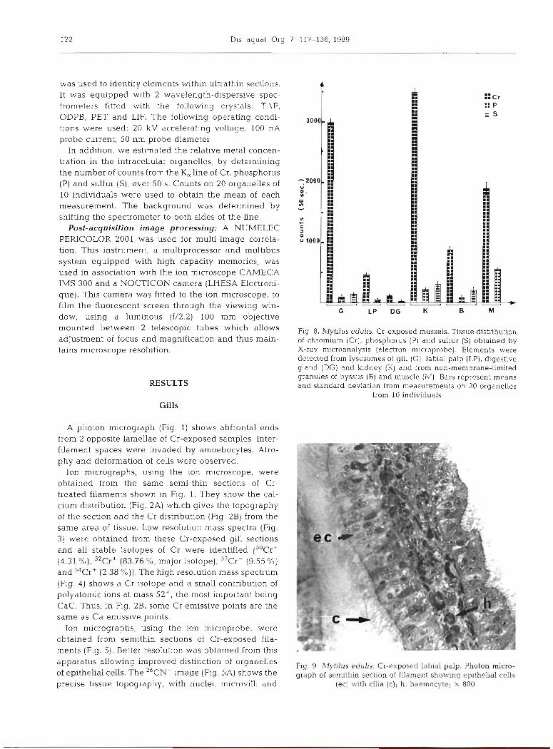
Fig. 9 Mytilus edults. Cr-exposed l ab~a l palp Photon micro- graph of semithln section of filament showing ep~thelial cells
(ec] with cllia ( c ) ~ h: haemocyte; x 800
Chassard-Bouchaud et al. Chromiunl In Mytilus edul f s
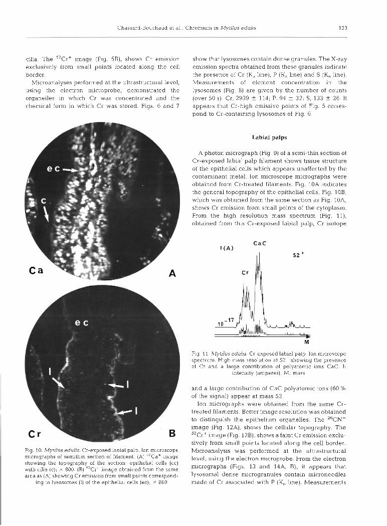
cilia. The 52Cr+ image (Fig. 5B), shows Cr emission exclusively from small points located along the cell border.
Microanalyses performed at the ultrastructural level, using the electron microprobe, demonstrated the organelles in w h ~ c h Cr was concentrated and the chemical form in which Cr was stored. Figs. 6 and 7
show that lysosomes contain dense granules. The X-ray em~ssion spectra obtained from these granules indicate the presence of Cr (K, line), P (K, line) and S (K, line). Measurements of element concentration in the lysosomes (Fig. 8) are glven by the number of counts (over 50 S): Cr, 2939 + 114; P, 94 i 32; S, 133 ? 26. It appears that Cr-high emisslve points of Fig. 5 corres- pond to Cr-containing lysosomes of Fig. 6.
Labial palps
A photon micrograph (Fig. 9) of a semi-thin section of Cr-exposed labial palp filament shows hssue structure of the epithelial cells which appears unaffected by the contaminant metal. Ion microscope micrographs were obtained from Cr-treated filaments. Fig. 10A Indicates the general topography of the epithelial cells. Fig. 10B, which was obtained from the same section as Fig. 10A, shows Cr emission from small points of the cytoplasm.
l From the high resolution mass spectrum (Fig. l l ) , obtained from this Cr-exposed labial palp, Cr isotope
Fig 11 MytJus e d u l ~ s Cr-exposed labial palp. Ion microscope spectrum High mass resolution at 52+ showing the presence of Cr and a large contribution of polyatomic ions CaC. I :
intensity (amperes); M mass
and a large contribution of CaC polyatomic ions (60 % of the signal) appear at mass 52.
Ion micrographs were obtained from the same Cr- treated filaments. Better image resolution was obtained to distinguish the epithelium organelles. The 26CN- image (Fig. 12A), shows the cellular topography. The "Crt image (Fig. 12B), shows a faint Cr emission exclu- sively from small points located along the cell border.
Fig. 10. Mytllus eduhs. Cr-exposed labial palp. Ion microscope Mlcroanalysis was gerformed at the ultrastructural rmcrographs of semlthln section of filament (A) 4 0 ~ a + image level, using the election mlcroprobe From the electron showing the topography of the section ep~thelial cells (ec) with cllia (c), X 800 (B) 52Cr+ Image obtained from the same micrographs (Figs 13 and 14A, B) , i t appears that
area as (A) showng Cr emission from small points correspond- ~ Y S O S O ~ ~ ~ dense mlcrogranules contain mlcroneedles ing to lysosomes (l) of the epithelia1 cells (ec), x 800 made of Cr assoc~ated wlth P (K, line) Measurements

124 Dis. aquat. Org. 7: 117-136, 1989
Fig. 12. Mytilus edulis. Cr-exposed labial palp. Ion microprobe Fig. 13. ~Vytilus edulis. Cr-exposed labial palp. Electron mic- micrographs of semithin section of filament. (A) ' CN- image rograph showing epithelial cells with cilia (c), mitochondria showing the topography of the section: epithelial cells (ec) (m), nuclei (n), autophagic vacuole (av) and lysosomes (1) with cilia (c) and nuclei (n); X 2000. (B) j2Cr - image obtained located along the cell border and containing Cr and P; X 7800 from the same area as (A) showing Cr emission from small points corresponding to lysosornes (I) located along the border
of the epithelia1 cells (ec) ; X 2000 vations of the faint Cr emission from a few points of the epithelial cells. The ion spectrum (Fig. 16) confirms this
of element concentration in these lysosomes (Fig. 8) are observation by exhibiting a small peak of Cr associated given by a number of 50 s counts: Cr 478 f 49; P, 39 f with a higher peak corresponding to the contribution of 20; S was below the detection limit. polyatomic ions CaC. These observations were addi-
tionally confirmed by ultrastructural observations since the emissive points of the ion image are lysosomes (Fig.
Digestive gland l?) of the epithelia1 cells, containing a few granules which, analysed by X-ray spectrometry, were shown to
The ion microprobe images obtalned from digestive consist of both elements: Cr, 139 f 31; P, 25 k 12; S was cells of the digestive diverticula (Fig. 15) allowed obser- below the detection limit (Fig. 8).


126 Dis. aquat. Org. 7: 117-136, 1989
Fig. 15. Mytilus edulis. Cr-exposed digestive cells. Ion micro- probe micrographs of a semithin section. (A) 2 6 ~ ~ - image showing the topography of the sectlon; ec: epithellal cell; X
2000. (B) 5 2 ~ r + image obtained from the same area as (A), showing faint chromium emission from the scarce lysosomes
(1) of the epithelia1 cells (ec); X 2000
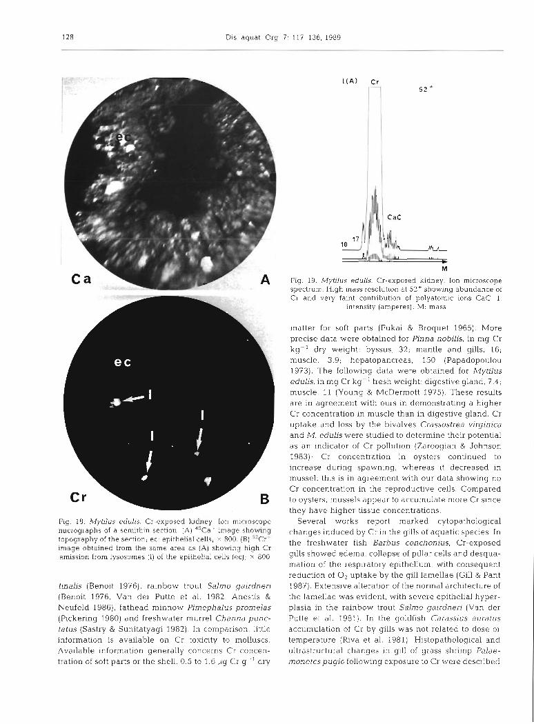
Kidney
The kidney takes the form of very highly convoluted and branched diverticula surrounded by a blood sinus containing occasional amoebocytes. The ion microscope image of this organ (Fig. 28), indicates very bright Cr emlssive points. The ion spectrum (Fig. 19) confirms this result by showing an important peak of Cr associated with a small one corresponding to polyatomic ions CaC. On the electron micrograph (Fig. 20), lysosomes corres- pond to the emissive points of the ion image; these dense lysosomal granules can be seen together wlth
Fig. 16. Wlytilus eduhs. Cr-exposed digestive cells. Ion micro- scope spectrum. High mass resolution at 52' showlng pre- sence of Cr and a high contribution of polyatomic ions CaC.
I : intensity (amperes); M: mass
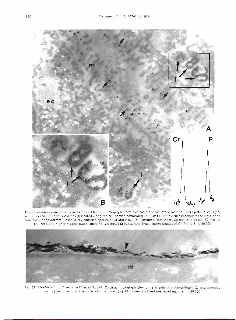
exocytotic vesicles by which excretory products are shed from the kidney. X-ray emission spectra obtained from the exocytotic vesicles and from the lysosomes indicate the presence (Fig. 8) of Cr, 3475 t 134, P, 215 + 39, and S, 307 & 36. When compared to the measure- ments obtained from the other tissues, Cr levels are highest in the kidney. An electron micrograph of non- osmicated and unstained material (Fig. 21) reveals the precise location of elements within lysosomes and within the dense precipitates shed from the kidney.
Byssus
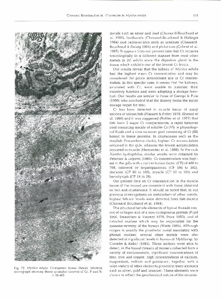
The thread 'elastic region', according to the defini- tion given by Benedict & Waite (1986), was investi- gated. It IS composed of electron-dense microfilaments embedded in electron-lucent matrix forming a sinuous electron-dense wavy pattern. This region of the byssal threads was examined and analyzed by X-ray spec- trometry at the ultrastructural level. Two sites of Cr concentration were detected. The first consisted of a thin sheath of electron-dense microneedles adsorbed onto the surface of the threads (Fig. 22). The second consisted of electron-dense microgranules incorpo- rated into the thread. The electron micrograph (Fig. 23) shows these non-membrane-limited dense granules distributed among the matrix. Using the electron mi- croprobe, it was possible to demonstrate that these granules contain the 3 clements Cr, 864 + 65; P, 63 + 37; and S, 203 k 32 (Fig. 8). The surface microneedles contained only Cr at a very high level: 1540 f 114.
Muscle
Samples of posterior adductor muscle were examined and analyzed at the ultrastructural level. Fig. 24 indi-
Chassard-Bouchaud et a1 Chromium ~n Mytllus e d u l ~ s 127
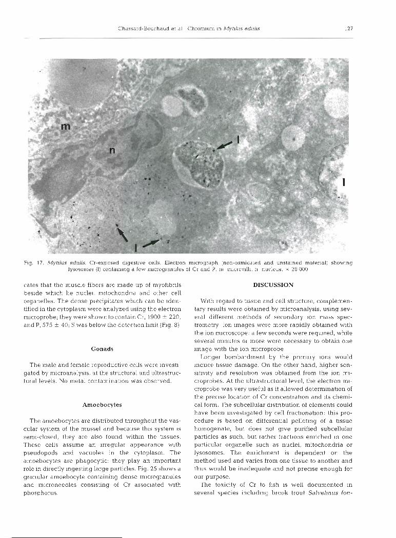
Fig. 17. Mytilus edulis. Cr-exposea aigestive cells. Electron micrograph [non-osmicated and unstained material) showng lysosomes (1) containing a few microgranules of Cr and P, m microvill~, n nucleus, X 20 000
cates that the muscle fibers are made up of niyofibrils beside which lie nuclei, mitochondria and other cell organelles. The dense precipitates which can be iden- tified in the cytoplasm were analyzed using the electron microprobe; they were shown to contain Cr, 1900 + 220, and P, 575 ? 40; S was below the detection limit (Fig. 8)
Gonads
The male and female reproductive cells were investi- gated by microanalysis, at the structural and ultrastruc- tural levels No metal contamination was observed.
Amoebocytes
The amoebocytes are distributed throughout the vas- cular system of the mussel and because this system IS
semi-closed, they are also found within the tissues. These cells assume an irregular appearance wlth pseudopods and vacuoles in the cytoplasm. The amoebocytes are phagocytic: they play an important role in dli-ectly ingesting large particles. Fig. 25 shows a granular amoebocyte containing dense microgranules and microneedles consisting of Cr associated with phosphorus.
DISCUSSION
With regard to tissue and cell structure, complemen- tary results were obtained by microanalysis, using sev- eral different methods of secondary ion mass spec- trometry Ion images were more rapidly obtained with the ion microscope: a few seconds were required, while several minutes or more were necessary to obtain one image with the ion microprobe.
Longer bombardment by the pnmary ions would ~ n d u c e tissue damage. On the other hand, higher sen- sitivity and resolution was obtained from the ion m]- croprobes. At the ultrastructural level, the electron mi- croprobe was very useful as it allowed determination of the precise location of Cr concentration and its chemi- cal form. The subcellular distribution of elements could have been investigated by cell fractionation: this pro- cedure is based on differential pelleting of a tissue homogenate, but does not give purified subcellular particles as such, but rather fractions enriched in one particular organelle such as nuclei, mitochondria or lysosomes. The enrichment is dependent on the method used and varies from one tissue to another and thus would be inadequate and not precise enough for our purpose.
The toxicity of Cr to fish is well documented in several species including brook trout Salvellnus fon-
128 Dis. aquat. Org.
Fig. 18. Mytdus edulis. Cr-exposed kidney. Ion microscope nucrographs of a sernithin section. (A) 40Caf image showing topography of the section, ec: epithelial cells; X 800. (B) "Cr- mmge obtained from the same area as (A) show~ng high Cr emission from lysosomes (1) of the epithelial cells (W) ; X 800
tinalis (Beno~t 1976), rainbow trout Saln~o gairdnen (Benoit 1976, Van der Putte et al. 1982, Anestis &
Neufeld 1986), fathead minnow Pirnephalus promelas (Pickering 1980) and freshwater murrel Channa punc- tatus (Sastry & Sunitatyagi 1982). In companson, little information is available on Cr toxicity to molluscs. Available information generally concerns Cr concen- tration of soft parts or the shell, 0.5 to 1.6 pg Cr g-I dry
b M
Fig. 19. Mytilus edulis. Cr-exposed kidney Ion microscope spectrum. High mass resolution at 52+ showing abundance of Cr and very faint contribution of polyatomlc ions CaC. I:
intensity (amperes); M: mass
matter for soft parts (Fukai & Broquet 1965). More precise data were obtained for Pinna nobilis, in mg Cr kg-' dry weight: byssus, 32; mantle and gills, 16; muscle. 3.9; hepatopancreas, 150 (Papadopoulou 1973). The following data were obtained for nilytilus edulis, in mg Cr kg-' fresh weight: digestive gland, 7.4; muscle. 11 (Young & McDermott 1975). These results are in agreement with ours in demonstrating a higher Cr concentration in muscle than in digestive gland. Cr uptake and loss by the bivalves Crassostrea virginica and M. edulis were studied to determine their potential as an indicator of Cr pollution (Zaroogian & Johnson 1983); Cr concentration in oysters continued to increase during spawning, whereas it decreased in mussel: this is in agreement with our data showing no Cr concentration in the reproductive cells. Compared to oysters, mussels appear to accumulate more Cr since they have higher tissue concentrations.
Several works report marked cytopathological changes induced by Cr in the gills of aquatic species. In the freshwater fish Barbus coachonius, Cr-exposed gills showed edema, collapse of pillar cells and desqua- mation of the respiratory epithelium, with consequent reduction of O2 uptake by the gi.ll lamellae (Gill & Pant 1987). Extensive alteration of the normal architecture of the lamellae was evident, with severe epithelial hyper- plasia in the rainbow trout Salmo gairdneri (Van der Putte et al.. 1981) In the goldfish Carassius auratus accumulation of Cr by gills was not related to dose or temperature (Riva et al. 1981). Histopathological and ultrastructural changes in gill of grass shrimp Palae- monetespugio following exposure to Cr were described

Chassard-Bouchaud et a1 Chromium in Mytllus eduhs
Fig. 20. Mytilus eduljs. Cr-exposed kidney Electron mlcrogrdph showlng eplthel~al cells (ec) w ~ t h mlcrovilli ( m ] and lysosomes [l) containing Cr, P and S. Note presence of exocytosls veslcles (stars) by whlch metals are shed from the kldney: x 26 000
as follows (Doughtie & Rao 1984): irregular profiles of gill lamellae indicating pronounced cellular distension,
cellular disorganization, increased number of ly- sosomes and distended cisternae of rough endoplas- mic reticulum. In the shrimp, major degenerative changes due to Cr contamination were restricted to the gills, with overall decrease in epithelia1 cytoplasmic density and a loss of euchromatin denslty Thus, it appears that the cytopathological changes we were able to observe in the mussel may be compared to the ones observed in fish and shrimps. The gill seems to be, in all these organisms, the predominant slte of Cr toxic action. Gill lesions were also observed in samples of Mytilus edulis collected from a harbour polluted by discharges of an iron and steel factory (probably con-
tainlng Cr) by Sunlla (1987) In these specimens part of the lnterfilamentar cillary lunctlons were replaced by metaplastic cellular connechons and chronic ~nflam- matory reacbon was observed. These gill lesions associated wlth heavy metal stress affect gas exchange and food transport.
The digestive gland is commonly known to be the maln organ of metal concentration in many inverte- brates. Using microanalytical techniques we investl- gated several organs (digestive gland, kidney, gill, labial palp) of Mytilus eduhs. We were able to demon- strate that the digestive gland of mussel 1s the target organ for the storage of many elements: lithium, the lightest metal (Chassard-Bouchaud et al. 1984), aluminium (Chassard-Bouchaud & Galle 1986), heavy
DIS dquat Org 7 117-136, 1989
Flg. 21 Myllus edulis Cr-exposed kldney Electron micrographs (non-osmicated and unstained material). (A) Eplthetial cells (ec) w ~ t h mlcrovllll (m) and lysosomes (I) located along the cell border containing Cr, P and S Note dense precipitates of metal shed from the k~dney (arrows) Inset X-ray emlssion spectra of CT and P (Ka fine] obtalned from these lysosames; X 3 2 000 (B) Part of
( A ) , seen at a hlgher magnification, showing lysosames [ I ) containing dense mlcrogranula ol Cr. P and Si X 66000
Fig 22 Myhlus eduhs. CI-exposed byssal thread. Ebctran micrograph show~ng a sheath of e1-n-dense Cc m~croneedles [arrow) adsorbed onto the surface of the matnx Irn) Non-osm~cated and unstained material; x 40000
Chassard-Bouchaud et al.: Chromium in Mytilus edulis
Fig. 23 Mytilus edulls Cr-exposed byssal thread. Electron micrograph show~ng dense granules (arrows) of Cr. P and S;
X 16400
metals such as silver and lead (Chassard-Bouchaud et al. 1985), lanthanids (Chassard-Bouchaud & Hallegot 1984) and radionuclides such as uranium (Chassard- Bouchaud & Escaig 1984) and plutonium (Calmet et al. 1987). It appears from our present data that Cr behaves toxicologically in a different manner from most other metals in M. edulis since the digestive gland is the tissue which exhibits one of the lowest Cr levels.
Our results reveal that the kidney of Mytilus edulis had the highest mean Cr concentration and may be considered the prime determinant site of Cr concen- tration. In this specific case, it seems that the kidneys, saturated with Cr, were unable to maintain their excretory function and were adopting a storage func- tion. Our results are similar to those of George & Pirie (1980) who concluded that the kidney forms the major storage organ for zinc.
Cr has been detected in muscle tissue of many species of teleost fish (Plaskett & Potter 1979, Elwood et al. 1980) and it was suggested (Buhler et al. 1977) that fish have 2 major Cr compartments, a rapid turnover pool consisting mainly of soluble Cr (VI) in physiologi- cal fluids and a slow turnover pool consisting of Cr (111) bound to tissue proteins. In crustaceans such as the crayfish Procambarus clarkii, highest Cr accumulation occurred in the gills, whereas the lowest accumulation occurred in muscle (Hernandez et al. 1986). In the crab Xantho hydrophilus, similar results were obtained by Peternac & Legovic (1986): Cr concentration was high- est in the gills with a concentration factor (CF) of 400 to 798, followed by hepatopancreas (CF 196 to 392), skeleton (CF 80 to 160), muscle (CF 52 to 104) and hemolyinph (CF 14 to 28).
Our present data on Cr concentration in the muscle tissue of the mussel are consistent with those obtained on fish and crustaceans. It should be noted that, in our previous investigations on metabolism of other metals, highest lithium levels were detected from fish muscle (Chassard-Bouchaud et al. 1984).
The structural tensile elements of byssal threads con- sist of collagen and of a non-collagenous protein (Pujol 1966, Smeathers & Vincent 1979, Price 1983), and of catechol oxidase which may be responsible for the quinone-tanning of the byssus (Waite 1985). Although copper is usually the prosthetic metal associated with phenol oxidase, several other metals were also detected at significant levels in byssus of Mytilus sp. by Coombs & Keller (1981). These authors were able to detect, in the byssal threads of mussels collected from a variety of environments, significant concentrations of zinc, iron and copper, high concentrations of calcium, magnesium, sodium and potassium, together with a wide variety of other elements present in trace amounts such as silver, gold and uranium. These elements were shown to reflect the geochemical nature of the environ-
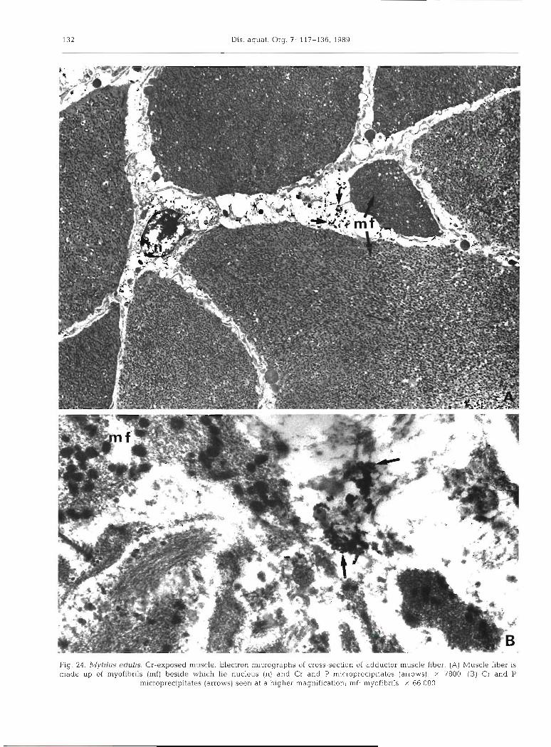
000 g9 X !s~~iq!jor(.ur :ju !uo1les1)1u6eru ~aq61q e le uaas (SMOJJP) sa~e~~dpa~do~s!~ d pue 13 (8) 008~ X !(s~orie) salel!d!sa.~do~~~ur d pue 13 pue (U) snalsnu ay q31q~ ap~saq (p) s~uq!jodur jo dn apem SI mqyj alssnm (V) 'raq!j alssnm lolsnppe jo uogsas-sso~s 30 sqde.1601srur uoJlsal3 .apsnm pasodxa-13 ,srlnpa sn[gA141 61-3

Chassard-Bouchaud et al.. Chromium in Mytilus edulis
Fig. 25. A4ytilus edulis. Cr-exposed macrophage amoebocyte. Electron micrograph showing heterogenous content of the cell, among which Cr microgranules and microneedles are visible (arrows); c: cilia; n: nucleus; X 32 000
ment. For the radionuclide, the concentration in the byssal threads was 10-fold higher than in the rest of the soft tissues. The same authors question whether the metals would be either adsorbed or complexed directly from the surrounding waters onto the threads, or incor- porated within the tissues. Our data, obtained by mi- croanalytical imaging techniques, clearly support the existence of both mechanisms for Cr concentration in
the byssal threads of the mussel. An iron excretion role for the byssus has been suggested (George et al. 1976), involving transfer from the other tissues to the byssus via the haernolymph. In experiments on factors influencing the flux of arsenic through Mytilus gallo- provincjalis, Unlii & Fowler (1979) found the highest concentration of 7 4 ~ s in the byssus. We can conclude with these authors that the byssal threads may consti-
Dis. aquat. Org. 7 117-136, 1989
tute a significant pathway for the elimination of metals. This process would provide a simple and convenient method for monitoring long-term environmental changes in metal toxicants.
Granular amoebocytes of Mytilus edulis were shown to contain Cr microparticles. In bivalves, these cells can perform both intra and extracellular digestion of for- eign particles. They migrate to the digestive gland, kidney and other epithelia1 linings where they are discharged to the exterior. These cells are involved in the phagocytic clearance of pollutants (Read & Read 1972) as they are enzymatically equipped with lysosomal phosphatase. Our results are in agreement with this concept, according to which granular amoebocytes perform detoxication of metals, such as iiac, iii the oysier Zstrea eduks (George et G!. 1978) and in the mussel Mytilus edulis (George & Pirie 1980).
At the subcellular level, we were able to investigate nuclei, mitochondria and lysosomes of all the organs and tissues in order to locate more precisely the sites of Cr concentration as well as detennine the insoluble chemical form in which it was precipitated. The lyso- some appears to be the target organelle of Cr concen- tration, as no metal was detected from other organelles. These data are in agreement with our previous results showing that lysosomes of Mytilus edulis are the target sites of several metals such as aluminium (Chassard- Bouchaud & Galle 1986), lanthanum. (Chassard- Bouchaud & Hallegot 1984) and uranium (Chassard- Bouchaud 1983, Chassard-Bouchaud & Escaig 1984). We agree with George e t al. (1982) who proposed that Mytilus may b e a useful model system for the study of intralysosomal metal accumulation.
In the lysosomes, Cr was concentrated in an insolu- ble from and associated wi.th phosphorus and sulfur. Chromium phosphate was the result of a n enzymatic reaction of acid phosphatase activity, as previously demonstrated by a method based on its microanalytical visualisation (Berry et al. 1982). The presence of sulfur, associated with Cr in the lysosome, may be explained by the possible existence of thionein-like protein. Metallothioneins in mussels were first described by Noel-Larnbot (1976). Since then numerous papers describing different metallothioneins in mussels have been published (George & Pirie 1979, George et al. 1979, Viarengo et al. 1986). By sequestering Cr, lysosomes play a defensive role by preventing the diffusion of the toxic metal throughout the cell.
In conclusion, Cr uptake by Mytilus edulis occurs via the gills where high levels of metal were detected, inducing major cellular degenerative changes there. Contaminated water is then conveyed towards the labial palps before entering the digestive system. The main storage tissue appears to be muscle, while the digestive gland plays a very minor role in the metal
concentration. Byssal threads are involved in Cr stor- age and excretion which however is chiefly performed by the kidney which exhibited the highest Cr levels. Granular amoebocytes are involved in Cr uptake, stor- age and excretion. The target organelle of Cr concen- tration was the lysosome where the metal was associ- ated with phosphorus and sulfur, in an insoluble form.
From our data, it appears that Mytilus edulis may be considered as a biological indicator of Cr pollution. Investigations of byssal threads, using several micro- analytical techniques, could provide, easily and rapidly, useful information on tissue levels relative to environmental contamination.
Acknowledgements. This work was financially supported by the Food and Agriculture Organization of the United Nations: MED POL 11 Programme (contract FRA 24 C), by the National Science Foundation of the United States (grant BBS-8610518) and by INSERM France (S.C.27). We thank F. Escaig, P. Boumati, F. Kleinbauer and J. Brissard for their technical assistance, and G . Plessard for drafting the drawings. Electron microscopy was performed in Laboratoire de Microscopie Electronique appliquee B la Biologie, 105 Bd. Raspail, F-75006 Paris, France.
LITERATURE CITED
Anestis, I . , Neufeld, R . J (1986). Avoidance preference reac- tions of rainbow trout (Salmo gairdneri) after prolonged exposure to chromum VI. Wat. Res. 20: 1233-1241
Benedict, C. V., Waite, J. H. (1986). Composition and ultra- structure of the byssus of Mytilus edulis. J. ':iorph. 189: 261-270
Benoit, D. A. (1976). Toxic effects of hexavalent chromium on brook trout (Salvelinus fontinalis) and rainbow trout (Salmo gairdneri). Wat. Res. 10: 497-500
Berry, J. P . , Hourdry, J., Sternberg, M., Gallc, P. (1982). Aluminium phosphate visualisation of a c ~ d phosphatase activity: a biochemical and X-ray microanalysis study. J. Histochem. Cytochem. 30: 86-90
Bryan, G. W. (1980). Recent trends in research on heavy metal contamination in the sea. Helgolander Meeresunters. 33: 6-25
Buhler, D. R., Stokes, R. M., Cadwell, R. S. (1977). Tissue accumulation and enzymatic effects of hexavalent chromium in rainbow trout (Salmo gairdneri). J. Fish. Res. Bd Can. 34: 9-18
Calmet, D.. Charmasson, S., Willemot, J. M., Verry, M. , Chas- sard-Bouchaud, C , Inglebert, R. L., Outrequin, M., Galle, P. (1987). Suivi des niveaux de plutonium 239-240 dans des moules Mytilus sp. prklevees sur le littoral francais (1983-1984): Ctudes radiochimique et microanalytique. C.r. hebd. Seanc. Acad. Sci., Paris 304 (Ser. 111) 9: 199-206
Cavellier, J. F., Escaig, F., Boumati, P., Gaurne, P., Hallegot, P. (1988). Numerisation and digital processing of images in secondary ion microscopy. In: Benninghoven, A. , Huber, A. M,, Werner, H W. (eds.) Secondary lun Mass Spec- trometry SlMS V1 John Wiley and Sons, New York, p. 385-388
Chassard-Bouchaud, C. (1983). Role des lysosomes et des spherocristaux dans le phenomene de concentration de I'uranium chez la moule fvlytilus edulis. M~croanalyse par

C h r o n ~ ~ u n l ln Mytilus edulis 135
spectrographie dcs rayons X. C.r. hebd. Seanc. Acad. Sci., Paris 296 (Sbr. 111): 581-586
Chassard-Bouchaud, C. (1987). lon microscopes and micro- probes In n~. i r ine pollution research. Analytica chim. .4cta 195: 307- 315
Chassard-Bouchaud, C. (1988). Current trends and applica- tlons of seconddl y ion microscopy in n ~ e d ~ c i n < . iind bioloqy :
a revlew. In Benninghoven, A., Huber, A M., Werner, H. W. (eds.) Secondary Ion hfass Sprctrometry SI',,IS VI. John tb'ile)' and Sons, New York, p. 855-863
Chassard-Bouchaud. C., Escaig, F. (1984). Uptake, storage and excretion of uranlum by Mytilus edulis. A structural, ultrastructural and microanalytical study by secondary ion mass and X-ray spectrometry. J . Phvs., Paris 45: 545-548
Chassard-Bouchaud, C., Galle, P. (1986). Bioaccumulation d'aluminlum par les organismes marins. Mise en evidence par microscop~e corpusculaire analytique C.r hebd. Seanc. Acad. Sci , Paris 302 (Sdr 111) 2: 55-61
Chassard-Bouchaud, C. , Galle, P. (1988) Sites cellulaires d e concentration du chrome chez la moule Mytrlus edulis: donnees preliminaires. C.r. hebd. Seanc. Acad. Sci., Paris 306 (Ser. 111): 4 6 7 4 7 3
Chassard-Bouchaud, C.. Gallc, P,, Escaig, F. (1985). Mise e n evidence d 'une contamination par l 'argent et le plomb d e I'huitre Crassostrea gigas et d e la moule Mytilus edulis dans les eaux cotieres franqaises. Etude microanalytique par enlission ionique secondaire. C.r. hebd. Seanc. Acad. Sci., Paris 300 (Ser 111) 1 . 3-8
Chassard-Bouchaud, C , Galle, P . , Escaig, F . , Miyawaki, M. (1984). Bioaccumulation de lithium par les 01-ganismes marins des zones cbtieres europeenes, americaines et asiatiques: etude microanalytique par emission ionique secondaire. C.r. hebd. Seanc. Sci., Paris: 299 (Ser 111) 18: 719-724
Chassard-Bouchaud, C.. Hallegot, P. (1984). Bioaccumulation d e lanthanc par des moules Mytilus edulis recoltees sur les cBtes f ran~aises . Microanalyse par spectrographie des ray- ons X et par emission ionique secondaire. C. r hebd. Seanc. Acad. Sci , Paris 298 (Ser. 111) 20: 567-572
Coombs, T L , Kcllrr, P J. (1981). A4ytilus byssal threads as an environmental n~drkel- for metals. Aq.uat. Toxic. t . 291-300
Douqhtie, D. G., Rao, R K . (1984). Histopathofoglcal and ultrastructural changes in the antenna1 gland, ~nidgut , hepatopancreas and gill of grass shrimp following expo- sure to hexavalent chromium. J . Invcrtebr Pathol. 43: 89-108
Elwood, J . W.. Beauchamp. J . J., Allen. C. P. (1980). Chromium levels in fish from a lake chronically contami- nated with chromates from cooling towers. Int. J. envir! Stud. 14: 289-298
Fukai, R., Broquet, D (1965). Distribution of chrom~um in marine organisms Bull Inst. oceanogr Monaco 65 (1336): 3-19
George, S. G.. Carpene, E., Coombs, T. L., Overnell, J . , Youngson, A. (1979). Characterisation of cadmium-bind- ing proteins from mussels, Myfilus edulis (L.), exposed to cadmium. Biochim. Biophys. Acta 580: 225-233
George, S. G., Coombs, T L., Pirie, B. J. S. (1982). Character- isation of metal-containing granules from the kidney of the common mussel Mytjlus edulis. Biochim. Biophys. Acta 716: 61-71
George, S. G . . Pirie, B J. S. (1979). The occurrence of cad- mium in sub-cellular particles in the kidney of the marine mussel, Mytilus edulls, exposed to cadn~iunl. The use of electron microprobe analysis. Blochim. Biophys Acta 580: 234-244
George, S. G., Pirie, B. J . S. (1980). Metabolism of zinc in the
mussel .A,lytilus edulis (L.): a comb~ned ultrastructural and b ioch~mical study. J. mar biol. Ass. U. K. 60: 575-590
Georgc, S. G., Pirie, B. J . S., Cheyne, A. R., Coombs, T L., Grant, P. T. (1978). Detoxication of metals by marine bival- ves: an ultrastructural study of the compar tn~entdt~on of copper and zlnc in the oyster Ostrea edulis. Mar. Biol. 45: 147-156
George, S. G. , Pirie, B. J . S . , Coombs, T L. (1976). The kinetics of accunlulation and excretion of fernc hydroxide in Mytilus edulis (L.) and its distribution in the tissues. J . exp. mar. Biol. Ecol. 23: 71-84
Gill, T. S., Pant, J . C. (1987). Hematological and pathological effects of chromium toxicosis in the freshwater fish Barbus conchonius Ham. Wat. Air Soil Poll. 35: 241-250
Giinter, T., Ruhe, B., Schmalbeck, J., Tehrani, N. (1974). Blochemistry of trace elements Zn, Cu, Cr and CO: dis- tnbution, binding and regulation by adrenal hormones. Z. khn Chem. klin. Biochem. 12. 327-335
Hernandez, F . , Diaz, J . , Medina, J. , Delramo, J . , Pastor, A. (1986). Detcrriiination of chromium treated crayfish Pro- cambarus clarkii by clectro-thermal AAS. study of Cr accumulation in different tissues. Bull. envir Contam. Toxic. 36: 851-857
Karbe, L.. Schnier, C. H., Siewers, H. 0. (1977). Trace ele- ments in mussel Mytilus edulis from coastal areas of the North Sea and the Baltic. Multielement analyses using instrumental neutron activation analysis. J. Radioanal. Chem 37. 927-943
Lande, E (1977) Heavy metal pollution in Trondheimsfjorden Norway and the recorded effects on the fauna and flora. Envir. Pollut. 12: 187-198
Levi-Setti, R., Chabala, J . , Wang, Y. L. (1988) Aspects of high resolution imaging with a scannlng ion microprobe Ultramicroscopy 24: 97-1 14
Mertz, W. (1969). Chromlum occurrence and function in biological svstems. Phys~ol. Rev. 49: 163-239
Noel-Lambot. F. (1976). Distribution of cadmium, zinc ant1 copper in the mussel Mytilus edulis. Existence of cad- mium-b~nding proteins similar to metallothioneins. Experientia 32. 324-325
Nriagu, J . O., Pacyna, J . M. (1988). Quant~tative assessment of .rvorldw~de contamination of air, water and so~ l s by trace metals Kdture, Lond. 333: 134-139
Qutrequin, I . , Bernard, J. L., Inglebert, R. L. (1988). Detection limit optimization in the new MIQ 256 mlcroprobe. In: Benninghoven, A., Huber, A. M., Werner. H. W (eds.) Secondary Ion Mass Spectrometry SIMS VI. John Wiley and Sons, New York, p . 165-168
Papadopoulu, C. (1973). The elementary composition of marine invertebrates as a contribution to the sea pollution investigation Pi-oc. Mambo meeting, Castellabate, Italy p. 1-18
Peternac, B., Legovlc, T (1986). Uptake, distribution and loss of chromium in the crab Xantho hydroph~lus Mar. Biol. 91: 467-471
Pickering. Q. H. (1980). Chronic toxicity of hexavalent chromium to the fathead minnow (Pimephalus promelas). Archs envir. Contam. Toxic. 9: 4 0 5 4 1 0
Plaskett. D., Potter. I. C. (1979). Heavy metal concentrations in the muscle tissue of 12 species of Teleost from Cockburn Sound, Western Australia. Aust. J . mar. Freshwat. Res. 30: 607-616
Price, H. A (1983). Structure and formation of the byssus con~plex in Mytilus (Mollusca Bi\ralvia) J. mollusc. Stud. 49: 9-17
Pujol, J P (1966). Le complexe byssogene des Mollusques bivalves Hlstochi~nie comparde des secre t~ons chez

136 Dis. aquat. Org. 7: 117-136, 1989
Mytdus edulis et Pinna nobiljs L. Bul. Soc. Linn. Normandie l 0 (8): 308-332
Read, P., Read, E. (1972). Phagocytosis in Invertebrates. 11. The clearance of carbon particles by the cldln Tridacna max- ima. Res. J . Reticuloendothelial Soc. 12: 349-360
M e y , J P . , Chester, R. (1971). Introduction to marine cbemis- try. Academic Press, New York
Riva, M. C., Flos, R., Crespi, M,, Balasch, J. (1981). Lethal potassium dichromate and whitening (Blankophor) expo- sure of goldfish (Carassius auratus): chromium levels in gills. Comp. Biochem. Physiol 68C: 161-165
Sastry, K. V., Sunitatyagi, M. (1982). Toxic effects of chromium in a freshwater teleost fish Channa punctatus. Toxic. Lett. 11: 17-21
Schulz-Baldes, M,, Rehm, E., Farke, H. (1983). Field experi- ments on the fate of lead and chromium in an intertidal benthic mesocosm, the Bremerhaven Caisson. Mar. Biol 75: 307-318
Sehwartz. K., ?-icrtz. W. (1959). C h r ~ m i u m [!!!! 3nd the 9111- cose tolerance factor. Archs Biochem. 85: 292-295
Smeathers, J. E., Vincent, J. F. V. (1979). Mechanical proper- ties of mussel byssus threads J. mollusc. Stud. 45: 219-230
Strik, J. J., De Longh, H. H., Vannjn, J. W., Wuite, T. P. (1975) Sublethal effects of toxic chemicals on aquatic animals. Elsevier, Amsterdam
Sunila. I. (1987). Histopathology of mussels (Mytilus edulis)
Responsible Subject Editor: Dr A. K. Sparks, Seattle, Washington, USA
from the Tvdrminne area, the Gulf of Finland (Baltic Sea). Annls zool. fenn. 24: 55-69
Unlii. M. Y , Fowler, S. W. (1979). Factors affecting the flux of arsenic through the mussel Mytilus galloprovincialis. Mar. Biol. 51: 209-219
Van der Putte, I . , Brinkhorst, M . A., Koeman, J. H. (1981). Effects of pH on the acute toxicity of hexavalent chromium to rainbow trout (Salrno gairdnerj). Aquat TOXIC. 1. 129-142
Van der Putte, I., van der Galien, W., Strik. J. J. (1982). Effects of hexavalent chromium in rainbow trout (Salmo gairdneq after prolonged exposure at two different pH levels. Ecotoxicol. Envir. Saf. 6: 246-257
Viarengo, A.. Moore, M. N., Pertica, M.. Mancinelli. G., Zanic- chi, G. (1985). Detoxification of copper in the cells of the digestive gland of mussel: the role of lysosomes and thio- neins Sci. total envir. 44: 135-145
Waite, J. H. (1985). Catechol oxidase in the byssus of the common mussel Mvtilus edulis L. J. mar. biol. Ass. U.K. 65: 359-37 1
Young, D. R., McDermott, M. (1975). Trace metal in harbor mussels. A. Rep. S. Calif. Coast. Water Res. Proj., El Segundo, California, p. 139-142
Zaroogian, G. E., Johnson, M. (1983). Chromium uptake and loss in the bivalves Crassostrea virginica and Mytilus edulis. Mar. Ecol. Prog. Ser. 12: 167-173
Manuscript first received. December 28, 1988 Revised version accepted: July 6, 1989