Embed Size (px)
Citation preview
DISEASES OF AQUATIC ORGANISMS Dis. aquat. Org. 1 Published April 14
NOTE
Macroscopic and microscopic features of ulcerative stomatitis in farmed Atlantic salmon Salmo salar
'Department of Veterinary Pathobiology, College of Veterinary Medicine, Texas A&M University, College Station, Texas 77843, USA
'Battelle Marine Research Laboratory. 439 West Sequim Bay Rd. Sequim, Washington 98382. USA
ABSTRACT. Ulcerative stomatitis is an economically unpor- tant disease of salmon srnolts In net-pen farms of Puget Sound, Washington, USA. Gross lesions and losses from this disease typically occur 3 to 8 wk following introduction to sea water and may result in a cumulative mortality of up to 30 O/u in some pens. The disease occurs seasonally and has been present on one farm since it began production in 1987 Ulcerated lesions are limited to the oral cavity, vary in size, and are associated with areas of dentition. Mats of Cytophaga-like filamentous bacteria are associated w ~ t h areas of ulceration and necrosis and often extend into the un- derlying bone Scanning electron mlcroscopy demonstrated mats of filarnentous bacteria overlying ulcerated areas and biofilms of f~lamentous bacter~a adhering to tooth surfaces at the ginglval-enameloid interface and invading the gingival epithelium. Our findings suggest the filamentous bacterium plays a major role in the pathogenesis of ulcerative stomatitis in salmon smolts.
KEY WORDS: Atlantic salmon - Salmo salar . Bacterial d ~ s - edse . Stornatitis . Cytophaga-l~ke . Flexibacter-like
Filamentous, nonphotosynthetic, nonfruiting, Gram- negative bacteria that are pathogenic to fish and are able to demonstrate a slow, gliding type of motility are included in the order Cytophagales (Larkin 1989). Although many species of bacteria within this order are pathogenic to freshwater fish (Frerichs & Roberts 1989), only a few pathogenic species have been identi- fied from the marine environment (Sawyer 1976, Masumura & Wakabayashi 1977, Wakabayashi et al. 1984, Kent et al. 1988, Dungan et al. 1989, Bernardet et al. 1990).
Mortality of Atlantic salmon Salmo salar smolts up to 6 mo after introduction to sea water has been attribu- ted to various causes including inadequate smoltifica- tion, poor feeding, and several infectious diseases. A disease survey of net-pen reared salmon from Puget
Sound, Washington, USA, during 1990 and 1991 re- vealed an association of dead and moribund smolts with a subacute, multifocal, ulcerative stomatitis. The disease occurred on 4 farms representing 5 sites with a combined annual production of over 1500 tons and was considered by farm managers to be the most severe in- fectious disease in 1990 and 1991. Reports from fish farmers in British Colun~bia indicated the loss of ap- proximately 139 000 smolts in 1989 associated wlth an apparently identical disease (M. Shephard pers. comm). Initial leslons of the disease proved to have a uniform character and were associated with a filamen- tous Cytophaga-like bacterium. The purpose of this report is to provide a detailed morphologic description of the disease and to discuss the findings in relation to pathogenesis of the disease. Additional studies re- garding b~ochemical and n~olecular characterization of the associated organism are under way and will be re- ported in a subsequent paper.
Material and methods. Affected smolts recently in- troduced to sea water were obtained from 4 net-pen farms in Puget Sound, Washington, USA, from April to July 1990 and 1991. Affected smolts were obtained from pens where oral lesions had been directly asso- ciated with mortalities. The 7 to 12 mo old smolts were directly introduced to sea water net-pens at a density of approximately 25 kg m-3 and weighed between 50 and 80 g when stocked.
Tissues were selected for necropsy from 8 dead, moribund, or clinically ill Atlantic salmon demon- strating characteristic oral lesions. Fish from 3 farms were examined in detail. Tissues were fixed in David- son's solution (Humason 1979), processed, and stained with hematoxylin and eosin for morphologic examina- tion. Selected tissues were stained with a Brown- Hopp's modified Gram stain (Sheehan & Hrapchak
O Inter-Research 199A
228 Dis. aquat. Org. 18: 227-231. 1994
Species 1990 1991 No. examined "/u affected No. examined 'Yo affected
AS 626 13.4 670 9 CS 227 0 14 0 RT 260 11.5 26 9.5
AS = Atlantic salmon Salmo salar; CS = chinook salmon Oncorhynchus tshawytscha; RT = rainbow trout Oncorhynchus mykiss
Table 1 Results of necropsy survey for necrotizing stomatitis from 3 net- containing an overlying layer of yellow to pen salmon farms in Puget Sound, Washington, USA. tan friable material. Lesions were characte-
ristically associated with regions of denti- tion including the premaxilla, dentary, vomer, and palatine bones. Tooth loss was often present in the affected areas. In many cases the opposing surfaces of the dentary and premaxilla bones were affected. Gram- stained smears of the ulcerated areas de- monstrated large numbers of uniform, Gram-negative 2.5 to 13 pm long filamen- tous organisms.
1980). Ti.ssues of the oral cavity and tissues from the Successful culture of the presumptive causative heart, gills, liver, spleen, anterior and posterior kidney, agent of this disease required careful dissection and and brain were also examined. washing of the lesions and serial-dilution plating due
Oral tissues from 3 of the smolts containing the typi- to the expected surface contaminants of the lesions. cal lesions and 2 macroscopically normal smolts were Once this method was refined, pure cultures of a esaixined by scanning electron microscopy (SEM) single colony type were obtained. Cells in the colo- These tissues were fixed in 4 % glutaraldehyde in nies were thin, Gram-negative rods typical of a Cyto- 0.1 M sodium cacodylate adjusted to pH 7.2 and then phaga sp. rinsed in 0.1 M cacodylate. Tissues were postfixed in Microscopic findings: Oral tissues from 1 smolt de- l % osmium tetroxide containing sodium cacodylate monstrated what we interpreted as the initial lesion, buffer, dehydrated in acetone, critical point dried with which was characterized by spongiosis of the epithelial carbon dioxide, sputter coated with gold, and exa- layer due to interepithelial edema with separation of mined on a JEOL JSM.25S scanning electron micro- individual epithelial cells and transepithelial inflam- scope. matory cell migration. The epithelium was intact but
Impression smears of oral lesions were stained with contained an overlying mat of filamentous bacteria. Giemsa and Gram stains and examined microscopi- Patchy areas in the stratum spongiosum contained cally. neutrophils and mononuclear cells. A single lymphatic
Lesions from selected smolts were dissected, minced contained an organized thrombus. in sterile 50 % sea water, and dilution-plated onto both In the remaining smolts the predominant oral lesion cytophaga medium (Difco) made with 50 % sea water consisted of a focal ulcerated area that extended to the and marine agar (Difco) (Anacker & Ordal 1959). stratum spongiosum or stratum compactum. The ulcer
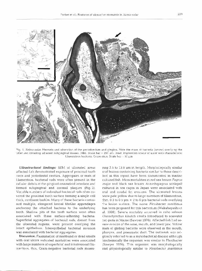
Results. Disease outbreaks occurred from April to frequently had an overlying layer of dense bacteria July when water temperatures were between 8 and mixed with necrotic debris (Fig. 1). With the Gram 12°C and salinity was between 29 and 32 %O Mortality stain, dense masses of filamentous, Gram-negative and morbidity were observed in smolts 3 to 8 wk fol- bacterial rods in the overlying debris and variable lowing sea water introduction. Cumulative pen morta- numbers of similar rods invading the underlying lities of smolts with characteristic oral 1esi.ons during stroma were evident. An inflammatory reaction con- the first 6 tvk post introduction were typically between sisting of mononuclear cells and polymorphonuclear 5 and 10 %, but occasionally reached as high as 30 'X,, cells surrounded the underlying edematous areas,
From Aprll to August in 1990 and 1991, 10 % and occasionally extending to the underlying bone and 9 %, respectively, of the 1113 and 710 fish necropsied laterally beyond the edge of the ulcer In one case, the demonstrated lesions of ulcerative stomatitis. In 1990, filamentous bacteria formed a compact linear zone 13 % of the Atlantic salmon, and 12 'h of rainbow trout aligned perpendicular to the axis of the bone. With the Oncorhynchus mykiss examined were affected. In Gram stain, it was evident that the bacteria had pene- 1991, 9 % of the Atlantic salmon, and 10 % of rainbow trated the superf~cial layer of bone but that there was trout examined were affected. The disease was not little inflammatory cell infiltration of the bone. ident~fied m any of the 241 ch.inook salmon On- In some smolts, osteonecrosis of the jawbone under- corhynchus tshawytscha examined (Table 1). lying the ulcerated foci was present and characterized
Macroscopic findings and bacteriology: Macro- by loss of osteocytcs and osteoblasts. Osteoclasts were scopic lesions occurred in various parts of the oral ca- often adjacent to necrotlc bone with irregular scallo- vity and consisted of an acute necrotizing ulcerative ping of the bone surface. gingivitis, glossitis and/or stomatitis. These lesions No lesions were present in the other organs exam- were 1 to 5 mm, round to spherical ulcerated areas ined.
Frelier et al.: Features of ulcerative stornatltis in Salmo salar
Fig. 1. Salrno salar. Necrosis and ulceration of the periodontiurn and gingiva. Note the mass of bacteria (arrow) overly~ng the ulcer clnd invading adjacent subgingival tissues. H&E. Scale bar - 250 pm. Inset: impression smear of ulcer with characteristic
filamentous bacteria. Gram stain. Scale bar = 10 pm
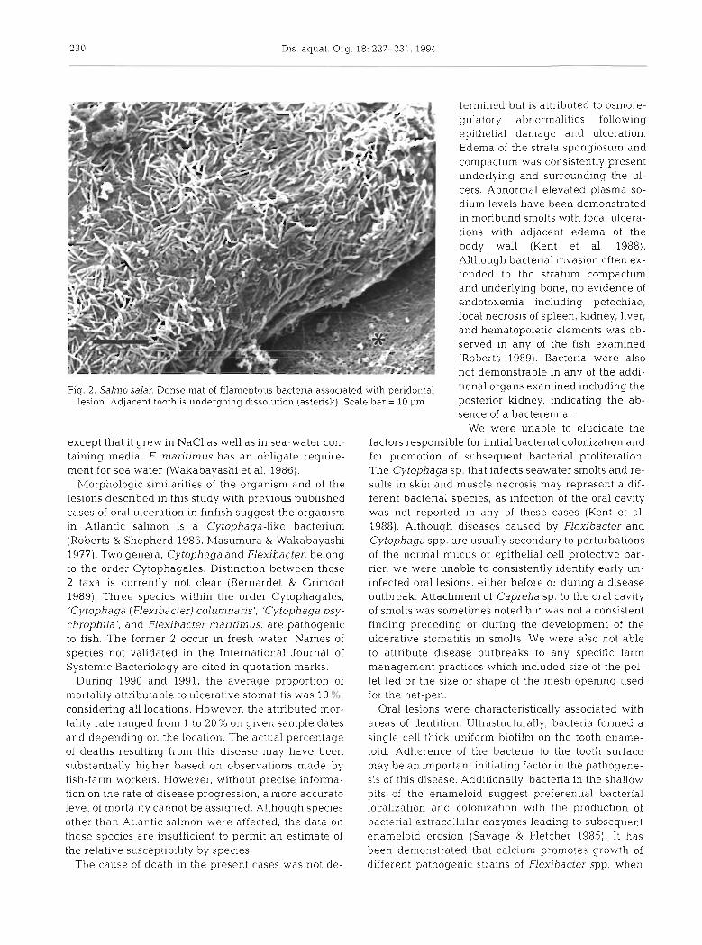
Ultrastructural findings: SEM of ulcerated areas affected fish demonstrated exposure of proximal tooth roots and periodontal cavities. Aggregates or mats of filamentous, bacterial rods were often present in the cellular debris at the gingival-enameloid interface and formed subgingival and corona1 plaques (Fig. 2). Variable numbers of individual bacterial rods often co- vered the proximal tooth surface forming a single cell thick, confluent biofilm. Many of these bacteria contai- ned multiple, elongated lateral fibrillar appendages anchoring the attached bacteria to the underlying tooth. Shallow pits of the tooth surface were often associated with these surface-adhering bacteria. Superficial aggregates of bacterial rods, distant from the ulcerated regions, were present overlying the intact epithelium. Interepithelial bacterial invasion was associated with bacterial aggregates.
Discussion. Examination of moribund or dead smolts with oral ulcers indicated mortalities were associated with large numbers of superficial and intralesional fila- mentous, thin, Gram-negative bacterial rods measu-
ring 2.5 to 13.0 pm in length. Morphologically similar oral lesions containing bacteria similar to those descri- bed in this report have been documented in marine cultured fish. Mass mortalities in red sea bream Pagrus major and black sea bream Acanthopayrus schlegeli cultured in sea cages in Japan were associated with oral and caudal fin erosions. The ulcerated lesions were pale yellow due to large numbers of filamentous, thin, 0.3 to 0.5 pm X 2 to 6 pm bacterial rods overlying the lesion surface. The name Flexibacter maritinius has been proposed for this bacterium (Wakabayashi et al. 1986). Severe mortality occurred in coho salmon Oncorhynchus kisutch smolts introduced to seawater net-pens in Maine (Sawyer 1976). Affected fish had se- vere erosion of the nose, mouth, and lower jaw. Yellow mats of gliding bacteria were observed in the mouth, pharynx, and pneumatic duct. The outbreak was ori- ginally referred to as a myxobacterial disease although biochemically the organism was similar to Flexibacter (Sawyer 1976). This organism was morphologically and physiologically similar to Flexibacter maritimus
230 Dis. aquat. Org. 18: 227-231, 1994
termined but is attributed to osmore- gulatory abnormalities following epithelial damage and ulceration. Edema of the strata spongiosum and compactum was consistently present underlying and surrounding the ul- cers. Abnormal elevated plasma so- dium levels have been demonstrated in moribund smolts with focal ulcera- tions with adjacent edema of the body wall (Kent et al. 1988). Although bacterial invasion often ex- tended to the stratum compactum and underlying bone, no evidence of endotoxemia including petechiae, focal necrosis of spleen, kidney, liver, and hematopoietic elements was ob- served in any of the fish examined (Roberts 1989). Bacteria were also not demonstrable in any of the addi-
Fig. 2. Salmo salar. Dense mat of filamentous bacteria associated with peridontal tional Organs examined the lesion. Adjacent tooth is undergoing dissolution (asterisk). Scale bar = 10 pm posterior kidney, indicating the ab-
sence of a bacteremia. We were unable to elucidate the
except that it grew in NaCl as well as in sea-water con- factors responsible for initial bacterial colonization and taining media. E maritimus has an obligate require- for promotion of subsequent bacterial proliferation. ment for sea water (Wakabayashi et al. 1986). The Cytophaga sp. that infects seawater smolts and re-
Morphologic similarities of the organism and of the sults in skin and muscle necrosis may represent a dif- lesions described in this study with previous published ferent bacterial species, as infection of the oral cavity cases of oral ulceration in finfish suggest the organism was not reported in any of these cases (Kent et al. in Atlantic salmon is a Cytophaga-like bacterium 1988). Although diseases caused by Flexibacter and (Roberts & Shepherd 1986, Masumura & Wakabayashi Cytophaga spp. are usually secondary to perturbations 1977). Two genera, Cytophaga and Flexibacter, belong of the normal mucus or epithelia1 cell protective bar- to the order Cytophagales. Distinction between these rier, we were unable to consistently identify early un- 2 taxa is currently not clear (Bernardet & Grimont infected oral lesions, either before or during a disease 1989). Three species within the order Cytophagales, outbreak. Attachment of Caprella sp. to the oral cavity 'Cytophaga (Flexibacter) columnans', 'Cytophaga psy- of smolts was sometimes noted but was not a consistent chrophila', and Flexibacter maritimus, are pathogenic finding preceding or during the development of the to fish. The former 2 occur in fresh water. Names of ulcerative stomatitis in smolts. We were also not able species not validated in the International Journal of to attribute disease outbreaks to any specific farm Systemic Bacteriology are cited in quotation marks. management practices which included size of the pel-
During 1990 and 1991, the average proportion of let fed or the size or shape of the mesh opening used mortality attributable to ulcerative stomatitis was 10 %, for the net-pen. considering all locations. However, the attributed mor- Oral lesions were characteristically associated with tality rate ranged from 1 to 20% on given sample dates areas of dentition. Ultrastucturally, bacteria formed a and depending on the location. The actual percentage single cell thick uniform biofilm on the tooth ename- of deaths resulting from this disease may have been loid. Adherence of the bacteria to the tooth surface substantially higher based on observations made by may be an important initiating factor in the pathogene- fish-farm workers. However, without precise informa- sis of this disease. Additionally, bacteria in the shallow tion on the rate of disease progression, a more accurate pits of the enameloid suggest preferential bacterial level of mortality cannot be assigned. Although species localization and colonization with the production of other than Atlantic sal.mon were affected, the data on bacterial extracellular enzymes leading to subsequent th.ese species are insufficient to permit a.n estimate of enameloid erosion (Savage & Fletcher 1985). It has the relative susceptibility by species. been demonstrated that calcium promotes growth of
The cause of death in the present cases was not de- different pathogenic strains of Flexibacter spp. when
Frelier et al.. Features of ulcerative stomatitis in Salmo salar 231
added to the culture medium (Hikida et al. 1979). The enameloid of teleost teeth is highly mineralized and could serve as a source of calcium to the bacteria (Shellis & Miles 1974). The mechanism of attachment to the enameloid surface may be similar to that of other bacteria associated with dental plaques and involve direct adsorption to enamel hydroxyapatite (Savage & Fletcher 1985). Once established on the tooth surface in a favorable microenvironment, these fllanientous bacteria could proliferate and invade the adjacent sub- gingiva and gingiva as occurs in man (Listgarten et al. 1975).
Our findings suggest the filamentous bacterium plays a major role in the pathogenesis of the disease and that the pathogenesis of ulcerative stomatitis in salmonid smolts may be similar to gingival diseases in man. Additional biochemical and molecular studies are in progress to identify the filamentous bacterium.
Acknowledgements. This work was supported by a Saltonstall-Kennedy grant NA26FD0448 from the National Marine Fisheries Service, U.S. Department of Commerce.
LITERATURE CITED
Bernardet, J . F., Campbell, A. C , Buswell, J. A (1990). Flex~bacter rnantimus is the agent of 'black patch necro- s ~ s ' In Dover sole in Scotland. Dis. aquat. Org. 8: 233-237
Bernardet, J. F., Gnmont, P. (1989). Deoxyribonucleic acid relatedness phenotypic characterization of Flexibacter columnaris sp. nov., nom. rev., Flexibacter psychrophilus sp nov., nom. rev., and Flexibacter rnantimus. Int J . Syst. Bacteriol. 39: 346-354
Dungan, C. F., Elston, R. A., Schiewe, M. H. (1989). Evidence for colonization and destruction of hinge ligaments in cul- tured juvenile Pacific oysters (Crassostrea gigas) by
Responsible Subject Editor: 7: Evelyn, Nanaimo, B.C.. Canada
Cytophaga-like bacteria. Appl. environ. Microbiol. 55: 1128-1135
Hikida, M., Wakabayashi, H., Egusa, S., Masumura, K. (1979). Flexibacter sp., a gliding bacterium pathogenic to some marine fishes in Japan Bull. Jap. Soc scient. Fish 45: 421-428
Humason, G. L. (1979). Animal tissue techniques. W. H. Freeman, San Francisco
Kent, M. L., Dungan, C. F., Elston, R. A., Holt, R. A. (1988). Cytophaya sp. (Cytophagales) intection in seawater pen- reared Atlantic salmon Salmo salar. DIS aquat. Org 4 : 173-179
Larkin, J. M. (1989). Order Cytophagales. In: Staley, J. T., Bryant. M. P., Fennig, N. (eds.) Bergey's manual of syste- matic bacteriology, Vol. 3. Williams & Wilkins, Baltimore, p. 2010-2050
Listgarten, M A., Mayo, H. E . , Tremblay, R. (1975). Development of dental plaque on epoxy resin crowns in man. J. Periodontol. 46: 10-26
Masumura, K., Wakabayashi, H. (1977). An outbreak of gliding bacterial disease in hatchery-born red sea bream (Pagrus major) and gilthead (Acanthopagrus schlegel~) fry in Hiroshima. Fish Pathol. 12: 171-177
Roberts, R. J., Shepherd, C. J. (1986). Handbook of trout and salmon diseases. Fishing News Books Ltd. Surrey
Savage, D. S., Fletcher, M. (1985). Bacterial adhesion: mecha- nisms and physiological significance. Plenum Press, New York
Sawyer, E. S. (1976). An outbreak of myxobacterial disease In coho salmon (Oncorhynchus kisutch) reared in a Maine estuary. J . Wildl. Dis. 12: 575-578
Sheehan, D. C., Hrapchak, B. B. (1980). Theory and practice of histotechnology. C. V. Mosby Co., St Louis
Shellis, R. P., Miles, A. E . (1974). Autoradiographic study of the formation of enameloid and dentine matrices in teleost fishes using tritiated amino acids. Proc. R. Soc. Lond. B 185: 51-72
Wakabayashi, H., Hikida, M., Masumura, K. (1984). Flexi- bacter infection in cultured marine fish in Japan. Helgolander Meeresunters 37: 587-593
Wakabayashi, H., Hikida, M.. Masumura, K. (1986). Flexi- bactermaritimussp. nov., a pathogen of marine fishes. Int. J. Syst. Bacteriol. 36: 396-398
Manuscript first received: December 4, 1992 Revised version accepted: Novem ber 2, 1993