Embed Size (px)
Citation preview

Journal of Clinical InvestigationVol. 41, No. 4, 1962
ORIGIN OF LIPID AND CHOLESTEROLIN EXPERIMENTALTHROMBOATHEROSCLEROSIS*
By MEYERFRIEDMAN, SANFORD0. BYERSAND SHIRLEY ST. GEORGEWITHTHE TECHNICAL ASSISTANCE OF CLARENCEOMOTO,WARRENHAYASHI
AND BETTY WANG
(From the Harold Brunn Institute, Mount Zion Hospital and Medical Center,San Francisco, Calif.)
(Submitted for publication August 30, 1961; accepted December 14, 1961)
Recently we have described (1) the sequenceof phenomena that followed the experimentalplacement of a thrombus in the aorta of a hyper-cholesterolemic rabbit. It was found that thethrombus invariably elicited a hyperplastic intimalresponse leading eventually to a plaque (see ColorFigure 1) which in its lipid and tissue character-istics bore a striking resemblance to the humanatherosclerotic plaque. Moreover, necrosis, lique-faction, and even calcification of the central por-tions of the plaque were commonly observed.
The sudanophilia and also the excess accumula-tion of cholesterol occurring in these plaques firstbecame detectable about 14 days after the induc-tion of the thrombus itself. Although hundredsof sections of these plaques were examined be-fore, during, and shortly after this 14-day period,the first accumulation of the lipid never wasobserved in the superficially located intimal tissuewhich was in closest contact with the luminalblood (i.e., the intimal tissue encircling theperiphery of the thrombus). Invariably suchlipid made its first appearance in the deeper areasof the new intimal tissue immediately adjacentto the underlying media. Indeed, not infre-quently, the first appearance of lipid was detectedin the inner portion of the media underlying thenewly formed plaque. These observations clearlysuggested to us that the excess lipid (and pre-sumably its cholesterol congener) was not derivedfrom luminal blood. Its accumulation, therefore,appeared to be due to its transport by bloodvessels newly derived from the adventitia or toits excess production in situ in the newly forming
*Aided by Grants from The Life Insurance MedicalResearch Fund, The National Institutes of Health(H-119), San Francisco Heart Association, SacramentoCounty Heart Association, Contra Costa County HeartAssociation, and Humboldt County Heart Association.
intimal tissue. Which of these two possible phe-nomena was responsible for the accumulation wasnot determined in our first study.
However, because of the theoretical as well aspractical importance of discovering the manner inwhich lipid and cholesterol accumulated in con-tinually increasing excess in this new intimaltissue, a second study was initiated to determinethis point. The results obtained by a variety oftechniques left little doubt that the excess accumu-lation of lipid and cholesterol was due primarilyto an excess escape of these substances from thenewly formed intimal blood vessels because ofthe latter's increased permeability and frequentrupture. Subsequent retention of the lipid andcholesterol moieties, which had infiltrated, ap-peared to be due to cellular sequestration by hy-perplastic intimal tissue.
METHODS
A. General histological study of the early developingplaqueIn order to verify the earlier finding of initial sudano-
philia, beginning in the basal areas of the newly develop-ing intimal tissue, and also to determine if excess muco-polysaccharide and hemorrhages were present, thrombiwere induced in the aortas of 18 rabbits by the introduc-tion of zinc chloride-treated magnesium alloy spirals (2).Half of these rabbits were given Wayne rabbit chow dietplus added cholesterol (2 per cent) and cottonseed oil(2 per cent), and the remaining half, only the chow diet.Five of the rabbits on each of the two diets were sac-rificed at the end of 18 days, and the remaining rabbits,at the end of 28 days. Blood samples obtained at thetime of insertion of the coil and again at time of sacrificewere analyzed for cholesterol content (3).
Three sections were obtained from each developingplaque and the portion of artery wall subjacent and im-mediately adjacent to it. These were stained with SudanIV, hematoxylin and eosin, and the Rinehart stain formucopolysaccharide (4), respectively.
828

LIPID AND CHOLESTEROLIN EXPERIMENTALTHROMBOATHEROSCLEROSIS
A plaque developing in a normocholesterolemic rabbit,hence exhibiting no sudanophilia (1), will be describedherein as a thromnbosclerotic plaque. A similar plaquedeveloping in a hypercholesterolemic rabbit, hence con-taining excess lipid and cholesterol (1), will be referredto as a tlzromtboatherosclerotic plaque.
B. Study of hyperplastic intimal tissue under avascularconditions
In order to investigate the relevance of the newlyformed blood vessels to the sudanophilia or lipid accumu-lation observed in the basal portions of the early thrombo-atherosclerotic plaque, this tissue was studied in a milieudevoid of blood vessels.
Cylinders were fabricated by us of Millipore 1 (filtersheet having a pore size of 3.0 A) by approximating thetwo ends of a flat section of the material (2.5 X 2.0 cm)and then sealing this juncture and one of the two openends with an "Epoxy" glue.2 At this time also, a shortlength of silk thread was incorporated in the "Epoxy"seal. Such capsules were capable of holding approxi-mately 0.2 to 0.3 ml of fluid.
Four normocholesterolemic rabbits, into whose ab-dominal aorta a thrombus-inducing spiral had been in-troduced 21 days previously, were killed. Under asepticconditions, the thrombosclerotic plaques were removed,pooled, and then finely minced with a razor blade. Aftersamples of this material had been taken for chemical andhistological analysis, part of the remainder was insertedinto each of 12 Millipore capsules. The open ends of thecapsules were closed with the Epoxy and the capsuleswere partially immersed in warm Tyrode's solution forseveral hours as the Epoxy seal hardened (these aredesignated as "intact" in Table II). For control pur-poses, the Millipore portions of 5 of the capsules weregently pierced in 8 to 10 separate areas with a 25 gaugehypodermic needle (these are designated as "open" inTable II).
One normocholesterolemic and two hypercholesterolemicrabbits, after initial blood samples had been obtained,were operated upon, and 12 intact and open capsules con-taining the minced intimal tissue were inserted and se-cured by suture to the posterior wall of the peritonealcavity. The number and type of capsules inserted intoeach rabbit are given in Table II. In addition, 1 intactMillipore capsule, containing only Tyrode's solution, wasplaced in the abdominal cavity of each hypercholes-terolemic rabbit.
At the end of 8 days, these animals were bled againand then were reoperated. A sample of peritoneal fluidwas obtained from the two hypercholesterolemic rabbitsand then the capsules were removed from all of the rab-bits. Tissue for histological study was obtained from all12 capsules and there was sufficient material available in9 of the capsules for cholesterol analysis (5). Also,
1 WSWPFilters, Nylon netting, white; Millipore Fil-ter Corp., Bedford, Mass.
2 Devcon "2-Ton" Epoxy; Devcon Corp., Danvers,Mass.
fluid was aspirated from each of the 2 capsules initiallycontaining only Tyrode's solution and this fluid was ana-lyzed for cholesterol content. Finally, the Millipore wallsof two of the closed (unpunctured or intact) capsulesalso were analyzed for cholesterol content.
C. Studies of the synthesis and deposition of cholesterolin the early-developing plaque
In order to determine the mechanism responsible forthe excess accumulation of cholesterol previously ob-served (1) in the developing thromboatheroscleroticplaque of the hypercholesterolemic rabbit, studies employ-ing acetate-l-C1' and cholesterol-4-C" were done.
1. Acetate-i-C' studies. Aortic thrombi were inducedby insertion of the spiral in two normal rabbits. Theserabbits ingested only Wayne rabbit chow diet. The ani-mals were anesthetized with pentobarbital (Nembutal)21 and 28 days, respectively, after insertion of the spiral,and each abdominal aorta was excised and placed immedi-ately in a small volume of ice cold 0.1 M potassiumphosphate buffer, pH 7.4. Potassium rather than sodiumwas used for the buffer to offset the tendency of excisedaortic tissue to lose excessive potassium (because of itslow endogenous metabolism), hence a lag in its synthesisof cholesterol (6). The tissues were kept cold from thattime until put into Warburg flasks.
The adventitia was rapidly removed before cuttingopen the aorta longitudinally to expose the plaque con-sisting of residual thrombus and hyperplastic intimal tis-sue. This plaque together with the minimal proximalarea of uninvolved aorta was used for study. The sampleswere approximately 9 mmin length and 3 mmin width.The thrombointimal portion of the thromboaortic sampleswas approximately twice the thickness of the media.
Small sections were taken for histological study, thenthe samples were sliced "free-hand" to a thickness lessthan 0.5 mm, and incubated at 370 C in Warburg flasks(in duplicate) for 130 to 150 minutes during the periodof steady oxygen uptake. All flasks contained in additionto tissue: nicotinamide 0.3 M in 0.011 M sodium EDTA,0.3 ml; 0.16 Mmagnesium sulfate, 0.1 ml; 0.02 Msodiumacetate-i-C14, 0.2 ml (2 uc); 0.1 M potassium phosphatebuffer, pH 7.1, 2.2 ml; and 0.2 ml 20 per cent potassiumhydroxide in the center well. Respiration was terminatedby the addition of 2.8 ml 50 per cent KOHfor digestionof the tissue. Fifty per cent KOHwas added to threeflasks prior to incubation to serve as a control on pos-sible tracer contamination of the isolated cholesterol digi-tonide. The activity of all three samples was identicalwith background.
Aliquots for nitrogen analysis were taken from eachdigested sample. Cholesterol was extracted from the di-gest by partitioning with ethanol: ether (1: 4), then pre-cipitated with digitonin, washed, and finally the choles-terol digitonide dissolved in glacial acetic acid. An ali-quot was removed for micro-scale cholesterol analysis(7) and the remainder dried in planchets to yield cho-lesterol digitonides of infinite thinness. Counting wasdone using a Tracerlab Mylar end-window TG C' tube.
829

MEYERFRIEDMAN, SANFORD0. BYERSAND SHIRLEY ST. GEORGE
TABLE I
Accumulation of lipid and mucopolysaccharide, and occurrence of hemorrhage in developingthromboatherosclerotic and thromboscierotic plaques (18 and 28 days)
No. with lipid (sudano- No. with excess mucopoly-philia) in: saccharide in: No. with hemorrhage in:
Intimal Intimal IntimalNo. of tissue tissue tissuerabbits Av.
with serum Age of Super- Adven- Super- Adven- Super- Adven-plaque cholest.* plaques Basal ficial Media titia Basal ficial Media titia Basal ficial Media titia
mg/100 ml daysA. Hypercholesterolemic rabbits
5 391 18 5 0 3 2 5 2 2 0 4 0 2 24 985 28 4 0 2 1 4 2 2 0 2 0 1 1
B. Normocholesterolemic rabbits5 38 18 0 0 0 0 5 2 2 0 0 0 0 04 28 28 0 0 0 0 4 2 2 0 1 0 0 0
* Average serum cholesterol calculated as the mean of serum cholesterol values at beginning and end of experiment.
Counts per minute were corrected for a background valueof 11 cpm (11.0 to 11.2 cpm). All samples were countedto a minimum 95 per cent confidence level (p < 0.05) or400 counts.
2. Cholesterol-4-C"4 studies. Aortic thrombi were in-duced in four rabbits, then 28 days later, each was given5 ,uc of cholesterol-4-C1' (dissolved in 2 ml of olive oil)by gastric intubation. The rabbits subsequently weregiven a diet containing 2 per cent cottonseed oil for 72hours to facilitate the absorption of the administered ra-dioactive cholesterol. They then were fed the ordinarylaboratory rabbit chow.
These four rabbits came to autopsy at 8, 22, 38, and66 days after the intubation. The plaque area of eachrabbit was stripped from the subjacent aorta and a sampleof nearby, but uninvolved, aorta also was obtained. Thethrombointimal portion of the thromboaortic samples wasapproximately the same width as that of the media. Di-gestion of the samples, nitrogen analysis, and cholesterolextraction and counting procedures were done as describedabove.
D. Studies of the permeability of newly formed bloodvessels int hyperplastic intimal tissue
The observed early accumulation of lipid in the basalportions of the intimal tissue growing in response tothrombus induction suggested the possibility that thenewly formed capillaries attending this intimal growthwere unduly permeable to the lipoprotein aggregates pres-ent in plasma. In order to determine the existence of ex-cessive permeability, the following studies were done.
Thrombi were induced in 87 normal rabbits subsequentlymaintained on ordinary rabbit chow. Then, at 14, 21, 42,and 84 days after such induction, some of the animalswere injected intravenously either with solutions ofEvans blue, Trypan blue, or with suspensions of Thoro-trast or fine iron particles derived from decompositionof iron carbonyl.3
3 Carbonyl iron powder (average diameter, 3 As); A. D.Mackay, Inc., New York, N. Y.
Thirty-seven such rabbits received injections of Evansblue (10 mg per day for 3 days) either at 14, 21, 42, or84 days after induction of the thrombus. Five of therabbits were injected with a single dose of Trypan blue(15 mg) 21 days after thrombus induction. Two of therabbits received a single injection of Thorotrast (2.5 ml)21 days after thrombus formation. The remaining 43rabbits were injected with a suspension containing 0.3 gof carbonyl iron particles (approximate diameter, 3 is)twice daily for 3 days beginning either at 14, 21, 42, or 84days after thrombus induction.
All rabbits were sacrificed 24 hours after the last in-jection and the aorta and the thrombosclerotic plaque in-spected grossly for differences in dye or particle accumu-lation. In addition, the plaques and samples of uninvolvedaorta of the rabbits receiving iron or Thorotrast weresectioned and stained with Berlin blue and Sudan, re-spectively.
RESULTS
A. Observations concerning some of the phe-nomena involved in the early-developing plaque.As was noted before (1), the early plaques ofall nine hypercholesterolemic rabbits, whetherthey were examined 18 or 28 days after thrombusinduction, exhibited sudanophilia in the basallayer of the newly developing hyperplastic intimaltissue. The stained lipid appeared to be depositedboth intra- and extracellularly. In addition(Table I and Color Figure 2), five of these rab-bits exhibited sudanophilic infiltration of the innerthird of the media underlying the plaque. In-deed, the internal elastic membrane as early as18 days after thrombus induction invariably ex-hibited splitting (Figure 1) or fragmentation.The adventitial area also was not spared, in thatthree of the nine hypercholesterolemic rabbits
830

LIPID AND CHOLESTEROLIN EXPERIMENTALTHROMBOATHEROSCLEROSIS
*A'i: - X . * S : .... Law.;.... w s>~~~~~~~~~~~~~~~~~~~~~~~~~~~~~~~~~~~~~~~~~~~~~~~~~......0-
A~~~~~~~~~~~~~~
* . .: .. X .....~~~~~~~~~~~~~~~~~~~~~~......... .....
intimal*tissue andmedi isshw.Tesltigo apat ofth ineral elsi mebrn
can~eail be dicene. Th dar patils contie wihi this reulcto.r mase o.+u8^fi;._
* * ~~~~~~~~~~~~~~~~~~~~~~~~~~~~~~~o 5.P;
p * it' ; ^ - .:os .. .... ,~~~~~~~~~~~~~~~~~~~~~~~~~~~~~~~~~~~~~~~~~~..4
lIpiGakn the SudanT stainNeeoten the diffusely scateredi exraasatedf redbloofd celles inthe
intirnal tissue, indicating a previous hemorrhage.
exhibited a similar but somewhat less sudano-philic infiltration of the adventitia opposite theplaque. None of these nine plaques, however,exhibited any sudanophilia in the superficial in-timal areas adjacent to luminal blood.
On the other hand, no significant sudanophiliawas observed in any portion of the plaque oraortic wall of the nine normocholesterolemic rab-bits, either in those sacrificed at 18 or 28 daysafter thrombus induction. However, similarsplitting and fragmentation of the internal elasticmembrane were observed.
The plaques of all rabbits, both normo- andhypercholesterolemic, exhibited excess mucopoly-saccharide in the basal layer of the new intimaltissue (Table I and Color Figure 3). The innermedia, but not the adventitia, of most of theserabbits likewise showed some excess of mucopoly-saccharide. It should be pointed out that suchexcess material usually was found in the sameareas of the plaque which exhibited sudanophilia.
Usually, too, it was in this same basal areaof new intimal tissue that new blood vessels could
be observed most frequently. Most of these ves-sels (Figure 2) appeared to be larger in sizethan the ordinary capillary, but they lacked thevarious cellular layers of the arteriole. Fre-quently, also, many vessels of this type wereconjoined to form a labyrinth of sinusoidal chan-nels. Such capillaries always were found to enterthe base of the plaque via an area of disorganizedmedial tissue. In no section, however, was thereevidence of the luminal origin of any capillary.
Hemorrhage (as evidenced by large accumula-tions of erythrocytes lying free in the tissue) oc-curred (Table I) in the plaques of six of the ninehypercholesterolemic rabbits, but in only one ofthe nine normocholesterolemic rabbits. Suchhemorrhages were observed early and once againusually were found chiefly in the same basal areaof the new intimal tissue (Color Figure 4) or inthe inner, disorganized third of the media wheresudanophilia and dense mucopolysaccharide stain-ing also were observed. However, hemorrhage(as evidenced by pools of erythrocytes) in the ad-ventitia immediately beneath the media (Figure
831

MEYERFRIEDMAN, SANFORD0. BYERSAND SHIRLEY ST. GEORGE
COLORFIG. 1. RABBIT RR-19. Fully developed throm-boatherosclerotic plaque, 84 days after insertion of throm-bus-inducing coil in a cholesterol-fed rabbit (Sudan IVstain, X25). The sudanophilia involving the basal andlateral areas of the plaque is clearly shown. Note toothe slight sudanophilic infiltration of the adventitia un-derlying the plaque. Medial atrophy below the plaque isquite striking. The similarity of this plaque to thatfound in the diseased human coronary artery already hasbeen pointed out (1).
COLORFIG. 2. RABBIT 0-2. Developing thromboathero-sclerotic plaque of a rabbit fed cholesterol-oil, 28 days af-ter intraaortic coil insertion (Sudan IV stain, X 100).The sudanophilia involving the basal intimal tissue, por-tions of the inner third of media, and parts of the sub-jacent adventitia is clearly depicted. Metallic salt frag-ments can still be seen. Between and over such areas,newly growing intimal tissue almost devoid of sudano-philia can be observed. The transmedial source of theSudan-stained lipid can be adduced from this photograph.
COLORFIG. 3. RABBIT 0-5. Developing thromboathero-sclerotic plaque in a rabbit fed cholesterol-oil, 28 daysafter insertion of coil [Rinehart and Abul-Haj (4) stain,X25]. The excess mucopolysaccharide (staining blue)can be seen to occupy chiefly the same basal area of newintimal tissue that the Sudan-staining lipid occupies inFigure 2. The superficial intimal tissue covering theoriginal thrombus (it has been torn from the base on theleft) also exhibits some excess mucopolysaccharide. Theadjacent uninvolved aorta is free of this blue stain.
COLORFIG. 4. RABBIT 0-3. Portion of a developing 28day old thromboatherosclerotic plaque (Sudan IV stain,X400). The area illustrated is a basal portion of thenewly developed intimal tissue lying upon a partiallydisintegrated inner media. The hemorrhage indicated bythe extravasated red blood cells (pale cells with heavy,circular outlines) is precisely in the area where Sudan-staining material abounds. Note the presence of bothred blood cells and sudanophilia in the media also.
COLORFIG. 5. RABBITS W2-W5. Sample of the pooledtissues obtained from 21 day old thrombosclerotic plaquesof four normocholesterolemic rabbits that later was placedin Millipore capsules (Sudan IV stain, X160). Thespindle-shaped cells with elongated nuclei and the com-plete absence of sudanophilia can easily be seen.
COLORFIG. 6. RABBIT 24. Portion of the pooled tis-sues shown in Color Figure 5 after 8 days in an "intact"Millipore capsule (Sudan IV stain, X160). The tissueresembles quite closely that shown in Color Figure 5. Nocapillaries or sudanophilia can be detected.
COLORFIG. 7. RABBIT 24. Portion of the pooled tis-sues shown in Color Figure 5 after 8 days in an "open"Millipore capsule (Sudan IV stain, X 160). Note theintense, intracellularly situated sudanophila and the pres-ence of capillaries. These findings contrast stronglywith those observed in the same tissue kept in the "intact"capsule (Color Figure 6).
COLORFIG. 8. RABBIT 42. Photograph of the openedabdominal aorta and a 14 day old thrombosclerotic plaqueof a normocholesterolemic rabbit previously injected withEvans blue dye. The thrombus exhibiting a large resi-due of metallic fragments has been detached from theaorta revealing, at this latter site, the presence of severalareas of extravasated Evans blue dye (blue).
COLOR FIG. 9. RABBIT 16. Developing 21 day oldthrombosclerotic plaque of a normocholesterolemic rabbitpreviously injected with colloidal iron (Berlin blue stain,X65). The blue-staining iron deposits can be observedin the basal and lateral areas of the intimal tissue abuttingupon the media.
COLORFIG. 10. RABBIT 36. Mature 84 day old throm-bosclerotic plaque of a normocholesterolemic rabbit previ-ously injected with colloidal iron (Berlin blue stain, X65).Unlike the deposit observed in the 21 day old plaque(Color Figure 9), no iron can be detected anywhere inthis older plaque.
832

> ~~~56
Jv-girl" r' 'L rwi
2 -lie
:2 4~~~~~~~~~~~~~~~~~~~~4
It4Y4~~~~~~~~~~~~~~~~
X,
, ate~~~~~~~~~~~~~~~~~~~~~~~~~~~~~~~~~~~~~~~~~~~~~~~~~~~~~~~~~~~~~~~~~X
.;~~~~~~~~~~~~~~~~~~~~~~~~~~~~~~~~~~~~~~~~~~~~~~~~~~~~~~~.
9t f
XjY£.TS7t_ 104 r.S'.isk:t4Kn X'4e s _; i )sl SE /
4K 9 :~~
S__!sED ^ ;t~~~~~~~~~~~~~~~~~~~~~~~~~~~~~~~~~~~~~~~~~~s
.i- t - - - 't

LIPID AND CHOLESTEROLIN EXPERIMENTALTHROMBOATHEROSCLEROSISi=., . . ^- *A,~~~'
fa
*
S.
g:B
* .
A .... ....
_s; - ..' s^o: a t .'. F
.:0::::-: ..j_ . .>ZA .e-m :.I'½_.
FIG. 2. RABBIT 0-4. Another plaque similar in type and age to that shown in Figure 1
(H & E stain, X400). Two vessels can be seen in the basal intimal tissue exhibiting a wide lu-men yet possessing capillary-like walls. Note the internal elastic membrane and its beginningfragmentation.
FIG. 3. RABBIT 0-4. Portion of adventitia and outer part of media subjacent to the plaqueillustrated in Figure 2 (Sudan IV stain). Here a large hemorrhage (indicated by the masses
of extravasated red blood cells) has occurred in the adventitia (below) immediately external to
the media (above).
833
4t
.ALA..

834~~MEYERFRIEDMAN, SANFORD0. BYERSAND SHIRLEY ST. GEORGE
3) occasionally was observed near the plaquesborne by the hypercholesterolemic rabbit. ,,~
B. Observations concerning the lipid and cho- _1 11 00
lesterol accumulation occurring in avascular hy-perplastic intimal tissue. The hyperplastic intimaltissue obtained from plaques borne by normo- -cholesterolemic rabbits and transferred to "in- 0
tact" Millipore capsules (which then were in- M
serted in the peritoneal cavity of each of twohyper- and one normocholesterolemic rabbits)survived during the 8-day period in every instancebut one. The same tissue transferred to the o~
open" capsules and inserted also into the peni- V
toneal cavity of the same three rabbits survived 0 0
in every instance.Observation at autopsy revealed that all of the
capsules were covered with highly vascular con-nective tissue. Moreover, this latter tissue was .observed to have penetrated each of the "open"Ccapsules through the openings that had been made.The "intact" capsules, however, had not been 0
penetrated from the outside. When the capsuleswere opened, the tissues contained in "open"capsules exhibited a rich vascularity as observed t
by means of the dissecting microscope. The tis-sues in the "intact" capsules, however, appeared -to be totally devoid of vascularity.
A second marked difference was observed be- - I
tween the tissue contained in the "intact" capsules 4.Ae 4, 0~~~~~~
and that in the "open"~ capsules. The tissues ob- ,tamned from. the four "intact" capsules inserted .into the hypercholesterolemic rabbits histologicallyresembled without exception (see and compare -Color Figure 6 with Color Figure 5), the initial
0H4proliferating intimal tissue. Both types of tissue .consisted predominantly of spindle-shaped cells and -mboth appeared totally devoid of sudanophilia. It 6 O.! ~. towas of interest, however, that the outer half of theCZQMillipore material 'which had been in contact with 0
hypechoestrolmic peritoneal fluid, itself stainedO.red with the Sudan stain. No capillaries couldbe detected on microscopic examination in thesamples of intimal tissue that had been in the4"intact" Millipore capsules for the 8-day period. *dThe sampe ointimal tissue contained in the 00C
"intact" capsules placed in the normocholestero- u2Elemic rabbit appeared identical with those described E
in the "intact" capsules of the hypercholestero- C1 1 *11 +
lemic animals.
834

LIPID AND CHOLESTEROLIN EXPERIMENTALTHROMBOATHEROSCLEROSIS
~4 c-l,
U) -
-. U-) 1-4 --(- vv v_ 4 \0 %o00 00_- _4 O
0o o 0 ')
eN etm4C
00 00
U)
-4in -I_.4IP
00 0o d' t_4 _-
-o0%4CNf % _--4 _ Cen m _
U 0o
tTo
1~ -0 ll
114 C')0 0
tko r-1-
00 U) U) Ul)r- U) U) \0N0 0% %0 %0
m' Ild NO %00-. NO C' 00
Io0% to0%C') C4 -% -4
0 (.- -
("'4 0-- 0' -'-. 0 ) 00
U) U) 0 (6666.
0%K (.-q -490t'0ot 00 0
0~~~~~0
0- m00
Cd 4)'0
t
b4U) ('-9malU
0) -V
c;
¢ U_
(-.9
C')
0-4
00%
t--
6
10-
0--
Ul)
00
oiN en 'o
o00 \000) 0
c to
-. Ul)0-9¢1: x- l)t-t
On the other hand, the three tissue samplesobtained from the "open" capsules inserted in thesame two hypercholesterolemic rabbits uniformlyshowed on microscopic examination (see ColorFigure 7) a rich supply of capillaries but nohemorrhages and an intense sudanophilia. Thestained lipid appeared to be exclusively intra-cellular. The tissues obtained from the "open"capsules inserted into the normocholesterolemicrabbit also exhibited the same rich vascularity. butno hemorrhages and no sudanophilia.
Analyses of the fluid remaining in the two "in-tact" capsules initially filled with Tyrode's solu-tion (Table II), however, indicated that con-siderable cholesterol, ostensibly from the peri-toneal fluid or from the blood supply of the newconnective tissue covering the outside of the cap-
4 sule, had been able to enter the capsules. More-o over, the Millipore material itself was found
to have adsorbed considerable cholesterol (Table:o II).0CO0
Cl-o
._
00)
la0
COla00
"._
e)
9..
00
00
0-
U'
._200)
0U)
00
* 4.
Cholesterol analyses of some of these capsulartissues agreed in general with the histological re-sults. Whereas the average cholesterol contentof two tissue samples placed in "intact" capsulescarried by a hypercholesterolemic rabbit was2,905 mg per 100 g (Table II), the averagecholesterol content of three similar tissues in"open" capsules carried by hypercholesterolemicrabbits was 7,810 mg per 100 g. It was ofinterest that the tissues contained in the "intact"capsules carried by the hypercholesterolemic rab-bit did not exhibit a much higher cholesterol thanthat exhibited by tissues contained in the "open"capsules placed in the normocholesterolemic rab-bit. Nor did such isolated tissues in the hyper-cholesterolemic rabbits exhibit a dramatic in-crease in cholesterol as compared to that present(2,140 mg per 100 g) in the intimal tissue asinitially placed in the capsule.
These results appeared to us to indicate thatthe sudanophilia and the intense cholesterol ac-cumulation noted in the early-developing plaquewere primarily dependent upon the presence ofthe new vascular supply of this same plaque; forin the absence of such a new vascular supply,both phenomena failed to occur.
C. Cholesterol synthesis and deposition in thedeveloping plaque. Table III documents the
zo
835
z <
a a5
0
. aw !!
COO'-40d0g4=
C:
"0
0
CO. s
0CO
p0
CID
-
- 9-
¢4
CttH s
CO
0
C-) C
0
Cd
of
cd a
To0 4!
"-CO
0C ¢
: Q- .0
bo- T:
I

MEYERFRIEDMAN, SANFORD0. BYERSAND SHIRLEY ST. GEORGE
TABLE IV
Extravasation of dyes in developingthrombosclerotic plaques
No. of plaquesexhibiting gross
dye extravasationNo. of
rabbits Super-with Plaque Basal ficial
plaque age area area
daysA. After injection of Evans blue
6 14 6 214 21 14 212 42 2 0
5 84 0 0
B. After injection of Trypan blue5 21 5 0
fact that the living tissue (i.e., the hyperplasticintimal tissue) of the early-developing plaquewas capable of incorporating acetate-i-C14 intocholesterol. Moreover, it also is evident from thedata in this table that this hyperplastic tissueappeared to incorporate as much radioactive ace-
tate into cholesterol as the control, uninvolvedsection of aorta, although the rate of incorpora-tion was quite slow in both types of tissue. Atfirst glance, the much higher specific activity ofthe cholesterol isolated from the normal aortasuggests that this tissue is capable of incorporat-ing more acetate-1-C14 into cholesterol than is theplaque tissue. However, the plethora of choles-terol already present in the plaques (1) prior tothe study would tend to dilute the newly formedradioactive cholesterol as compared with thatformed in the normal aorta. A comparison, there-fore, of the respective specific activities of thecholesterol finally isolated would not reveal the re-
spective degrees of acetate incorporation. How-ever, when the counts per minute (cpm) of thetotal cholesterol content are correlated with thenitrogen content of both tissues, it can be seen
(Table III) that the ratios are essentially thesame. In short, these results strongly suggestedthat the newly formed intimal tissue was synthe-sizing neither more nor less cholesterol than thenormal aorta. The excess cholesterol observedaccumulating in these plaques, therefore, did notappear to be due to an increased cholesterol syn-
thesis in situ.The results obtained from the administration of
cholesterol-4-C14 to plaque-bearing animals, how-
ever, strongly suggested (Table III) that the tis-sue making up these plaques differed markedlyfrom normal aortic tissue in its ability to extractor retain an excess of cholesterol from the blood,or both. Thus, at whatever age the plaque-bearinganimals were sacrificed, although the cholesterolmoiety of the normal aorta exhibited very minimalradioactivity per milligram of tissue nitrogen, thatof the plaques exhibited significant activity (atleast ten times as much as that of aortic choles-terol). It also was of interest that the specific ac-tivity of the cholesterol contained in the plaqueswas the same as or slightly higher than that of thenormal aorta (Table III), despite the dilutingeffect of the relatively enormous amount of non-radioactive cholesterol that had accumulated be-fore, and continued to accumulate after, the ad-ministration of the radioactive cholesterol. Theseresults, therefore, when considered with thoseobtained from the acetate-i-C14 experiments,strongly suggested that the excess cholesterolfound in the thromboatherosclerotic plaque wasbeing obtained primarily and chiefly from theblood supplying this tissue.
D. Permeability of the newly formed bloodvessels in hyperplastic intimal tissue. The studiesdescribed above left little doubt that the excesslipid and cholesterol accumulating in the thrombo-atherosclerotic plaque were not formed in situ,but were being brought to the area by its newsupply of blood vessels, arriving by way of theadventitia. It seemed probable to us that thesesame vessels, at least in the earlier stages ofplaque development, were abnormally permeableto large size molecules such as lipoproteins. Ifsuch increased permeability were present, itshould be possible to demonstrate it by othertechniques.
The results obtained with the dyes, Evans blueand Trypan blue, and with the iron and Thoro-trast suspensions made it clear indeed that thesevessels were excessively permeable. After theinjection of Evans blue and sacrifice of plaque-bearing rabbits, it was observed grossly (TableIV, A) that those plaques which were 14 and21 days old invariably exhibited a considerableextravasation and condensation of the dye at theirbase; i.e., at the point where they were attachedto the aorta (Color Figure 8). This area, ofcourse, is precisely that region where lipid and
836
LIPID AND CHOLESTEROLIN EXPERIMENTALTHROMBOATHEROSCLEROSIS
cholesterol were observed to be initially and later,maximally deposited. It is also the area whereinthe majority of blood vessels was first observed.On the other hand, the peripheral intimal tissuecovering the initial thrombus infrequently ex-hibited extravasated dye (Table IV, A). Also,no extravasation of the dye was ever detected inthe intima of the nearby normal uninvolved aorta.The 21 day old plaques of the five rabbits in-jected with Trypan blue similarly revealed (TableIV, B1) extravasation and condensation of thedye at the base of the thrombosclerotic plaque.Similar extravasation of dyes in the adjacent un-involved aorta was never seen.
However, 42 day old thrombosclerotic plaquesinfrequently exhibited, and 84 day old plaquesnever exhibited (see Table IV, A) the basalextravasation of Evans blue dye after its ante-mortem injection. Apparently the increased per-meability of these intimal vessels was a phenome-non limited to the first few weeks or months oftheir development.
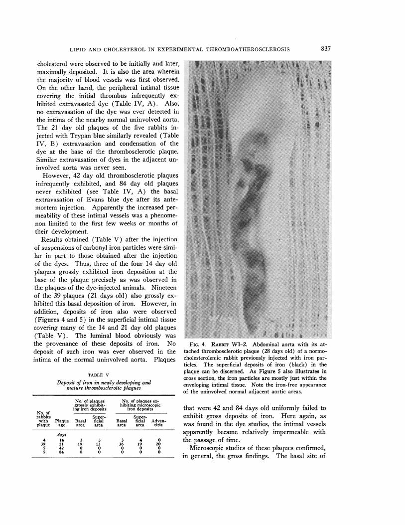
Results obtained (Table V) after the injectionof suspensions of carbonyl iron particles were simi-lar in part to those obtained after the injectionof the dyes. Thus, three of the four 14 day oldplaques grossly exhibited iron deposition at thebase of the plaque precisely as was observed inthe plaques of the dye-injected animals. Nineteenof the 39 plaques (21 days old) also grossly ex-hibited this basal deposition of iron. However, inaddition, deposits of iron also were observed(Figures 4 and 5) in the superficial intimal tissuecovering many of the 14 and 21 day old plaques(Table V). The luminal blood obviously wasthe provenance of these deposits of iron. Nodeposit of such iron was ever observed in theintima of the normal uninvolved aorta. Plaques
TABLE V
Deposit of iron in newly developing andmature thromboscierotic plaques
No. of plaques No. of plaques ex-grossly exhibit- hibiting microscopic
ing iron deposits iron depositsNo. ofrabbits Super- Super-
with Plaque Basal ficial Basal ficial Adven-plaque age area area area area titia
days4 14 3 3 3 4 0
39 21 19 13 36 19 205 42 0 0 0 0 05 84 0 0 0 0 0
FIG. 4. RABBIT WI 2. Abdominal aorta with its at-tached thrombosclerotic plaque (28 days old) of a normo-cholesterolemic rabbit previously injected with iron par-ticles. The superficial deposits of iron (black) in theplaque can be discerned. As Figure 5 also illustrates incross section, the iron particles are mostly just within theenveloping intimal tissue. Note the iron-free appearanceof the uninvolved normal adjacent aortic areas.
that were 42 and 84 days old uniformly failed toexhibit gross deposits of iron. Here again, aswas found in the dye studies, the intimal vesselsapparently became relatively impermeable withthe passage of time.
Microscopic studies of these plaques confirmed,in general, the gross findings. The basal site of
837

MEYERFRIEDMAN, SANFORD0. BYERSAND SHIRLEY ST. GEORGE
FIG. 5. RABBIT W1-2. Microscopic view of thrombosclerotic plaque alsoillustrated in Figure 4 (Berlin blue stain, X65). The superficial deposits ofiron (black) in the enveloping intimal cells are clearly shown. Note alsothe dispersed iron particles in the adventitial area subjacent to the total plaqueprocess. The deposits of iron usually observed in the basal intimal areadid not occur in this plaque.
iron deposit in the early developing plaque was
clearly observed (Color Figure 9) and its ab-sence as clearly noted in the fully developed or
84 day old plaque (Color Figure 10). Again ina number of the 14 and 21 day old plaques, de-posits of iron in the more peripheral areas ofintimal tissue encircling the thrombus could beobserved (Figure 5). Also, in more than half ofthe 21 day old plaques, iron deposits were ob-served in that portion of the adventitia underlyingthe plaque (Figure 5). It was our impressionthat these latter iron particles were being retainedby the endothelial cells of smaller, possibly newlyformed adventitial vessels. In several instances,we were certain that we could detect actual phago-cytosis of iron particles by the lining cells ofadventitial capillaries.
The two rabbits injected with Thorotrast 21days after thrombus induction exhibited, on sacri-fice, plaques whose sections, when stained withSudan, exhibited the characteristic minute par-
ticles of thorium (dioxide) again and only in the
extravascular areas of the basal areas of thehyperplastic intima.
DISCUSSION
The preceding studies appear to us to answerthe question posed in our first study of experi-mental thromboatherosclerosis (1) concerning theorigin of the excess lipid and cholesterol detectedvery early in the basal layers of hyperplastic inti-mal tissue in response to an induced thrombus.
The present study confirmed the findings of theinitial study in respect to the initial site of thelipid and cholesterol accumulation and the im-probability of their derivation from the luminalblood flowing past the intimal process. Thus, thegreatest mass of new blood vessels (chiefly capil-laries) occurred in the same area that was ob-served to accumulate lipid and cholesterol. Thefrequent rupture of these vessels (especially inthe hypercholesterolemic animal) was observed,and again in the same area in which lipid andcholesterol first accumulated in excess. Also, inti-
838

LIPID AND CHOLESTEROLIN EXPERIMENTALTHROMBOATHEROSCLEROSIS
mal tissue, transplanted in such a manner as toensure the absence of these blood vessels, failedto exhibit either excess lipid or cholesterol. Theradioactive studies, moreover, indicated that thehyperplastic intimal tissue, while not capable ofsynthesizing more cholesterol than is normal aortictissue, nevertheless appeared to extract or retainoverwhelmingly more cholesterol from plasmathan did normal aortic tissue. Finally, the experi-mental results obtained from the dye and colloidalsuspensions suggested that "leakage" from thenewly formed adventitia-derived capillaries wassufficiently gross to allow the escape of particleshaving a diameter of at least 3 Au (e.g., the car-bonyl iron particles). Such "leakiness" or in-creased permeability of these vessels was, how-ever, a temporary phenomenon observed onlyduring the first few weeks of plaque development.It was of interest, too, that the superficial hyper-plastic intimal tissue itself in its earliest phasesof growth appeared capable of phagocytosis, asrevealed by the iron injection studies.
Considered as a whole, these results suggestthat the thromboatherosclerotic plaque developingin the hypercholesterolemic rabbit accumulateslipid coming from capillaries almost certainly ofadventitial origin. The early or initial structureof these capillaries appears to be such that theirfrequent gross fracture and excessive permeabilityallow this accumulation. However, important ashemorrhage from these vessels probably is in thisprocess of lipid and cholesterol deposition, it isnot the chief provender. Excessive lipid and cho-lesterol deposit also occurred in the intimal tissueallowed to survive in the "open" Millipore cap-sules, although in no instance was hemorrhageever observed in these transplanted tissues. Inshort, hemorrhage undoubtedly increases the lipidand cholesterol accumulation, but such accumula-tion will still take place even in the absence ofhemorrhage.
Additional evidence that increased capillarypermeability was the chief cause of the excessdeposit of lipid and cholesterol in the formingthromboatherosclerotic plaque was provided bythe following observation. Capillaries of a throm-bosclerotic plaque (i.e., an aortic plaque placedin a normocholesterolemic rabbit) failed to ex-hibit this abnormal permeability to dyes and col-loidal suspensions after 6 or more weeks (1).
Such mature plaques also were observed (1) toexhibit extreme resistance to the accumulation ofexcess lipid and cholesterol if the rabbits bearingsuch plaques later were made hypercholesterolemiconly after the relative maturation of the plaque.
There was little doubt when these studies hadbeen completed that the lipid and cholesterolaccumulating in the plaque of the hypercholes-terolemic rabbit had escaped from abnormally per-meable and frequently ruptured capillaries, orpossibly from capillaries whose walls were not asyet fully formed. Nevertheless, the hyperplasticintimal tissue in its turn probably was capable ofpreferentially retaining lipid and cholesterol. Themarked phagocytic or retentive capacity of thistissue observed in respect to escaped iron particlesprobably is the cause also of the frequently ob-served intracellular sequestration, hence probableretention of the escaped lipoproteins. Frequently,extracellular masses of Sudan-stained materialalso were observed even in young plaques, butinvariably there was associated hemorrhage ornecrosis of intimal cells. Certainly the lipid wasexclusively intracellular in the intimal tissuewithin the "open" Millipore capsules. Such trans-planted intimal tissue, moreover, never exhibitedhemorrhage.
Besides the phagocytic property of new intimaltissue, a further cause of retention of the escapedlipoproteins could be the observed excess of muco-polysaccharide accumulating in the basal areas ofthe new intimal growth. Such mucopolysaccha-ride has been reported (8) to be capable of bind-ing ,-lipoprotein moieties. Finally, the possibleabsence of lympatic vessels in this newly growingtissue might also favor the accumulation of es-caped lipoproteins therein.
The demonstration of the role of the intimalcapillary in the pathogenesis of the excess lipidand cholesterol found to be accumulating in thethromboatherosclerotic plaque of the hypercholes-terolemic rabbit in no manner controverts thefindings of those investigators (9-14) who havefound aortic tissue and even plaques capable ofsynthesis of certain lipids and cholesterol. In thepresent study we also found the aorta and plaquecapable of cholesterol synthesis. Nevertheless, webelieve our present results amply illustrate that,in this form of thromboatherosclerosis, the excesstriglyceride and cholesterol are not derived pri-
839

MEYERFRIEDMAN, SANFORD0. BYERSAND SHIRLEY ST. GEORGE
marily from local processes of synthesis. It is ofinterest that the plaques in animals made athero-sclerotic by feeding of cholesterol also have beenfound to extract relatively large quantities ofeither labeled cholesterol (14-16) or triglyceride(17) when either substance is administered. Theatherosclerotic aorta of man also has been ob-served (18) to extract and retain administeredlabeled cholesterol.
In our first study of experimental thrombo-atherosclerosis, we stressed our belief that theessential sameness of all experimental athero-sclerotic plaques may be due, not to the samenessof initiating agent, but to the sameness of reactiveintimal hyperplasia and its characteristic proper-ties which are elicited by various agents or ac-tions; e.g., thrombus formation (19-21), hemor-rhage (22), direct mechanical or chemical injuryof the artery (23-28), spontaneous fragmentationof the internal elastic membrane (29, 30), im-plantation of synthetic grafts (31, 32), experi-mental hypercholesterolemia, and so forth. Twoof these characteristic properties are now foundto be: 1) increased permeability of the capillariesaccompanying the intimal hyperplasia, and 2)phagocytosis by intimal cells. Both of these prop-erties acting conjointly serve as an adequate ex-planation for the observed avidity with which thisintimal tissue obtains and retains excess lipid andcholesterol in experimental thromboatherosclero-sis. It is our opinion that these processes are alsoactive in the pathogenesis of human atheroscleroticplaques, again regardless of the possible varietyof agents that may initiate intimal hyperplasia inthe human species.
The present study leaves unanswered the ques-tion of why the newly formed intimal capillariesexhibit increased permeability or "leakage" in theearlier stages of the development of a thrombo-sclerotic plaque and a loss of this property in thelatter's more mature stage. As stated before (1),we are uncertain at this time whether such in-creased permeability is induced by some atypicalinflammatory process directly injuring the capil-laries, such as seemingly occurred in the arteriallesions which Waters (27, 28) produced by in-jections of pressor chemicals. Nor are we certainthat this capillary defect also will disappear, per--haps more slowly, in a thromboatheroscleroticplaque because the latter type of plaque contains
an excess of lipid and cholesterol. These twosubstances (or possibly only cholesterol) havebeen found (1) to lead to necrosis and liquefactionof the central area of plaques and are found inthis present study to be associated with increasedfragmentation of newly formed plaque capillaries.Thus, these substances may themselves act aschronic stimulants for compensatory intimal hy-perplasia with a persistence or even intensificationof the two properties characteristic of early intimalhyperplasia which were found in this study toplay such a major role in the accumulation oflipid and cholesterol.
SUMMARY
The pathogenesis of the excess accumulation oflipid and cholesterol occurring in experimentalthromboatherosclerotic plaques was studied bymeans of various techniques.
It was observed that the principal causes of theaccumulation of excess lipid and cholesterol were:1) an increased permeability of the newly formedintimal capillaries, associated with 2) a phago-cytic capacity of the new intimal tissue, leadingto sequestration and retention of the escaped ex-cess lipoprotein molecules. The tissue of theearly developing plaque did not appear to synthe-size cholesterol any more rapidly than adjacentnormal aortic tissue, but it was found capable ofextracting much more cholesterol from the cir-culating plasma than was uninvolved aortic tissue.This last capacity was considered to be due pri-marily to the increased permeability of the newlyformed intimal capillaries.
The possible role of hyperplastic intimal tissuein the pathogenesis of human atherosclerosis isdiscussed.
ACKNOWLEDGMENT
The authors wish to express their thanks to BurroughsWellcome & Company for underwriting the cost of colorreproductions published with this study.
REFERENCES
1. Friedman, M., and Byers, S. 0. Experimental throm-bo-atherosclerosis. J. clin. Invest. 1961, 40, 1139.
2. Friedman, M., Byers, S. O., and Pearl, F. Experi-mental production of intra-arterial and intravenousthrombi in the rabbit and rat. Amer. J. Physiol.1960, 199, 770.
840
LIPID AND CHOLESTEROLIN EXPERIMENTALTHROMBOATHEROSCLEROSIS
3. Saifer, A., and Kammerer, 0. F. Photometric de-termination of total cholesterol in plasma or se-rum by a modified Liebermann-Burchard reaction.J. biol. Chem. 1946, 164, 657.
4. Rinehart, J. F., and Abul-Haj, S. K. An improvedmethod for histologic demonstration of acid muco-polysaccharides in tissues. A. M. A. Arch. Path.1951, 52, 189.
5. Friedman, M., and Byers, S. 0. Observations con-cerning both the induction and regression of lipidand cholesterol infiltration in occular implants ofrabbit aorta. Circulat. Res. 1959, 7, 179.
6. Bucher, N. L. R., and McGarrahan, K. The biosyn-thesis of cholesterol from acetate-l-C1 by cellularfractions of rat liver. J. biol. Chem. 1956, 222, 1.
7. Rosenthal, H. L., Pfluke, M. L., and Buscaglia, S.A stable iron reagent for the determination of cho-lesterol. J. Lab. clin. Med. 1957, 50, 318.
8. Gero, S., Gergely, J., Devenyi, T., Jakab, L., Szekely,J., and Virag, S. Role of mucoid substances of theaorta in the deposition of lipids. Nature (Lond.)1960, 187, 152.
9. Siperstein, M. D., Chaikoff, I. L., and Chernick, S. S.Significance of endogenous cholesterol in arterio-sclerosis: Synthesis in arterial tissue. Science1951, 113, 747.
10. Schwenk, E., and Werthessen, N. T. Studies on bio-synthesis of cholesterol. III. Purification of C14-cholesterol from perfusions of livers and other or-gans. Arch. Biochem. 1952, 40, 2.
11. Zilversmit, D. B., Shore, M. L., and Ackerman, R. F.The origin of aortic phospholipid in rabbit athero-matosis. Circulation 1954, 9, 581.
12. Newman, H. A., and Zilversmit, D. B. Origin ofvarious lipids in atheromatous lesions of rabbits(abstract). Circulation 1959, 20, 967.
13. Zilversmit, D. B., McCandless, E. L., Jordan, P. H.,Jr., Henly, W. S., and Ackerman, R. F. The syn-thesis of phospholipids in human atheromatouslesions. Circulation 1961, 23, 370.
14. Dayton, S. Turnover of cholesterol in the arterywalls of normal chickens. Circulat. Res. 1959,7, 468.
15. Biggs, M. W., and Kritchevsky, D. Observationswith radioactive hydrogen (HW) in experimentalatherosclerosis. Circulation 1951, 4, 34.
16. Schwenk, E., and Stevens, D. F. Deposition ofcholesterol in experimental rabbit atherosclerosis.Proc. Soc. exp. Biol. (N. Y.) 1960, 103, 614.
17. Friedman, M., Byers, S. O., Felton, L., and Cady, P.Localization and retention of I' from fed triolein inthe atherosclerotic infiltration of rabbit aortas. J.clin. Invest. 1959, 38, 539.
18. Biggs, M. WV., Kritchevsky, D., Colman, D., Gofman,
J. W., Jones, H. B., Lindgren, F. T., Hyde, G.,and Lyon, T. P. Observations on the fate of in-gested cholesterol in man. Circulation 1952, 6,359.
19. Duguid, J. B. Thrombosis as a factor in the patho-genesis of coronary atherosclerosis. J. Path. Bact.1946, 58, 207.
20. Duguid, J. B. Thrombosis as a factor in the patho-genesis of aortic atherosclerosis. J. Path. Bact.1948, 60, 57.
21. Duguid, J. B. Pathogenesis of atherosclerosis.Lancet 1949, 2, 925.
22. Paterson, J. C., and Moffatt, T. The demonstrationof iron in early atherosclerotic plaques (abstract).Circulation 1954, 10, 609.
23. Ssolowjew, A. Experimentelle Untersuchungen uiberdie Bedeutung von lokaler Schadigung fur dieLipoidablagerung in der Arterienwand. Z. ges. exp.Med. 1930, 69, 94.
24. Taylor, C. B., Baldwin, D., and Hass, G. M. Local-ized arteriosclerotic lesions induced in the aortaof the juvenile rabbit by freezing. Arch. Path.1950, 49, 623.
25. Kelly, F. B., Jr., Taylor, C. B., and Hass, G. M.Experimental atheroarteriosclerosis. Localizationof lipids in experimental arterial lesions of rab-bits with hypercholesteremia. Arch. Path. 1952,53, 419.
26. Prior, J. T., and Hartmann, W. H. The effect ofhyercholesteremia upon intimal repair of the aortaof the rabbit following experimental trauma.Amer. J. Path. 1956, 32, 417.
27. Waters, L. L. Studies on the pathogenesis of vascu-lar disease: The effect of intravenous egg-yolkemulsions on inflammatory lesions of the aorta andcoronary arteries of dogs. Yale J. Biol. Med. 1956,29, 9.
28. Waters, L. L. Studies on the pathogenesis of vas-cular disease: The effect of intravenously injectedhuman plasma and of lipid-rich human plasmaglobulins on inflammatory lesions of the coronaryarteries of dogs. Yale. J. Biol. Med. 1957, 30, 57.
29. Moon, H. D., and Rinehart, J. F. Histogenesis ofcoronary arteriosclerosis. Circulation 1952, 6,481.
30. Moon, H. D. Coronary arteries in fetuses, infants,and juveniles. Circulation 1957, 16, 263.
31. Tarizzo, R. A., Alexander, R. W., Beattie, E. J., Jr.,and Economou, S. G. Atherosclerosis in syntheticvascular grafts. Arch. Surg. 1961, 82, 826.
32. Florey, H. W., Greer, S. J., Poole, J. C. F., andWerthessen, N. T. The pseudointima lining fabricgrafts of the aorta. Brit. J. exp. Path. 1961, 42,236.
841