Embed Size (px)
Citation preview
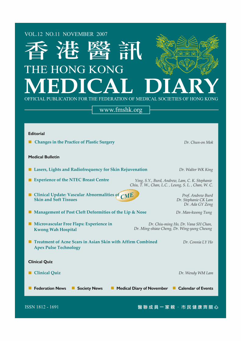
VOL.12 NO.11 NOVEMBER 2007
ISSN 1812 - 1691
THE HONG KONG
MEDICAL DIARYOFFICIAL PUBLICATION FOR THE FEDERATION OF MEDICAL SOCIETIES OF HONG KONG
www.fmshk.org
Editorial
� Federation News � Society News � Medical Diary of November � Calendar of Events
�
Medical Bulletin
�
Changes in the Practice of Plastic Surgery
Clinical Quiz
� Clinical Quiz Dr. Wendy WM Lam
Lasers, Lights and Radiofrequency for Skin Rejuvenation
Dr. Chun-on Mok
� Experience of the NTEC Breast Centre
Dr. Walter WK King
� Clinical Update: Vascular Abnormalities ofSkin and Soft Tissues
Dr. Man-kwong Tung� Management of Post Cleft Deformities of the Lip & Nose
� Microvascular Free Flaps: Experience inKwong Wah Hospital
Ying, S.Y., Burd, Andrew, Lam, C. K. StephanieChiu, T. W., Chan, L.C. , Leung, S. L. , Chan, W. C.
Prof. Andrew BurdDr. Stephanie CK Lam
Dr. Ada GY Zeng
Dr. Chiu-ming Ho, Dr. Vana SH Chan,Dr. Ming-shiaw Cheng, Dr. Wing-yung Cheung
� Treatment of Acne Scars in Asian Skin with Affirm CombinedApex Pulse Technology
Dr. Connia LY Ho
ContentsVOL.12 NO.11 NOVEMBER 2007
ContentsEditorial
� Changes in the Practice of Plastic Surgery 2
Medical Bulletin
� News from Member Societies
Society News 33
Federation News 32
33
� Clinical Quiz
Clinical Quiz
31
Medical Diary of November 34
Calendar of Events
� Courses 3636� Meetings
PresidentDr. FONG To-sang, Dawson 方道生醫生
1st Vice- PresidentDr. CHAN Chi-kuen 陳志權醫生
2nd Vice- PresidentDr. LO Sze-ching, Susanna 盧時楨醫生
Hon. SecretaryDr. LO See-kit, Raymond 勞思傑醫生
Deputy Hon. SecretaryDr. CHAN Sai-kwing 陳世炯醫生
Hon. TreasurerMr. LAM Lop-chi, Nelson 林立志先生
Deputy Hon. TreasurerMr. LEE Cheung-mei, Benjamin 李祥美先生
Executive Committee MembersDr. CHAN Chi-fung, Godfrey 陳志峰醫生Dr. CHIM Chor-sang, James 詹楚生醫生Dr. HO Chung-ping 何仲平醫生Dr. HUNG Kwan-ngai 洪君毅醫生Dr. KINGWing-keung, Walter 金永強醫生Dr. LEE Man-piu, Albert 李文彪醫生Dr. LI Fuk-him, Dominic 李福謙醫生Ms. MAN Bo-lin, Manbo 文保蓮女士Dr. MAN Chi-wai 文志衛醫生Dr. MOK Chun-on 莫鎮安醫生Mr. TO Yuen-yuk, Peter 杜元郁先生
Executive ManagerMs. CHENG Shu-yue, Sue 鄭淑儀小姐
The Federation of Medical Societies of Hong Kong4/F Duke of Windsor Social Service Building,15 Hennessy Road, Wanchai, Hong KongTel: 2527 8898 Fax: 2865 0345
PresidentDr. CHOI Kin 蔡 堅醫生
Vice- PresidentsDr. CHU Kin-wah 朱建華醫生Dr. SHIH Tai-cho 史泰袓醫生
Hon. SecretaryDr. LEUNG Chi-chiu 梁子超醫生
Hon. TreasurerDr. CHOW Pak-Chin 周伯展醫生
Council Rep.Dr. CHAN Yee-shing 陳以誠醫生Dr. HO Chung-ping 何仲平醫生
Chief ExecutiveMrs. Yvonne LEUNG 梁周月美女士Tel: 2527 8285 (General Office)
2527 8324 / 2536 9388 (Club House in Wanchai / Central)Fax: 2865 0943 (Wanchai), 2536 9398 (Central)Email: [email protected]: http://www.hkma.org
PresidentDr. Jason BROCKWELL
Vice-PresidentProf. David ANDERSON
Hon. SecretaryDr. LO See-kit, Raymond 勞思傑醫生
Hon. TreasurerDr. Alex YIP
Council Rep.Dr. Jason BROCKWELLDr. CHEUNG Tse-ming 張子明醫生Tel: 2527 8898 Fax: 2865 0345
Board of DirectorsPresident
Dr. FONG To-sang, Dawson 方道生醫生1st Vice- President
Dr. CHAN Chi-kuen 陳志權醫生2nd Vice- President
Dr. LO Sze-ching, Susanna 盧時楨醫生Hon. Secretary
Dr. LO See-kit, Raymond 勞思傑醫生Hon. Treasurer
Mr. LAM Lop-chi, Nelson 林立志先生Directors
Dr. CHIM Chor-sang, James 詹楚生醫生Dr. KINGWing-keung, Walter 金永強醫生Dr. LI Fuk-him, Dominic 李福謙醫生Mr. CHAN Yan-chi, Samuel 陳恩賜先生Ms. MAN Bo-lin, Manbo 文保蓮女士
Dr. Walter WK King
� Lasers, Lights and Radiofrequency forSkin Rejuvenation
4
� Experience of the NTEC Breast Centre 8
� Clinical Update: Vascular Abnormalities ofSkin and Soft Tissues
12
Dr. Man-kwong Tung
� Management of Post Cleft Deformities of theLip &Nose
20
� Microvascular Free Flaps: Experience inKwongWahHospital
23
Dr. Wendy WM Lam
Dr. Chun-on Mok
� MCHKCME Programme Self-assessment Questions 19
Ying, S.Y., Burd, Andrew, Lam, C. K. StephanieChiu, T. W., Chan, L.C. , Leung, S. L. , Chan, W. C.
Prof. Andrew BurdDr. Stephanie CK LamDr. Ada GY Zeng
Dr. Chiu-ming Ho, Dr. Vana SH Chan,Dr. Ming-shiaw Cheng, Dr. Wing-yung Cheung
� Treatment of Acne Scars in Asian Skin with AffirmCombined Apex Pulse Technology
29
Dr. Connia LY Ho
Editorial VOL.12 NO.11 NOVEMBER 2007
2
As Hong Kong becomes more affluent and the local people are morereceptive to the so called 'life style medicine', the demand for plasticsurgery is on the rise. Recently, massive advertisement in the mediaand TV programmes by institutes to solicit the public for plasticsurgery-related procedures which are poorly monitored andregulated. These entrepreneurial business and advertising strategiesexploit vulnerable patients and give over optimistic prediction butfail to highlight the indications and potential risks and complicationsinvolved. In return, advertising generates unrealistic expectation inmany patients. The professional and ethical standard would becompromised as the surgical practice would undoubtedly bedetermined primarily by the capacity of the patient to pay ratherthan the clinical benefit.
To combat this unhealthy trend, plastic surgery colleagues shouldendeavour more efforts in educating the public and disseminatemore updated plastic surgery knowledge to medical and healthcolleagues. This kind of promotion is different from advertisementas objective, factual information including the indications, realisticresult, potential risks and complications are fully revealed to ourcolleagues and the public. In the past, our specialty has not doneenough to make ourselves known to even our medical colleagues,not to mention the public. I am happy to see that this issue will beone of the many efforts in the future to disseminate realistic, wellregulated plastic surgery information to the medical profession aswell as the public.
Lastly, I would like to thank the authors, Professor Andrew Burd,Dr. Ho Chiu Ming, Dr. Walter King, Dr. Tung Man Kwong and Dr.Ying Shun Yuen for they have contributed excellent scientific articleswhich are the results of many years of hard working andexperiences.
Dr. PDr. Chun-on Mok
Specialist in Plastic SurgeryEditor
Changes in the Practiceof Plastic SurgeryDr. Chun-onMok
Published byThe Federation of Medical Societies of Hong Kong
EDITOR-IN-CHIEFDr. KINGWing-keung, Walter 金永強醫生
EDITORSDr. CHAN Chi-fung, Godfrey陳志峰醫生 (Paediatrics)Dr. CHAN Chun-hon, Edmond陳振漢醫生 (General Practice)Dr. MOK Chun-on莫鎮安醫生 (Plastic Surgery)
EDITORIAL BOARDDr. AU-YEUNG Cheuk-lun, Henry區陽卓倫醫生 (Paediatrics)Dr. CHAN Chi-wai, Angus陳志偉醫生 (General Surgery)Dr. Norman CHAN陳諾醫生 (Diabetes, Endocrinology & Metabolism)Dr. CHIANG Chung-seung蔣忠想醫生 (Cardiology)Dr. CHONG Lai-yin莊禮賢醫生 (Dermatology & Venereolgy)Dr. CHUH An-tung, Antonio許晏冬醫生 (Family Medicine)Dr. FAN Yiu-wah范耀華醫生 (Neurosurgery)Dr. FONG Ka-yeung方嘉揚醫生 (Neurology)Dr. FUNG Ho-chuen, Alec馮浩泉醫生 (Dermatology & Venereolgy)Dr. FUNG Yee-pong, Adrian馮貽邦醫生 (Dermatology & Venereolgy)Dr. HO Pak-leung何 良醫生 (Clinical Microbiology and Infections)Dr. KWOK Kwan-ho, Alvin郭坤豪醫生 (Ophthalmology)Dr. KWOK Po-yin, Samuel郭寶賢醫生 (General Surgery)Dr. LAI Cheuk Seen, Edward黎卓先醫生 (General Surgery)Dr. LAI Kei-wai, Christopher賴奇偉醫生 (Respiratory Medicine)Dr. LAI Sik-to, Thomas黎錫滔醫生 (Gastroenterology & Hepatology)Dr. LAM Tat-chung, Paul林達聰 醫生 (Psychiatry)Dr. LAMWai-man, Wendy林慧文醫生 (Radiology)Dr. LAO Tzu-his, Terence勞子僖醫生 (Obstetrics & Gynaecology)Dr. LEE Man-piu, Albert李文彪醫生 (Dentistry)Dr. LEUNGWai-tong, Thomas梁惠棠醫生 (Clinical Oncology)Dr. LO See-kit, Raymond勞思傑醫生 (Geriatrics)Dr. MAN Chi-wai文志衛醫生 (Urology)Dr. MOKMo-yin莫慕賢醫生 (Rheumatology)Dr. MONG Hoi-Keung蒙海強醫生 (Forensic Pathology)Dr. TEO Man-lung, Peter張文龍醫生 (Clinical Oncology)Dr. TSANGWai-kay曾偉基醫生 (Nephrology)Dr. TSE Tak-fu謝德富醫生 (Cardiology)Prof. William I WEI韋霖 教授 (Otorhinolaryngology)Dr. WONG Bun-lap, Bernard黃品立 醫生 (Cardiology)Dr. WUWing-cheung, Stephen胡永祥醫生 (Orthopaedics & Traumatology)
EXECUTIVE MANAGERMs. CHENG Shu-yue, Sue 鄭淑儀小姐
MBBS (HK), FRCS (Edin), FRACS, FCSHK,FHKAM (Surgery)
Medical Bulletin
4
VOL.12 NO.11 NOVEMBER 2007
Clinically, photo-acoustic waves are generated withinthe chromophobe targets causing cavitation of thepigment particles which are removed by phagocytosisby macrophages. On the other hand, long pulsed laserscoupled with higher fluences and larger spot size maybe more effective in heating a larger volume of tissue ata deeper depth. (The larger the spot size, the less tissuescattering), a feature that is preferred in the laser orlight treatment of unwanted body hair. In general, lasertreatment of skin condition is safer than IPL treatments,contrary to popular belief since the learning curve forIPL treatment is long and steep.
Commonly used laser systems belong to one of eightcategories:
I. Pigment Laser / Light SystemsCommonly used pigment reducing laser systemsinclude QS NdYAG 1064, QS Alexandrite 755, QS-532,VP-532, long pulsed Alexandrite 755, IPL, IPL andradiofrequency (Aurora)(Fig.1). Clinical indicationsinclude the treatment of freckles, lentigines, naevus,melasma, naevus spilus, naevus of Ota, Becker naevus,eyebrow liner, eyelid liner, body tattoo, postinflammatory hyperpigmentation and pigmented scars.
Not all types of pigmentation will respond to laser / IPLtreatments. In general, the response rate varies from25% to 75% and the number of treatments varies from 3to 15 at intervals of 4 - 6 weeks.
II. Vascular Laser / Light SystemsCommonly used laser / light systems for the treatmentof vascular anomalies include pulsed dye 590/585,VP532, IPL, IPL and radiofrequency (Aurora), laser andradiofrequency (Polaris)(Fig.2), long pulsed Alexandrite755. Clinical indications include port wine stain,rosacea, telangiectasia, poikiloderma civette, angioma,haemangioma, hypertrophic scars and leg veins. Port
I. Pigment Laser / Light SystemsII. Vascular Laser / Light SystemsIII. Non-ablative Rejuvenation Laser / Light /
Radiofrequency SystemsIV. Hair Reduction Laser / Light SystemsV. Ablative Resurfacing Laser SystemsVI. Microthermal Rejuvenation Laser SystemsVII. Surgical Laser Systems
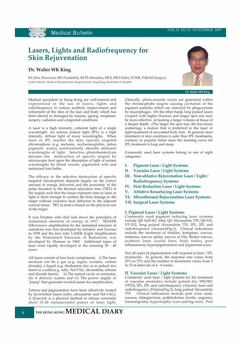
Medical specialists in Hong Kong are well-trained andexperienced in the use of lasers, lights andradiofrequency to induce aesthetic improvement andrefinement of the skin of the face and body which hasbeen altered or damaged by trauma, ageing, neoplasm,surgery, radiation and congenital conditions.
A laser is a high intensity, coherent light of a singlewavelength. An intense pulsed light (IPL) is a highintensity, diffuse light of many wavelengths. Whenlaser or IPL reaches the skin, specific targetedchromophore (e.g. melanin, oxyhaemoglobin, tattoopigment, water) preferentially absorbs differentwavelengths of light. Selective photothermolysisdenotes the destruction of specific targets bymicroscopic heat upon the absorption of light of certainwavelengths by blood vessels, pigmented cells andmelanised hair bulbs.
The efficacy in the selective destruction of specifictargeted chromophore depends largely on the correctamount of energy delivered and the proximity of thepulse duration to the thermal relaxation time (TRT) ofthe targets such that the tissue exposure time to laser orlight is short enough to confine the heat damage to thetarget without excessive heat diffusion to the adjacentnormal tissue. TRT in term is based on the physical sizeof the target.
It was Einstein who first laid down the principles ofstimulated emission of energy in 1917. MASER(Microwave amplification by the simulated emission ofradiation) was first developed by Schalow and Townesin 1958 and the first ruby LASER (Light Amplificationby the Stimulated Emission of Radiation) wasdeveloped by Maiman in 1960. Additional types oflaser were rapidly developed in the ensuring 30 - 40years.
All lasers consist of four basic components. i) The lasermedium can be a gas (e.g. argon, excimer, carbondioxide), a liquid (e.g. rhodamine dye as in pulsed dyelaser) or a solid (e.g. ruby, Nd:YAG, alexandrite, erbiumand dioxide lasers). ii) The optical cavity or resonator,iii) A delivery system and iv) The power supply or"pump" that generates excited atoms for amplification.
Tattoos and pigmentation have been effectively treatedby Q-switched lasers (ruby, alexandrite and Nd-YAG).A Q-switch is a physical method to release extremelyshort (5-20 nanoseconds) pulses of laser light.
Lasers, Lights and Radiofrequency forSkin RejuvenationDr.WalterWKKingBA (Hon, Wisconsin), MD (Vanderbilt), MCPS (Manitoba), FRCS, FRCS (Edin), FCSHK, FHKAM (Surgery)Centre Director, Plastic & Reconstructive Surgery Centre, Hong Kong Sanatorium&Hospital
Dr. Walter WK King
VOL.11 NO.5 MAY 2006 Medical Bulletin
5
VOL.12 NO.11 NOVEMBER 2007
wine stain may require 20 to 40 treatments andclearance may not be complete. Laser treatment of thickor deep haemangioma will require long pulsedAlexandrite or NdYAG lasers. Laser / IPL treatment ofsmall leg veins gives treatment results equivalent tosclerotherapy both of which have associated sideeffects.
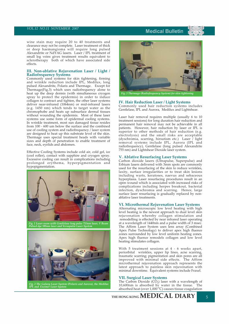
III. Non-ablative Rejuvenation Laser / Light /Radiofrequency SystemsCommonly used systems for skin tightening, firmingand wrinkle reduction include IPL, Medilux, longpulsed Alexandrite, Polaris and Thermage. Except forThermage(Fig.3) which uses radiofrequency alone toheat up the deep dermis (with simultaneous cryogenspray to protect the epidermis) in order to inducecollagen to contract and tighten, the other laser systemsdeliver near-infrared (1064nm) or mid-infrared lasers(e.g. 1450 nm) which tends to target water as thechromophobe and heats up subsurface dermal tissueswithout wounding the epidermis. Most of these lasersystems use some form of epidermal cooling systems.In wrinkle treatment, most sun damaged tissue residesfrom 100 - 600 um below the surface and the combineduse of cooling system and radiofrequency / laser systemare designed to heat up this substrate level of the skin.Thermage uses special treatment heads with variablesizes and depth of penetration to enable treatment offace, neck, eyelids and abdomen.
Effective Cooling Systems include cold air, cold gel, ice(cool roller), contact with sapphire and cryogen spray.Excessive cooling can result in complications includingprolonged erythema, hyperpigmentation andhypopigmentation.
IV. Hair Reduction Laser / Light SystemsCommonly used hair reduction systems includesGentlelase, IPL and Aurora, Medilux and Lightshear.
Laser hair removal requires multiple (usually 6 to 10treatment sessions) for long duration hair reduction andpermanent hair removal may not be achievable in allpatients. However, hair reduction by laser or IPL issuperior to other methods of hair reduction (e.g.electrolysis) and the small risks are acceptable(dyschromia, scarring, hirsutism etc.) Laser / lightremoval systems include IPL, Aurora (IPL andradiofrequency), Gentlelase (long pulsed Alexandrite755 nm) and Lightshear Dioxide laser system.
V. Ablative Resurfacing Laser SystemsCarbon dioxide lasers (Ultrapulse, Superpulse) andErbium lasers delivered with 3mm spots are commonlyused for the resurfacing of the skin to reduce wrinkles,laxity, surface irregularities or to treat skin lesionsincluding warts, keratoses, naevus and sebaceoushyperplasia. Laser resurfacing procedures result in anopen wound which is associated with increased risks ofcomplications including herpes breakout, bacterialinfection, dyschromia and scarring. Hence, largesurface laser resurfacing is gradually replaced by non-ablative laser treatments.
VI. Microthermal Rejuvenation Laser SystemsAlternating microscopic low level heating with highlevel heating is the newest approach to dual level skinrejuvenation whereby collagen stimulation andremodelling is effected by near infrared laser operatingat a wavelength of 1440nm and a pulse width of 3 msec.The Affirm Laser System uses lens array (CombinedApex Pulse Technology) to deliver apex high fluencezones surrounded by low level uniform heating zones.Apex high fluence remodels collagen and low levelheating stimulates collagen.
With 5 treatment sessions at 4 - 6 weeks apart,periorbital wrinkles, upper lip lines, acne scarring,traumatic scarring; pigmentation and skin pores are allimproved with minimal side effects. The Affirmmicrothermal rejuvenation approach represents thelatest approach to painless skin rejuvenation withminimal downtime. Equivalent systems include Fraxel.
VII. Surgical Laser SystemsThe Carbon Dioxide (CO2) laser with a wavelength of10,600nm is absorbed by water in the tissue. Theabsorbed heat (over 1,000 oC) causes tissue coagulation
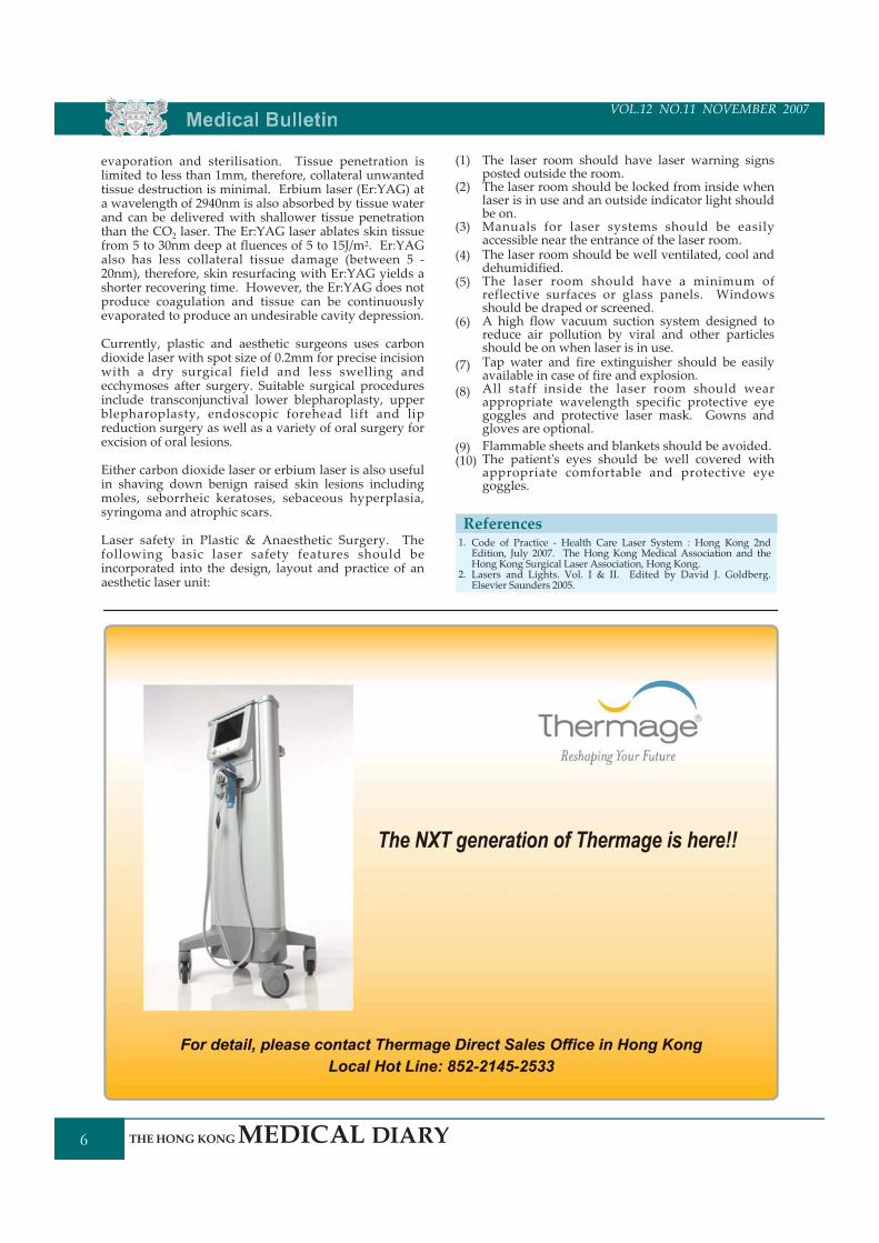
Fig. 1 Laser room with Gentlelase long pulse 755nm laser, V BeamPulsed dye 595nm laser and Versapulse Laser System
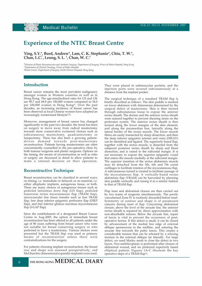
Fig. 2 The Galaxy Laser System (Polaris and Aurora), the MediluxIPL and Excimer Laser System
Fig. 3 Thermage (Radiofrequency System) for skin tightening
Medical Bulletin
6
VOL.12 NO.11 NOVEMBER 2007
Flammable sheets and blankets should be avoided.The patient's eyes should be well covered withappropriate comfortable and protective eyegoggles.
(9)(10)
Code of Practice - Health Care Laser System : Hong Kong 2ndEdition, July 2007. The Hong Kong Medical Association and theHong Kong Surgical Laser Association, Hong Kong.Lasers and Lights. Vol. I & II. Edited by David J. Goldberg.Elsevier Saunders 2005.
1.
2.
References
Tap water and fire extinguisher should be easilyavailable in case of fire and explosion.All staff inside the laser room should wearappropriate wavelength specific protective eyegoggles and protective laser mask. Gowns andgloves are optional.
evaporation and sterilisation. Tissue penetration islimited to less than 1mm, therefore, collateral unwantedtissue destruction is minimal. Erbium laser (Er:YAG) ata wavelength of 2940nm is also absorbed by tissue waterand can be delivered with shallower tissue penetrationthan the CO2 laser. The Er:YAG laser ablates skin tissuefrom 5 to 30nm deep at fluences of 5 to 15J/m2. Er:YAGalso has less collateral tissue damage (between 5 -20nm), therefore, skin resurfacing with Er:YAG yields ashorter recovering time. However, the Er:YAG does notproduce coagulation and tissue can be continuouslyevaporated to produce an undesirable cavity depression.
Currently, plastic and aesthetic surgeons uses carbondioxide laser with spot size of 0.2mm for precise incisionwith a dry surgical field and less swelling andecchymoses after surgery. Suitable surgical proceduresinclude transconjunctival lower blepharoplasty, upperblepharoplasty, endoscopic forehead lift and lipreduction surgery as well as a variety of oral surgery forexcision of oral lesions.
Either carbon dioxide laser or erbium laser is also usefulin shaving down benign raised skin lesions includingmoles, seborrheic keratoses, sebaceous hyperplasia,syringoma and atrophic scars.
Laser safety in Plastic & Anaesthetic Surgery. Thefollowing basic laser safety features should beincorporated into the design, layout and practice of anaesthetic laser unit:
(7)
(8)
The laser room should have laser warning signsposted outside the room.The laser room should be locked from inside whenlaser is in use and an outside indicator light shouldbe on.Manuals for laser systems should be easilyaccessible near the entrance of the laser room.
(1)
(2)
(3)
The laser room should be well ventilated, cool anddehumidified.The laser room should have a minimum ofreflective surfaces or glass panels. Windowsshould be draped or screened.A high flow vacuum suction system designed toreduce air pollution by viral and other particlesshould be on when laser is in use.
(4)
(5)
(6)

Medical Bulletin
8
VOL.12 NO.11 NOVEMBER 2007
IntroductionBreast cancer remains the most prevalent malignancyamongst women in Western countries as well as inHong Kong. The age-standardised rates for US and UKare 90.7 and 68.8 per 100,000 women compared to 34.0per 100,000 women in Hong Kong21. Over the pastdecades, an increasing incidence of breast cancer hasbeen observed as local Chinese women have adopted anincreasingly westernised lifestyle5;38.
Moreover, management of breast cancer has changedsignificantly in the past two decades: the trend has beenon surgery to move away from radical mastectomytowards more conservative excisional choices such assubcutaneous mastectomy, quadrantectomy orlumpectomy. There has also been a growing patient-dr iven demand towards post -mastectomyreconstruction. Patients having mastectomies are oftenconcomitantly counselled in the pre-operative clinic byboth tumour surgeons and plastic surgeons. Options onthe timing, the means of reconstruction as well as risksof surgery are discussed in detail to allow patients tomake a rational decision on their operation.
Reconstructive TechniqueBreast reconstruction can be classified in several ways;on timing, i.e. immediate or delayed; or on material, i.e.either alloplastic implants, autogenous tissue, or both.There are many choices of autogenous tissues such aspedicled latissimus dorsi flap (LD flap), pedicledtransverse rectus myocutaneous flap (TRAM flap),microvascular free tissue transfer such as free TRAMflap, free deep inferior epigastric perforator flap (DIEPflap), and free inferior gluteus maximus myocutaneousflap (I-GAP flap).
Since the establishment of a designated Breast CancerCentre in Aug-2005, the option of immediate breastreconstruction has been offered to all patients under theage of 80 years suffering from breast cancer who werenot suitable for breast conserving surgery or whopreferred to have a mastectomy. Various choices werepresented but the TRAM flap was used as primarymeans of reconstruction unless there werecontraindications for the surgery.
For patients choosing implant reconstruction, the breastsize and shape was assessed preoperatively, andMcGhanbio-dimensional expander implantswereused.
Experience of the NTEC Breast Centre
Ying, S.Y.*, Burd, Andrew*, Lam, C. K. Stephanie*, Chiu, T. W.*,Chan, L.C. , Leung, S. L. #, Chan,W. C.#
*Division of Plastic Reconstructive and Aesthetic Surgery, Department of Surgery, Prince of Wales Hospital, Hong KongDepartment of Clinical Oncology, Prince of Wales Hospital#Breast Centre, Department of Surgery, North District Hospital, Hong Kong
They were placed in submuscular pockets, and theinjection ports were secured subcutaneously at adistance from the implant pocket.
The surgical technique of a standard TRAM flap isbriefly described as follows. The skin paddle is markedon lower abdomen with dimensions determined by thesurgical defect of mastectomy. Skin is then incisedthrough subcutaneous tissue to expose the anteriorrectus sheath. The dermis and the anterior rectus sheathwere sutured together to prevent shearing strain on theperforator system. The anterior rectus sheath is thenincised along the lower margins of the skin element,and along the paramedian line, laterally along thelateral border of the rectus muscle. The lower musclefibres are easily transected by sharp dissection, and thenthe deep inferior epigastric arteries and veins (DIEAV)can be identified and ligated. The superiorly based flap,together with the rectus muscle, is dissected from thesubjacent posterior rectus sheath by sharp and bluntdissection, and is raised to the subcostal margin. It isnot necessary to expose the superior epigastric vesselthat enters the muscle medially at the subcostal margin.The superior insertion of the rectus abdominis musclemay be detached from the 5th, 6th and 7th costalcartilages to facilitate rotation of the myocutaneous flap.A subcutaneous tunnel is created to facilitate passage ofthe myocutaneous flap. A vertically-based rectusabdominis flap (VRAM) can be harvested by planningskin paddle vertically and raising it in a similar fashionto that of TRAM flap.
Flap inset and abdominal closure are then carried outby two teams of surgeons simultaneously. The poorlyvascularised Zone IV is routinely discarded before inset.Symmetry of contour and shape is of paramountconcern during inset of flap. Concerning abdominalclosure, above the level of the arcuate line, the anteriorrectus sheath is repaired by direct approximation withnon-absorbable sutures. Below the arcuate line, repairof fascia is vital to prevent the occurrence of post-operative hernia. If this defect is small, it can be closedby advancement of the medial free edge of externaloblique aponeurosis to the midline, and suturing thearcuate line towards the pubic fascia. This creates aconsiderable tension that can be released by a relaxingincision in the external oblique far laterally. For largedefect, we used Marlex mesh to close the defect in twolayers. Neo-umbilicoplasty is performed after closure ofabdominal wound, and we preferred superiorly basedelliptical pattern. Figures 1A-C illustrate the keyoperative steps of a TRAM flap14.
VOL.11 NO.5 MAY 2006 Medical Bulletin
9
VOL.12 NO.11 NOVEMBER 2007
ResultsFrom August 2005 to January 2007, 255 newly presentedbreast cancer patients were treated at Breast CancerCentre of New Territories East Cluster based at NorthDistrict Hospital. The choice of breast reconstructionwas offered to every newly diagnosed breast cancerpatient who chose to have mastectomy. Post-mastectomy patients followed up at our clinic were alsooffered the choice of reconstruction. In total, only 55patients (21.6%) opted for breast reconstruction out of255 patients during the period. After reconstructivesurgery, patients were followed-up prospectively inCombined Breast Cancer clinic where they were jointlyattended by surgeons and oncologists. The need foradjuvant therapy were determined by tumour'soncologic status, while patient's subjective satisfactionwere assessed half year after operation time. Threepatients defaulted follow up. One patient died as aresult of brain metastasis one year after operation.
Fifty-four patients were Chinese, one came fromPhilippines. The age of patients ranged from 29.2 to 78.2years with a mean SD of 48.9 8.6 years. Patients'body mass index (BMI) ranged from 17.0 to 31.6 with amean SD of 22.9 3.7. Forty patients (72.7%) weremarried and twelve (21.8%) were either divorced orsingle. Thirty-two patients (58.2%) were employed,while twenty patients (36.4%) were unemployed.Twenty-four patients (43.6%) received less than high
school education, twenty-two (40%) patients hadcompleted high school education, six patients (10.9%)received graduate or above level of education. Allpatients were non-smoker. Twenty-two patients (40.0%)had background medical illnesses. One patient had abackground of diabetes mellitus. Six patients (10.9%)had first-degree relatives with a history of breast cancer.Table 1 summarises patient statistics. Forty patients(72.7%) had early breast cancers (DCIS or <T3). Table 2summarises patients' tumour type and sizes.
Postoperatively, the patient is nursed in head up tilt 45degree and hip flexion position for 3 days. All patientsare advised not to engage in heavy domestic activitiesfor 3 weeks, and refrain from strenuous exercise for 3months. Adjuvant radiotherapy, chemotherapy andhormonal therapy were prescribed according tooncologic status of the tumour after surgery.
Figure 1 A-C, Illustration of concept of unipedicled TRAMflap. (Courtesy to Hartrampf, C. R., Jr., and Bennett, G. K.Ann. Surg. 205: 508-519, 1987.)
Table 1. Patient demographics.
Patient Details standarddeviation
AgeBody mass index (BMI)Marital StatusMarriedDivorced or SingleMissingEmployment StatusEmployedUnemployedMissingEducation level<High schoolHigh schoolGraduate or aboveMissingFamily history of breast cancerYesNoMissingMedical illnessesNilAllergic rhinitisAsthmaDepressionGraves' diseaseHepatitis BHypertensionDiabetes mellitusOvarian cystObstructive sleep apnoea syndromeThallassaemiaMissing
8.63.7
mean n
40123
32203
242263
6463
3411152312113
48.922.9
Twelve patients (21.8%) required post-operative adjuvantradiotherapy for tumour clearance. Seven patients(12.7%) developed local recurrences during one year'sfollow up. Thirty-eight patients (69.1%) opted forimmediate reconstruction, seventeen (30.9%) chosedelayed reconstruction. The mean SD interval of delaywas 4.9 5.4 years. Forty-six (83.7%) patients chose tohave autogenous tissue reconstruction and nine (16.3%)had reconstruction by implants. Fourteen patients(25.5%) chose to have further nipple-areolar complexreconstruction. Seven patients (15.2%) suffered frompostoperative fat necrosis, and these patients had beenreconstructed by unipedicled TRAM flap. There was nofat necrosis among the seven bipedicled TRAM flaps.Adjuvant therapy for one patient has to be delayed as aresult of fat necrosis. Table 3 summarises complication ofoperation. Risk factors for surgical complications weretested by applying nonparametric tests against patientdemographic parameters and surgical parameters. It is
Table 2. Summary of tumour types and size.
Tumour Type Count
Breast Cancer
Phalloides tumourSarcomaTotal
121120623155
Stage
DCIS1234
Medical Bulletin
10
VOL.12 NO.11 NOVEMBER 2007
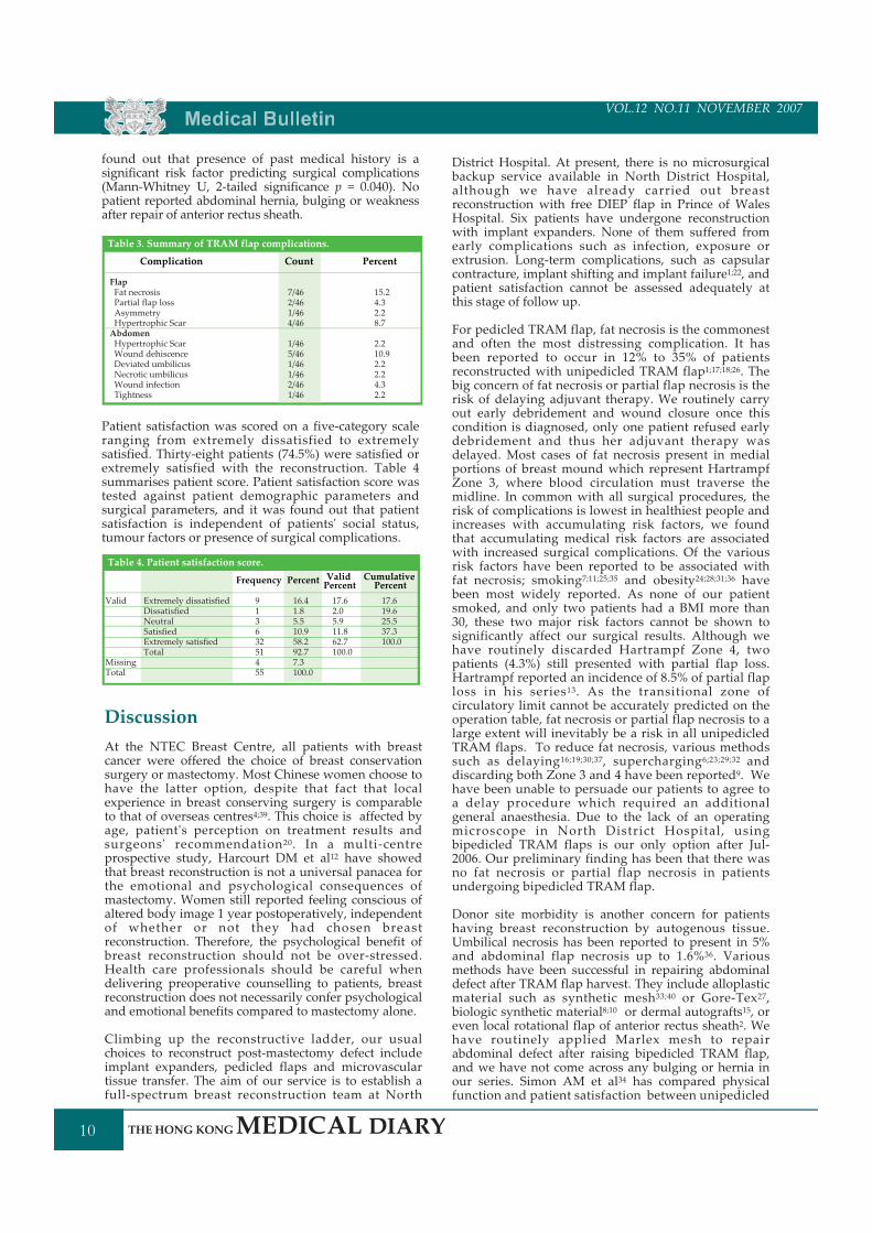
Table 3. Summary of TRAM flap complications.
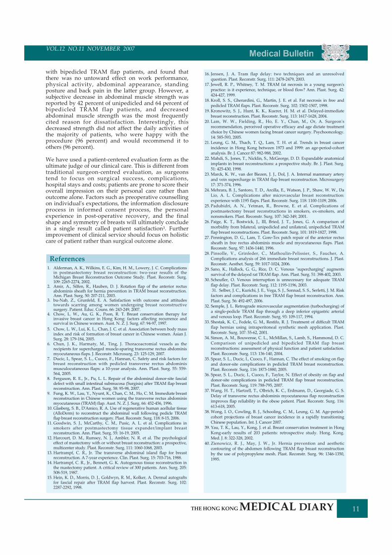
Patient satisfaction was scored on a five-category scaleranging from extremely dissatisfied to extremelysatisfied. Thirty-eight patients (74.5%) were satisfied orextremely satisfied with the reconstruction. Table 4summarises patient score. Patient satisfaction score wastested against patient demographic parameters andsurgical parameters, and it was found out that patientsatisfaction is independent of patients' social status,tumour factors or presence of surgical complications.
found out that presence of past medical history is asignificant risk factor predicting surgical complications(Mann-Whitney U, 2-tailed significance p = 0.040). Nopatient reported abdominal hernia, bulging or weaknessafter repair of anterior rectus sheath.
District Hospital. At present, there is no microsurgicalbackup service available in North District Hospital,although we have already carried out breastreconstruction with free DIEP flap in Prince of WalesHospital. Six patients have undergone reconstructionwith implant expanders. None of them suffered fromearly complications such as infection, exposure orextrusion. Long-term complications, such as capsularcontracture, implant shifting and implant failure1;22, andpatient satisfaction cannot be assessed adequately atthis stage of follow up.
For pedicled TRAM flap, fat necrosis is the commonestand often the most distressing complication. It hasbeen reported to occur in 12% to 35% of patientsreconstructed with unipedicled TRAM flap1;17;18;26. Thebig concern of fat necrosis or partial flap necrosis is therisk of delaying adjuvant therapy. We routinely carryout early debridement and wound closure once thiscondition is diagnosed, only one patient refused earlydebridement and thus her adjuvant therapy wasdelayed. Most cases of fat necrosis present in medialportions of breast mound which represent HartrampfZone 3, where blood circulation must traverse themidline. In common with all surgical procedures, therisk of complications is lowest in healthiest people andincreases with accumulating risk factors, we foundthat accumulating medical risk factors are associatedwith increased surgical complications. Of the variousrisk factors have been reported to be associated withfat necrosis; smoking7;11;25;35 and obesity24;28;31;36 havebeen most widely reported. As none of our patientsmoked, and only two patients had a BMI more than30, these two major risk factors cannot be shown tosignificantly affect our surgical results. Although wehave routinely discarded Hartrampf Zone 4, twopatients (4.3%) still presented with partial flap loss.Hartrampf reported an incidence of 8.5% of partial flaploss in his series13. As the transitional zone ofcirculatory limit cannot be accurately predicted on theoperation table, fat necrosis or partial flap necrosis to alarge extent will inevitably be a risk in all unipedicledTRAM flaps. To reduce fat necrosis, various methodssuch as delaying16;19;30;37, supercharging6;23;29;32 anddiscarding both Zone 3 and 4 have been reported9. Wehave been unable to persuade our patients to agree toa delay procedure which required an additionalgeneral anaesthesia. Due to the lack of an operatingmicroscope in North District Hospital, usingbipedicled TRAM flaps is our only option after Jul-2006. Our preliminary finding has been that there wasno fat necrosis or partial flap necrosis in patientsundergoing bipedicled TRAM flap.
Donor site morbidity is another concern for patientshaving breast reconstruction by autogenous tissue.Umbilical necrosis has been reported to present in 5%and abdominal flap necrosis up to 1.6%36. Variousmethods have been successful in repairing abdominaldefect after TRAM flap harvest. They include alloplasticmaterial such as synthetic mesh33;40 or Gore-Tex27,biologic synthetic material8;10 or dermal autografts15, oreven local rotational flap of anterior rectus sheath2. Wehave routinely applied Marlex mesh to repairabdominal defect after raising bipedicled TRAM flap,and we have not come across any bulging or hernia inour series. Simon AM et al34 has compared physicalfunction and patient satisfaction between unipedicled
DiscussionAt the NTEC Breast Centre, all patients with breastcancer were offered the choice of breast conservationsurgery or mastectomy. Most Chinese women choose tohave the latter option, despite that fact that localexperience in breast conserving surgery is comparableto that of overseas centres4;39. This choice is affected byage, patient's perception on treatment results andsurgeons' recommendation20. In a multi-centreprospective study, Harcourt DM et al12 have showedthat breast reconstruction is not a universal panacea forthe emotional and psychological consequences ofmastectomy. Women still reported feeling conscious ofaltered body image 1 year postoperatively, independentof whether or not they had chosen breastreconstruction. Therefore, the psychological benefit ofbreast reconstruction should not be over-stressed.Health care professionals should be careful whendelivering preoperative counselling to patients, breastreconstruction does not necessarily confer psychologicaland emotional benefits compared to mastectomy alone.
Climbing up the reconstructive ladder, our usualchoices to reconstruct post-mastectomy defect includeimplant expanders, pedicled flaps and microvasculartissue transfer. The aim of our service is to establish afull-spectrum breast reconstruction team at North
Complication Percent
FlapFat necrosisPartial flap lossAsymmetryHypertrophic ScarAbdomenHypertrophic ScarWound dehiscenceDeviated umbilicusNecrotic umbilicusWound infectionTightness
15.24.32.28.7
2.210.92.22.24.32.2
Count
7/462/461/464/46
1/465/461/461/462/461/46
Table 4. Patient satisfaction score.
Valid
MissingTotal
Frequency ValidPercent
CumulativePercent
Extremely dissatisfiedDissatisfiedNeutralSatisfiedExtremely satisfiedTotal
91363251455
16.41.85.510.958.292.77.3100.0
17.62.05.911.862.7100.0
17.619.625.537.3100.0
Percent
VOL.11 NO.5 MAY 2006 Medical Bulletin
11
VOL.12 NO.11 NOVEMBER 2007
Alderman, A. K., Wilkins, E. G., Kim, H. M., Lowery, J. C. Complicationsin postmastectomy breast reconstruction: two-year results of theMichigan Breast Reconstruction Outcome Study. Plast. Reconstr. Surg.109: 2265-2274, 2002.Amir, A., Silfen, R., Hauben, D. J. Rotation flap of the anterior rectusabdominis sheath for hernia prevention in TRAM breast reconstruction.Ann. Plast. Surg. 50: 207-211, 2003.bu-Nab, Z., Grunfeld, E. A. Satisfaction with outcome and attitudestowards scarring among women undergoing breast reconstructivesurgery. Patient. Educ. Couns. 66: 243-249, 2007.Chow, L. W., Au, G. K., Poon, R. T. Breast conservation therapy forinvasive breast cancer in Hong Kong: factors affecting recurrence andsurvival in Chinese women. Aust. N. Z. J. Surg. 67: 94-97, 1997.
1.
2.
3.
4.
Jensen, J. A. Tram flap delay: two techniques and an unresolvedquestion. Plast. Reconstr. Surg. 111: 2478-2479, 2003.Jewell, R. P., Whitney, T. M. TRAM fat necrosis in a young surgeon'spractice: is it experience, technique, or blood flow? Ann. Plast. Surg. 42:424-427, 1999.Kroll, S. S., Gherardini, G., Martin, J. E. et al. Fat necrosis in free andpedicled TRAM flaps. Plast. Reconstr. Surg. 102: 1502-1507, 1998.Kronowitz, S. J., Hunt, K. K., Kuerer, H. M. et al. Delayed-immediatebreast reconstruction. Plast. Reconstr. Surg. 113: 1617-1628, 2004.Lam, W. W., Fielding, R., Ho, E. Y., Chan, M., Or, A. Surgeon'srecommendation, perceived operative efficacy and age dictate treatmentchoice by Chinese women facing breast cancer surgery. Psychooncology.14: 585-593, 2005.Leung, G. M., Thach, T. Q., Lam, T. H. et al. Trends in breast cancerincidence in Hong Kong between 1973 and 1999: an age-period-cohortanalysis. Br. J. Cancer 87: 982-988, 2002.Mahdi, S., Jones, T., Nicklin, S., McGeorge, D. D. Expandable anatomicalimplants in breast reconstructions: a prospective study. Br. J. Plast. Surg.51: 425-430, 1998.Marck, K. W., van der Biezen, J. J., Dol, J. A. Internal mammary arteryand vein supercharge in TRAM flap breast reconstruction. Microsurgery17: 371-374, 1996.Mehrara, B. J., Santoro, T. D., Arcilla, E., Watson, J. P., Shaw, W. W., DaLio, A. L. Complications after microvascular breast reconstruction:experience with 1195 flaps. Plast. Reconstr. Surg. 118: 1100-1109, 2006.Padubidri, A. N., Yetman, R., Browne, E. et al. Complications ofpostmastectomy breast reconstructions in smokers, ex-smokers, andnonsmokers. Plast. Reconstr. Surg. 107: 342-349, 2001.Paige, K. T., Bostwick, J., III, Bried, J. T., Jones, G. A comparison ofmorbidity from bilateral, unipedicled and unilateral, unipedicled TRAMflap breast reconstructions. Plast. Reconstr. Surg. 101: 1819-1827, 1998.Pennington, D. G., Lam, T. Gore-Tex patch repair of the anterior rectussheath in free rectus abdominis muscle and myocutaneous flaps. Plast.Reconstr. Surg. 97: 1436-1440, 1996.Pinsolle, V., Grinfeder, C., Mathoulin-Pelissier, S., Faucher, A.Complications analysis of 266 immediate breast reconstructions. J. Plast.Reconstr. Aesthet. Surg. 59: 1017-1024, 2006.Sano, K., Hallock, G. G., Rice, D. C. Venous "supercharging" augmentssurvival of the delayed rat TRAM flap. Ann. Plast. Surg. 51: 398-402, 2003.Scheufler, O. Venous interruption is unnecessary for adequate TRAMflap delay. Plast. Reconstr. Surg. 112: 1195-1196, 2003.31. Selber, J. C., Kurichi, J. E., Vega, S. J., Sonnad, S. S., Serletti, J. M. Riskfactors and complications in free TRAM flap breast reconstruction. Ann.Plast. Surg. 56: 492-497, 2006.Semple, J. L. Retrograde microvascular augmentation (turbocharging) ofa single-pedicle TRAM flap through a deep inferior epigastric arterialand venous loop. Plast. Reconstr. Surg. 93: 109-117, 1994.Shestak, K. C., Fedele, G. M., Restifo, R. J. Treatment of difficult TRAMflap hernias using intraperitoneal synthetic mesh application. Plast.Reconstr. Surg. 107: 55-62, 2001.Simon, A. M., Bouwense, C. L., McMillan, S., Lamb, S., Hammond, D. C.Comparison of unipedicled and bipedicled TRAM flap breastreconstructions: assessment of physical function and patient satisfaction.Plast. Reconstr. Surg. 113: 136-140, 2004.Spear, S. L., Ducic, I., Cuoco, F., Hannan, C. The effect of smoking on flapand donor-site complications in pedicled TRAM breast reconstruction.Plast. Reconstr. Surg. 116: 1873-1880, 2005.Spear, S. L., Ducic, I., Cuoco, F., Taylor, N. Effect of obesity on flap anddonor-site complications in pedicled TRAM flap breast reconstruction.Plast. Reconstr. Surg. 119: 788-795, 2007.Wang, H. T., Hartzell, T., Olbrich, K. C., Erdmann, D., Georgiade, G. S.Delay of transverse rectus abdominis myocutaneous flap reconstructionimproves flap reliability in the obese patient. Plast. Reconstr. Surg. 116:613-618, 2005.Wong, I. O., Cowling, B. J., Schooling, C. M., Leung, G. M. Age-period-cohort projections of breast cancer incidence in a rapidly transitioningChinese population. Int. J. Cancer 2007.Yau, T. K., Lau, Y., Kong, J. et al. Breast conservation treatment in HongKong-early results of 203 patients: retrospective study. Hong. Kong.Med. J. 8: 322-328, 2002.Zienowicz, R. J., May, J. W., Jr. Hernia prevention and aestheticcontouring of the abdomen following TRAM flap breast reconstructionby the use of polypropylene mesh. Plast. Reconstr. Surg. 96: 1346-1350,1995.
16.
17.
18.
19.
20.
21.
22.
23.
24.
25.
26.
27.
28.
29.
30.
32.
33.
34.
35.
36.
37.
38.
39.
40.
Chow, L. W., Lui, K. L., Chan, J. C. et al. Association between body massindex and risk of formation of breast cancer in Chinese women. Asian J.Surg. 28: 179-184, 2005.Chun, J. K., Harmaty, M., Ting, J. Thoracoacromial vessels as therecipients for supercharged muscle-sparing transverse rectus abdominismyocutaneous flaps. J. Reconstr. Microsurg. 23: 125-129, 2007.Ducic, I., Spear, S. L., Cuoco, F., Hannan, C. Safety and risk factors forbreast reconstruction with pedicled transverse rectus abdominismusculocutaneous flaps: a 10-year analysis. Ann. Plast. Surg. 55: 559-564, 2005.Ferguson, R. E., Jr., Pu, L. L. Repair of the abdominal donor-site fascialdefect with small intestinal submucosa (Surgisis) after TRAM flap breastreconstruction. Ann. Plast. Surg. 58: 95-98, 2007.Fung, K. W., Lau, Y., Nyunt, K., Chan, C. M., Ho, C. M. Immediate breastreconstruction in Chinese women using the transverse rectus abdominismyocutaneous (TRAM) flap. Aust. N. Z. J. Surg. 66: 452-456, 1996.Glasberg, S. B., D'Amico, R. A. Use of regenerative human acellular tissue(AlloDerm) to reconstruct the abdominal wall following pedicle TRAMflap breast reconstruction surgery. Plast. Reconstr. Surg. 118: 8-15, 2006.Goodwin, S. J., McCarthy, C. M., Pusic, A. L. et al. Complications insmokers after postmastectomy tissue expander/implant breastreconstruction. Ann. Plast. Surg. 55: 16-19, 2005.Harcourt, D. M., Rumsey, N. J., Ambler, N. R. et al. The psychologicaleffect of mastectomy with or without breast reconstruction: a prospective,multicenter study. Plast. Reconstr. Surg. 111: 1060-1068, 2003.Hartrampf, C. R., Jr. The transverse abdominal island flap for breastreconstruction. A 7-year experience. Clin. Plast. Surg. 15: 703-716, 1988.Hartrampf, C. R., Jr., Bennett, G. K. Autogenous tissue reconstruction inthe mastectomy patient. A critical review of 300 patients. Ann. Surg. 205:508-519, 1987.Hein, K. D., Morris, D. J., Goldwyn, R. M., Kolker, A. Dermal autograftsfor fascial repair after TRAM flap harvest. Plast. Reconstr. Surg. 102:2287-2292, 1998.
5.
6.
7.
8.
9.
10.
11.
12.
13.
14.
15.
References
with bipedicled TRAM flap patients, and found thatthere was no untoward effect on work performance,physical activity, abdominal appearance, standingposture and back pain in the latter group. However, asubjective decrease in abdominal muscle strength wasreported by 42 percent of unipedicled and 64 percent ofbipedicled TRAM flap patients, and decreasedabdominal muscle strength was the most frequentlycited reason for dissatisfaction. Interestingly, thisdecreased strength did not affect the daily activities ofthe majority of patients, who were happy with theprocedure (96 percent) and would recommend it toothers (96 percent).
We have used a patient-centered evaluation form as theultimate judge of our clinical care. This is different fromtraditional surgeon-centred evaluation, as surgeonstend to focus on surgical success, complications,hospital stays and costs; patients are prone to score theiroverall impression on their personal care rather thanoutcome alone. Factors such as preoperative counsellingon individual's expectations, the information disclosureprocess in informed consent process, the personalexperience in post-operative recovery, and the finalshape and symmetry of breasts will ultimately concludein a single result called patient satisfaction3. Furtherimprovement of clinical service should focus on holisticcare of patient rather than surgical outcome alone.
Medical Bulletin
12
VOL.12 NO.11 NOVEMBER 2007
IntroductionAlthough it has been over twenty years since Mullikenand Glowacki wrote their classic paper regarding theclinical distinction of haemangiomas and vascularmalformations1-2 it is still not uncommon to meetpatients in their late teens who are still waiting for'haemangiomas' to spontaneously resolve. Table 1summarises the main distinctions between the twotypes of vascular abnormalities. It should be noted thatalthough vascular malformations will be present atbirth they may not always be clinically apparent at birthand indeed may not be noticed until several years afterbirth. The most important differentiating feature is therapid growth of haemangiomas in the first year of lifeand the subsequent involution. This distinction isshown in Figure 1.
HaemangiomaHaemangiomas are the most common tumour ofinfancy and typically appear as a small reddish macule3.80% occur within the first month of life. The maculequickly grows and becomes raised and lobulated. Theappearance is not unlike a strawberry hence the namestrawberry naevus. At a cellular level the vascularendothelium in the proliferating haemangioma iscycling at a very high rate4. The mitotic index isextremely high and the cycling time can be as short astwenty four hours. VEGF is expressed very highly inthese cells and can be detected in the circulating blood.bFGF expression is also significantly upregulated. Thepattern of subsequent cytokine expression and mitoticindex is shown in Figure 2.
Whilst the vast majority of haemangiomas involve theskin, they can occur subcutaneously, appearing as abluish patch under the skin. This can cause a rapidlydeveloping swelling which then involutes as in thecutaneous lesion. Most haemangiomas can be treatedconservatively with an expectant strategy. The parentsneed to be reassured that the lesion(s) will involute andin most cases surgery is relegated to some minorprocedures to remove the redundant skin.
There are occasions, however, when haemangiomas do
need more active intervention. Table II shows intrinsicand extrinsic causes for concern. Of note, the 'Kasabach-Merritt' syndrome has been described as a rarepresentation of thrombocytopenia associated withhaemangiomas. This association has been erroneouslymade and the syndrome is but a feature of the rarekaposiform haemangioendothelioma. The managementof rapidly growing haemangiomas which are causingvisual field obstruction is an indication for urgentintervention. There are a number of both non-invasiveand invasive options depicted in Tables III. The first lineof treatment with a rapidly developing lesion causingvisual field obstruction is oral steroid therapy. Theresponse to oral steroids is variable and will depend inpart on the stage at which they are used. The response in30-40% of cases can be quite rapid; in another 30-40% ofcases the response still occurs but is slower indeveloping. In a smaller number there is no response5.
Interferon alpha 2a has been used as a subcutaneousinjection in life threatening haemangiomas. Althoughthere are reports of clinical control and regression of thelesions, there are also reports of spastic diplegia arisingas a complication of this treatment6.
Compression therapy has been tried for haemangiomasbut there is no evidence to indicate that this speeds upnatural involution of the lesion.
With regard to the invasive, medical treatment,intralesional steroids have been tried but they are nomore effective than systemic steroids. OK-432 is adenatured streptococcal protein which acts supposedlyby stimulating the immune response and speeding upthe resolution of the haemangioma7. Sclerosing agentshave been tried but the indications for such interventionhave to be clearly understood. The correct diagnosishas to be made and the decision to intervene has to bebalanced against non-intervention and spontaneousinvolution. The outcome has to be considered andtreatment with sclerosants should be limited tohaemangiomas where the site is not of a major aestheticconcern. Surgical debulking has to be considered in thesame context with regard to the long term effects.Surgery involving extensive scarring should be avoidedif the alternative is awaiting natural resolution andlesser scarring8.
Clinical Update: Vascular Abnormalities ofSkin and Soft Tissues1Prof. Andrew BurdMD, FRCS, FHKAM(Surgery)
1Dr. Stephanie CK LamMBChB, MRCS
2Dr. Ada GY ZengMBChB, MRCS
1Division of Plastic & Reconstructive and Aesthetic Surgery, Department of Surgery,The Chinese University of Hong Kong, Prince of Wales Hospital2Department of Surgery, TuenMunHospital
Prof. Andrew Burd
This article has been selected by the Editorial Board of the Hong Kong Medical Diary for participants in the CME programme of theMedical Council of Hong Kong (MCHK) to complete the following self-assessment questions in order to be awarded one CME creditunder the programme upon returning the completed answer sheet to the Federation Secretariat on or before 30 November 2007.
VOL.11 NO.5 MAY 2006 Medical Bulletin
13
VOL.12 NO.11 NOVEMBER 2007
Laser treatment for haemangiomas is generallyunsatisfactory. The biological mechanism of action isreferred to as selective photothermolysis. Theoxyhaemoglobin complex is selectively targeted by laserenergy close to the third absorption spectral peak(577nm). Laser energy is converted to heat whichdissipates to destroy the surrounding vessel wall. Thelimited penetration of the laser energy means that onlysuperficial lesions are accessible to treatment. Becauseof the physical dispersion of the thermal energy, laserworks best in slow flow lesions with small vessels.Nevertheless lasers can be effective in small lesions andthe application in early haemangiomas has yet to beevaluated9.
Vascular Malformations (V.M.)The term V.M. refers to lesions where the anatomy andmorphology of the vessels are abnormal although thevascular endothelium is normal. These lesions caneither be high, low or mixed flow lesions (Table IV).
Capillary MalformationsThese are typically referred to as Port Wine Stains andcan be classified according to the Waner Gradingsystem10.
The present treatment of choice is laser therapy.Previously other treatments have been tried, inparticular radiation. This unfortunately resulted inlonger term post-radiation skin stigmata (Figure 3).Although laser has improved the outcome of patientswith port-wine stains the results are unpredictable andthe patients must be counselled at the outset thatcomplete clearing of the lesions is most unlikely andthat the response to treatment will plateau (Figure 4).The management of the resistant port wine stain is verydifficult and the options are limited; using cosmeticcamouflage or excising and reconstructing with graftsor flaps.
Venous LesionsThe key to the management of venous lesions isaccurate diagnosis. History and clinical examinationwill differentiate haemangiomas from vascularmalformations. Ultrasound examination by a welltrained operator can give information about the flowcharacteristics and the architecture of the lesion. Theamount of parenchyma and the nature of the vascularspaces, (for example 'lakes' or 'honey-comb') caninfluence the treatment. MRI is used for assessing theanatomical extent of the lesion. In the low flow venouslesion it is acceptable to surgically debulk it removing
Grade I lesions: vessel diameters are in the 80 gmrange. These lesions are light pink macules.Grade II lesions: vessel diameters measure up to 120 gm. these lesions are darker pink macules.Grade III lesions: vessel diameters measure up to 150 gm. These lesions are red macules.Grade IV lesions: vessel diameters are greater than150 gm. These lesions are purple and may becomepapular.
only that part which is giving rise to the most concern.Figure 5a-d shows a vascular malformation thatinvolved the lower lip. There were some problems withoral continence and speech. Two years after limitedbulking shows the patient with good oral continence,clear speech but still with residual (not recurrent)malformation involving the right cheek.
The management of diffuse venous malformations canbe very challenging as the symptoms are usually mildand the major concern is the appearance. The generalstrategies used are to treat these lesions with sclerosantsor sutures which will produce intralesional scarring andreduction in size of the lesion. There have been reportsof producing targeted scleroses using intralesionalcopper wires11,12.
Lymphatic LesionsThese may be microcystic, macrocystic or a combinationof the two. The lymphangioma is an example of amicrocystic lesion and the architecture is characterisedby a 'honey comb' of connective tissue septacompartmentalising the cystic lesion. The cystichygroma is an example of a macrocystic lymphaticmalformation.
Most lymphatic malformations are present at birth orappear within the first two years of life. They rarelyinvolute spontaneously. They may enlarge if theybecome infected or bleeding occurs inside the lesion.
Lymphatic malformations can be treated byintralesional injection of OK-432. If surgery isperformed it is important to fully excise the lesion toprevent recurrence.
High Flow LesionsThese are either arterial or arterio-venousmalformations. The solitary arterial malformations areuncommon and may present as pulsatile subcutaneouslesions which turn out to be aneurysms or collections oftortuous vessels.
The arterio-venous malformations are more commonand whilst the anatomical abnormality will be presentat birth the lesions may only become clinically apparentlater in infancy or childhood. The lesions areconveniently classified according to the modifiedSchobinger classification13 (Table V).
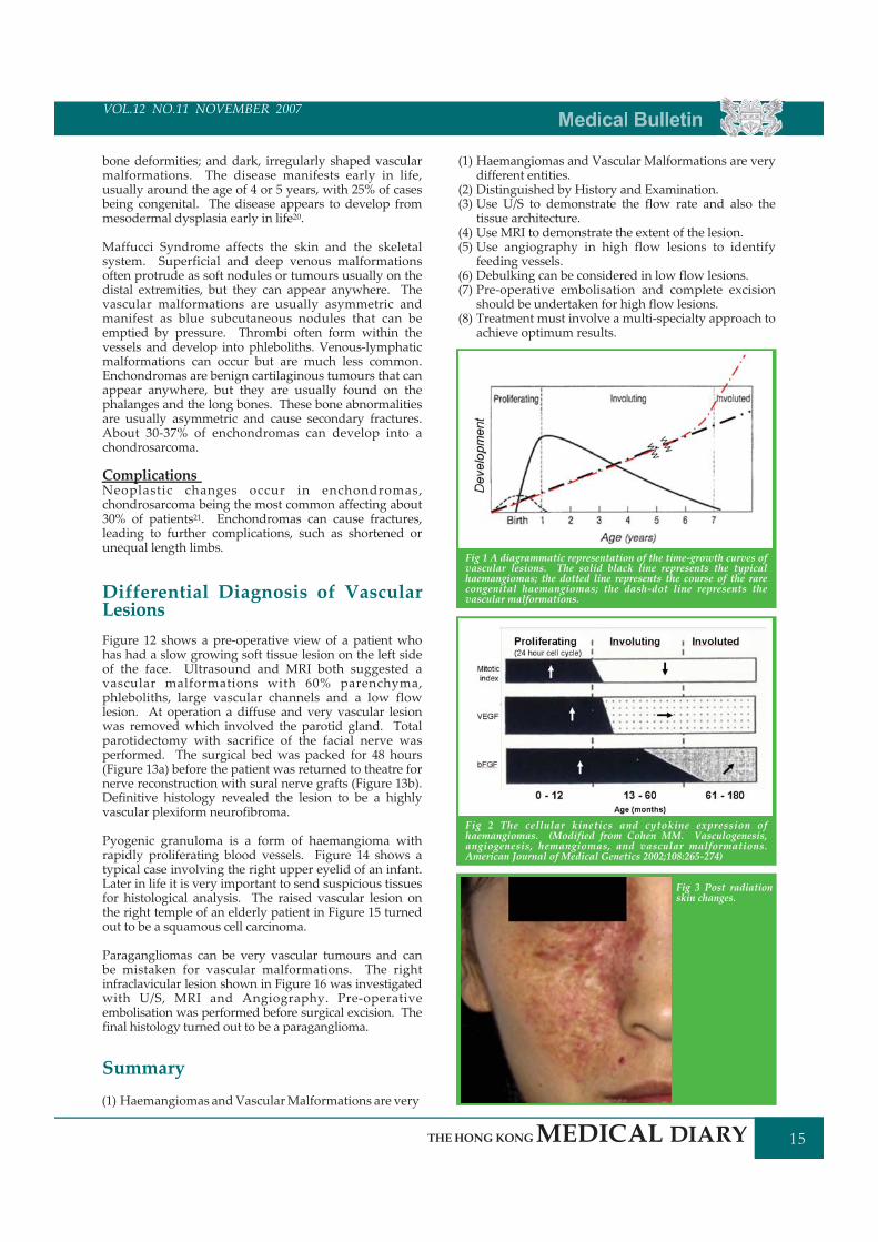
When lesions reach stage 4 they are life threatening.Figure 6a-b shows the left leg of a patient admitted withhigh output cardiac failure associated with an AVM.The MRI scan shows that the increase in leg size ispredominantly associated with the enlargement of thesubcutaneous tissues. When history, examination,ultrasound and MRI indicate that there is a high flowAVM, a diagnostic angiogram should be performed toidentify the feeding vessels. Interventional radiology isan essential part of the treatment of such lesions. Aftercorrecting clotting abnormalities, pre-operativeembolisation should be performed. Vascularembolisation can be achieved using a variety ofsubstances including silicone spheres, stainless steel
Medical Bulletin
14
VOL.12 NO.11 NOVEMBER 2007
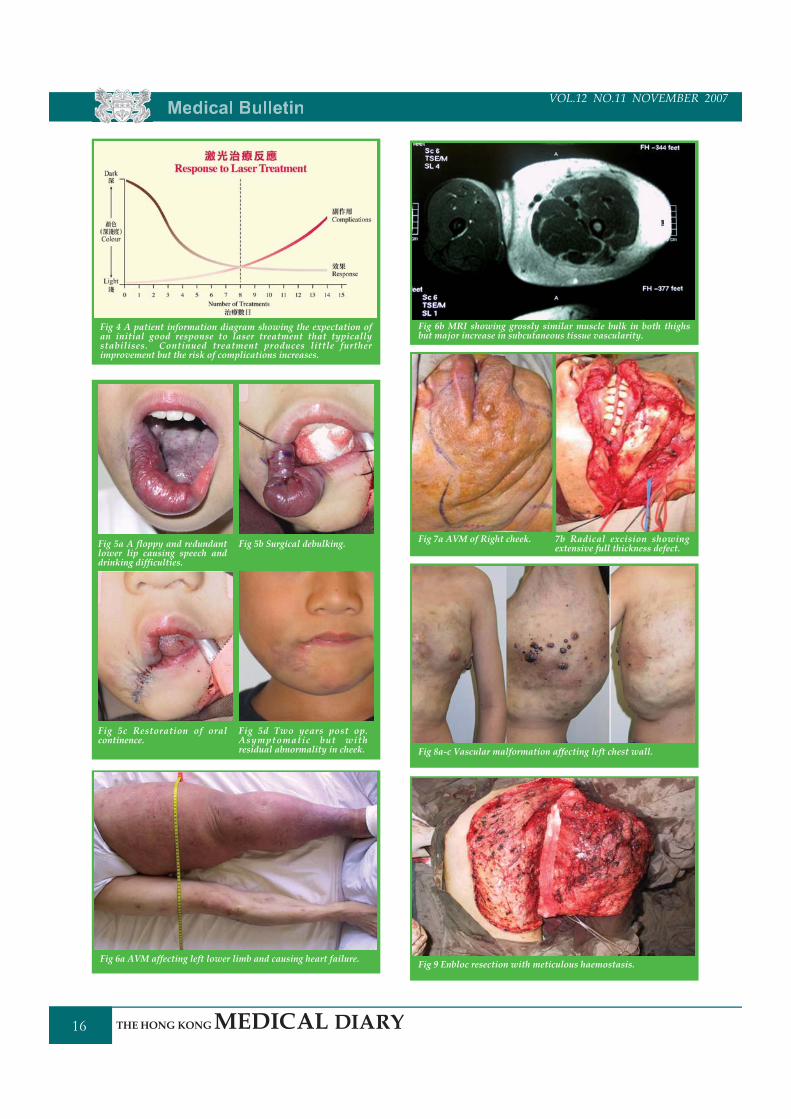
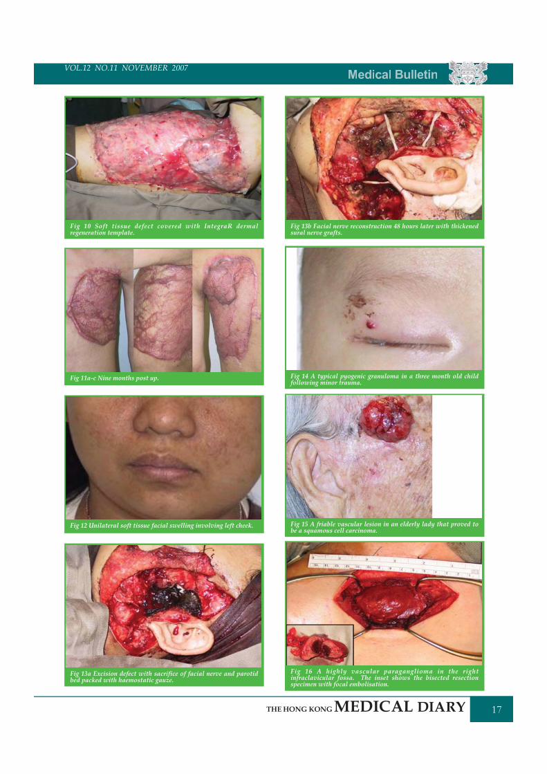
coils, foam and glue. The role of embolisation is tofacilitate surgery and the surgical resection should be ascomplete as possible to prevent recurrence of the AVM.With extensive resection, complex reconstructions willbe needed (Figure 7a-d). These reconstructions willoften need to be staged to optimise both the aestheticand functional aspects14. There are occasions where thelesions are so extensive that complete resection is notpossible. Figure 8a-c shows an extensive AVM of theleft chest wall that involved skin, subcutaneous tissues,muscles, pleura and pericardial tissues. Surgery hadbeen deferred due to the extent of the lesion but withthe onset of puberty the lesion grew rapidly and therewas daily bleeding from the cutaneous nodules. Afterextensive counselling and discussions regarding therisks of surgery the patient underwent pre-operativeembolisation of perforators from the internal mammaryartery. Embolisation was not carried out of the lumbarartery perforators due to the extensive vascular plexi inthe longitudinal spinal muscles and the risk of spinalcord ischaemia. Excisional surgery was performedleaving the intercostal muscles (Figure 9) and the defectreconstructed with Integra (Figure 10). The patientremains well eighteen months after surgery althoughthe possibility of recurrence remains (Figure 11a-c).
Surgical treatment in vascular abnormalities can rangefrom a minor skin excision to an ultramajor excisionwith very real risks of intra-operative mortality. Thereare no shortcuts in such surgery and a multi-specialtyapproach is essential. Special points of note in thepreoperative consent are listed in Table VI.
Syndromes Associated with VascularLesionsSturge-Weber SyndromeThe Sturge-Weber Syndrome (SWS) is aneurocutaneous disorder with angiomas involving theleptomeninges and skin of the face, typically in the V1and V2 distributions of the trigeminal nerve. It is alsocalled encephalotrigeminal angiomatosis. SWS iscaused by residual embryonal blood vessels and theirsecondary effects on surrounding brain tissue. SWS isreferred to as complete when both CNS and facialangiomas are present and incomplete when only onearea is affected. Because of possible associatedanomalies these patients should be referred forspecialist investigation and management.
Klippel-Trenaunay-Weber SyndromeKlippel-Trenaunay-Weber Syndrome is characterisedby a triad of port wine stain, varicose veins, and bonyand soft tissue hypertrophy involving an extremity.The exact cause of Klippel-Trenaunay-Weber Syndrome(KTWS) is unknown. Most cases are sporadic, althougha few cases in the literature report an autosomaldominant pattern of inheritance15.
KTWS generally affects a single extremity, althoughcases of multiple affected limbs have been reported.The leg is the most common site followed by the arms,the trunk, and rarely the head and the neck. Mostpatients demonstrate all 3 signs of the clinicalsyndrome: port wine stain, varicose veins, and bonyand soft tissue hypertrophies.
Other features include lymphatic obstruction, spinabifida, hypospadias, polydactyly, syndactyly,oligodactyly, hyperhidrosis, hypertrichosis, paresthesia,decalcification of involved bones, chronic venousinsufficiency, stasis dermatitis, poor wound healing,ulceration, thrombosis, and emboli16. As with Sturge-Weber syndrome these patients need specialisedinvestigation and management.
Osler-Weber-Rendu SyndromeOsler-Weber-Rendu Syndrome, also known ashereditary haemorrhagic telangiectasia, is an autosomaldominant disorder identified typically by the triad oftelangiectasia, recurrent epistaxis, and a positive familyhistory for the disorder. The disease is caused by aninherited defect. Currently 2 loci have been identifiedassociated with Osler-Weber-Rendu Syndrome, one onchromosome arm 9q33-q34 and a second onchromosome arm 12q. The major cause of morbidityand mortality due to this disorder lies in the presence ofmultiorgan arterio-venous malformations (AVMs) andthe associated haemorrhage that may accompany them.The disease has a wide spectrum of presentations;patients may be asymptomatic or have multiple organinvolvement, presenting at any age. The clinicalmanifestations of Osler-Weber-Rendu disease arecaused by the development of abnormal vasculature,including telangiectases, AVMs, and aneurysms17.
HistoryPresentation will be influenced by the site of thepathology.
Maffucci's SyndromeMaffucci Syndrome (enchondroma with multipleangiomas) is a rare genetic disorder. It is characteriszedby benign enlargements of cartilage (enchondromas);
Nasal mucosa: Epistaxis is the most commonmanifestation of the disease and occurs in up to 90%of affected patients.GI tract: Recurrent painless GI bleeding occurs in10-40% of patients and generally occurs later in lifethan epistaxis.Pulmonary vasculaturePulmonary AVMs are present in 15-20% of patientswith the disease18. Haemoptysis results from eithertelangiectasia of the trachea and bronchi orpulmonary arterio-venous (AV) fistulas.Brain involvementNeurologic involvement occurs in 8-12% of patientswith Osler-Weber Rendu Syndrome. A history ofheadache, seizures, and focal neurologic symptomsmay be found on questioning19. Stroke and brainabscess are more common in these patients.Fatigue may be elicited on history and may be dueto an iron deficiency anaemia caused by recurrentblood loss. Liver involvement may cause rightupper quadrant (RUQ) pain, jaundice, symptoms ofhigh-output cardiac failure, and bleeding fromoesophageal varices. Migraine headaches arepresent in 50% of patients. Although the reason isunclear, the headaches seem to be associated withpulmonary AVMs.
1.
2.
3.
4.
VOL.11 NO.5 MAY 2006 Medical Bulletin
15
VOL.12 NO.11 NOVEMBER 2007
bone deformities; and dark, irregularly shaped vascularmalformations. The disease manifests early in life,usually around the age of 4 or 5 years, with 25% of casesbeing congenital. The disease appears to develop frommesodermal dysplasia early in life20.
Maffucci Syndrome affects the skin and the skeletalsystem. Superficial and deep venous malformationsoften protrude as soft nodules or tumours usually on thedistal extremities, but they can appear anywhere. Thevascular malformations are usually asymmetric andmanifest as blue subcutaneous nodules that can beemptied by pressure. Thrombi often form within thevessels and develop into phleboliths. Venous-lymphaticmalformations can occur but are much less common.Enchondromas are benign cartilaginous tumours that canappear anywhere, but they are usually found on thephalanges and the long bones. These bone abnormalitiesare usually asymmetric and cause secondary fractures.About 30-37% of enchondromas can develop into achondrosarcoma.
ComplicationsNeoplastic changes occur in enchondromas,chondrosarcoma being the most common affecting about30% of patients21. Enchondromas can cause fractures,leading to further complications, such as shortened orunequal length limbs.
Differential Diagnosis of VascularLesionsFigure 12 shows a pre-operative view of a patient whohas had a slow growing soft tissue lesion on the left sideof the face. Ultrasound and MRI both suggested avascular malformations with 60% parenchyma,phleboliths, large vascular channels and a low flowlesion. At operation a diffuse and very vascular lesionwas removed which involved the parotid gland. Totalparotidectomy with sacrifice of the facial nerve wasperformed. The surgical bed was packed for 48 hours(Figure 13a) before the patient was returned to theatre fornerve reconstruction with sural nerve grafts (Figure 13b).Definitive histology revealed the lesion to be a highlyvascular plexiform neurofibroma.
Pyogenic granuloma is a form of haemangioma withrapidly proliferating blood vessels. Figure 14 shows atypical case involving the right upper eyelid of an infant.Later in life it is very important to send suspicious tissuesfor histological analysis. The raised vascular lesion onthe right temple of an elderly patient in Figure 15 turnedout to be a squamous cell carcinoma.
Paragangliomas can be very vascular tumours and canbe mistaken for vascular malformations. The rightinfraclavicular lesion shown in Figure 16 was investigatedwith U/S, MRI and Angiography. Pre-operativeembolisation was performed before surgical excision. Thefinal histology turned out to be a paraganglioma.
Summary
Haemangiomas and Vascular Malformations are verydifferent entities.Distinguished by History and Examination.Use U/S to demonstrate the flow rate and also thetissue architecture.Use MRI to demonstrate the extent of the lesion.Use angiography in high flow lesions to identifyfeeding vessels.Debulking can be considered in low flow lesions.Pre-operative embolisation and complete excisionshould be undertaken for high flow lesions.Treatment must involve a multi-specialty approach toachieve optimum results.
(1)
(2)(3)
(4)(5)
(6)(7)
(8)
Haemangiomas andVascularMalformations are very(1)
Fig 1 A diagrammatic representation of the time-growth curves ofvascular lesions. The solid black line represents the typicalhaemangiomas; the dotted line represents the course of the rarecongenital haemangiomas; the dash-dot line represents thevascular malformations.
Fig 2 The cellular kinetics and cytokine expression ofhaemangiomas. (Modified from Cohen MM. Vasculogenesis,angiogenesis, hemangiomas, and vascular malformations.American Journal of Medical Genetics 2002;108:265-274)
Fig 3 Post radiationskin changes.
Medical Bulletin
16
VOL.12 NO.11 NOVEMBER 2007
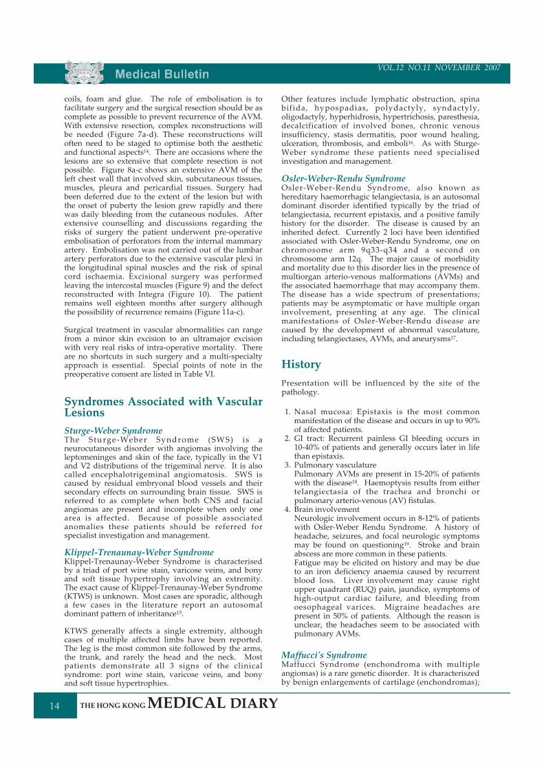
Fig 4 A patient information diagram showing the expectation ofan initial good response to laser treatment that typicallystabilises. Continued treatment produces little furtherimprovement but the risk of complications increases.
Fig 6a AVM affecting left lower limb and causing heart failure.
Fig 6b MRI showing grossly similar muscle bulk in both thighsbut major increase in subcutaneous tissue vascularity.
Fig 8a-c Vascular malformation affecting left chest wall.
Fig 9 Enbloc resection with meticulous haemostasis.
Fig 7a AVM of Right cheek. 7b Radical excision showingextensive full thickness defect.Fig 5a A floppy and redundant
lower lip causing speech anddrinking difficulties.
Fig 5b Surgical debulking.
Fig 5c Restoration of oralcontinence.
Fig 5d Two years post op.Asymptomatic but withresidual abnormality in cheek.
VOL.11 NO.5 MAY 2006 Medical Bulletin
17
VOL.12 NO.11 NOVEMBER 2007
Fig 10 Soft tissue defect covered with IntegraR dermalregeneration template.
Fig 11a-c Nine months post up.
Fig 13b Facial nerve reconstruction 48 hours later with thickenedsural nerve grafts.
Fig 14 A typical pyogenic granuloma in a three month old childfollowing minor trauma.
Fig 15 A friable vascular lesion in an elderly lady that proved tobe a squamous cell carcinoma.
Fig 16 A highly vascular paraganglioma in the rightinfraclavicular fossa. The inset shows the bisected resectionspecimen with focal embolisation.
Fig 12 Unilateral soft tissue facial swelling involving left cheek.
Fig 13a Excision defect with sacrifice of facial nerve and parotidbed packed with haemostatic gauze.
Medical Bulletin
18
VOL.12 NO.11 NOVEMBER 2007
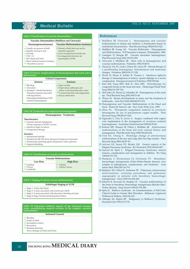
Table I Classification of vascular abnormalities.
Vascular Abnormalities (Mulliken and Glowacki)Vascular Malformations (anatomy)
Usually not present at birthRapidly increase in sizeInvoluteF:M = 3:160% Head and NeckMost common tumour of infancy"Strawberry naevus"
Present at birth (but may not beclinically apparent)Grow in proportion to body sizeCan degenerate but also canhypertrophy (AVM)
Haemangiomas(tumour)
Table II Serious complications of haemangiomas that need activeintervention.
Critical CompromiseExtrinsic
BleedingUlcerationKasabach - Merritt SyndromeThrombocytopenia associatedwith kaposiformhaemangioendothelioma NOTcommon haemangioma.
Upper eyelidsDeprivation amblyopia andfailure to develop binocular vision.Corneal distortion and astigmatism.Airway obstruction
Intrinsic
Table III Treatments for complicated haemangiomas.
Haemangiomas - Treatments
Systemic steroids 2mg/kg/daySome response in 50-90% of cases
Interferon alpha 2a subcutCompression therapy
Non-Invasive
InvasiveIntralesional steroidsIntralesional OK-432 (from denatured streptococcal protein)Intralesional sclerosing agents (Hypertonic saline, glucose)Surgical debulking
Table V Staging of arterio-venous malformations
Schobinger Staging of AVMStage 1: A blue-skin blushStage 2: A mass associated with a bruit and a thrillStage 3: A mass associated with ulceration, bleeding and painStage 4: Stage 3 lesions producing heart failure
Table VI Important addition aspects of the informed consentwhen contemplating surgical excision of the complex vascularmalformation.
Informed ConsentBleedingDeath on tableIncomplete excisionRecurrenceResidual deformityNerve damage (in Head and Neck)
Table IV Classification of vascular malformations.
Vascular MalformationsHigh Flow
CapillaryVenousLymphatic
ArterialArterio-venous
Low Flow
Mulliken JB, Glowacki J. Hemangiomas and vascularmalformations in infants and children: a classification based onendothelial characteristics. Plast Reconst Surg 1982;69:412-422.Mulliken JB, Young AE. Vascular Birthmarks - Hemangiomasand Malformations. W B Saunders Company: Philadelphia, 1988.Gampper TJ, Morgan RF. Vascular anomalies: hemangiomas.Plast Reconstr Surg 2002;110:572-585.Glowacki J, Mulliken JB. Mast cells in hemangiomas andvascular malformations. Pediatrics 1982;70:48-51.Hasan Q, Tan ST, Gush J, Peters SG, Davis PF. Steroid therapy ofa proliferating hemangioma: histochemical and molecularchanges. Pediatrics 2000;105:117-120.Worle H, Maass E, Kohler B, Treuner J. Interferon alpha-2atherapy in haemangiomas of infancy: spastic diplegia as a severecomplication. European Journal of Pediatrics 1999;158:344.Kim KH, Sung MW, Roh JL, Han MH. Sclerotherapy forcongenital lesions in the head and neck. Otolaryngol Head NeckSurg 2004;131:307-316.McCarthy JG, Borud LJ, Schreiber JS. Hemangiomas of the nasaltip. Plast Reconstr Surg 2002;109:31-40.Waner M. Recent developments in lasers and the treatment ofbirthmarks. Arch Dis Child 2003;88:372-374.Hemangiomas and Vascular Malformations of the Head andNeck. Waner M, Suen JY, eds. New York: Wiley-Liss, 1999.Zhou PL. Therapeutic coagulation induced in cavernoushemangioma by use of percutaneous copper needles. PlastReconstr Surg 1992;89:613-622.Ogunsalu C, Fray D, Lewis A. Surgery combined with copperwire implantation in the management of cavernous orofacialhaemangiomas. Australian Dental Journal 2000;45:55-60.Kohout MP, Hansen M, Pribaz J, Mulliken JB. Arteriovenousmalformations of the head and neck: natural history andmanagement. Plast Reconstr Surg 1998;102:643-654.Tark KC, Chung S. Histologic change of arteriovenousmalformations of the face and scalp after free flap transfer. PlastReconstr Surg 2000;106:87-93.Aelvoet GE, Jorens PG, Roelen LM. Genetic aspects of theKlippel-Trenaunay Syndrome. Br J Dermatol 1992;126:603-607.Samuel M, Spitz L. Klippel-Trenaunay Syndrome: clinicalfeatures, complications and management in children. Br J Surg1995;82:757-761.Haitjema T, Westermann CJ, Overtoom TT. Hereditaryhemorrhagic telangiectasia (Osler-Weber-Rendu disease): newinsights in pathogenesis, complications, and treatment. ArchIntern Med 1996;156:714-719.Kjeldsen AD, Oxhoj H, Andersen PE. Pulmonary arteriovenousmalformations: screening procedures and pulmonaryangiography in patients with hereditary hemorrhagictelangiectasia. Chest 1999;116:432-439.Kikuchi K, Kowada M, Sasajima H. Vascular malformations ofthe brain in hereditary hemorrhagic telangiectasia (Rendu-Osler-Weber disease). Surg Neurol 1994;41:374-380.Spitz JL. Maffucci syndrome. In: Genodermatosis, A Full-ColorClinical Guide to Genetic Skin Disorders. Baltimore: LippincottWilliams & Wilkins, 1995;106-107.Albregts AE, Rapini RP. Malignancy in Maffucci's Syndrome.Dermatol Clin 1995;13:73-78.
1.
2.
3.
4.
5.
6.
7.
8.
9.
10.
11.
12.
13.
14.
15.
16.
17.
18.
19.
20.
21.
References
VOL.11 NO.5 MAY 2006 Medical Bulletin
19
VOL.12 NO.11 NOVEMBER 2007
MCHK CME Programme Self-assessment QuestionsPlease read the article entitled "Clinical Update: Vascular Abnormalities of Skin and Soft Tissues" by Prof. AndrewBurd, Dr. Ada GY Zeng and Dr. Stephanie CK Lam, and complete the following self-assessment questions.Participants in the MCHK CME Programme will be awarded 1 CME credit under the Programme for returningcompleted answer sheets via fax (2865 0345) or by mail to the Federation Secretariat on or before 30 November 2007.Answers to questions will be provided in the next issue of The Hong Kong Medical Diary.
Questions 1-10: Please answer T (true) or F (false)1. Both vascular malformations and hemangiomas will be apparent at birth.2. Because of its eventual involution, hemangioma never needs surgical intervention.3. Spastic diplegia is a complication of oral steroid therapy.4. Laser works best in slow flow lesions with small vessels.5. Laser for port-wine stain can usually completely clear the lesion.6. For venous lesions, ultrasound is used to assess flow while MRI is used to assess extent.7. Sturge-Weber syndrome is also called encephalotrigeminal angiomatosis.8. 60% of enchondroma inMaffucci's syndrome develop into chondrosarcoma.9. Differential diagnosis of vascular lesions include plexiform neurofibroma, paraganglioma, pyogenic granuloma andsquamous cell carcinoma.
10. Schobinger staging of arterio-venous malformations divide them into four categories according to signs andsymptoms.
Please return the completed answer sheet to the Federation Secretariat on or before 30 November 2007 fordocumentation. 1 CME point will be awarded for answering the MCHK CME programme (for non-specialists)self-assessment questions.
1 2 3 4 5
Name (block letters):____________________________________ HKMA No.:
___ ___ - ___ ___ ___ ___ X X (x)HKID No.:
____________ ______________________
____________ ______________________Other Membership No.
ContactTelNo.:_________________________________________________
6 7 8 9 10
ANSWER SHEET FOR NOVEMBER 2007
Answers to October 2007 issue
Quality of life and orthodontic treatment need related to Occlusal indices
1 . T 2 . F 3 . F 4 . T 5 . F 6 . T 7 . F 8 . F 9 . T 10 . F
(please indicate):
Clinical Update: Vascular Abnormalities of Skin and Soft Tissues1Prof. Andrew BurdMD, FRCS, FHKAM(Surgery)
1Dr. Stephanie CK LamMBChB, MRCS
2Dr. Ada GY ZengMBChB, MRCS
1Division of Plastic & Reconstructive and Aesthetic Surgery, Department of Surgery,The Chinese University of Hong Kong, Prince of Wales Hospital2Department of Surgery, TuenMunHospital
Medical Bulletin
20
VOL.12 NO.11 NOVEMBER 2007
to make sure whether there is any dental problem thatneeds to be corrected first. A referral to theOromaxillofacial Surgeon will be able to get an overallpicture in the dental aspect & he will coordinate therelevant surgery & relevant Orthodontic treatment.Soft tissue correction before dental treatment canresult in very embarrassing outlook that may not becorrectable later.
This paper is a brief summary of the methods used todeal with the post-cleft deformities of the lip & nose -the non-dental part. The correct functional positioningof related perioral muscles can be performed in thesame session.
Operations for post cleft deformitiesof the lip & nose
Mal-alignment of vermillion borderThis is one of the commonest problems & can becorrected under local anaesthesia.Z-plasty (see diagram)
Excision of excess tissue + realignmentAs part of the whole lip revision operation
Whistle deformity of upper lipSingle or multiple Z-plasty (see diagram)
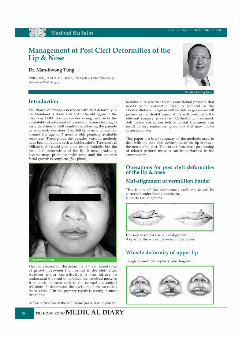
IntroductionThe chance of having a newborn with cleft deformity inthe Mainland is about 1 in 1100. The old figure in theSAR was 1:800. The ratio is decreasing because of theavailability of advanced ultrasound machines leading toearly detection of cleft conditions; allowing the parentsto make early decisions! The cleft lip is usually repairedaround the age of 3 months old; pending availableresources. Throughout the decades various methodshave been in favour; such as LeMesurier's, Tennison's &Millard's. All could give good results initially, but thepost cleft deformities of the lip & nose graduallybecame more prominent with time until the patient'sfacial growth is complete. (See photo)
The main reason for the deformity is the different ratesof growth between the normal & the cleft side.Another major contribution is the failure tounderstand the need to mobilize the involved muscles& to position them back to the normal anatomicalrelations. Furthermore, the excision of the so-called"excess tissue" in the primary repair is wrong in mostsituations.
Before correction of the soft tissue parts, it is important
Management of Post Cleft Deformities of theLip & NoseDr.Man-kwong TungMBBS(HKU), FCSHK, FRCS(Edin), FRCS(Glas), FHKAM(Surgery)Specialist in Plastic Surgery.
Dr. Man-kwong Tung
Preoperative view
VOL.11 NO.5 MAY 2006 Medical Bulletin
21
VOL.12 NO.11 NOVEMBER 2007
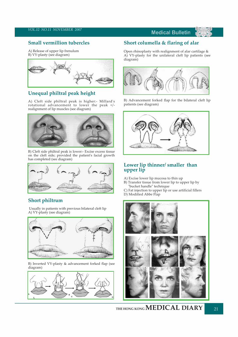
Short columella & flaring of alarOpen rhinoplasty with realignment of alar cartilage &A) VY-plasty for the unilateral cleft lip patients (seediagram)
B) Advancement forked flap for the bilateral cleft lippatients (see diagram)
Lower lip thinner/ smaller thanupper lipA) Excise lower lip mucosa to thin upB) Transfer tissue from lower lip to upper lip by
"bucket handle" techniqueC) Fat injection to upper lip or use artificial fillersD) Modified Abbe Flap
Small vermillion tuberclesA) Release of upper lip frenulumB) VY-plasty (see diagram)
Unequal philtral peak heightA) Cleft side philtral peak is higher:- Millard'srotational advancement to lower the peak +/-realignment of lip muscles (see diagram)
Short philtrumUsually in patients with previous bilateral cleft lipA) VY-plasty (see diagram)
B) Inverted VY-plasty & advancement forked flap (seediagram)
B) Cleft side philtral peak is lower:- Excise excess tissueon the cleft side; provided the patient's facial growthhas completed (see diagram)
Medical Bulletin
22
VOL.12 NO.11 NOVEMBER 2007
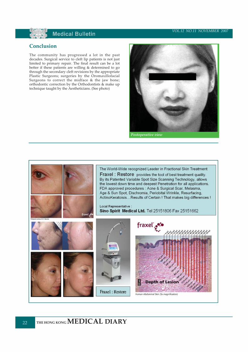
ConclusionThe community has progressed a lot in the pastdecades. Surgical service to cleft lip patients is not justlimited to primary repair. The final result can be a lotbetter if these patients are willing & determined to gothrough the secondary cleft revisions by the appropriatePlastic Surgeons; surgeries by the OromaxillofacialSurgeons to correct the midface & the jaw bone;orthodontic correction by the Orthodontists & make uptechnique taught by the Aestheticians. (See photo)
Postoperative view
VOL.11 NO.5 MAY 2006 Medical Bulletin
23
VOL.12 NO.11 NOVEMBER 2007
IntroductionThe use of flaps with an intact blood supply is theessence of Plastic Surgery. The ability to performsuccessful microvascular repair of vessels as small as1mm enables the transfer of composite tissue from itsdonor site basing on an isolated vascular pedicle andthe flap subsequently revascularises with microvascularanastomosis. This is termed microvascular free tissuetransfer or free flap as the tissue is transplanted fromone part of the body to another.
Since its introduction to clinical use more than 30 yearsago, the use of microvascular free tissue transfer gainspopularity exponentially and now becomes a routinereconstructive procedure even in non-academicinstitutions. Microvascular free tissue transfer is apowerful tool as it allows an almost unlimited choice ofreconstructive methods, replacement of lost tissue withsimilar components and optimal selection of donor sitewith minimal morbidity.
The major deterring factor to embark on the use of freetissue transfer is the need for technical expertise andspecial resources such as operating microscope andmicro-instruments. Microsurgical procedures tend to belengthy and failure will result in a total loss of the flap.The operative experience of the surgical team is acritical factor influencing the success rates and mostsurgeons have to go through a learning curve.
With improvements in the surgical skills, bettermicrosurgical instruments and appropriateidentification of donor sites, the failure rate of free flapsdropped over the past 2 decades. The success rate of theflap reported in recent series ranged from 94.3 to 99.2percent, whereas in earlier series, it ranged from 74% to91% (Khouri 1992, Bui 2007). Failures and vascularcomplications, however still occur, even in experiencedhands. A technical error can be blamed in most casesbut no obvious reason can be located in some.
Over the past 2 decades, the focus of research onmicrovascular surgery shifted from flap anatomy andflap survival towards refinements and choice of flap. Afree flap should have the right components and fulfilthe functional and aesthetic requirements of thereconstructed defect. Plastic Surgeons are no longercontemplated with plugging a hole but thereconstruction has to match the recipient site not only insize but also in texture, form and colour. Attention wasalso paid to minimise the morbidity of the donor site by
a wider choice of donor sites, new minimally invasiveand endoscopic harvesting of flaps and the use ofperforator flaps.
Perforator flaps evolve from musculocutaneous flaps inwhich the muscle carrier is not included because it isnot necessary for the survival of the flap (Geddes 2003).Perforator flaps would have less donor site morbidity,less functional deficit as the muscle is spared andquicker postoperative recovery for the patient.However, it is technically more demanding andmeticulous dissection of the small perforator 1mm orless in diameter is required. It takes longer to harvest aflap and the perforator is more easily damaged than thevascular pedicle of a conventional free flap.
We would like to share our experience of microvascularfree tissue transfer in Kwong Wah Hospital over thepast 12 years.
Patients and MethodsPatient dataThe Division of Plastic Surgery, Department of Surgery,Kwong Wah Hospital performed 412 free tissuetransfers in 395 patients between December 1994 toDecember 2006. There was slight female predominance(228 vs. 167; 58% vs. 42%). The age ranged from 2 to 92,with a median of 53. Eight patients (2%) were youngerthan 18. Eighty-five patients (21%) were 70 or above ofwhom 15 (4%) were 80 or above. Majority of thepatients were non-smoker (83%). There was a history ofirradiation prior to operation in 14% of the patients. Sixpercent had diabetes mellitus (DM) and another 2% hadboth DM and occlusive peripheral vascular disease.
Seventeen patients had 2 free flap operations. In ninepatients a second free flap was performed in the samehospital admission: four had a second free flap forsalvage of flap failure, four had bilateral breastreconstruction and one had a rapid local recurrence oforal cavity cancer that required wide resection andanother free flap for soft tissue coverage. In eightpatients a second free flap was performed in a separatehospital admission: four had resection of localrecurrence, two had another primary tumour and twohad soft tissue problems after irradiation.
Indications for microvascular free tissuetransferIn themajority of the patients, microvascular free tissue
Microvascular Free Flaps: Experience inKwong Wah HospitalDr. Chiu-ming Ho, Dr. Vana SHChan,Dr. Ming-shiaw Cheng, Dr. Wing-yung CheungDivision of Plastic Surgery, Department of Surgery, Kwong Wah Hospital, Kowloon
Dr. Chiu-ming Ho
Medical Bulletin
24
VOL.12 NO.11 NOVEMBER 2007
transfer was used to reconstruct defects or deformitiesafter oncological resection of malignant tumours(381/412, 92%). (Table 1) Head and neck cancer wasthe most common (218/381, 57%), followed by breastcancer (147/381, 39%) and skin cancer (16/381 4%).
The commonest indication for free flaps in non-malignant conditions was the coverage of complicatedwounds (Table 1). This could be an elective treatmentof difficult scars and contractures after burn andtrauma. It was also required in acute situations likeischaemic ulcers in peripheral vascular disease andinfection in diabetes mellitus, where coverage of thewound in the lower extremity was needed to avoidamputation.
The commonest site requiring free flap reconstructionwas the head and neck region (58%), followed by thebreast (36%) and the limbs (6%).
Free flap dataThe different types of flaps according to their vascularpattern and components and the site of origin aretabulated in Tables 2 and 3, respectively. The mostfrequently used free flaps in Kwong Wah Hospital wasthe rectus myocutaneous flap (122), which was usuallyused for breast reconstruction after total mastectomy(Figure 1) The second was fibular flap (72), which wasused for reconstruction after composite oromandibularresection (Figures 2 and 3). The deep inferiorepigastric perforator flap (DIEP) was used for breastreconstruction (58) (Figures 4 and 5) and theanterolateral thigh flap (ALT) (55) was usually usedfor reconstruction of soft tissue defect in the oral cavityand oropharynx (Figures 6 and 7). The visceral freeflap was the jejunal flap used for bridging acircumferential pharyngeal defect between theoropharynx and the oesophagus (Figure 8).
The number of free flaps performed each year was inan increasing trend. The perforator flap wasintroduced in 2001 and gained popularity steadily. Itrepresented 36% of the free flaps used in 2006 and 27%of the free flaps in this series.
ResultsThe overall complication rate was 40% and could bebroken down into general complications (61, 15%), flapcirculation-related complications (24, 6%), andcomplications related to the recipient site (71, 17%)and donor site (33, 8%). (Table 4) Most of thecomplications were minor and could be managedconservatively. Forty three patients (10%) required asecond operation because of the complication whichwas usually due to compromised circulation of theflap (56%) (Table 5)..
Four patients (1%) died in the same admission for freeflap surgery. Two patients died of cardiac events andone had a carotid blow-out 2 weeks after surgery. Thelast patient was an 87 year old patient who had aseries of general complication including pneumonia,urinary tract infection and cerebrovascular accident.He died 2 months after the operation.
The overall success rate of free flaps in our series was95.6%. There were 18 (4.4%) total loss and 8 (2%)partial loss of the flap. Four flap failures weredetected intra-operatively because of failure toestablish an adequate arterial inflow and 14 patientsoccurred postoperatively. In 24 patients (5.8%), re-exploration was needed postoperatively because ofcompromised circulation of the flaps (Table 5). Seventypercent of the flaps were explored within 72 hourspost-operatively. Salvage of the flap was successful in6 (25%) patients and loss of the flap in 14 patients.Partial loss of the flap occurred in 4 patients.
There was neither partial nor total loss of flap inirradiated patients. The rate of partial flap loss andtotal flap loss in diabetic patient was 16% and 4%respectively, compared to 1% and 4% for those whohad DM. For those patients who had both DM andocclusive peripheral vascular disease, the rate ofpartial flap loss and total flap loss was 13% and 25%respectively, much higher than those who did not havesuch diseases. (Table 6)
Two hundred and thirty eight free flaps wereperformed in head and neck region. There were 5partial losses and 9 total losses. The overall successrate was 96.2%. In 148 free flaps for breastreconstruction, there was 1 partial loss and 5 failedflaps and the overall success rate was 96.6%. Thenumber for limb reconstruction was relatively small,22 for lower limb and 4 for upper limb. There were 2partial losses and 4 total losses in lower limb. Thatrepresented 84.6% of overall success rate.
Further wound coverage after failing of flaps wasnecessary in 19 patients (5%). Ten patients requiredregional flaps for coverage, pectorails majormyocutaneous flap in 7, latissimus dorsi flap in 2 anddelto-pectoral flap in 1. Four patients needed skingrafting and 4 patients needed another free flap. Onepatient eventually needed above-knee amputation forsalvage.
DiscussionMicrovascular free flap revolutionised reconstructivesurgery and was indispensable in surgical oncology. Itwas the first choice of reconstructive options incomplex defects after extensive resection of malignantlesions involving multiple tissue components such asskin, mucosa, soft tissue and bone.
The success rate and re-exploration rate of free flaps inthis series was 95.6% and 5.8%, respectively. This wasconsistent with the recent large series reported by wellknown microsurgery centres in the world (Bui 2007). Aprospective study of 493 microvascular free flapsperformed by 23 world famous microvascularsurgeons over 6 months showed a flap failure rate of4.1% and a postoperative thrombosis rate requiring re-exploration of 9.9% (Khouri 1998).
The success rates in the head and neck region and thebreast were comparable but the success rate in limbswas only 84.6%. This was probably related to the fact
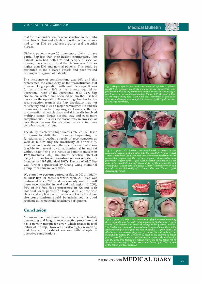
VOL.11 NO.5 MAY 2006 Medical Bulletin
25
VOL.12 NO.11 NOVEMBER 2007
that the main indication for reconstruction in the limbswas chronic ulcer and a high proportion of the patientshad either DM or occlusive peripheral vasculardisease.
Diabetic patients were 20 times more likely to havepartial flap loss than their healthy counterparts. Forpatients who had both DM and peripheral vasculardisease, the chance of total flap failure was 6 timeshigher than DM and normal patients. This could beattributed to the diseased vessels and poor woundhealing in this group of patients.
The incidence of complications was 40% and thisrepresented the complexity of the reconstruction thatinvolved long operation with multiple steps. It wasfortunate that only 10% of the patients required re-operation. Most of the operations (56%) were flapcirculation- related and operated within the first fewdays after the operation. It was a huge burden for thereconstruction team if the flap circulation was notsatisfactory and it was a major commitment to embarkon microvascular free flap surgery. However, the useof conventional pedicle flaps and skin grafts involvedmultiple stages, longer hospital stay and even morecomplications. This was the reason why microvascularfree flaps became the standard of care in thesecomplex reconstructions.
The ability to achieve a high success rate led the PlasticSurgeons to shift their focus on improving thefunctional and aesthetic result of reconstruction aswell as minimising the morbidity of donor site.Koshima and Soeda were the first to show that it wasfeasible to harvest lower abdominal skin and fatwithout sacrificing the rectus abdominis muscle in1989 (Koshima 1989). The clinical beneficial effect ofusing DIEP for breast reconstruction was reported byBlondeel in 1997 (Blondeel 1997). The use of ALT flapwas further popularised by Chang Gung Memorialgroup from Taiwan (Wei 2002).
We started to perform perforator flap in 2001, initiallyas DIEP flap for breast reconstruction. ALT flap wasperformed since 2003 and was mainly used for softtissue reconstruction in head and neck region. In 2006,36% of the free flaps performed in Kwong WahHospital were perforator flaps. With appropriatechoice and application of free flaps not only the donorsite complications could be minimised, a goodaesthetic outcome could be achieved (Figure 9).
ConclusionMicrovascular free tissue transfer is a complicated,demanding and lengthy reconstructive procedure thathas a narrow margin for error, which results in totalfailure of the flap. However it is also highly rewardingand has a high rate of success with acceptableoperative complications.
Fig. 1 (Upper left) Patient with carcinoma of left breast. (Upperright) Skin-sparing mastectomy and axilla dissection wasperformed followed by immediate breast reconstruction using afree transverse rectus abdominis flap. (Lower left) Reconstructionof the nipple using local skin flaps was performed a year laterafter chemotherapy was completed. (Lower right) Nipple areolartattoo was performed.
Fig. 2 (Upper left) Patient presented with a tumor in theretromolar trigone with invasion into the mandible. (Upper center)Tumor approached from the neck and composite resection of theretromolar trigone together with a segment of mandible wasperformed. (Upper right) Defect after resection showing the 2 cutends of the mandible and soft tissue defect of the oral cavity andoropharynx. (Lower left and lower center) Anteroposterior andlateral contour deformity after tumor ablation. (Lower right)Resected specimen.
Fig. 3 (Upper left) Fibular osteocutaneous flap harvested includingthe skin paddle and the underlying segment of fibular bone. (Uppercenter) Flap isolated and elevated basing on the peroneal vessels.The fibular bone was osteotomised into 3 segments and fixed withtitanium miniplates to form the new"mandible". (Upper right) Thefibular osteocutaneous flap was fixed to the cut ends of themandible to restore the occlusion as well as the contour of lowerjaw. (Lower left) A water-tight closure to separate the oral cavityand the neck was formed by suturing the skin of the lower leg tothe cut mucosal edges. (Lower center and lower right) The contourof the lower jaw was restored.
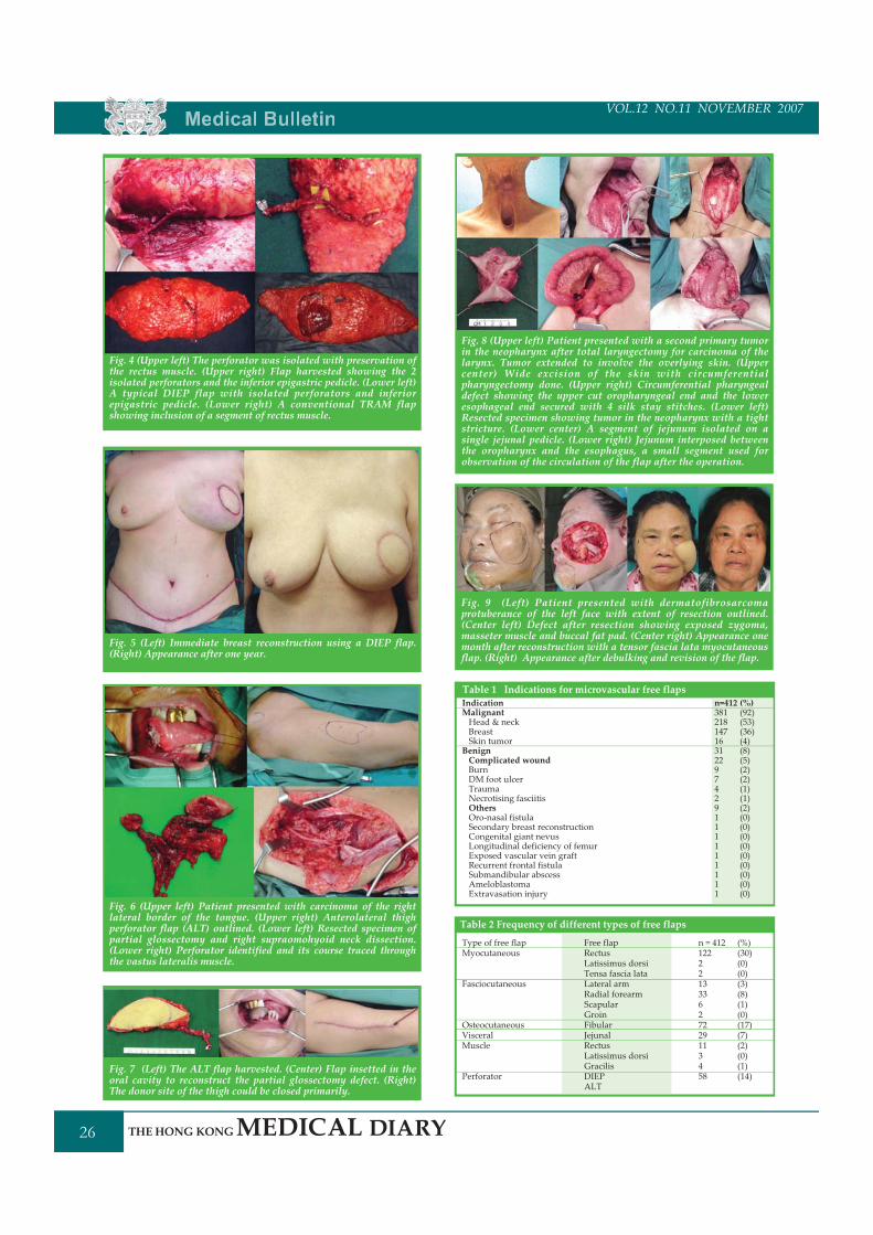
Medical Bulletin
26
VOL.12 NO.11 NOVEMBER 2007
Fig. 8 (Upper left) Patient presented with a second primary tumorin the neopharynx after total laryngectomy for carcinoma of thelarynx. Tumor extended to involve the overlying skin. (Uppercenter) Wide excision of the skin with circumferentialpharyngectomy done. (Upper right) Circumferential pharyngealdefect showing the upper cut oropharyngeal end and the loweresophageal end secured with 4 silk stay stitches. (Lower left)Resected specimen showing tumor in the neopharynx with a tightstricture. (Lower center) A segment of jejunum isolated on asingle jejunal pedicle. (Lower right) Jejunum interposed betweenthe oropharynx and the esophagus, a small segment used forobservation of the circulation of the flap after the operation.
Fig. 4 (Upper left) The perforator was isolated with preservation ofthe rectus muscle. (Upper right) Flap harvested showing the 2isolated perforators and the inferior epigastric pedicle. (Lower left)A typical DIEP flap with isolated perforators and inferiorepigastric pedicle. (Lower right) A conventional TRAM flapshowing inclusion of a segment of rectus muscle.
Fig. 5 (Left) Immediate breast reconstruction using a DIEP flap.(Right) Appearance after one year.
Fig. 6 (Upper left) Patient presented with carcinoma of the rightlateral border of the tongue. (Upper right) Anterolateral thighperforator flap (ALT) outlined. (Lower left) Resected specimen ofpartial glossectomy and right supraomohyoid neck dissection.(Lower right) Perforator identified and its course traced throughthe vastus lateralis muscle.
Fig. 7 (Left) The ALT flap harvested. (Center) Flap insetted in theoral cavity to reconstruct the partial glossectomy defect. (Right)The donor site of the thigh could be closed primarily.
Fig. 9 (Left) Patient presented with dermatofibrosarcomaprotuberance of the left face with extent of resection outlined.(Center left) Defect after resection showing exposed zygoma,masseter muscle and buccal fat pad. (Center right) Appearance onemonth after reconstruction with a tensor fascia lata myocutaneousflap. (Right) Appearance after debulking and revision of the flap.
Table 1 Indications for microvascular free flapsIndicationMalignantHead & neckBreastSkin tumorBenignComplicated woundBurnDM foot ulcerTraumaNecrotising fasciitisOthersOro-nasal fistulaSecondary breast reconstructionCongenital giant nevusLongitudinal deficiency of femurExposed vascular vein graftRecurrent frontal fistulaSubmandibular abscessAmeloblastomaExtravasation injury
n=412 (%)381 (92)218 (53)147 (36)16 (4)31 (8)22 (5)9 (2)7 (2)4 (1)2 (1)9 (2)1 (0)1 (0)1 (0)1 (0)1 (0)1 (0)1 (0)1 (0)1 (0)
Table 2 Frequency of different types of free flaps
Type of free flapMyocutaneous
Fasciocutaneous
OsteocutaneousVisceralMuscle
Perforator
Free flapRectusLatissimus dorsiTensa fascia lataLateral armRadial forearmScapularGroinFibularJejunalRectusLatissimus dorsiGracilisDIEPALT
n = 412 (%)122 (30)2 (0)2 (0)13 (3)33 (8)6 (1)2 (0)72 (17)29 (7)11 (2)3 (0)4 (1)58 (14)

VOL.11 NO.5 MAY 2006 Medical Bulletin
27
VOL.12 NO.11 NOVEMBER 2007
Table 3 Frequency of different types of microvascular free flapsperformed between 1994 and 2006Year
1994199519961997199819992000200120022003200420052006Total
General
Flap circulation-related
Recipient site
Donor site
n = 412 (%)61 (15)23 (6)12 (3)6 (1)6 (1)3 (1)3 (1)2 (0)2 (0)6 (1)24 (5)12 (2)9 (2)2 (0)1 (0)71 (17)25 (6)16 (4)12 (3)10 (2)7 (2)1 (0)33 (8)19 (5)7 (2)4 (1)2 (0)1 (0)
Type of flapOsteocutaneous
02336595711371172
Myocutaneous1918271012545191220124
Fasciocutaneous065886546111354
Visceral024244132131229
Muscle000203313420220
n
1193017252830314245513459412
Perforator0000000132023231321113
Table 4 Complication rates of microvascular free flapsComplication
Chest infectionUrinary tract infectionCardiac arrythmiaMyocardiac infarctionCerbrovascular accidentPleural effusionPneumothoraxAcute retention of urineMiscellaneous
Flap congestionFlap ischemia/failureSkin island necrosisAnastomotic blow-out
Wound infectionWound edge necrosisSeromaHematomaFistula/anastomotic leakMiscellaneous
Wound infectionLoss of skin graftSkin necrosis/dehiscenceFat necrosisMiscellaneous
Risk factor
Past healthDMDM + PVDControl
SiteHead & neckBreastLower limbUpper limb
Type of flapMyocutaneousFasciocutaneousOsteocutaneousVisceralMusclePerforator
Table 6 Risk factors with reference to complicationsn=412
258379
238148224
12454722920113
Partial flap loss (%)84 (16)1 (13)3 (1)85 (2)1 (1)2 (10)0 (0)83 (2)1 (2)2 (3)0 (0)2 (10)0 (0)
Total flap loss (%)181 (4)2 (25)15 (4)189 (4)5 (3)4 (19)0 (0)183 (2)5 (9)3 (4)1 (3)1 (5)5 (4)
Flap circulation-relatedFlap congestionFlap ischemia/failureSkin island necrosisAnastomotic blow-out
Recipient sitePartial flap lossFistula/wound infectionOsteomyelitisWound necrosisChyle leakageAvulsed sentinel jejunal loop
Donor siteWound infection/necrosis
GeneralGIB / acalculous cholecystitis
n=43 (%)24 (56)1292115 (35)5422113 (7)31 (2)1
Table 5 Indications for re-operation in microvascular free flapsn=43 (%)24 (56)1292115 (35)5422113 (7)31 (2)1
Complication
Blondeel PN, Vanderstraeten GG, Monstrey SJ et al. The donor sitemorbidity of free DIEP flaps and free TRAM flaps for breastreconstruction. Br J Plast Surg 50:322, 1997.Bui DT, Cordeiro PG, Hu QY, Disa JJ, Pusic A, Mehrara BJ. Freeflap re-exploration: indications, treatment, and outcomes in 1193free flaps. Plast Reconstr Surg 119:2092-100, 2007.Geddes CR, Morris SF, Neligan PC. Perforator flaps: evolution,classification and applications. Ann Plast Surg 50:90-9, 2003.Khouri R. Avoiding free flap failure. Clin Plast Surg 19:773 1992.Khouri RK, Cooley BC, Kunselman AR, Landis JR, Yeramian P,Ingram D, Natarajan N, Bened CO, Wallemark C. A prospectivestudy of microvascular free-flap surgery and outcome. Plastreconstr Surg 102:711-21, 1998.Koshima I, Soeda S. Inferior epigastric artery skin flap withoutrectus abdominis muscle. Br J Plast Surg 42:645-648, 1989.Wei FC, Jain V, Celik N et al. Have we found an ideal soft tissueflap? An experience with 672 anterolateral thigh flaps. PlastReconst Surg 109:2219, 2002.
References
Upcoming Certificate Courses of the Federation of Medical Societies of Hong Kong
22 Nov 07 - 13 Dec 07
3 Jan 08 - 31 Jan 08
Nil Medical Health Professionals &Practitioners
Clinical Management of VertigoC126
C127
Date Coursrr e No Co-organiserCourse Name TaTT rget Participants
VacanciesRentalOffice VacanciesFor Sale(flat, car, office, etc.)
Please contact the Federation Secretariat at 25278898 for placement of classified advertisement.( Charge: HK$ 600 per 30 word unit, HK$50 peradditional 10 word unit)
Classified Advertisement

VOL.11 NO.5 MAY 2006 Medical Bulletin
29
VOL.12 NO.11 NOVEMBER 2007
Figure 1 -Improvement of left lateral acne scars, before (left) andpost-treatment (right)
Figure 2 -Improvement of right peri-orbital acne scars, before(left) and post-treatment (right)
IntroductionAcne is a common disease and has been estimated toaffect 95-100% of 16-17 year old boys and 83-85% of 16-17 year old girls. Acne resolves in the vast majority by23-25 years of age, but 1% of males and 5% of femalescontinue to exhibit acne lesions at 40 years of age.1-5
There are many factors that influence the onset of acneand the eventual sequela of acne scarring. There doesappear to be a hereditary component to acne, but it alsois strongly influenced by external factors.6-7
Acne is highly embarrassing for adolescents. Post-acnescarring is particularly devastating. These scars appearas multiple depressive scars, cause problemscosmetically and impact self-confidence, especially inyoung people.8
Post-acne scars are dermal depressions that are mostcommonly caused by collagen damage. Many differenttreatments including chemical peels, surgical excision,punch grafting, dermabrasion, and tissue augmentationwith a variety of filler substances, have been used totreat post-acne scars with varying degrees ofsuccess.9-10
Recontouring of post-acne scars with CO2 and Er: YAGlasers have become popular over the last decade.Though more effective than other methods due to itsprecision with depth control and variable methods ofsurface cutting, laser resurfacing has an extended post-laser recovery period and the potential risks of delayedwound healing, pigmentary changes, and scarring.8, 11
These procedures have not been widely accepted inAsian countries due to the increase in post inflammatoryhyperpigmentation. In fact, these complications anddowntime can sometimes last more than one year. Somepost inflammatory hyperpigmentation may bepermanent. For these reasons Asian patients have beenless willing to undergo ablative laser skin resurfacing.12-13
In general, Asian skin types have a tendency to developpigmentation and other complications with mostconventional skin rejuvenation procedures.
In a previous study conducted at the National SkinCenter in Singapore a 1450 nm nonablative diode laserwas found to result in clinical improvement of atrophicacne scars in Asian skin types after 4-6 treatments witha fluence of 11-12 J/cm2.
However, in this study the side effect of postlaserhyperpigmentation was significant. The possiblecontributing factor to this postlaser hyperpigmentationmay be the high fluence used in the aforementionedstudy. Pain experienced during treatment was alsosignificant in this study despite the use of pre-treatmentanesthetic cream and cryogen precooling.15
It is for these reasons that novel treatments for acne scarsin Asian skin types have been sought.14
MethodsThe objective of this study is to evaluate the performanceof the Affirm, 1440 nm Nd: YAG laser device withCombined Apex Pulse (CAP) technology in the treatmentof ten subjects of Asian skin type presenting with acnescars.
Subjects were evaluated and photographed prior to theinitiation of treatment, prior to each subsequent
Treatment of Acne Scars in Asian Skin with AffirmCombined Apex Pulse TechnologyDr. Connia LYHo
Specialist in DermatologyMB,BS, MRCP, FHKCP, FHKAM(Med), Dip. Derm(London)
Medical Bulletin
30
VOL.12 NO.11 NOVEMBER 2007
treatment, and at one and three months following thefinal treatment. Subjects received five treatments at 3-4week intervals. Prior to treatment the skin was cleanedand any makeup was removed.
Subjects were treated using the Affirm laser system. Thestrong absorption of the 1440 nm wavelength makes theAffirm a safe and effective treatment of the area of solarelastosis (100-300 microns) and deeper penetration intothe tissue is rendered unnecessary. The CAP array in the1440 nm Nd:YAG Affirm laser system was developed tocombine the effects of both high-fluence aggressiveheating resulting in collagen remodeling and themechanism of low-fluence low level heating resulting incollagen stimulation.
The CAP array consists of a special lens which createsapexes of high-fluence regions surrounded by low-fluence treatment zones. The high-fluence apexes create apattern of columns surrounded by low-fluencebackground heat. This combination improves treatmentefficacy while maintaining the existing side effects profile.
The Affirm 1440 nm wavelength was delivered using the10 mm spot size with the CAP array and a 3 msec pulseduration. Treatment fluences ranged from 2.5-4.0 J/cm2,and 1-2 Hz pulse repetition rate.
Initial subject treatment fluences were 2.5-3.0 J/cm2.Treatment fluences increased with each subsequenttreatment based upon patient tolerance. Subjectsreceived two treatment passes. All treatments weredelivered in conjunction with SmartCoolTM (Cynosure,Inc., Westford, MA) cold-air cooling, with a fan speed of3 or 4. No additional anesthesia was required. The CAParray tip requires full contact with the skin for safe andeffective treatment. When treating acne scars,compression is used to flatten the scar, for more directdelivery of the laser energy to the area. Subjects couldresume normal activities following treatment.
Ten subjects participated in this investigation. Nine ofthe ten subjects completed all five treatmentprocedures. The remaining one patient completed herfourth treatment. All ten subjects have returned fortheir one-month post-treatment evaluation. As of 7May 2007 two patients have returned for their three-month post-treatment evaluation.
ResultsWhile evaluating the treatment of ten subjects usingthe Affirm laser system at 1440 nm wavelength animprovement in acne scars was noted.
Improvement in enlarged pores, dyschromia, postinflammatory hyperpigmentation (PIH), skin color,pigmentation and skin texture also was noted. Patientstolerated the procedures well and were very satisfiedwith the outcome.
Of the ten subjects treated eighteen distinct treatmentareas were evaluated. Treatment area photos weregraded according to the following scale: 0 = noimprovement, 1 = mild improvement, 2 = moderateimprovement, 3 = good improvement, 4 = excellentimprove-ment. Of these twenty seven treatment areasall showed some improvement. No treatment areasshowed less than a 25% improvement. Seven treatmentareas showed a 25-50% improvement, seven showed a50-75% improvement and four showed a 75-100%improvement. The overall average improve-ment forall treatment areas was 50-75%.
Pain experienced during the procedure was minimalto moderate. Patient discomfort was evaluated on ascale of 0-5. Only one patient reported experiencingdiscomfort of 4 during just one of the treatments.Patients reported an average discomfort level of 2.3.No topical anesthesia was used during treatments.
Postlaser hyperpigmentation was minimal with manypatients seeing improvement in existing postinflammatory hyperpigmentation.
DiscussionTreatment with the Affirm laser system at 1440 nmwavelength is an effective treatment for acne scars.Improvement can also be observed in enlarged pores,dyschromia, postinflammatory hyperpigmentation,
Figure 3 -Improvement of left lateral acne scars, before (left) andpost-treatment (right)
Figure 5 - Improvement of left lateral acne scars,before (left) and post-treatment (right)
Figure 4 -Improvement of right peri-orbital scars, before (left)and post-treatment (right)
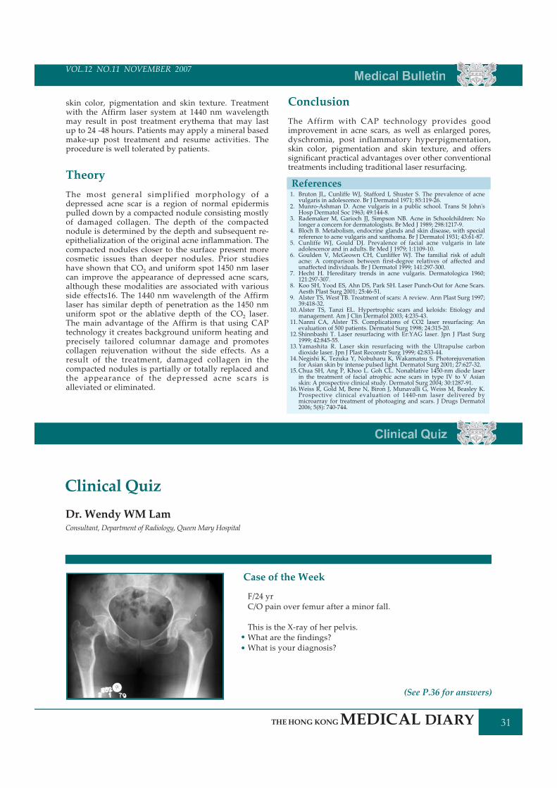
VOL.11 NO.5 MAY 2006 Medical Bulletin
31
VOL.12 NO.11 NOVEMBER 2007
Bruton JL, Cunliffe WJ, Stafford I, Shuster S. The prevalence of acnevulgaris in adolescence. Br J Dermatol 1971; 85:119-26.Munro-Ashman D. Acne vulgaris in a public school. Trans St John'sHosp Dermatol Soc 1963; 49:144-8.Rademaker M, Garioch JJ, Simpson NB. Acne in Schoolchildren: Nolonger a concern for dermatologists. Br Med J 1989; 298:1217-9.Bloch B. Metabolism, endocrine glands and skin disease, with specialreference to acne vulgaris and xanthoma. Br J Dermatol 1931; 43:61-87.Cunliffe WJ, Gould DJ. Prevalence of facial acne vulgaris in lateadolescence and in adults. Br Med J 1979; 1:1109-10.Goulden V, McGeown CH, Cunliffer WJ. The familial risk of adultacne: A comparison between first-degree relatives of affected andunaffected individuals. Br J Dermatol 1999; 141:297-300.Hecht H. Hereditary trends in acne vulgaris. Dermatologica 1960;121:297-307.Koo SH, Yood ES, Ahn DS, Park SH. Laser Punch-Out for Acne Scars.Aesth Plast Surg 2001; 25:46-51.Alster TS, West TB. Treatment of scars: A review. Ann Plast Surg 1997;39:418-32.Alster TS, Tanzi EL. Hypertrophic scars and keloids: Etiology andmanagement. Am J Clin Dermatol 2003; 4:235-43.Nanni CA, Alster TS. Complications of CO2 laser resurfacing: Anevaluation of 500 patients. Dermatol Surg 1998; 24:315-20.Shinnbashi T. Laser resurfacing with Er:YAG laser. Jpn J Plast Surg1999; 42:845-55.Yamashita R. Laser skin resurfacing with the Ultrapulse carbondioxide laser. Jpn J Plast Reconstr Surg 1999; 42:833-44.Negishi K, Tezuka Y, Nobuharu K, Wakamatsu S. Photorejuvenationfor Asian skin by intense pulsed light. Dermatol Surg 2001; 27:627-32.Chua SH, Ang P, Khoo L. Goh CL. Nonablative 1450-nm diode laserin the treatment of facial atrophic acne scars in type IV to V Asianskin: A prospective clinical study. Dermatol Surg 2004; 30:1287-91.Weiss R, Gold M, Bene N, Biron J, Munavalli G, Weiss M, Beasley K.Prospective clinical evaluation of 1440-nm laser delivered bymicroarray for treatment of photoaging and scars. J Drugs Dermatol2006; 5(8): 740-744.
1.
2.
3.
4.
5.
6.
7.
8.
9.
10.
11.
12.
13.
14.
15.
16.
References
skin color, pigmentation and skin texture. Treatmentwith the Affirm laser system at 1440 nm wavelengthmay result in post treatment erythema that may lastup to 24 -48 hours. Patients may apply a mineral basedmake-up post treatment and resume activities. Theprocedure is well tolerated by patients.
TheoryThe most general simplified morphology of adepressed acne scar is a region of normal epidermispulled down by a compacted nodule consisting mostlyof damaged collagen. The depth of the compactednodule is determined by the depth and subsequent re-epithelialization of the original acne inflammation. Thecompacted nodules closer to the surface present morecosmetic issues than deeper nodules. Prior studieshave shown that CO2 and uniform spot 1450 nm lasercan improve the appearance of depressed acne scars,although these modalities are associated with variousside effects16. The 1440 nm wavelength of the Affirmlaser has similar depth of penetration as the 1450 nmuniform spot or the ablative depth of the CO2 laser.The main advantage of the Affirm is that using CAPtechnology it creates background uniform heating andprecisely tailored columnar damage and promotescollagen rejuvenation without the side effects. As aresult of the treatment, damaged collagen in thecompacted nodules is partially or totally replaced andthe appearance of the depressed acne scars isalleviated or eliminated.
ConclusionThe Affirm with CAP technology provides goodimprovement in acne scars, as well as enlarged pores,dyschromia, post inflammatory hyperpigmentation,skin color, pigmentation and skin texture, and offerssignificant practical advantages over other conventionaltreatments including traditional laser resurfacing.
VOL.11 NO.5 MAY 2006 Clinical Quiz
(See P.36 for answers)
Clinical QuizDr. WendyWM LamConsultant, Department of Radiology, QueenMary Hospital
F/24 yrC/O pain over femur after a minor fall.
This is the X-ray of her pelvis.What are the findings?What is your diagnosis?
Case of the Week
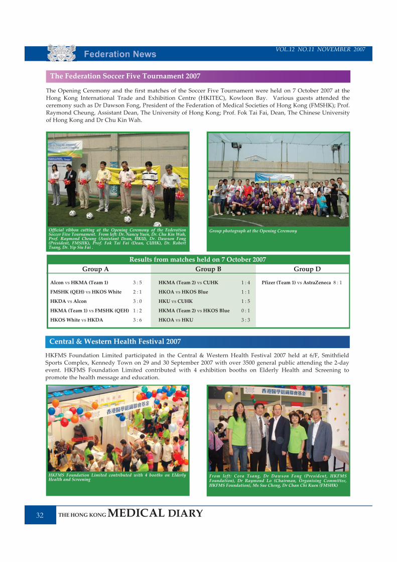
Federation News
32
VOL.12 NO.11 NOVEMBER 2007
The Federation Soccer Five Tournament 2007
The Opening Ceremony and the first matches of the Soccer Five Tournament were held on 7 October 2007 at theHong Kong International Trade and Exhibition Centre (HKITEC), Kowloon Bay. Various guests attended theceremony such as Dr Dawson Fong, President of the Federation of Medical Societies of Hong Kong (FMSHK); Prof.Raymond Cheung, Assistant Dean, The University of Hong Kong; Prof. Fok Tai Fai, Dean, The Chinese Universityof Hong Kong and Dr Chu Kin Wah.
Group photograph at the Opening Ceremony
Central & Western Health Festival 2007
HKFMS Foundation Limited participated in the Central & Western Health Festival 2007 held at 6/F, SmithfieldSports Complex, Kennedy Town on 29 and 30 September 2007 with over 3500 general public attending the 2-dayevent. HKFMS Foundation Limited contributed with 4 exhibition booths on Elderly Health and Screening topromote the health message and education.
HKFMS Foundation Limited contributed with 4 booths on ElderlyHealth and Screening
From left: Cora Tsang, Dr Dawson Fong (President, HKFMSFoundation), Dr Raymond Lo (Chairman, Organising Committee,HKFMS Foundation), Ms Sue Cheng, Dr Chan Chi Kuen (FMSHK)
Results from matches held on 7 October 2007Group A Group B Group D
Alcon vs HKMA (Team 1) 3 : 5
FMSHK (QEH) vs HKOSWhite 2 : 1
HKDA vs Alcon 3 : 0
HKMA (Team 1) vs FMSHK (QEH) 1 : 2
HKOSWhite vs HKDA 3 : 6
HKMA (Team 2) vs CUHK 1 : 4
HKOA vs HKOS Blue 1 : 1
HKU vs CUHK 1 : 5
HKMA (Team 2) vs HKOS Blue 0 : 1
HKOA vs HKU 3 : 3
Pfizer (Team 1) vs AstraZeneca 8 : 1
Official ribbon cutting at the Opening Ceremony of the FederationSoccer Five Tournament. From left: Dr. Nancy Yuen, Dr. Chu Kin Wah,Prof. Raymond Cheung (Assistant Dean, HKU), Dr. Dawson Fong(President, FMSHK), Prof. Fok Tai Fai (Dean, CUHK), Dr. RobertTsang, Dr. Yip Siu Fai .
VOL.11 NO.5 MAY 2006 Federarr tion News
Societytt News
33
VOL.12 NO.11 NOVEMBER 2007
News fromMember Societies:
Hong Kong Paediatric Haematology and Oncology Study Group
Updated office-bearers for the year 2007-2008 are as follows: Chairman: Dr. CHIANG Kwok-shing, Alan, Vice-Chairman: Dr. YAU Ping-wa, Secretary: Dr Vincent LEE, Treasurer: Dr. LI Chak-ho, Rever, Council Members: DrCHAN Chi-fung, Godfrey, Dr LING Siu-cheung, Dr SHING Ming-kong, Dr YUEN Hui-leung
To promote the study and advancement of the science and practice of psychiatry andancillary sciences and branches of medicineTo contribute to the improvement of mental health care for Hong Kong citizensthrough the provision of specially trained psychiatristsTo ensure the highest professional standard of competence and ethical integrity inPsychiatry
The forerunner of the Hong Kong College of Psychiatrists was the Hong KongPsychiatric Association. The College was founded in September 1990 with the followingobjectives:
Apart from the above objectives, the College has been, in the past year, devoting more emphasis on publiceducation with a view to raising public awareness on the importance of mental health as well as contributingtowards de-stigmatisation of mental disorder. In addition, the College has also engaged the government andsubmitted our views on the future development of mental health development in Hong Kong to the Secretary ofFood and Health and one of the suggestions is enhanced collaboration with the primary care sector to ensurequality psychiatric service at all levels.
(1)
(2)
(3)Dr.Se-fong Hung
Founded in 1981, the Hong Kong Geriatrics Society is the only local professional society for doctors practicinggeriatric medicine. There are up to now 180 members with 42 associate members. Geriatricians areresponsible for the management of acute and chronic illnesses, including severe disability and terminalconditions in older people. The Society's objectives are to promote research, training and development ingeriatric medicine, and to improve the standard of care for older people.
In order to attract more young people to develop an interest in care for the elderly to meet the growingdemand of a rapidly aging population, the Society have now set up scholarship prizes, one for each of the twolocal medical schools, to facilitate medical students to undergo elective period in geriatric medicine overseas.With the introduction of the sponsorship, we are hoping to "catch them young" and sow seeds in their heartsby facilitating medical students' earlier exposure to reputable academic as well as service geriatric units.Applications should be made via the scholarship application offices of the 2 universities respectively.
Hong Kong Geriatrics Society
The Hong Kong College of Psychiatrists
The new FMSHK Team (Administrative Officer - Miss. Paulina P.K. Tang)
Miss Paulina Tang has been appointed as the Administrative Officer of FMSHK since 8October 2007, a role she comes to after 13 years experiences in the banking industry. Shehas been active in volunteer services, thus she is bringing along with her invaluableexperience and skills that can be an asset to our Federation.
Miss Tang is energetic and dedicated to serve the evolving needs of our member societies.
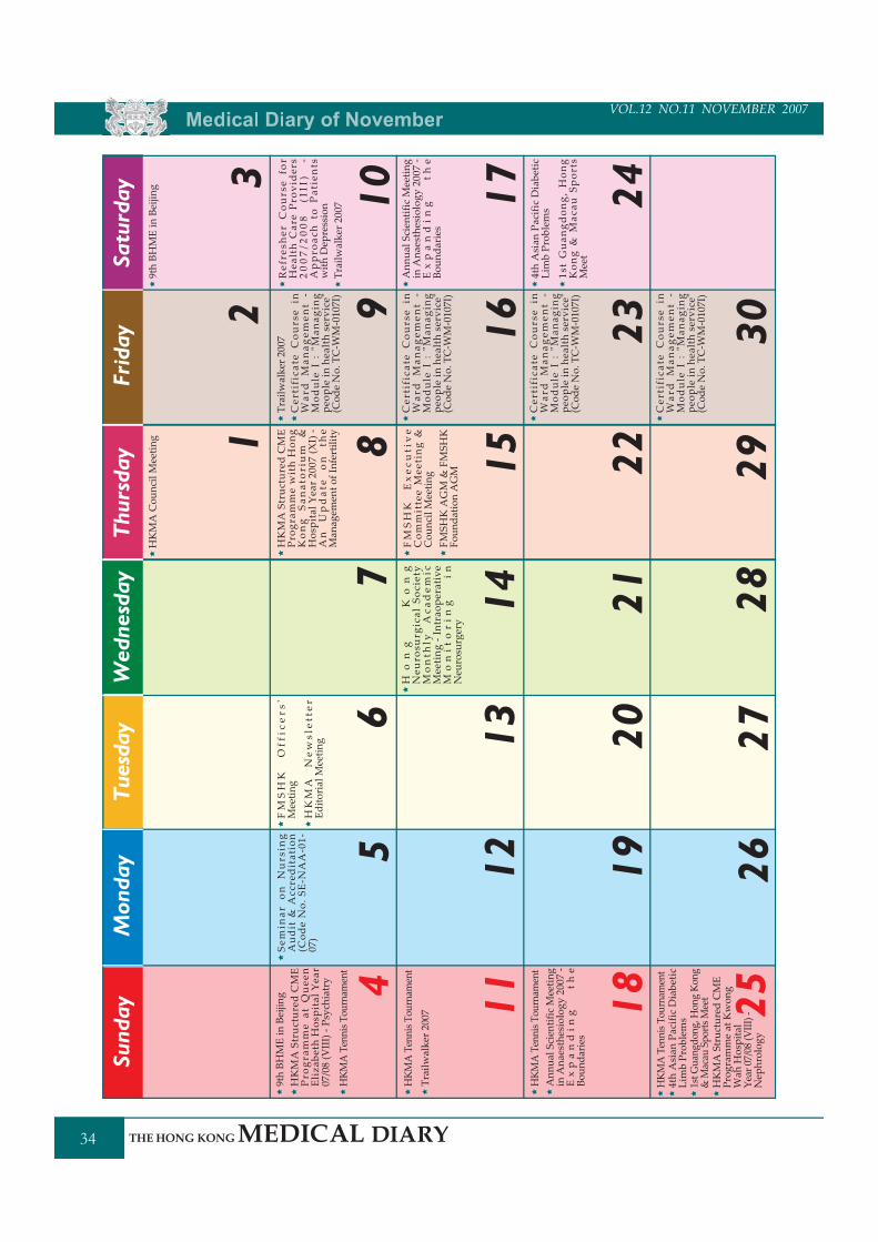
Medical Diaryrr of November
34
VOL.12 NO.11 NOVEMBER 2007Sunday
Monday
Tuesday
Wednesday
Thursday
Friday
Saturday
1516
17
18 25
1920
10
1112
13
7 14
89
45
21
6
31
2
2223
2829
30
24
2627
HKMACou
ncilMeetin
g9thBH
MEin
Beijing
Trailw
alker2
007
Trailw
alker2
007
Cer
tifica
teCou
rse
inW
ard
Man
agem
ent-
Mod
ule
I:"M
anag
ing
peop
lein
health
service"
(Cod
eNo.TC
-WM-0107I)
Cer
tifica
teCou
rse
inW
ard
Man
agem
ent-
Mod
ule
I:"M
anag
ing
peop
lein
health
service"
(Cod
eNo.TC
-WM-0107I)
Ann
ualS
cientificMeetin
gin
Ana
esthesiology
2007
-Expandin
gth
eBo
unda
ries
4thAsian
Pacific
Diabetic
LimbProb
lems
1stGuan
gdon
g,Hon
gKon
g&
Mac
auSp
orts
Meet
4thAsian
Pacific
Diabetic
LimbProb
lems
1stG
uang
dong
,Hon
gKon
g&Macau
SportsMeet
HKMAStructured
CME
Prog
rammeat
Kwon
gWah
Hospital
Year
07/08(VIII)-
Nep
hrolog
y
Cer
tifica
teCou
rse
inW
ard
Man
agem
ent-
Mod
ule
I:"M
anag
ing
peop
lein
health
service"
(Cod
eNo.TC
-WM-0107I)
Cer
tifica
teCou
rse
inW
ard
Man
agem
ent-
Mod
ule
I:"M
anag
ing
peop
lein
health
service"
(Cod
eNo.TC
-WM-0107I)
Refre
sher
Cou
rse
for
Hea
lthCarePro
viders
2007/2
008
(III
)-
Appro
ach
toPatients
with
Dep
ression
Trailw
alker2
007
9thBH
MEin
Beijing
HKMA
Stru
ctur
edCME
Pro
gram
me
atQuee
nElizab
ethHospitalY
ear
07/08(VIII)-P
sychiatry
Ann
ualScientificMeetin
gin
Ana
esthesiology
2007
-Expandin
gth
eBo
unda
ries
Semin
aron
Nursin
gAudit
&Accreditation
(Cod
eNo.
SE-N
AA-01-
07)
FM
SHK
Off
icers
'Meetin
gHKMA
Stru
ctur
edCME
Prog
rammewithHon
gKong
Sanat
orium
&HospitalY
ear2007
(XI)-
An
Updat
eon
the
Man
agem
ento
fInfertility
FM
SHK
Execu
tive
Com
mitteeMee
ting
&Cou
ncilMeetin
gFM
SHKAGM
&FM
SHK
Foun
datio
nAGM
Ho
ng
Ko
ng
Neu
rosu
rgical
Society
Month
lyAca
dem
icMeeting
-Intraop
erative
Monit
orin
gin
Neu
rosurgery
HKM
ANew
slet
ter
Edito
rialMeetin
g
HKM
ATenn
isTo
urnament
HKM
ATenn
isTo
urnament
HKM
ATenn
isTo
urnament
HKM
ATenn
isTo
urnament
VOL.11 NO.5 MAY 2006 Medical Diaryrr of November
35
VOL.12 NO.11 NOVEMBER 2007
FMSHK AGM & FMSHK Foundation AGMOrganised by: The Federation of Medical Societies of Hong Kong # Lecture Hall, 4/F.,Duke of Windsor Social Service Building, 15 Hennessy Road, Wanchai, Hong Kong
SecretariatTel: 2527 8898 Fax: 2865 0345
8:45 pm - 10:00 pm
8 THUMiss Viviane LAMTel: 2527 8452(Registration fee is required)1 CME Point
HKMA Structured CME Programme with Hong Kong Sanatorium & Hospital Year2007 (XI) - An Update on the Management of InfertilityOrganised by: The Hong Kong Medical Association & Hong Kong Sanatorium &Hospital Speaker: Dr. TANG Oi Shan # HKMA Dr. Li Shu Pui Professional EducationCentre, 2/F., Chinese Club Building, 21-22 Connaught Road Central, Hong Kong
2:00 pm
15 THUFMSHK Executive Committee Meeting & Council MeetingOrganised by: The Federation of Medical Societies of Hong Kong # Council Chambers,4/F., Duke of Windsor Social Service Building, 15 Hennessy Road, Wanchai, Hong Kong
SecretariatTel: 2527 8898 Fax: 2865 0345
(4)
(11,18,25)
(10,11)
25 SUNHKMA Structured CME Programme at Kwong Wah Hospital Year 07/08 (VIII) -NephrologyOrganised by: The Hong Kong Medical Association & Kwong Wah Hospital Speaker:Dr. LO Kin Yee & Dr. TONGMei Wah # Lecture Theatre, 10/F., Yu Chun KeungMemorial Medical Centre, Kwong Wah Hospital
2:00 pm Miss Viviane LAMTel: 2527 8452(Registration fee is required)3 CME Points
1st Guangdong, Hong Kong & Macau Sports MeetOrganised by: The Hong Kong Medical Association Chairman: Dr. H YEUNG # Macau
Ms. Dora HOTel: 2527 8285
24 SAT4th Asian Pacific Diabetic Limb ProblemsOrganised by: Various # Wiliam MWMong Block, Li Ka Shing Faculty of Medicine, TheUniversity of Hong Kong, 21 Sassoon Road, Hong Kong
Website:http://www.diabeticlimb.hk/
17 SATAnnual Scientific Meeting in Anaesthesiology 2007 - Expanding the BoundariesOrganised by: The Hong Kong College of Anaesthesiology & The Society ofAnaesthetists of Hong Kong # Hong Kong Convention and Exhibition Centre
Secretariat (CMPMedica PacificLimited)Tel: 2559 5888 Fax: 2559 6910Email:[email protected]:www.hkca.edu.hk/asm2007.htm
14 WEDHong Kong Neurosurgical Society Monthly Academic Meeting - IntraoperativeMonitoring in NeurosurgeryOrganised by: Hong Kong Neurosurgical Society Chairman: Dr. TSE Tat Shing Speaker:Dr. LAW Hing Yuen # Seminar Room, G/F, Block A, Queen Elizabeth Hospital,Kowloon
Dr. Y.C. POTel: 2990 3788 Fax: 2990 37892 CME PointsCollege of Surgeons of HK
10 SATRefresher Course for Health Care Providers 2007/2008 (III) - Approach to Patientswith DepressionOrganised by: The Hong Kong Medical Association & Our Lady of Maryknoll HospitalSpeaker: Dr. CHAN Sai Yin # Training Room II, 1/F., OPD Block, Our Lady ofMaryknoll Hospital, 118 Shatin Pass Road, Wong Tai Sin, Kowloon
Ms. Clara TSANGTel: 2354 24402 CME Points
3 SATMs. Candy YUENTel: 2527 8285
9th BHME in BeijingOrganised by: The Hong Kong Medical Association Chairman: Dr. T.C. SHIH & Dr.S.W. LI
5 MONSecretariatTel: 2572 9255 Fax: 2838 62803 CNE Points
Seminar on Nursing Audit & Accreditation (Code No. SE-NAA-01-07)Organised by: College of Nursing, Hong Kong
Date / Time Function Enquiry / Remarks
1 THUMs. Christine WONGTel: 2527 8285
HKMA Council MeetingOrganised by: The Hong Kong Medical Association Chairman: Dr. K CHOI # HKMAHeadOffice, 5/F., Duke of Windsor Social Service Building, 15 Hennessy Road, Wanchai, Hong Kong
8:00 pm
4 SUNMiss Viviane LAMTel: 2527 8452(Registration fee is required)3 CME Points
HKMA Structured CME Programme at Queen Elizabeth Hospital Year 07/08 (VIII) -PsychiatryOrganised by: The Hong Kong Medical Association & Queen Elizabeth Hospital Speaker:Dr. CHU Lap Sun, Dr. WONGMan Tak & Dr. LAMWai Keung # Lecture Theatre, G/F.,Block M, Queen Elizabeth Hospital, Kowloon
2:00 pm
6:30pm to 9:30pm
6 TUESecretariatTel: 2527 8898 Fax: 2865 0345
FMSHK Officers' MeetingOrganised by: The Federation of Medical Societies of Hong Kong # Gallop, 2/F., HongKong Jockey Club Club House, Shan Kwong Road, Happy Valley, Hong Kong
Ms. Tammy TAMTel: 2527 8941
HKMA Newsletter Editorial MeetingOrganised by: The Hong Kong Medical Association Chairman: Dr. H.H. TSE # HKMAHead Office, 5/F., Duke of Windsor Social Service Building, 15 Hennessy Road,Wanchai, Hong Kong
9 FRIMs. Dora HOTel: 2527 8285
Trailwalker 2007Organised by: The Hong Kong Medical Association Chairman: Dr. Y.H. CHOW #MacLehose Trail
SecretariatTel: 2572 9255 Fax: 2838 628024 CNE Points
Certificate Course in Ward Management - Module I : "Managing people in healthservice" (Code No. TC-WM-0107I)Organised by: College of Nursing, Hong Kong
9:00 am
2:30 pm
7:30 am
7:00 pm - 8:45 pm
(16,23,30)
(18)
(25)
(25)
6:30pm to 9:30pm
8:00 pm - 10:00pm
8:00 pm
Ms. Dora HOTel: 2527 8285
HKMA Tennis TournamentOrganised by: The Hong Kong Medical Association Chairman: Dr. C.W. CHIN # KowloonTong Club
7:30 pm
Calendar of Events
36
VOL.12 NO.11 NOVEMBER 2007
MeetingsHong Kong Surgical Forum, Winter 2008Organised by: Department of Surgery, Li Ka Shing Faculty of Medicine, University of Hong Kong Medical Centre; Queen MaryHospital & Hong Kong Chapter of the American College of Surgeons # Underground Lecture Theatre, New Clinical Building,Queen Mary Hospital, Pokfulam, Hong Kong Enquiry: Forum Secretary Tel: 2855 4885 Fax: 2819 3416 Email:[email protected] Website: http://www.hku.hk/surgery
Hong Kong Surgical Forum, Summer 2008Organised by: Department of Surgery, Li Ka Shing Faculty of Medicine, University of Hong Kong Medical Centre; Queen MaryHospital & Hong Kong Chapter of the American College of Surgeons # Underground Lecture Theatre, New Clinical Building,Queen Mary Hospital, Pokfulam, Hong Kong Enquiry: Forum Secretary Tel: 2855 4885 Fax: 2819 3416 Email:[email protected] Website: http://www.hku.hk/surgery
11-12/1/2008
11-12/7/2008
CoursesWorkshop on Neonatal Touch and its updated information (Code No. SE-NT-0107)Organised by: College of Nursing, Hong Kong Enquiry: Secretariat Tel: 2572 9255 Fax: 2838 6280
Certificate Course in Ward Management - Module II: "Managing resources in health service" (Code No. TC-WM-0107II)Organised by: College of Nursing, Hong Kong Enquiry: Secretariat Tel: 2572 9255 Fax: 2838 6280
Certificate Course in Ward Management - Module III: "Managing risk at workplace" (Code No. TC-WM-0107III)Organised by: College of Nursing, Hong Kong Enquiry: Secretariat Tel: 2572 9255 Fax: 2838 6280
18/12/20076:30pm to 9:30pm
25/1/20081,15,22,29/2/20087,14,28/3/200811/4/20086:30pm to 9:30pm
2,9,16,23,30/5/20086,13,20,27/6/20086:30pm to 9:30pm
Dr. WendyWM LamConsultant, Department of Radiology, QueenMary Hospital
Answer to Clinical Quiz
Findings:Well-defined expansile cystic lesion with a sclerotic border is seen at Rt upper femur. Internal ground-glassappearance is noted. Pathological fracture is seen across the lesion. Mild shepherd crook deformity is seen at proximalfemur.
Diagnosis:Fibrous dysplasia involving Rt upper femur with pathological fracture.
Discussion:Fibrous dysplasia is a disease with unknown pathogenesis. Medullary bone is replaced by fibrous tissue. Thediagnosis is usually made between 3-15 yrs old. It may be monostotic or polyostotic. In polyostotic cases the lesionstend to be unilateral; if bilateral then asymmetrical. Most frequent sites are femur, pelvis, skull, mandible, ribs andhumerus. Other bones are less frequently affected.
Radiological changes include:
Cyst-like lesion in diaphysis or metaphysis with endosteal scalloping +/- bone expansion. No periosteal reaction.The epiphysis is only involved after fusion. Thick sclerotic border gives rise to 'rind' sign. Internally the lesionshows ground-glass appearance +/- irregular calcifications together with irregular sclerotic areas.Bone deformity, such as shepherd crook deformity of proximal femur.Growth disparity.Accelerated bone maturation.Skull shows mixed lucencies and sclerosis mainly on the convexity of calvarium and floor of anterior fossa.Associated endocrine abnormalities include sexual precocity in McCune-Albright syndrome, rarely acromegaly,Cushing's syndrome and parathyroid hyperplasia.
1.
2.3.4.5.6.
Answer :