Embed Size (px)
Citation preview

PerspectivesRecovery St ra teg i es F rom the OR to Home
Vol. 10, No.1
Continued on page 7
Jan Foster, RN, PhD, MSN, CCRNAssociate Professor of Nursing
Texas Woman’s University, Houston, TX
Mikel Gray, PhD, CUNP, CCCN, FAANProfessor and Nurse Practitioner
University of VirginiaDepartment of Urology and School of Nursing,
Charlottesville, VA
Tim Op’t Holt, EdD, RRT, AEC, FAARCProfessor, Dept. of Respiratory Care
and Cardiopulmonary SciencesUniversity of South Alabama,
Mobile, AL
Paul K. Merrel, RN, MSN, APN-2Advance Practice Nurse, Adult Critical Care
University of Virginia Health System,Charlottesville, NC
Jennifer A. Wooley, MS, RD, CNSDClinical Nutrition Manager
University of MichiganHealth SystemAnn Arbor, MI
Advisory Board
Dale Medical is committed to nursing education and is the proud sponsor of Perspectives, a source of free, quality CEs for nurses for the
past 15 years.
CEs for Nurses
By 2050, an estimated 27 million people will need some type of long-term care. Home healthcare and hospice agencies are the major providers
of community based long-term care. Currently, about 7.6 million people receive community-based care for post-acute and chronic conditions, often with multiple co-morbidities. This number is expected to increase as the population ages. This issue of Perspectives focuses on best practices in the management of home care patients, in particular patients with either an indwelling catheter or a trachesotomy.
Although there is ample research-based evidence regarding indwelling urinary catheter management in acute and long-term care settings, there is limited home care information. Dr. Wilde’s and Mr. Zhang’s article describes the application of evidence-based practices for the home care patient.
Care of the tracheostomy in the home is a growing trend due to the increased efforts to transition patients to less costly points of care, along with the technologic advances that allow caregivers to deliver limited forms of medical care in the home.
Indwelling urinary catheters can be used for short periods of time, such as post-hospitalization, or indefi-
nitely for persons with chronic urinary retention. Long-term catheters (LTC) are reserved for people who cannot per-form intermittent catheterization, have no one to do it for them, or cannot man-age any other way. Also, in certain indi-viduals a catheter might be a quality of life decision making it easier to travel or work. Nevertheless, use of an indwelling urinary catheter puts the person at risk for persistent catheter-related problems. The purpose of this article is to address the nursing management of people with indwelling urinary catheters, primarily those with long-term catheters, and to suggest best practices based on evidence to minimize or prevent catheter related problems.
Short and Long-term Catheters DefinedShort-term catheters are defined as
catheters that are used for less than one month. In actual home care practice, this time frame could be extended to a few months before the catheter is dis-continued. Sometimes a “trial without a catheter” by removal and monitoring voiding and residual urine is not suc-cessful and the catheter is reinserted. Short-term catheter users need to keep a closed system, which can minimize or delay bacteria in the urine (bacteriuria) and possibly symptomatic catheter-asso-ciated urinary tract infection (CAUTI);1
Best Practices in Managing the Indwelling Urinary Catheter for the Homecare Patient By Mary H. Wilde, RN, PhD and Feng Zhang, RN, BS
however all with a catheter will have bac-teriuria after 30 days.2 Irrigations should not be done unless they are prescribed by a physician for a specific purpose such as removal of blood clots. The cath-eter type can include polytetrafluoro-ethylene (PTFE or Teflon)-coated latex, which is soft but provides some protec-tion from latex. It can be used for up to a month. Coatings on catheters, such as silver alloy—but not silver oxide—might be of value in decreasing bacteria in urine for short-term use up to 2 weeks, but neither silver alloy nor antimicrobial catheters have prevented symptomatic CAUTI.3,4
Long-term catheter use requires different considerations. First, the deci-sion should be made about whether the catheter will remain in situ, and whether other options would be better. Persons with persistent retention, hand dexter-ity and mental capacity can be taught to use intermittent catheters, as well as family or caregivers. For men with se-vere incontinence and no retention, an external catheter could be an option. Sometimes a catheter has been placed inappropriately in the hospital,5 and the home care nurse should ask the physi-cian about removing the catheter if the indication is unclear. Catheter valves, which clamp off the catheter until time for voiding, are not FDA-approved and thus not available in the U.S.; however
2
Perspectives
Tracheostomy, the creation of an opening in the neck into the trachea, is performed in the event of airway obstruc-
tion, prolonged endotracheal intu-bation, and for tracheobronchial hy-giene.1 In adults, tracheostomy is done by traditional surgical opening of the trachea in the operating room and by the more recent percutaneous dilation-al tracheostomy technique at the bed-side, usually in the intensive care unit. At a local hospital in south Alabama, it was reported that a total of 191 trache-otomies had been performed in 2010 and 2011. Of those, 79 (41%) patients were discharged with the tube in place. It may be common for a patient to be discharged with the tube in place, put-ting the burden for care on the fam-ily and the responsibility for prepar-ing the family for this task on hospital educators. Care of the tracheostomy in the home is a growing trend due to increased economic pressures on acute-care medical facilities, efforts to transition patients to less costly points of care, technologic advances that al-low caregivers to deliver limited forms of medical care in the home, and the growing social acceptance of persons with disabilities.2 When the tracheos-tomy is no longer indicated, the tube is removed (decannulation), and the pa-tient resumes ventilation through the native airway.
Information from a large database of pediatric patients revealed that in 2009, 4341 tracheotomies were per-formed on children in the 46 states participating in the Healthcare Cost and Utilization Project (HCUP) Kid’s Inpatient Database, sponsored by the Agency for Healthcare Research and Quality.3 While data on the number of children discharged with a tracheosto-my are unavailable, many children are discharged to home with a tracheos-tomy, as evidenced by the numbers of health care workers engaged in teach-
Tracheostomy Care in the Home Care PatientTim Op’t Holt, EdD, RRT, AE-C, FAARC
Jennifer McDaniel, RRT-NPS
Kathleen Deakins, MSHA, RRT-NPS, FAARC
ing caregivers how to care for children with tracheostomy in the home.
Indications for TracheostomyTracheostomy is indicated for up-
per airway obstruction, prolonged me-chanical ventilation, and neuromus-cular disease. Airway obstruction may be caused by or related to infection, trauma, tumor, foreign body, obstruc-tive sleep apnea, or tracheal stenosis. Once the upper airway obstruction is bypassed by the tracheostomy, the tra-cheostomy tube is inserted and the pa-tient may be able to ventilate sponta-neously. After a period of time (2 days to several weeks of oral intubation) a tracheostomy is considered for pa-tients requiring prolonged mechani-cal ventilation, the timing of which is a topic of considerable controversy.4 In patients with respiratory failure, the tracheostomy facilitates tracheobron-chial hygiene and may speed the ven-
tilator liberation process, since airway patency is better assured. Likewise in neuromuscular disease, a tracheosto-my facilitates airway access for hygiene and attachment of oxygen and ventila-tory devices. The long-term presence of a tracheostomy is dictated by the failure of an acute tracheostomy situ-ation to resolve. For example, a post-trauma comatose patient may not re-gain consciousness and therefore has a chronically compromised airway. At-tempts to decannulate such a patient often results in the need for recannu-lation.
In premature infants, tracheosto-my has been reported in approximate-ly 2.8% of very low birthweight infants (361-1500 g), or about 1500 children/year.5 This is to facilitate long-term me-chanical ventilation, often associated with bronchopulmonary dysplasia. Tra-cheostomy is also indicated in infants born with severe maxillofacial abnor-malities or neurologic deficiency. Most children requiring a tracheostomy are younger than 1 year of age. In these children, tracheostomy is associated with better growth and development, better development of facial expres-sion and phonation, and improved ability to learn sucking and swallow-ing, They are also able to be cared for in the home environment as opposed to patients who remain orally intubat-ed. Other indications include trauma (usually closed head injury and need for mechanical ventilation), postop-erative complications following major cardiothoracic surgery, and infection/sepsis-related disease.
Issues in the Care of a Patient with a Long-Term Tracheostomy
There are several issues in the care of the patient with a long-term trache-ostomy. These issues are central to the airway itself and those pertaining to the mechanical ventilation that often accompany the presence of the trache-ostomy tube. Mechanical ventilation issues are beyond the scope of this pre-sentation. However, when considering the tracheostomy alone, cuff pressure, cuff leak, tube dislodgement, tube oc-clusion, infection, changing the tube, suctioning, and promoting communi-cation are important issues for the pa-tient and caregiver.
Cuff pressure and cuff leak are
In adults,
tracheostomy is done
by traditional surgical
opening of the trachea
in the operating room
and by the more
recent percutaneous
dilational
tracheostomy
3
Perspectives
most pertinent to patients receiving mechanical ventilation. In those pa-tients, cuff pressure should be kept between 20 and 25 cm H2O to prevent air leaks and aspiration of oropha-ryngeal contents. If the cuff is actu-ally leaking, the tube/cuff should be changed, as continued leak is likely to allow aspiration and promote nosoco-mial infection. In patients who are not being ventilated, the cuff may be left deflated if the patient has good upper airway control. (e.g. Since the patient is spontaneously ventilating, the tube is placed for airway patency.) Tube dislodgement means that the tube has either fallen out or has migrated into the pretracheal space. If the tube falls out, or is coughed out, it should not be difficult to insert a clean tube provided that the track is mature. A tube in the pretracheal space should be promptly repositioned into the trachea, to prevent suffocation. This problem should be prevented by us-ing appropriate securement. Tube narrowing or occlusion by inspissated secretions causes a significant increase in work of breathing or suffocation. In this event, the inner cannula must be removed and replaced, or the entire tracheostomy tube replaced. There-after, proper suctioning technique and inhaled gas humidification must be reinforced. Stoma site infection is common. The stoma is kept clean with hydrogen peroxide. Since there is no proven benefit to changing the tracheostomy tube, it should only be changed if there is a need to do so. A potential hazard of tube replacement is placement of the tube into the pre-tracheal space. Most patients with a tracheostomy tube require suction-ing, so the equipment necessary for this procedure must be available to all home tracheostomy patients. In the home, suctioning is done with a clean technique – a nonsterile catheter and clean gloves.6 Suctioning should be done as needed, and to a depth where the tip of the catheter is just beyond the end of the tube. Closed suction catheters are useful for patients who are being ventilated. Routine use of saline lavage is not recommended. Appropriate humidity must be provid-ed with a heated humidifier, aerosol nebulizer, or heat and moisture ex-change humidifier. A full description
of this issue is beyond the scope of this paper. Speech is facilitated by partial deflation of the cuff during mechani-cal ventilation. In patients who are not being ventilated, a fenestrated trache-ostomy tube can facilitate speech dur-ing exhalation. A speaking valve, such as the Passy-Muir valve allows airflow through the tube during inspiration, then routes the air through the vocal cords during expiration. A referenced, full explanation of these issues can be found elsewhere.4
Patient and Caregiver Education for Children with a Tracheostomy, from the Therapist’s Perspective
Note: Please refer to the “Parent Guide to Trach Care” at the following website: www.perspectivesinnursing.org. Most of these comments on care for the child with a tracheostomy also apply to care of the adult with a tracheostomy.
The teaching of home tracheos-tomy care should ideally begin prior to the procedure. Consideration should be given to the family’s level of educa-tion and their feelings of apprehen-sion or anxiety regarding the impend-ing procedure. Talking with the family about “what is a tracheostomy” and assuring them of their ability to care for their child is extremely important. Answer their questions truthfully and honestly and in the simplest manner possible. Be positive in all interactions with the family. Try not to overwhelm the family at this time with too much information—there will be plenty of time after the surgery to emphasize skills.
Some parents/caregivers wish to see another child with a tracheostomy. If there is another child in the hospital
with a tracheostomy, you must get the parent’s permission before bringing your new parents into the room. Also, be especially cautious with what type of patient you present to the parents. A parent with a child with an acute upper airway obstruction who is fairly normal in other aspects might become distressed at seeing a child in a chron-ic vegetative state with a continuously bubbling tracheostomy.
Leave educational booklets and in-formation and encourage the parents to look over and write their questions down prior to watching the tracheosto-my care video. It is very important dur-ing this anxious period for the thera-pist to be available to answer questions and offer reassurances. Encourage the review of all training materials. If par-ents have access to a computer, make them aware of Aaron’s Tracheostomy Page at www.tracheostomy.com.
During the first 24 hours after the surgery, parents may or may not be able to hold the child (usually not if on ventilator). Encourage parents to observe care during this time, but do not force this issue. Remember, to the parents the tracheostomy looks like a big, gaping, oozing hole through which a plastic tube has been inserted. Give the parents time to adjust and ac-cept the tracheostomy.
During the first week postopera-tively, encourage parents to observe and participate in tracheostomy care/tracheostomy tube tie changes/suc-tioning/manual ventilation. Remem-ber it is a scary thought to have to in-sert something inside your child, es-pecially if it makes the child cry while you are doing it.
Once the surgeon has changed the tracheostomy tube after at least 1 week, training for parents and care-givers should be increased. Meet with the social worker at this time to coor-dinate training schedules, prepare an equipment list for home care compa-nies, arrange rooming-in requirements and target a discharge date. Ideally, we would like to be able to discharge the patient within 1 week of the surgeon’s initial tracheostomy tube change or 2 weeks from the placement of the tra-cheostomy if that is the only apparent problem for the patient.
One of the first things to teach the parent is tracheostomy care and
In patients who are
not being ventilated,
the cuff may be left
deflated if the patient
has good upper airway
4
Perspectives
changing the tracheostomy tube ties or commercially available securement device. Work toward having the care-giver perform with your help in hold-ing the tracheostomy tube and the child. Once the parents are comfort-able donning sterile gloves, the goal is to work toward independent suction-ing of the tracheostomy tube. Since there is only one glove in the home suction kit, it is easy for the parents to know the difference between their clean suction catheter hand and their dirty hand, which will be the manual resuscitator hand. Until the parent is comfortable suctioning, the therapist will be the second person to manually ventilate. While suctioning, the parent should know when saline instillation is indicated, as this should not be done routinely. If the secretions are thick upon suctioning, encourage saline in-stillation until the secretions are easily removed by the suction catheter. Al-ways tell the parent that the last time they pass the catheter will be a dry pass. While suctioning, it is important to show the parent proper use of the manual resuscitation bag. Show the parents/caregiver how to check for bag function (including the pressure pop-off), how to ventilate the child and to look for chest rise. Chest rise is the most important thing to empha-size.
If the patient is to receive some form of therapy, medication treat-ments or chest physiotherapy, now is the time to stress the importance of this. Parents should be shown the medications, dosage, and frequency of therapy. This would be a good time to coordinate with the nursing staff and create a medication therapy schedule or chart to hang at the patient’s bed-side.
Parents should be made aware that humidity is important for a tracheos-tomy patient because the child is no longer breathing through their mouth and nose. The mouth and nose usually warms and filters the air we breathe so we must do this for the child with a tra-cheostomy. Humidity should be used at least 12 hours per day or while the child is sleeping.
Continue working on the above technical and decision-making skills. Coordinate with social services a dis-charge planning meeting to occur dur-
ing this time to involve parents and all disciplines (social services, respiratory therapy, physical and occupational therapy, speech therapy, nutritionist, physicians and home care coordina-tors, nursing and durable medical equipment [DME] providers). Have a final equipment list prepared for the DME provider. Finalize rooming-in requirements (coordinate with the DME for delivery of their equipment and of their training needs) and set a discharge date.
Parents should be now ready to change out the tracheostomy tube. It is probably best to let them change the tracheostomy tube with your assis-tance. Teach the parents to gather all the equipment and prepare the new tracheostomy tube. Once equipment has been gathered and the tracheosto-my tube change has been performed, the child should be monitored for col-or and/or respiratory distress. If there is ever any question about where the tube is, ventilate the patient and look for chest movement. If in doubt, re-move the tube and replace it, Once the tracheostomy is in, the tracheos-tomy tube ties are fastened, the tube is suctioned as needed and the tracheos-tomy site cleaned. The primary care-giver should change the tracheostomy tube 3 times before discharge and the secondary caregiver should do the same at least once.
Parents/caregivers should be taught how to assess their child’s re-spiratory status and what signs or
symptoms may indicate respiratory dis-tress. If the child is experiencing signs of respiratory distress, the child may need to be ventilated and suctioned, the tube may need to be changed, a breathing treatment may be indicated, or the child may need to be transport-ed to the hospital. Once parents have changed the tracheostomy tube at least once and can state or recognize signs of respiratory distress, begin teaching what to do in an emergency if the tube were to become plugged. Emergency tracheostomy tube procedures are probably the most important training the parents will receive. Review them daily with the parents/caregivers un-til the child is discharged. Caregivers need to be able to tell you what to do and do it without hesitation.
Once parents are comfortable with emergency tracheostomy procedures, you may begin teaching them CPR us-ing a mannequin for practice. The hos-pital educator is responsible for teach-ing and checking off CPR skills. Always make them do the 4 steps in emergen-cy tracheostomy procedures as their initials steps of CPR. It does absolutely no good to do compression if the air-way is not patent.
Review the contents of the travel bag with parents and emphasize the importance of taking the bag every-where the child goes. This should be a completely different bag than the diaper bag. Parents should be able to state the contents of the bag.
If the child is old enough to be-gin making sounds (usually 6-to-9 months), they should be considered for a speaking valve. Consult with ENT and speech therapy before plac-ing the valve. (A physician order is necessary to use a valve.) Because it is a 1-way valve with the patient breath-ing in through the tracheostomy and out through the mouth there must be a leak present around the tracheosto-my tube for air movement. Ideally we would like to place the valve in the hos-pital before the patient is discharged, but the patient can be brought in as an outpatient for valve placement.
Parents should be encouraged to treat the child as normally as possible. Parents may obtain hints for daily liv-ing from the reading materials, talking with other parents or children with a tracheostomy, or going online to Aar-
Parents/caregivers
should be taught
how to assess their
child’s respiratory
status and what signs
or symptoms may
indicate respiratory

5
Perspectives
on’s Tracheostomy Page, cited above.Once you have insured that all
training has been completed and doc-umented, it is now time to send the baby home. Answer any questions the parent/caregivers may have or address any problems that may have arisen during rooming in. Contact the DME home therapist to make sure all equip-ment is in place and nothing else is needed. Work with the social worker, resident and nursing to write prescrip-tions for equipment and therapy. Re-view home nursing orders to ensure respiratory concerns are addressed. Congratulate the caregivers, wish them well and walk the patient and family to the door.
Securement and Decannulation of the Tracheostomy Tube
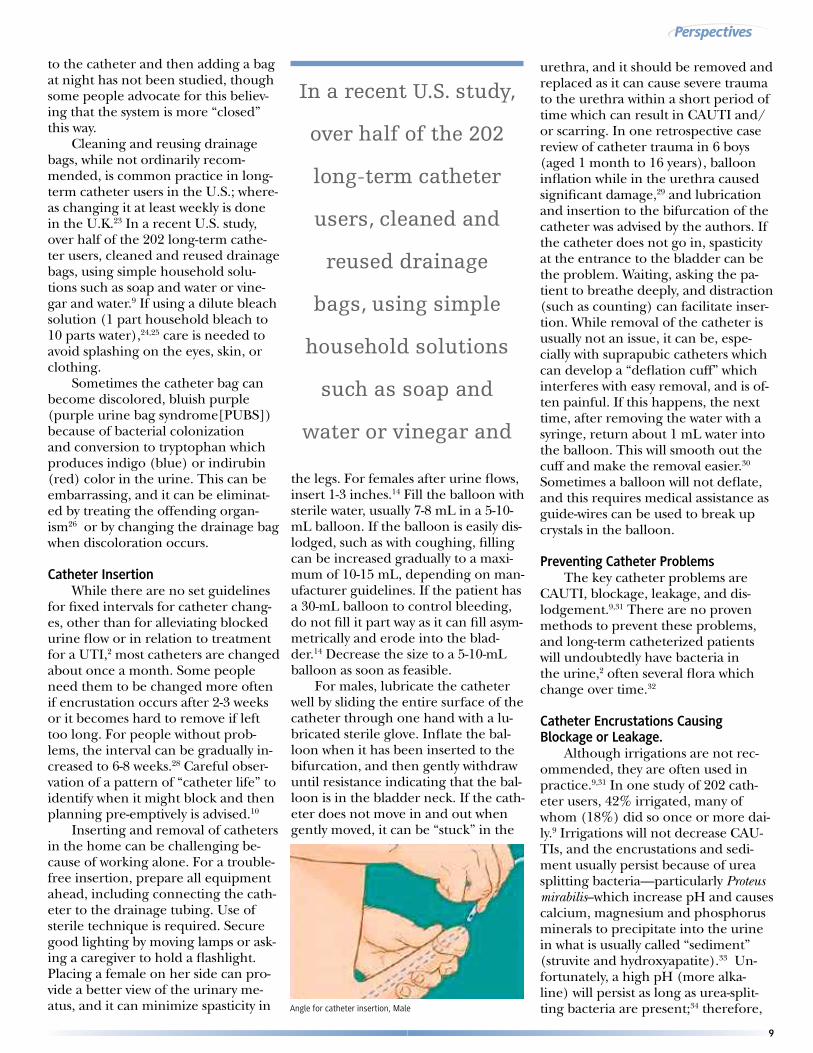
Three materials are used for secur-ing the tracheostomy tube to the pa-tient: cotton twill tape, commercially available Velcro® and felt (fabric) tra-cheostomy securement devices, and a beaded chain. An American Thoracic Society committee looking into the use of these three devices in children made the following observations.6
Cotton twill tape may trap mois-ture, becomes soiled frequently, may unravel, and subsequently irritate the skin. Velcro devices have less tendency to abrade the skin, as does twill tape. They are convenient and easily adjust-ed. One-person tie changes are easier with Velcro ties than with other tie materials (Figure 1). They are wider than cotton twill and chain, so they have less tendency to abrade the skin. They are easily adjusted and because of their padding material, should be quite comfortable. There is third par-ty coverage for these securement de-vices. A stainless steel beaded chain is durable, does not trap moisture, and
maintains a constant tension. Any se-curement device is kept tight enough to allow slipping a finger between the neck and device material. The consen-sus was that the most important as-pect of choosing a tracheostomy tie is how well the tie can be secured, rather than the material it is made from.6 While there does not seem to be any similar data avaialble for tracheostomy securement in adults, these same prin-ciples should apply.
The removal of the tracheostomy tube is referred to as decannulation. This implies that it is the intention to not reinsert the tube, as is the case with a tube change. Because of equip-ment concerns and the potential for a need for emergency airway manage-ment, decannulation is usually not done at home.7 None of the referenc-es recommended home decannula-tion.
Special Tracheostomy Issues for Children
Like adults, children sometimes require placement of a tracheostomy when there is a need for a short term or permanent airway, or if there is a need for long-term invasive ventila-tion. The indications for tracheos-tomy in children differ from adult patients and are widespread. Of the 16 characteristic indications for pe-diatric tracheostomy, the most com-mon are airway obstruction, need for airway protection due to aspiration or inability to manage secretions, evi-dence of subglottic stenosis, chronic lung disease patients requiring long term mechanical ventilation, and the presence of congenital abnormalities.8 Unlike adult patients who may be con-sidered candidates for tracheostomy after approximately 1 week of inva-sive mechanical ventilation, children
are often not considered candidates for tracheostomy until much later in their course, with the exception of those with anatomic airway abnormali-ties. Davis indicated that the average time of tracheostomy insertion in chil-dren was at about 65 days following initial intubation; and is often longer in some cases.8 Despite the variation in length of time a tracheostomy is needed, about two-thirds of pediat-ric patients are eventually decannu-lated.8 Because of the unpredictable duration of pediatric tracheostomy, patients and families/caregivers begin preparations for transition to home as soon as feasibly possible.
Children often remain in the in-tensive or specialized care units until the first tracheostomy change is com-pleted: around 5-7 days after inser-tion.1 Close observation is required because a large number of pediatric tracheostomy tubes are small and un-cuffed, and there is a greater chance for dislodgement or decannulation. It is recommended that specialized trained caregivers such as respiratory therapists or nurses trained in trache-ostomy care assist the surgeon during the initial tracheostomy tube change, as described in the previous section.1 Patients are typically transitioned to a lower level of care and an organized approach to patient and caregiver training begins.
Hospital stays for children with a tracheostomy vary amongst institu-tions. Some programs report length of stay at 5 to 472 hospital days while oth-ers are 14 to 280 days from initial ad-mission.6,8 Causes for these variations are multifactorial. Proactively prepar-ing the tracheostomy patient and fam-ily for discharge and care in the home setting is highly recommended.1 Suc-cessful discharge planning programs have had positive outcomes if educa-tion initiatives were started before the initial surgery.1 Because patient’s med-ical issues, or family social concerns often create barriers and play a role in delaying discharge, attempts are made to keep caregivers on an educa-tion and training timeline. This allows time for home assessment, equipment selection, repetition of skills and re-tention of concepts presented.6 The need for nursing care in the home frequently contributes to a prolonged Figure 1a. Dale Trachesotomy Tube Holder Figure 1. Dale Trachesotomy Tube Holder (Pediatric)

6
Perspectives
hospital stay. Pediatric patients requir-ing home nursing care are on me-chanical ventilators, have an unstable airway or have multiple complex medi-cal needs in addition to tracheostomy care. In some cases, an average of 8-24 hours of nursing care may be granted based on these requirements.6 The level of nursing care varies based on degree of medical complexity, medi-cal insurance benefits or state-based resources. Orchestrating these services is the greatest challenge and may con-sume a significant amount of time in this phase. In addition, complex train-ing requirements and patient and fam-ily education require additional re-sources. Most importantly, coordinated care, streamlined education and train-ing and shortened the length of stay from tracheostomy insertion to dis-charge are primary objectives of these programs.
In 2000, discharge planning pro-grams for pediatric patients were ini-tially created based on prior recom-mendations from the American Tho-racic Society’s Care of the Child with Chronic Tracheostomy.6 Since then, much attention has been focused on standardizing care of the pediatric tra-cheostomy patient prior to discharge. Graf et al attempted to emulate this by implementing a family education pro-gram for pediatric tracheostomy pa-tients. The Texas Children’s Hospital program reduced the length of stay fol-lowing tracheostomy to a mean of 6.5 days following an educational program (5-114 days total length of stay).9,10 In 2012, the American Academy of Oto-laryngology- Head and Neck Surgery created a clinical consensus statement to provide updated recommenda-tions for pediatric and adult patients with a tracheostomy.1 Some recom-mendations have been tested and have since become expectations of care for these patients. To prepare families or caregivers for caring for the pedi-atric tracheostomy patient at home, 2 caregivers are identified as soon as the decision is made to proceed with the procedure.6 Basic education is dis-seminated to caregivers via handouts, such as a specialized tracheostomy care booklets (or electronic versions if desired) as described above. Upon gradual completion of these compo-
nents, the caregiver is tested to as-sess knowledge and competency. The most unique element of a pediatric discharge planning program is a re-quirement for families to “room in,” or spend a 24-hour or other designat-ed period of time taking care of the child independently as if they were in a home setting while having the ability to raise any final questions and calm their fears or uncertainties prior to discharge.
Pediatric tracheostomy patients are cared for by families and nurses in the home and monitored on a regular basis by respiratory therapists. Physi-cians assess the airway to determine if changes in airway size is necessary, make modifications in mechanical ventilator settings or verify the time for liberation from all components. If decannulation is being considered, the patient must be free of aspira-tion episodes, not require mechanical ventilator support, have completed a tracheostomy capping trial if appro-priate, and have been evaluated by bronchoscopy within a few months of considering decannulation. Pedi-atric tracheostomy-ventilator clinics provide a vehicle to assess changes in the patient’s condition and allows for further evaluation. Streamlining pedi-
atric tracheostomy care is an ongoing challenge and a unique opportunity to make a difference!
References
1 Mitchell RB, Hussey HM, Setzen G, et al. Clinical consensus statement: tracheostomy care. Otolaryngol Head Neck Surg. 2013;148(1):6-20.
2 Lewarski JS. Long-term care of the patient with a tracheostomy. Respir Care. 2005;50(4):534-537.
3 HCUP Kids’ Inpatient Database (KID). Healthcare Cost and Utilization Project (HCUP). 2006 and 2009. Agency for Healthcare Research and Quality, Rockville, MD. www.hcup-us.ahrq.gov/kidoverview.jsp. Accessed April 10, 2013.
4 Dhand R, Johnson JC. Care of the chronic tracheostomy. Respir Care. 2006;51(9):984-1004.
5 Joseph RA. Tracheostomy in infants: Patient education for home care. Neonatal Network. 2012;30(4):231-242.
6 Sherman JM, Davis S, Albamonte-Petrick S, et al. Care of the child with a chronic tracheostomy. This official statement of the American Thoracic Society was adopted by the ATS Board of Directors, July 1999. Am J Respir Crit Care Med. 2000;161(1):297-308.
7 Johns Hopkins Medicine. Decannulation: http://www.hopkinsmedicine.org/tracheostomy/living/decannulation.html. Accessed April 16, 2013.
8 Davis GM. Tracheostomy in children. Paediatric Resp Reviews. 2006;7(Suppl 1):S206–S209.
9 Graf JM, Montagnino BA, Heuckel R, McPherson ML. Children With New Tracheostomies: Planning for family education and common impediments to discharge. Pediatric Pulmonology. 2008;43(8):788–794.
10 Graf JM, Montagnino BA, Heuckel R, McPherson ML. Pediatric tracheostomies: a recent experience for one academic medical center. Pediatr Crit Care Med. 2008;9(1):96-100.
Timothy B. Op’t Holt, EdD, RRT, AE-C, FAARC, is Director of “Breath of Life” COPD and the Asthma Edu-cation and Therapy Program at Victory Health Partners Clinic in Mobile, Alabama. At the University of South Alabama, he is Professor, Department of Respiratory Care and Cardiopulmonary Sciences. He is the author or co-author of 8 books and 30 studies in journals and has presented over 35 papers at international conferences.
Kathleen M. Deakins, MSHA, RRT-NPS, FAARC is Clinical Manager, Women’s and Children’s Respira-tory Care, Pediatric Pulmonary Function and Infant Monitoring Rainbow Babies & Children’s Hospital of University Hospitals, Cleveland, Ohio. She is an accred-ited neonatal-pediatric specialist with an interest in all facets of pediatric and neonatal respiratory care. She has published more than 50 publications on respira-tory medicine and lectured widely in her field. She is also a member of several professional organizations dedicated to respiratory medicine. Ms Deakins lives in Chardon, Ohio.
Jennifer D. McDaniel, RRT-NPS is currently a re-spiratory therapist at the USA and Children’s Hospital In Mobile, AL. In addition to her clinical duties in the neonatal intensive care, Ms. McDaniel teaches neonatal and pediatric respiratory care. Ms. McDaniels received her Bachelor of Science in Cardiorespiratory Care from the University of South Alabama.
Pediatric patients
requiring home
nursing care are
on mechanical
ventilators, have
an unstable airway
or have multiple
complex medical
needs in addition to

7
Perspectives
they are commonly used in select pa-tients in the United Kingdom (UK) and Europe.6-8 When a catheter is ex-pected to be used indefinitely, it can be with the person for many years. In one study of 202 long-term catheter users, the mean duration of use was 6 years (SD 7) and the median was 3.25 years.9 Therefore, patients and their families need to learn how to manage the catheter between nurse visits.
Catheter SelectionFor long-term catheter patients,
the catheter type should be hydro-philic or coated with silicone.10 Pure silicone is not as pliable but it has a larger inner lumen which is of value for people with large amounts of sedi-ment that blocks and causes frequent catheter changes. However, in one study, half the water evaporated with-in 3 weeks and 85% within 8 weeks.11 This can cause the catheter to “slip down,” causing irritation to the open-ing of the bladder and/or leaking (by-passing of urine). Latex allergy is of concern to many with long-term cath-eters as this can develop gradually over time,12 but latex softness can be of val-ue. In one long-term care facility, the staff attempted to create a latex-free environment, but in doing so, they in-advertently contributed to penile ero-sion of the urethra in men newly using rigid silicone catheters.13
For short- or long-term catheter patients, the catheter should be the smallest size diameter that permits good urine flow.2 This should be 12-16 Fr for men and 12-14 Fr for women. Children’s sizes can range from 5-6 Fr on newborns to 5-10 Fr on toddlers to children up to age 12.14 If a larger di-ameter was used to facilitate passage of blood clots, the size can be decreased by 2-4 Fr per catheter change until the optimal size is reached. Likewise, bal-loon sizes should be small, with 5- or 10-mL balloons, reserving the 30 mL only for bleeding episodes, usually postoperatively. Balloons of 2.5-5 mL can be used in children.14 Patients and their families should know the cath-eter and balloon size and the amount
of water instilled to promote continu-ity of care. In a study of 202 persons, 8% did not know their catheter size, and 23% did not know the balloon size.9
Catheter SecurementPatients who use long-term in-
dwelling urinary catheters commonly use catheter securement devices to prevent urethral and bladder trauma and inadvertent catheter dislodge-ment. Post-hospitalization, securement is used to control postoperative bleed-ing or to protect a surgical anastomo-sis. Securing a catheter might also in-crease comfort and reduce CAUTI.15 Over time, traction of the catheter can cause erosion of the urethra, and in men this damage can spread to the length of the penis.15 Therefore, all long-term catheters should be se-cured—in women to the thigh and in men to the lower anterior abdomen or high on the thigh.14 Suprapubic cath-eters also should be secured. In a case report, a non-secured suprapubic cath-eter balloon made its way to the out-side of the bladder and caused a urine-carrying fistula to the skin surface.16 Although nurses might say they be-lieve catheter securement is desirable, they may not always do it. In a survey of 82 nurses, including 8 continence specialists, 98% indicated they favor catheter securement, but in a previous prevalence study, only 4% had secured their patients’ catheters.17
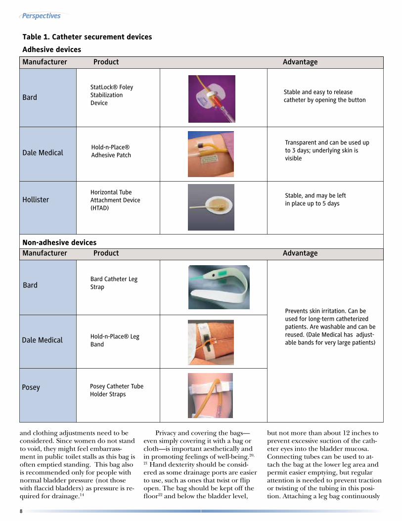
Catheter securement devices can be improvised or manufactured, in-
Best Practices in Managing the IndwellingUrinary Catheter for the Homecare Patient—Continued
Latex straps, which
are used to hold the
drainage bag in place
on the leg, often twist
and pull on the leg
hair, and the latex can
cause skin irritation or
cluding adhesive and non-adhesive devices. (See Table 1. Catheter Secure-ment Devices.) Improvised devices are made with adhesive tape, sometimes with safety pins and rubber bands; however, tape can be irritating to the skin and hard to apply and remove. An early randomized clinical trial (RCT)18 with 59 patients showed that the man-ufactured device is easier to apply and remove and can stay in place 2 days longer than tape.
Adhesive-backed devices are avail-able and these are particularly useful in short-term catheter use or for indi-viduals prone to dislodgement. How-ever, adhesive-backed devices also can irritate the skin and cause pain when removed. This should be an important consideration for geriatric patients or others with fragile skin. When using adhesive-backed securement devices, alcohol-based or other special wipes need to be used for removal to de-crease skin damage.
Most non-adhesive devices use a stretch band and a Velcro locking sys-tem to hold the catheter to the thigh. While these devices have adjustability and can even be used with large/obese patients, they still need to be used ac-cording to the manufacturer’s instruc-tions to be sure that blood flow is not restricted. Latex straps, which are used to hold the drainage bag in place on the leg, often twist and pull on the leg hair, and the latex can cause skin irri-tation or allergy. Placing a thin cloth underneath the strap can minimize irritation. Nurses should instruct pa-tients not to put any strap too tight and carefully monitor the skin condi-tion, and this especially true for pa-tients with peripheral vascular disease. Non-adhesive types also include hol-ster-like devices hung from the waist or netting/cloth supporting bags,19 which can be ordered through urological supply companies online.
Drainage Bags Urine drainage bags are available
as leg bags, in sizes from 270-1000 mL,14 which permit more freedom in movement and travel. Large sized overnight bags (2000-4000 mL) are of-ten used in the hospital or for people who are bedridden, e.g., at end of life. A “belly bag” has a 1000-mL capacity,
8
Perspectives
Table 1. Catheter securement devices
Manufacturer Product Advantage
Manufacturer Product Advantage
BardStatLock® Foley Stabilization Device
Stable and easy to release catheter by opening the button
Dale MedicalHold-n-Place® Adhesive Patch
Transparent and can be used up to 3 days; underlying skin is visible
HollisterHorizontal TubeAttachment Device (HTAD)
Stable, and may be left in place up to 5 days
Non-adhesive devices
BardBard Catheter Leg Strap
Prevents skin irritation. Can be used for long-term catheterized patients. Are washable and can be reused. (Dale Medical has adjust-able bands for very large patients)Dale Medical Hold-n-Place® Leg
Band
Posey Posey Catheter Tube Holder Straps
Adhesive devices
and clothing adjustments need to be considered. Since women do not stand to void, they might feel embarrass-ment in public toilet stalls as this bag is often emptied standing. This bag also is recommended only for people with normal bladder pressure (not those with flaccid bladders) as pressure is re-quired for drainage.14
Privacy and covering the bags—even simply covering it with a bag or cloth—is important aesthetically and in promoting feelings of well-being.20,
21 Hand dexterity should be consid-ered as some drainage ports are easier to use, such as ones that twist or flip open. The bag should be kept off the floor22 and below the bladder level,
but not more than about 12 inches to prevent excessive suction of the cath-eter eyes into the bladder mucosa. Connecting tubes can be used to at-tach the bag at the lower leg area and permit easier emptying, but regular attention is needed to prevent traction or twisting of the tubing in this posi-tion. Attaching a leg bag continuously
9
Perspectives
In a recent U.S. study,
over half of the 202
long-term catheter
users, cleaned and
reused drainage
bags, using simple
household solutions
such as soap and
water or vinegar and
to the catheter and then adding a bag at night has not been studied, though some people advocate for this believ-ing that the system is more “closed” this way.
Cleaning and reusing drainage bags, while not ordinarily recom-mended, is common practice in long-term catheter users in the U.S.; where-as changing it at least weekly is done in the U.K.23 In a recent U.S. study, over half of the 202 long-term cathe-ter users, cleaned and reused drainage bags, using simple household solu-tions such as soap and water or vine-gar and water.9 If using a dilute bleach solution (1 part household bleach to 10 parts water),24,25 care is needed to avoid splashing on the eyes, skin, or clothing.
Sometimes the catheter bag can become discolored, bluish purple (purple urine bag syndrome[PUBS]) because of bacterial colonization and conversion to tryptophan which produces indigo (blue) or indirubin (red) color in the urine. This can be embarrassing, and it can be eliminat-ed by treating the offending organ-ism26 or by changing the drainage bag when discoloration occurs.
Catheter Insertion While there are no set guidelines
for fixed intervals for catheter chang-es, other than for alleviating blocked urine flow or in relation to treatment for a UTI,2 most catheters are changed about once a month. Some people need them to be changed more often if encrustation occurs after 2-3 weeks or it becomes hard to remove if left too long. For people without prob-lems, the interval can be gradually in-creased to 6-8 weeks.28 Careful obser-vation of a pattern of “catheter life” to identify when it might block and then planning pre-emptively is advised.10
Inserting and removal of catheters in the home can be challenging be-cause of working alone. For a trouble-free insertion, prepare all equipment ahead, including connecting the cath-eter to the drainage tubing. Use of sterile technique is required. Secure good lighting by moving lamps or ask-ing a caregiver to hold a flashlight. Placing a female on her side can pro-vide a better view of the urinary me-atus, and it can minimize spasticity in
the legs. For females after urine flows, insert 1-3 inches.14 Fill the balloon with sterile water, usually 7-8 mL in a 5-10-mL balloon. If the balloon is easily dis-lodged, such as with coughing, filling can be increased gradually to a maxi-mum of 10-15 mL, depending on man-ufacturer guidelines. If the patient has a 30-mL balloon to control bleeding, do not fill it part way as it can fill asym-metrically and erode into the blad-der.14 Decrease the size to a 5-10-mL balloon as soon as feasible.
For males, lubricate the catheter well by sliding the entire surface of the catheter through one hand with a lu-bricated sterile glove. Inflate the bal-loon when it has been inserted to the bifurcation, and then gently withdraw until resistance indicating that the bal-loon is in the bladder neck. If the cath-eter does not move in and out when gently moved, it can be “stuck” in the
urethra, and it should be removed and replaced as it can cause severe trauma to the urethra within a short period of time which can result in CAUTI and/or scarring. In one retrospective case review of catheter trauma in 6 boys (aged 1 month to 16 years), balloon inflation while in the urethra caused significant damage,29 and lubrication and insertion to the bifurcation of the catheter was advised by the authors. If the catheter does not go in, spasticity at the entrance to the bladder can be the problem. Waiting, asking the pa-tient to breathe deeply, and distraction (such as counting) can facilitate inser-tion. While removal of the catheter is usually not an issue, it can be, espe-cially with suprapubic catheters which can develop a “deflation cuff” which interferes with easy removal, and is of-ten painful. If this happens, the next time, after removing the water with a syringe, return about 1 mL water into the balloon. This will smooth out the cuff and make the removal easier.30 Sometimes a balloon will not deflate, and this requires medical assistance as guide-wires can be used to break up crystals in the balloon.
Preventing Catheter ProblemsThe key catheter problems are
CAUTI, blockage, leakage, and dis-lodgement.9,31 There are no proven methods to prevent these problems, and long-term catheterized patients will undoubtedly have bacteria in the urine,2 often several flora which change over time.32
Catheter Encrustations Causing Blockage or Leakage.
Although irrigations are not rec-ommended, they are often used in practice.9,31 In one study of 202 cath-eter users, 42% irrigated, many of whom (18%) did so once or more dai-ly.9 Irrigations will not decrease CAU-TIs, and the encrustations and sedi-ment usually persist because of urea splitting bacteria—particularly Proteus mirabilis--which increase pH and causes calcium, magnesium and phosphorus minerals to precipitate into the urine in what is usually called “sediment” (struvite and hydroxyapatite).33 Un-fortunately, a high pH (more alka-line) will persist as long as urea-split-ting bacteria are present;34 therefore, Angle for catheter insertion, Male

10
Perspectives
Although irrigations
are not recommended,
they are often used in
practice. In one study
of 202 catheter users,
42% irrigated, many
of whom (18%) did so
once or more daily.9
an often suggested method to lower urine pH (acidify) with vitamin C is not likely to be of benefit. However, increased fluid intake can dilute the urine and sometimes helps in decreas-ing blockage and premature catheter replacement even when the pH is high because the pH point of mineral pre-cipitation is not the same as the urine pH.35 Bladder stones, often caused by the same minerals, need to be re-moved by cystoscopy, and if possible the offending organism should be eliminated through antibiotics. Never-theless, bacteria living in biofilms and protected by a crystalline matrix are not easy to eradicate.36 Some people, with healthcare provider approval, might be helped by taking cranberry juice tablets because it keeps bacteria from adhering to the bladder lining,37
but research in catheterized patients is limited and evidence of cranberry juice efficacy in other populations is also unclear.38 Moreover, the juice may be unacceptable to patients over long periods of time.38
Leakage (bypassing of urine) can be minimized by proper positioning of the catheter so that traction is not pull-ing the balloon against the bladder opening, antispasmodic medication, and by treating constipation.39 Smaller size catheters and balloons also cause less irritation to the bladder sphincter and thus less leakage. Paying Attention.
There is some research indicating that paying attention to fluid intake and preventing accidental dislodg-ment can be of benefit.40,41,42
Nurses can teach their patients to be more aware of their catheter posi-tion during transfers and instruct care-givers/family to keep the drainage bag from being run over by a wheel chair or pulled out when turning in bed. Noticing early signs of CAUTI could bring earlier treatment. Changes in the color or character of urine (odor, sediment) as well as malaise or weak-ness are the most common symptoms in long-term catheter patients.9 A pilot study teaching self-management, in-cluding optimal fluid intake and pre-venting dislodgement was effective, but further research is needed.41,42 Ap-propriate fluid intake levels vary, and while 30 mL/kg of body weight has been recommended,43 what is right for
an individual should be mutually de-termined with the patient and provid-er.
In conclusion, home care nurses often manage care for patients with indwelling urinary catheters, many of whom have the device for years. Know-ing catheter-related best practices is es-sential for their patients’ health.
References
1. Kunin CM, McCormack RC. Prevention of catheter-induced urinary-tract infections by sterile closed drainage. N Engl J Med. 1966;274:1155-1161.
2. Gould CV, Umscheid CA, Agarwal RK, Kuntz G, Pegues DA, and the Healthcare Infection Control Practices Advisory Committee (HICPAC)., eds. Guideline for Prevention of Catheter-Associated Urinary Tract Infections 2009. Atlanta, GA: Centers for Disease Control and Prevention; 2009.
3. Schumm K, Lam T. Types of urethral catheters for management of short-term voiding problems in hospitalised adults. Cochrane Database Syst Rev. 2008;(2):CD004013.
4. Parker D, Callan L, Harwood J, Thompson DL, Wilde M, Gray M. Nursing interventions to reduce the risk of catheter-associated urinary tract infection. Part 1: Catheter selection. J Wound Ostomy Continence Nurs. 2009;36:23-34.
5. Gokula RR, Hickner JA, Smith MA. Inappropriate use of urinary catheters in elderly patients at a midwestern community teaching hospital. Am J Infect Control. 2004;32:196-9.
6. Fader M, Pettersson L, Brooks R, Dean G, Cottenden A, Malone-Lee J. A multicentre comparative evaluation of catheter valves. BrJ Nursing. 1997;6:359-368.
7. van den Eijkel E, Griffiths P. Catheter valves for indwelling urinary catheters: A systematic review. Br J Community Nurs. 2006;11:111-114.
8. German K, Rowley P, Stone D, Kumar U, Blackford HN. A randomized cross-over study comparing the use of a catheter valve and a leg-bag in urethrally catheterized male patients. Br J Urol. 1997;79:96-8.
9. Wilde MH, McDonald MV, Brasch J, et al. Long-term urinary catheter users self-care practices and problems. J Clin Nurs. 2013;22:356-367.
10. Cottenden A, Bliss D, Fader M, Getliffe, K., Buckley, B., Wilde, M.H., & Pieters, R Management using Continence Products. Incontinence, 4th International Consultation in Incontinence. 4th ed. Paris: Health Publication Ltd.; 2009. Abrams P., Cardozo L., Khoury S. and Wein A., eds.
11. Barnes KE, Malone-Lee J. Long-term catheter management: minimizing the problem of premature replacement due to balloon deflation. J Adv Nurs. 1986;11:303-7.
12. Shenot P, Rivas DA, Kalman DD, Staas WE,Jr, Chancellor MB. Latex allergy manifested in urological surgery and care of adult spinal cord injured patients. Arch Phys Med Rehabil. 1994;75:1263-1265.
13. Bell MA. Severe indwelling urinary catheter-associated urethral erosion in four elderly men. Ostomy Wound Manage. 2010;56:36-39.
14. WOCN Society Clinical Practice Continence subcommittee. Indwelling urinary catheters: Best practice for clinicians. Wound Ostomy and Continence Nursing Society; 2009.
15. Gray ML. Securing the indwelling catheter. Am J Nurs. 2008;108:44-50; quiz 50.
16. Vaidyanathan S, Hughes PL, Soni BM. Unusual complication of suprapubic cystostomy in a male patient with tetraplegia: traction on Foley catheter leading to extrusion of Foley balloon from urinary bladder and suprapubic urinary fistula--importance of securely anchoring suprapubic catheter with adhesive tape or BioDerm tube holder. ScientificWorldJournal. 2007;7:1575-1578.
17. Siegel TJ. Do registered nurses perceive the anchoring of indwelling urinary catheters as a necessary aspect of nursing care?: a pilot study. J Wound Ostomy Continence Nurs. 2006;33:140-4.
18. Blaylock B, Clayton G, Milnes M, Foster C. Product notebook: a catheter anchoring device. Ostomy Wound Manage. 1993;39:36-41, 43.
19. Gray M, Joseph PAC, Mercer DM, Newman DK, Rovner E. Consensus and controversy in urinary drainage systems: Implications for improving patient safety. Safe Practices in Patient Care 2008; 4 (1) Available at www.Safe Practices.org. Accessed January 31, 2013.
20. Fraczyk L, Godfrey H, Feneley R. A pilot study of users’ experiences of urinary catheter drainage bags. Br J Community Nurs. 2003;8:104-111.
21. Wilde MH. Life with an indwelling urinary catheter: the dialectic of stigma and acceptance. Qual Health Res. 2003;13:1189-204.
22. Glahn BE, Braendstrup O, Olesen HP. Influence of drainage conditions on mucosal bladder damage by indwelling catheters. II. Histological study. Scand J Urol Nephrol. 1988;22:93-9.
23. Royal College of Nurses (England), ed. Catheter Care: RCN Guidance for Nurses. Pub. Code. 003-237 ed. www.rcn.org.uk.; 2008; No. March.

11
Perspectives
Perspectives is an education program distributed free of charge to health professionals. Perspec-tives is published by Saxe Healthcare Communica-tions and is funded through an educational grant from Dale Medical Products Inc. Perspectives’ objective is to provide health professionals with timely and relevant information on postoperative recovery strategies, focusing on the continuum of care from operating room to recovery room, ward, or home.
The opinions expressed in Perspectives are those of the authors and not necessarily of the editorial staff or Dale Medical Products Inc. The publisher and Dale Medical Products Inc. disclaim any re-sponsibility or liability for such material. Clinicians are encouraged to consult additional sources prior to forming a clinical decision.
Please direct your correspondence to:
Saxe Healthcare CommunicationsP.O. Box 1282, Burlington, VT 05402
Fax: (802) [email protected]© Copyright: Saxe Communications 1998-2013
After reading these articles, the learner should be able to:
1. Identify correct catheter and balloon sizes for adults and children.
2. Describe different types of catheter securement.
3. List the indications for tracheostomy in adults and children.
4. Identify the issues concerning care of the tracheostomy and tracheostomy tube for the patient at home.
To receive continuing education credit, simply do the following:
1. Read the educational offering (both articles).
2. Complete the post-test for the educational offering. Mark an X in the box next to the correct answer. (You may make copies of the answer form.)
3. Complete the learner evaluation.
4. You may take this test online at www.saxetesting.com, or you may mail or fax the completed learner evaluation and post-test to Saxe Communications.
5. To earn 2.0 contact hours of continuing education, you must achieve a score of 75% or more. If you do not pass the test, you may take it again 1 time.
6. Your results will be sent within 4 weeks after the form is received.
7. The administrative fee has been waived through an educational grant from Dale Medical Products, Inc.
8. Answer forms must be postmarked by May 1, 2017. Please visit www.perspectivesinnursing.org for renewal updates. Programs are generally renewed.
9. Faculty disclosures: No conflicts were disclosed.
* Approval does not imply ANCC or endorsement of any product.
Provider approved by the California board of Registered Nursing.
Provider #CEP 14477
Mary Wilde, RN, PhD is Associate Professor in the School of Nursing, University of Rochester, Rochester, New York. Her current research interests focus on the self-management of urine flow in long-term catheter use, quality of life in long-term urinary catheter users, and urinary drainage bag decontamination. Dr. Wilde is the author or coauthor of 37 articles, book chapters, abstracts, and other publications and has given many presentations on the subject of long-term urinary catheter care and management. She also sits on the review boards of several medical journals. She lives in Whitesboro, New York.
Feng Zhang, RN, is enrolled in the MS/PhD program at the School of Nursing, University of Rochester in Roches-ter, prior to which he worked as an RN in Bramlage House of Meadowlark Hill Retirement Community (Transitional Services), Manhattan, Kansas. In May, 2011, he received his BSc in Nursing at Washburn University, Topeka, Kansas. He has received several awards and honors, including the Loretta Lord Fellowship (2012-2013). Mr. Zhang lives in Rochester, New York.
24. Dille CA, Kirchhoff KT, Sullivan JJ, Larson E. Increasing the wearing time of vinyl urinary drainage bags by decontamination with bleach. Arch Phys Med Rehabil. 1993;74:431-7.
25. Dille CM, Kirchhoff KT. Decontamination of vinyl urinary drainage bags with bleach. Rehabil Nurs. 1993;18:292-5.
26. Tsumura H, Satoh T, Kurosaka S, Fujita T, Matsumoto K, Baba S. Clinical characteristics in patients with purple urine bag syndrome. Hinyokika Kiyo. 2008;54:185-188.
27. Vallejo-Manzur F, Mireles-Cabodevila E, Varon J. Purple urine bag syndrome. Am J Emerg Med. 2005;23:521-4.
28. Ferrie BG, Glen ES, Hunter B. Long-term urethral catheter drainage. Br Med J. 1979;2:1046-1047.
29. D’Cruz R, Soundappan SS, Cass DT, Smith G. Catheter balloon-related urethral trauma in children. J Paediatr Child Health. 2009;45:564-566.
30. Parkin J, Scanlan J, Woolley M, Grover D, Evans A, Feneley RC. Urinary catheter ‘deflation cuff’ formation: clinical audit and quantitative in vitro analysis. BJU Int. 2002;90:666-71.
31. Wilde MH, Brasch J, Getliffe K, et al. Study on the use of long-term urinary catheters in community-dwelling individuals. J Wound Ostomy Continence Nurs. 2010;37:301-310.
32. Warren JW, Tenney JH, Hoopes JM, Muncie HL, Anthony WC. A prospective microbiologic study of bacteriuria in patients with chronic indwelling urethral catheters. J Infect Dis. 1982;146:719-23.
33. Choong S, Wood S, Fry C, Whitfield H. Catheter associated urinary tract infection and encrustation. Int J Antimicrob Agents. 2001;17:305-10.
34. Bibby JM, Hukins DW. Acidification of urine is not a feasible method for preventing encrustation of indwelling urinary catheters. Scand J Urol Nephrol. 1993;27:63-5.
35. Khan A, Housami F, Melotti R, Timoney A, Stickler D. Strategy to control catheter encrustation with citrated drinks: a randomized crossover study. J Urol. 2010;183:1390-1394.
36. Stickler DJ, Feneley RC. The encrustation and blockage of long-term indwelling bladder catheters: a way forward in prevention and control. Spinal Cord. 2010;48:784-790.
37. Sobota AE. Inhibition of bacterial adherence by cranberry juice: potential use for the treatment of urinary tract infections. J Urol. 1984;131:1013-6.
38. Jepson RG, Craig JC. Cranberries for preventing urinary tract infections. Cochrane Database Syst Rev. 2008;(1):CD001321.
39. Switters DM. Assessing leakage from around the urethral catheter. Urol Nurs. 1989;9:8-10.
40. Wilde MH, Dougherty MC. Awareness of urine flow in people with long-term urinary catheters. Commentary by B. Roe. Journal of Wound, Ostomy, and Continence Nursing. 2006;33:164-75.
41. Wilde MH, Brasch J. Self-monitoring of urine flow in people with long-term urinary catheters. Res Nurs Health. 2008;31:490-500.
42. Wilde MH, Brasch J. Teaching Self-management to long-term urinary catheter users. International
Journal of Urological Nursing. 2008;2:62-71.
43. Gray M, Krissovich M. Does fluid intake influence the risk for urinary incontinence, urinary tract infection, and bladder cancer? J Wound Ostomy Continence Nurs. 2003;30:126-131.
You may take this test online at www.saxetesting.com
Name & Credentials
Position/Title
Address
City State Zip
Phone Fax
Email Address
AARC # (if applicable)
Participant’s Evaluation Questions
What is the highest degree you have earned 1. Diploma 2. Associate 3. Bachelor’s (circle one) ? 4. Master’s 5. Doctorate
Indicate to what degree you met the objectives for this program: Using 1 = strongly disagree to 6 = strongly agree rating scale. Please circle the number that best reflects the extent of your agreement to each statement.
Strongly Disagree Strongly Agree
1 2 3 4 5 6
1 2 3 4 5 6
1 2 3 4 5 6
1 2 3 4 5 6
Mark your answers with an X in the box next to the correct answer
A B C D
1
A B C D
2
A B C D
3
A B C D
4
A B C D
5
A B C D
6
A B C D
7
A B C D
8
A B C D
9
A B C D
10
For immediate results, take this test online at www.saxetesting.com
or mail to: Saxe Communications, PO Box 1282, Burlington, VT 05402 • Fax: (802) 872-7558 •
www.saxetesting.com
Vol. 10, No. 1 /10
1. Identify correct catheter and balloon sizes for adults and children.
2. Describe different types of catheter securement.
3. List the indications for tracheostomy in adults and children.
4. Identify the issues concerning care of the tracheos-tomy and tracheostomy tube for the patient at home.
1. Catheters for short-term use:
a. Include silver oxide coated types.b. Should not include use of coated latex
catheters, such as those with PTFE coatings.
c. Are used with a closed system.d. Are used for over 1 month.
2. Long-term catheter users:
a. Have persistent urinary retention and no other alternatives
b. Should irrigate the catheter to prevent CAUTI
c. Need to use a large size overnight bag and avoid leg bags to prevent bacteriuria
d. Are mostly people with severe incontinence
3. Pure silicone catheters:
a. Have a smaller internal lumen and thus promote better urine flow
b. Are softer than other catheter typesc. Have been known to contribute to
erosion of the urethra in mend. Can hold the water in the balloon
longer than coated latex catheters
4. Catheter securement:
a. Is not likely to be of benefit because patients don’t like it
b. Contributes to excessive latex allergyc. Is expensive as the devices are single
use and need to be changed dailyd. Can prevent urethral or bladder
damage
5. Drainage bags:
a. Should never be covered b. Are available in a wide range of sizes
with different types of emptying ports
c. Evidence indicates that leg bags must be connected to overnight bags
d. Tend to turn purple because of environmental conditions
6. When inserting a catheter in a male:
a. Insert the catheter about 2 inches after urine is returned
b. Removed if resistance is met at any time
c. Inserted to the bifurcationd. Use a minimum of lubricant which
could be irritating to the urethra
7. What percentage of tracheostomy patients has been reported as discharged to home with the tracheostomy in pace?
a. 12%b. 28%c. 41%d. 53%
8. What is a recommended tracheostomy tube cuff pressure?
a. 10-15 mm Hgb. 16-18 cm H
2O
c. 20-25 cm H2O
d. 28-32 mm Hg
9. Securement devices are commonly made of all of the following EXCEPT?
a. Cloth adhesive tapeb. Cotton twill tapec. Beaded chaind. Fabric with Velcro
10. Why are tracheostomy tubes in children more easily dislodged?
a. The child moves a lotb. The tube is uncuffed and smallc. Securement is difficultd. Ventilator tubing pulling on the
tracheostomy tube