Embed Size (px)
Citation preview
Karen Marzlin www.cardionursing.com 1
CCRN Review: Pulmonary
Karen Marzlin DNP, CCNS, ACNPBC-AG, CCRN-CMC, CHFN
www.cardionursing.com
12016
5
Physiology of Pulmonary System
• Ventilation and Perfusion
• Diffusion
• Relationship of Oxygen to Hemoglobin
• Oxygen Delivery to the Tissues
• Cellular Respiration
Karen Marzlin www.cardionursing.com 2
6
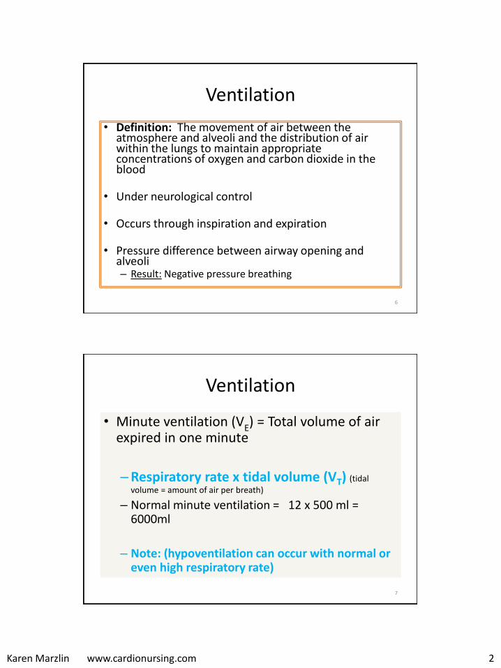
Ventilation
• Definition: The movement of air between the atmosphere and alveoli and the distribution of air within the lungs to maintain appropriate concentrations of oxygen and carbon dioxide in the blood
• Under neurological control
• Occurs through inspiration and expiration
• Pressure difference between airway opening and alveoli – Result: Negative pressure breathing
7
Ventilation
• Minute ventilation (VE) = Total volume of air expired in one minute
–Respiratory rate x tidal volume (VT) (tidal volume = amount of air per breath)
– Normal minute ventilation = 12 x 500 ml = 6000ml
– Note: (hypoventilation can occur with normal or even high respiratory rate)
Karen Marzlin www.cardionursing.com 3
8
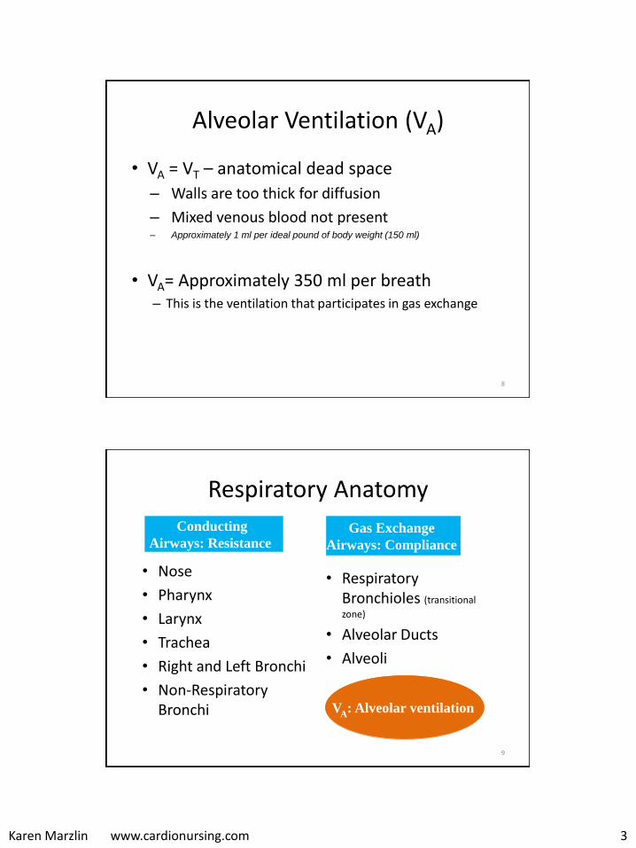
Alveolar Ventilation (VA)
• VA = VT – anatomical dead space
– Walls are too thick for diffusion
– Mixed venous blood not present– Approximately 1 ml per ideal pound of body weight (150 ml)
• VA= Approximately 350 ml per breath – This is the ventilation that participates in gas exchange
9
Respiratory Anatomy
• Nose
• Pharynx
• Larynx
• Trachea
• Right and Left Bronchi
• Non-Respiratory Bronchi
• Respiratory Bronchioles (transitional
zone)
• Alveolar Ducts
• Alveoli
Conducting
Airways: Resistance Gas Exchange
Airways: Compliance
VA: Alveolar ventilation
Karen Marzlin www.cardionursing.com 4
10
11
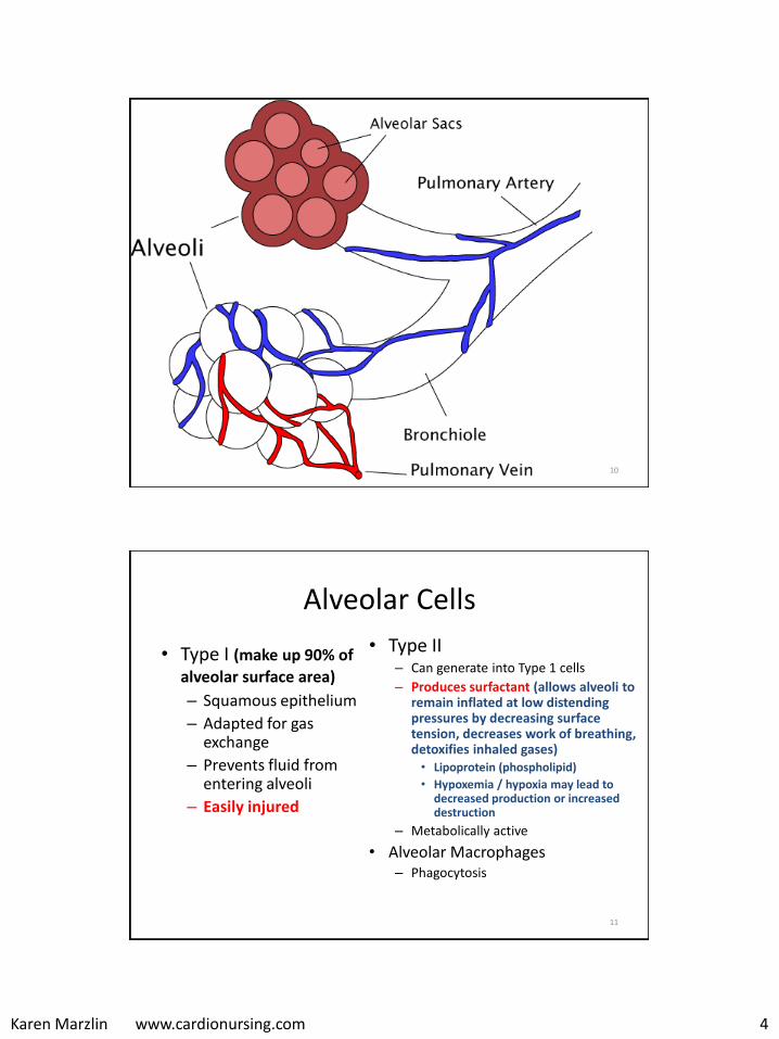
Alveolar Cells
• Type I (make up 90% of
alveolar surface area)
– Squamous epithelium
– Adapted for gas exchange
– Prevents fluid from entering alveoli
– Easily injured
• Type II – Can generate into Type 1 cells
– Produces surfactant (allows alveoli to remain inflated at low distending pressures by decreasing surface tension, decreases work of breathing, detoxifies inhaled gases) • Lipoprotein (phospholipid)
• Hypoxemia / hypoxia may lead to decreased production or increased destruction
– Metabolically active
• Alveolar Macrophages – Phagocytosis
Karen Marzlin www.cardionursing.com 5
12
13
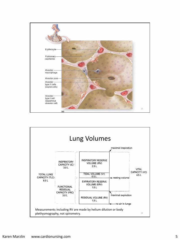
Lung Volumes
Measurements including RV are made by helium dilution or body plethysmography, not spirometry.
Karen Marzlin www.cardionursing.com 6
14
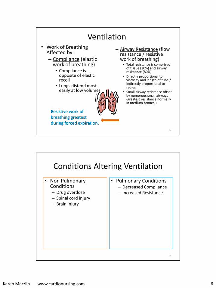
Ventilation • Work of Breathing
Affected by: – Compliance (elastic
work of breathing) • Compliance is
opposite of elastic recoil
• Lungs distend most easily at low volumes
– Airway Resistance (flow resistance / resistive work of breathing)• Total resistance is comprised
of tissue (20%) and airway resistance (80%)
• Directly proportional to viscosity and length of tube / indirectly proportional to radius
• Small airway resistance offset by numerous small airways (greatest resistance normally in medium bronchi)
Resistive work of breathing greatest during forced expiration.
Conditions Altering Ventilation
• Non Pulmonary Conditions– Drug overdose – Spinal cord injury – Brain injury
• Pulmonary Conditions – Decreased Compliance – Increased Resistance
15
Karen Marzlin www.cardionursing.com 7
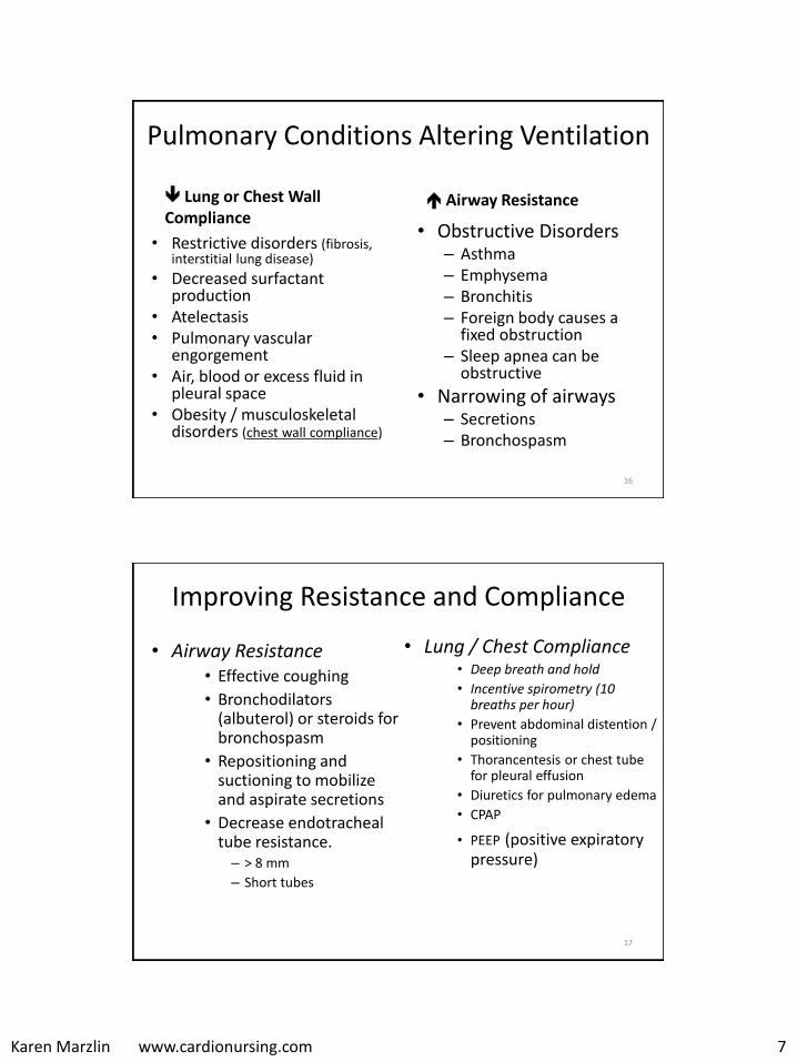
Pulmonary Conditions Altering Ventilation
Lung or Chest Wall Compliance
• Restrictive disorders (fibrosis, interstitial lung disease)
• Decreased surfactant production
• Atelectasis • Pulmonary vascular
engorgement • Air, blood or excess fluid in
pleural space • Obesity / musculoskeletal
disorders (chest wall compliance)
Airway Resistance
• Obstructive Disorders – Asthma– Emphysema– Bronchitis– Foreign body causes a
fixed obstruction– Sleep apnea can be
obstructive
• Narrowing of airways– Secretions – Bronchospasm
16
17
Improving Resistance and Compliance
• Airway Resistance • Effective coughing
• Bronchodilators (albuterol) or steroids for bronchospasm
• Repositioning and suctioning to mobilize and aspirate secretions
• Decrease endotracheal tube resistance.– > 8 mm
– Short tubes
• Lung / Chest Compliance • Deep breath and hold
• Incentive spirometry (10 breaths per hour)
• Prevent abdominal distention / positioning
• Thorancentesis or chest tube for pleural effusion
• Diuretics for pulmonary edema
• CPAP
• PEEP (positive expiratory pressure)
Karen Marzlin www.cardionursing.com 8
18
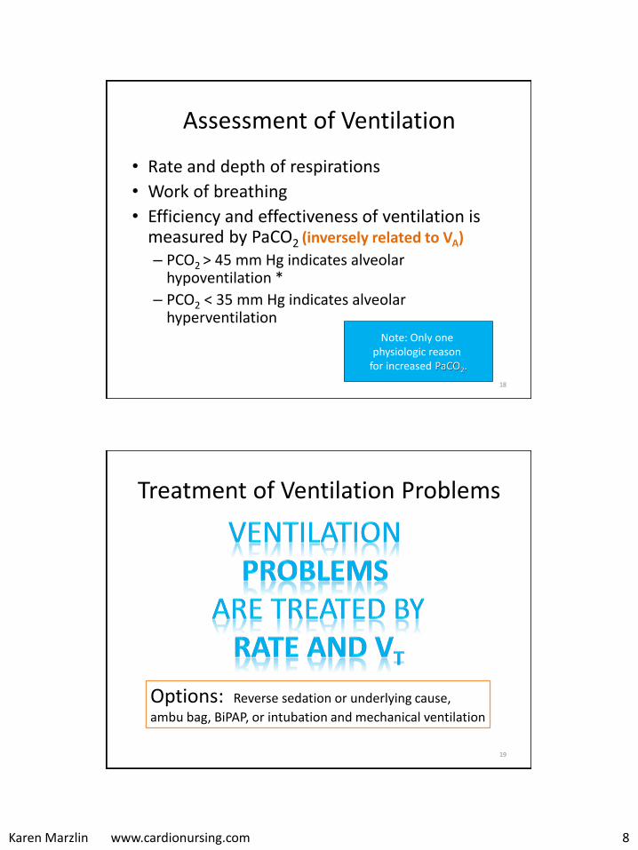
Assessment of Ventilation
• Rate and depth of respirations
• Work of breathing
• Efficiency and effectiveness of ventilation is measured by PaCO2 (inversely related to VA)
– PCO2 > 45 mm Hg indicates alveolar hypoventilation *
– PCO2 < 35 mm Hg indicates alveolar hyperventilation
Note: Only one physiologic reason
for increased PaCO2.
Treatment of Ventilation Problems
19
Options: Reverse sedation or underlying cause,
ambu bag, BiPAP, or intubation and mechanical ventilation
Karen Marzlin www.cardionursing.com 9
20
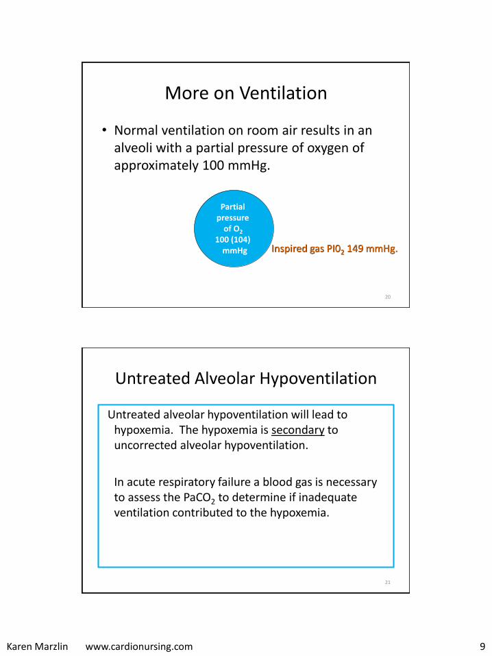
More on Ventilation
• Normal ventilation on room air results in an alveoli with a partial pressure of oxygen of approximately 100 mmHg.
Partial pressure
of O2
100 (104) mmHg Inspired gas PI02 149 mmHg.
21
Untreated Alveolar Hypoventilation
Untreated alveolar hypoventilation will lead to hypoxemia. The hypoxemia is secondary to uncorrected alveolar hypoventilation.
In acute respiratory failure a blood gas is necessary to assess the PaCO2 to determine if inadequate ventilation contributed to the hypoxemia.
Karen Marzlin www.cardionursing.com 10
22
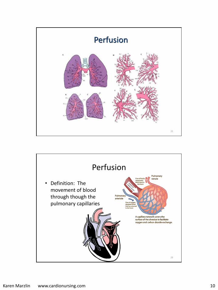
Perfusion
23
Perfusion
• Definition: The movement of blood through though the pulmonary capillaries
Karen Marzlin www.cardionursing.com 11
24
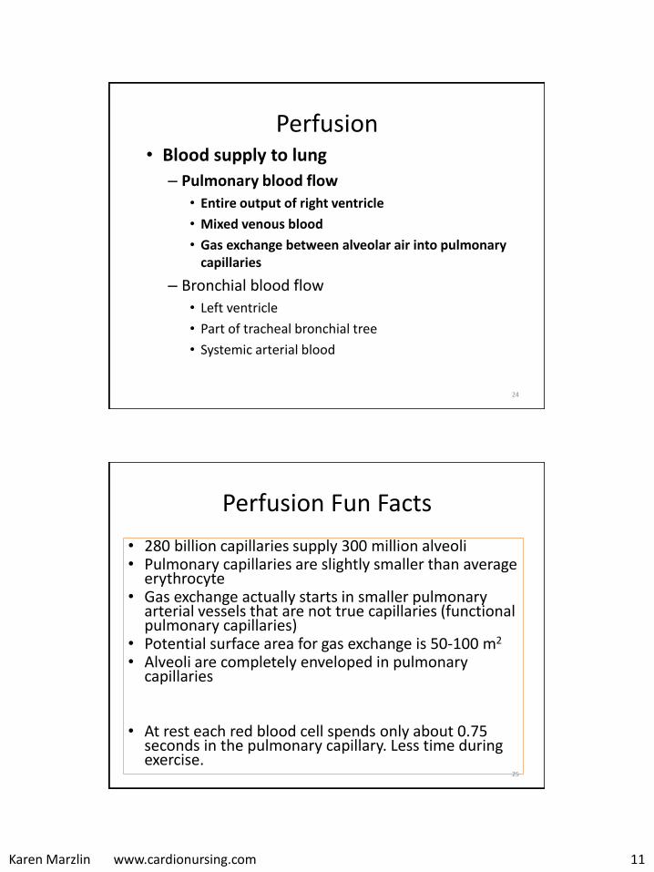
Perfusion • Blood supply to lung
– Pulmonary blood flow
• Entire output of right ventricle
• Mixed venous blood
• Gas exchange between alveolar air into pulmonary capillaries
– Bronchial blood flow
• Left ventricle
• Part of tracheal bronchial tree
• Systemic arterial blood
25
Perfusion Fun Facts
• 280 billion capillaries supply 300 million alveoli• Pulmonary capillaries are slightly smaller than average
erythrocyte • Gas exchange actually starts in smaller pulmonary
arterial vessels that are not true capillaries (functional pulmonary capillaries)
• Potential surface area for gas exchange is 50-100 m2
• Alveoli are completely enveloped in pulmonary capillaries
• At rest each red blood cell spends only about 0.75 seconds in the pulmonary capillary. Less time during exercise.
Karen Marzlin www.cardionursing.com 12
26
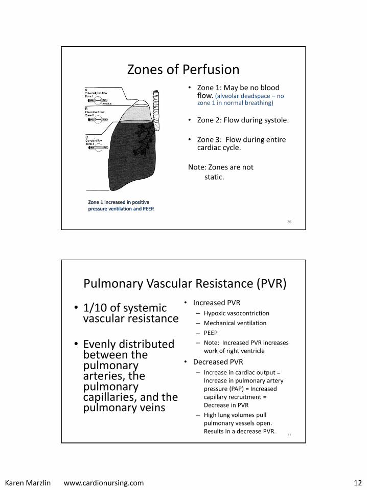
Zones of Perfusion • Zone 1: May be no blood
flow. (alveolar deadspace – no zone 1 in normal breathing)
• Zone 2: Flow during systole.
• Zone 3: Flow during entire cardiac cycle.
Note: Zones are notstatic.
Zone 1 increased in positive pressure ventilation and PEEP.
Pulmonary Vascular Resistance (PVR)
• 1/10 of systemic vascular resistance
• Evenly distributed between the pulmonary arteries, the pulmonary capillaries, and the pulmonary veins
• Increased PVR – Hypoxic vasocontriction
– Mechanical ventilation
– PEEP
– Note: Increased PVR increases work of right ventricle
• Decreased PVR– Increase in cardiac output =
Increase in pulmonary artery pressure (PAP) = Increased capillary recruitment = Decrease in PVR
– High lung volumes pull pulmonary vessels open. Results in a decrease PVR.
27
Karen Marzlin www.cardionursing.com 13
28
Hypoxic Pulmonary Vasoconstriction • Diverts blood away from poorly ventilated
alveoli– Also occurs in response to more global hypoxia
• Increases pulmonary artery pressure and recruits pulmonary capillaries to improve ventilation and perfusion matching
• Has limitations because of small amount of vascular smooth muscle in the pulmonary arteries
• The hypoxic vasoconstriction intended to help with V/Q mismatching increases the workload of right ventricle.
• Increases in PA pressures to recruit alveoli can also lead to pulmonary edema.
Conditions that Alter Pulmonary Perfusion
• #1 = pulmonary embolism
• Any decrease in cardiac output from right ventricle: shock
• Clinical Applications
– An increase in PVR for any reason can lead to right heart failure
– Any increase in pulmonary artery pressures can lead to pulmonary edema
29
Karen Marzlin www.cardionursing.com 14
30
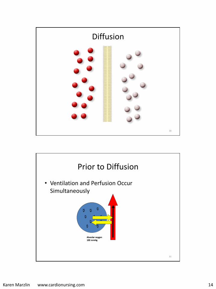
Diffusion
31
Prior to Diffusion
• Ventilation and Perfusion Occur Simultaneously
Alveolar oxygen 100 mmHg
Karen Marzlin www.cardionursing.com 15
32
Diffusion
• Movement of gases between the alveoli, plasma, and red blood cells
• Net movement of molecules from an area where the particular gas exerts a high partial pressure to an area where it exerts a lower partial pressure – Different gases each move according to their own partial
pressure gradients
• Diffusion of oxygen from alveoli to capillary determines the patient’s oxygenation status
33
Determinants of Diffusion • Surface Area: negatively affected by any type of
pulmonary resection; tumor, emphysema, pneumothorax
• Driving pressure: negatively affected by low inspired fraction of O2 (smoke inhalation) or by low barometric pressure (high altitudes) – Barometric pressure is the sum of the pressures of all the
gases it contains
• Thickness of alveolar capillary membrane (< 1 RBC):
negatively affected by pulmonary edema, pneumonia, or fibrosis
Karen Marzlin www.cardionursing.com 16
34
35
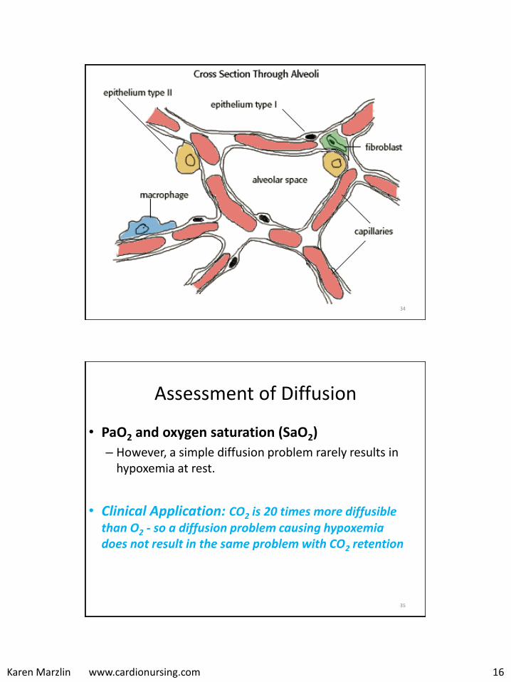
Assessment of Diffusion
• PaO2 and oxygen saturation (SaO2)
– However, a simple diffusion problem rarely results in hypoxemia at rest.
• Clinical Application: CO2 is 20 times more diffusible than O2 - so a diffusion problem causing hypoxemia does not result in the same problem with CO2 retention
Karen Marzlin www.cardionursing.com 17
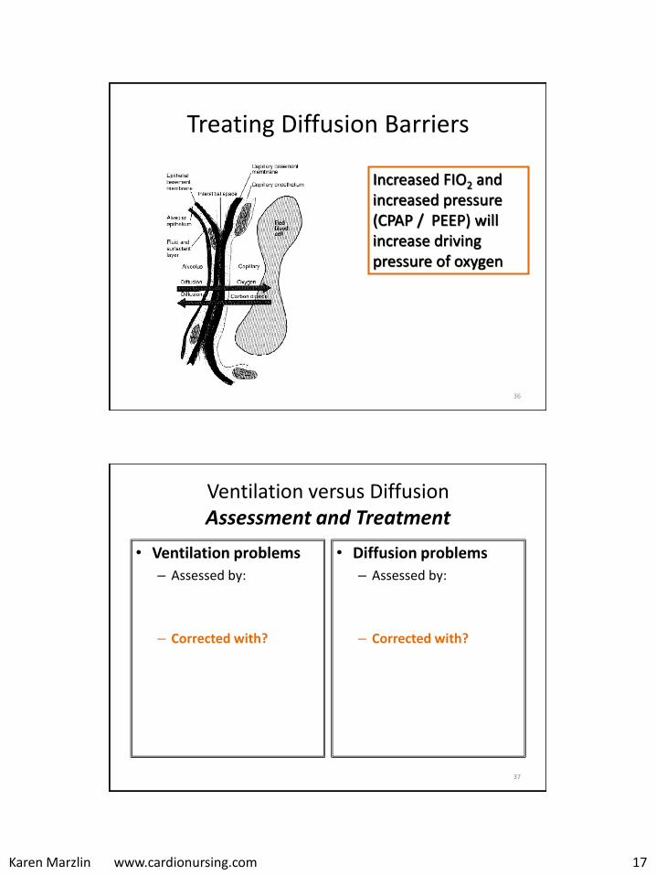
Treating Diffusion Barriers
36
Increased FIO2 and increased pressure (CPAP / PEEP) will increase driving pressure of oxygen
37
Ventilation versus Diffusion Assessment and Treatment
• Ventilation problems
– Assessed by:
– Corrected with?
• Diffusion problems
– Assessed by:
– Corrected with?
Karen Marzlin www.cardionursing.com 18
38
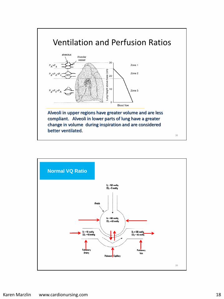
Ventilation and Perfusion Ratios
Alveoli in upper regions have greater volume and are less compliant. Alveoli in lower parts of lung have a greater change in volume during inspiration and are considered better ventilated.
39
Normal VQ Ratio
Karen Marzlin www.cardionursing.com 19
40
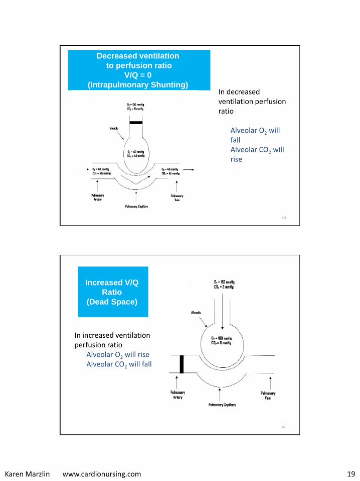
Decreased ventilation
to perfusion ratio
V/Q = 0
(Intrapulmonary Shunting)In decreased ventilation perfusion ratio
Alveolar O2 will fall Alveolar CO2 will rise
41
Increased V/Q
Ratio
(Dead Space)
In increased ventilation perfusion ratio
Alveolar O2 will riseAlveolar CO2 will fall
Karen Marzlin www.cardionursing.com 20
Causes of V/Q Mismatching• Non uniform ventilation
– Uneven resistance • Collapsed airways
(Emphysema) • Bronchoconstriction
(Asthma) • Inflammation (Bronchitis)
– Uneven compliance• Fibrosis • Pulmonary vascular
congestion • Atelectasis
42
Non uniform perfusion: •Pulmonary Emboli•Compression of pulmonary capillaries from high alveolar pressures •Tumors
43
Karen Marzlin www.cardionursing.com 21
44
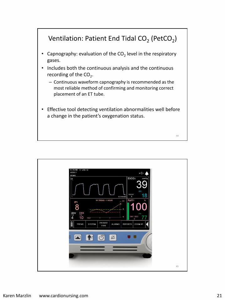
Ventilation: Patient End Tidal CO2 (PetCO2)
• Capnography: evaluation of the CO2 level in the respiratory gases.
• Includes both the continuous analysis and the continuous recording of the CO2.
– Continuous waveform capnography is recommended as the most reliable method of confirming and monitoring correct placement of an ET tube.
• Effective tool detecting ventilation abnormalities well before a change in the patient’s oxygenation status.
45
Karen Marzlin www.cardionursing.com 22
46
Assessing Oxygenation
• Cannot assess PaO2 (arterial) without considering alveolar oxygenation content (PA02)
• Increase in FIO2 will increase PAO2
• Increase in PACO2 will decrease PAO2
Note: With normal diffusion the majority of oxygen in the alveoli should diffuse across the alveolar capillary membrane.
47
PaO2 and FIO2 Ratio • An assessment and trending tool
• PaO2/ FIO2 ratio:
– Normal well above 300
– Acute lung injury < 300
– ARDS< or= 200
PaO2 of 60 mmHg with an FIO2 of 0.5 (50%)
represents a PaO2 /FIO2 ratio of
60 / 0.5 = 120.
This is a clinically significant intrapulmonary
shunt.
Karen Marzlin www.cardionursing.com 23
48
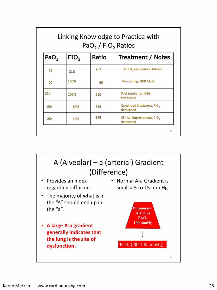
Linking Knowledge to Practice with PaO2 / FIO2 Ratios
PaO2 FIO2 Ratio Treatment / Notes
55 21%261
60 100% 60
210 100% 210
190 60% 316
150 40% 375
Admit; respiratory distress
Worsening; NRB Mask
Post intubation ABG, antibiotics
Continued treatment, FIO2
decreased
Clinical improvement, FIO2
decreased
49
A (Alveolar) – a (arterial) Gradient (Difference)
• Provides an index regarding diffusion.
• The majority of what is in the “A” should end up in the “a”.
• A large A-a gradient generally indicates that the lung is the site of dysfunction.
• Normal A-a Gradient is small = 5 to 15 mm Hg
PaO2 ( 80-100 mmHg)
Karen Marzlin www.cardionursing.com 24
Hypoxemia• Causes
– Diffusion abnormality
– Untreated alveolar hypoventilation
– Ventilation and perfusion mismatching
• Assessment Clues – PaO2 / SaO2: Will be low regardless of etiology
– Improvement with increased FIO2= Diffusion problem
– ↑ PaCO2 / increased work of breathing: Ventilation failure
– A-a gradient will be normal in ventilatory failure from neurological abnormality
– ↓PaO2 / FIO2 ratio: Suggests something more severe than simple diffusion abnormality (i.e. intra pulmonary shunting from decreased V/Q ratio
50
SpO2 (Pulse Oximetry)
• Used to estimate oxyhemoglobin.– The SpO2 generally correlates with the SaO2 - + or
- 2%.
• The goal equal to or greater than 92-94% in most patients being treated with oxygen.
• Requires the presence of a pleth wave detecting an accurate pulse.
51
Karen Marzlin www.cardionursing.com 25
Factors Affecting Accuracy of SpO2
(Pulse Oximetry)
• Hemoglobin < 5 g/dL or hematocrit <15%• Abnormal hemoglobin (carboxyhemoglobin or
methemoglobin)
• Other Factors – SpO2 below 70%– Low blood flow: hypotension or vasoconstriction– IV dyes, fingernail polish, some skin pigmentations– Administration of high fat content such as with
propofol or TPN can have a falsely high SpO2
52
53
Hypoxia and Hypoxemia
• Hypoxemia
– Insufficient oxygenation of the blood
– Mild: PaO2 < 80 mm Hg or SaO2 95%
– Moderate: PaO2 < 60 mmHg or SaO2 90%
– Severe: PaO2 < 40 mmHg or SaO2 75%
• Hypoxia
– Insufficient oxygenation of tissues
– Determined by oxygen delivery and cellular demand
Karen Marzlin www.cardionursing.com 26
57
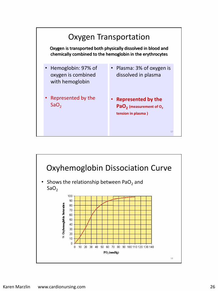
Oxygen Transportation
• Hemoglobin: 97% of oxygen is combined with hemoglobin
• Represented by the SaO2
• Plasma: 3% of oxygen is dissolved in plasma
• Represented by the PaO2 (measurement of O2
tension in plasma )
Oxygen is transported both physically dissolved in blood and chemically combined to the hemoglobin in the erythrocytes
58
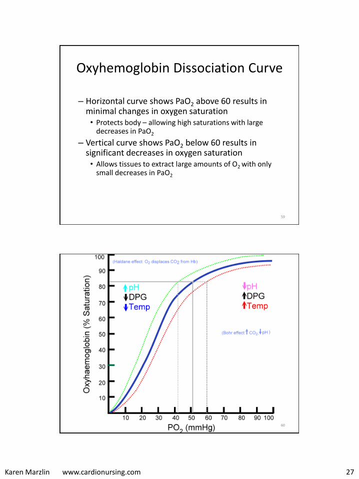
Oxyhemoglobin Dissociation Curve
• Shows the relationship between PaO2 and SaO2
Karen Marzlin www.cardionursing.com 27
59
Oxyhemoglobin Dissociation Curve
– Horizontal curve shows PaO2 above 60 results in minimal changes in oxygen saturation • Protects body – allowing high saturations with large
decreases in PaO2
– Vertical curve shows PaO2 below 60 results in significant decreases in oxygen saturation• Allows tissues to extract large amounts of O2 with only
small decreases in PaO2
60
Karen Marzlin www.cardionursing.com 28
61
Shifts in Oxyhemoglobin Curve
• Shift to the Left
– Easier to pick up at the lung level and more difficult to drop off (unload) at the tissue level
– Hemoglobin is more saturated for a given PaO2 and less oxygen is unloaded for a given Pao2
• Shift to the Right
– More difficult to pick up at the lung level but easier to drop off (unload) at the tissue level
– Hemoglobin is less saturated for a given PaO2 and more oxygen is unloaded for a given PaO2
62
Shifts in Oxyhemoglobin Curve
• Causes of Shift to Left
– Hypothermia
– Decreased 2,3 – DPG
– Hypocapnia
– Alkalemia
• Causes of Shift to Right
– Hyperthermia
– Increased 2,3 – DPG
– Hypercapnia
– Acidemia
Karen Marzlin www.cardionursing.com 29
63
Alterations in Oxyhemoglobin Curve
64
A Closer Look at 2,3-DPG
• 2,3-Diphosphoglycerate • Substance in the erythrocyte which affects the
affinity of hemoglobin for oxygen (binds to hemoglobin and decreases the affinity of hemoglobin for oxygen)
• Produced by erythrocytes during their normal glycolysis
• Increased – Chronic hypoxemia, anemia, hyperthyroidism
• Decreased – Massive transfusion of banked blood, hypophosphatemia,
hypothyroidism
Karen Marzlin www.cardionursing.com 30
65
Acid –Base Balance
69
Acid - Base Regulation
• Respiratory System
– Responds within minutes – fast but weak
– Regulates the excretion or retention of carbonic acid
• If pH is down: increase rate and depth of respiration to blow off PCO2
• If pH is up: decrease rate and depth of respiration to retain PCO2
Karen Marzlin www.cardionursing.com 31
70
Acid - Base Regulation
• Renal System
– Responds within 48 hours – slow but powerful
– Regulates excretion or retention of bicarbonate and the excretion of hydrogen and non-volatile acids
• If pH is down: kidney retains bicarbonate
• If pH is up: kidney excretes bicarbonate
71
ABG Analysis
• Evaluate ventilation: PaCO2
• Evaluate acid-base status: pH
• Evaluate source of abnormal pH: respiratory or metabolic
• Evaluate oxygenation: PaO2, SaO2
Karen Marzlin www.cardionursing.com 32
75
Compensation
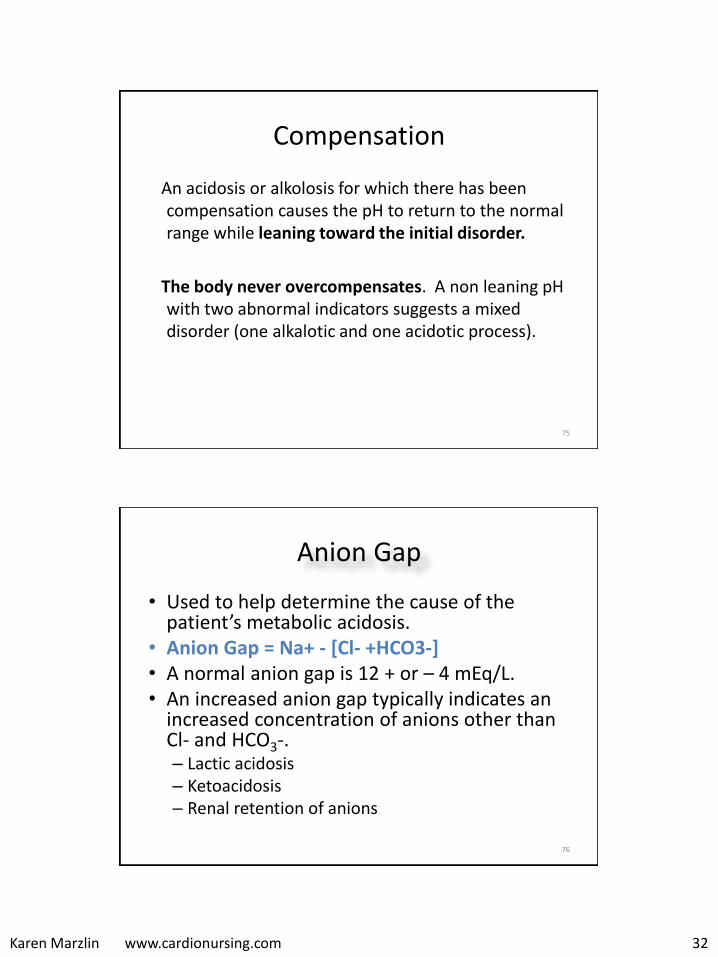
An acidosis or alkolosis for which there has been compensation causes the pH to return to the normal range while leaning toward the initial disorder.
The body never overcompensates. A non leaning pH with two abnormal indicators suggests a mixed disorder (one alkalotic and one acidotic process).
76
Anion Gap
• Used to help determine the cause of the patient’s metabolic acidosis.
• Anion Gap = Na+ - [Cl- +HCO3-]• A normal anion gap is 12 + or – 4 mEq/L. • An increased anion gap typically indicates an
increased concentration of anions other than Cl- and HCO3-. – Lactic acidosis – Ketoacidosis – Renal retention of anions
Karen Marzlin www.cardionursing.com 33
More on Anion Gap
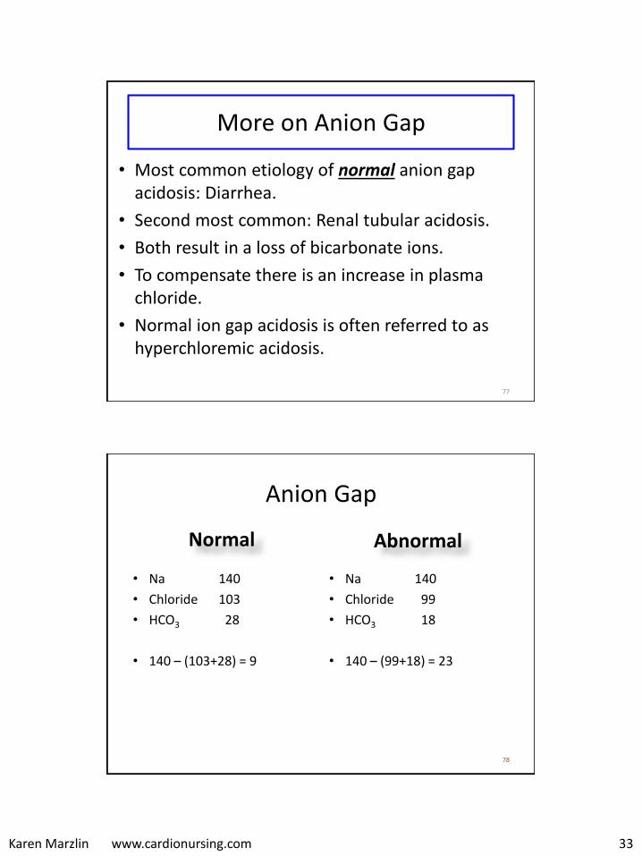
• Most common etiology of normal anion gap acidosis: Diarrhea.
• Second most common: Renal tubular acidosis.
• Both result in a loss of bicarbonate ions.
• To compensate there is an increase in plasma chloride.
• Normal ion gap acidosis is often referred to as hyperchloremic acidosis.
77
Anion Gap
Normal
• Na 140
• Chloride 103
• HCO3 28
• 140 – (103+28) = 9
Abnormal
• Na 140
• Chloride 99
• HCO3 18
• 140 – (99+18) = 23
78
Karen Marzlin www.cardionursing.com 34
79
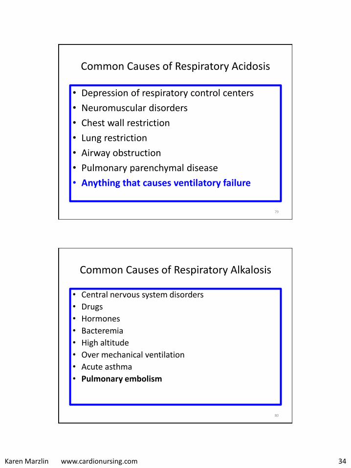
Common Causes of Respiratory Acidosis
• Depression of respiratory control centers
• Neuromuscular disorders
• Chest wall restriction
• Lung restriction
• Airway obstruction
• Pulmonary parenchymal disease
• Anything that causes ventilatory failure
80
Common Causes of Respiratory Alkalosis
• Central nervous system disorders
• Drugs
• Hormones
• Bacteremia
• High altitude
• Over mechanical ventilation
• Acute asthma
• Pulmonary embolism
Karen Marzlin www.cardionursing.com 35
81
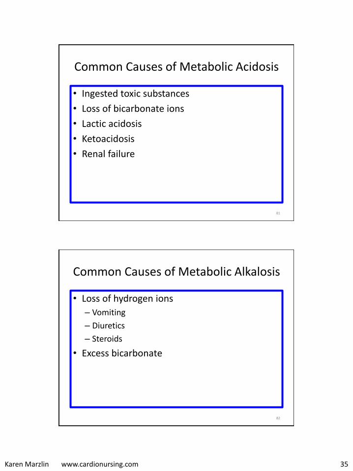
Common Causes of Metabolic Acidosis
• Ingested toxic substances
• Loss of bicarbonate ions
• Lactic acidosis
• Ketoacidosis
• Renal failure
82
Common Causes of Metabolic Alkalosis
• Loss of hydrogen ions
– Vomiting
– Diuretics
– Steroids
• Excess bicarbonate
Karen Marzlin www.cardionursing.com 36
83
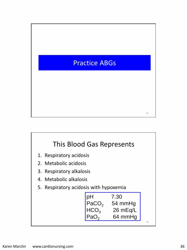
Practice ABGs
This Blood Gas Represents
84
1. Respiratory acidosis
2. Metabolic acidosis
3. Respiratory alkalosis
4. Metabolic alkalosis
5. Respiratory acidosis with hypoxemia
pH 7.30
PaCO2 54 mmHg
HCO3 26 mEq/L
PaO2 64 mmHg
Karen Marzlin www.cardionursing.com 37
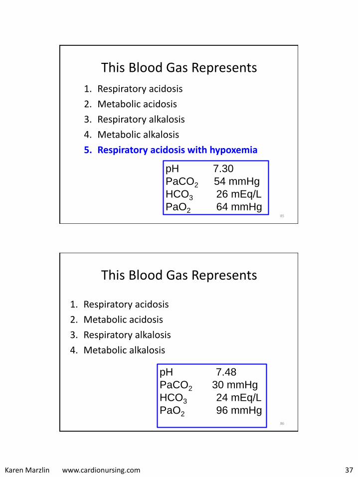
This Blood Gas Represents
85
1. Respiratory acidosis
2. Metabolic acidosis
3. Respiratory alkalosis
4. Metabolic alkalosis
5. Respiratory acidosis with hypoxemia
pH 7.30
PaCO2 54 mmHg
HCO3 26 mEq/L
PaO2 64 mmHg
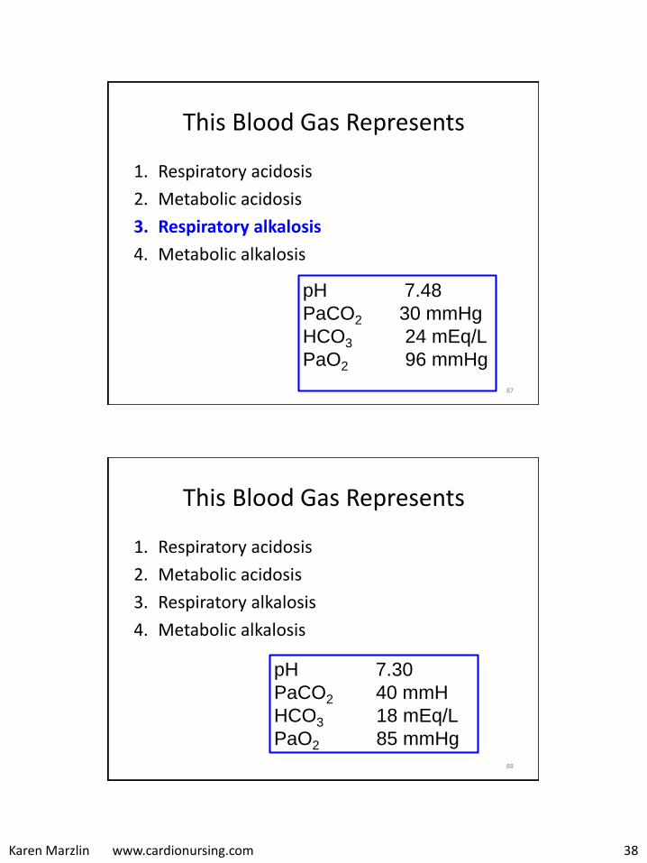
This Blood Gas Represents
86
1. Respiratory acidosis
2. Metabolic acidosis
3. Respiratory alkalosis
4. Metabolic alkalosis
pH 7.48
PaCO2 30 mmHg
HCO3 24 mEq/L
PaO2 96 mmHg
Karen Marzlin www.cardionursing.com 38
This Blood Gas Represents
87
1. Respiratory acidosis
2. Metabolic acidosis
3. Respiratory alkalosis
4. Metabolic alkalosis
pH 7.48
PaCO2 30 mmHg
HCO3 24 mEq/L
PaO2 96 mmHg
This Blood Gas Represents
88
1. Respiratory acidosis
2. Metabolic acidosis
3. Respiratory alkalosis
4. Metabolic alkalosis
pH 7.30
PaCO2 40 mmH
HCO3 18 mEq/L
PaO2 85 mmHg
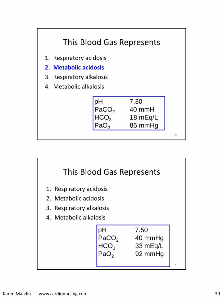
Karen Marzlin www.cardionursing.com 39
This Blood Gas Represents
89
1. Respiratory acidosis
2. Metabolic acidosis
3. Respiratory alkalosis
4. Metabolic alkalosis
pH 7.30
PaCO2 40 mmH
HCO3 18 mEq/L
PaO2 85 mmHg
This Blood Gas Represents
90
1. Respiratory acidosis
2. Metabolic acidosis
3. Respiratory alkalosis
4. Metabolic alkalosis
pH 7.50
PaCO2 40 mmHg
HCO3 33 mEq/L
PaO2 92 mmHg
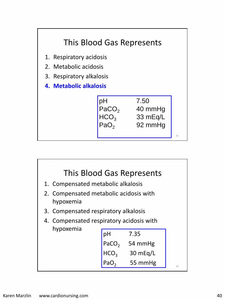
Karen Marzlin www.cardionursing.com 40
This Blood Gas Represents
91
1. Respiratory acidosis
2. Metabolic acidosis
3. Respiratory alkalosis
4. Metabolic alkalosis
pH 7.50
PaCO2 40 mmHg
HCO3 33 mEq/L
PaO2 92 mmHg
This Blood Gas Represents
92
1. Compensated metabolic alkalosis
2. Compensated metabolic acidosis with hypoxemia
3. Compensated respiratory alkalosis
4. Compensated respiratory acidosis with hypoxemia
pH 7.35
PaCO2 54 mmHg
HCO3 30 mEq/L
PaO2 55 mmHg
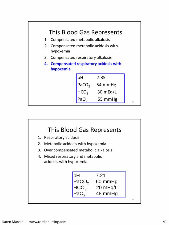
Karen Marzlin www.cardionursing.com 41
This Blood Gas Represents
93
1. Compensated metabolic alkalosis
2. Compensated metabolic acidosis with hypoxemia
3. Compensated respiratory alkalosis
4. Compensated respiratory acidosis with hypoxemia
pH 7.35
PaCO2 54 mmHg
HCO3 30 mEq/L
PaO2 55 mmHg
This Blood Gas Represents
94
1. Respiratory acidosis
2. Metabolic acidosis with hypoxemia
3. Over compensated metabolic alkalosis
4. Mixed respiratory and metabolic acidosis with hypoxemia
pH 7.21
PaCO2 60 mmHg
HCO3 20 mEq/L
PaO2 48 mmHg
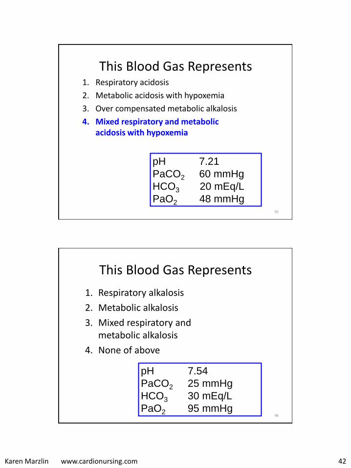
Karen Marzlin www.cardionursing.com 42
This Blood Gas Represents
95
1. Respiratory acidosis
2. Metabolic acidosis with hypoxemia
3. Over compensated metabolic alkalosis
4. Mixed respiratory and metabolic acidosis with hypoxemia
pH 7.21
PaCO2 60 mmHg
HCO3 20 mEq/L
PaO2 48 mmHg
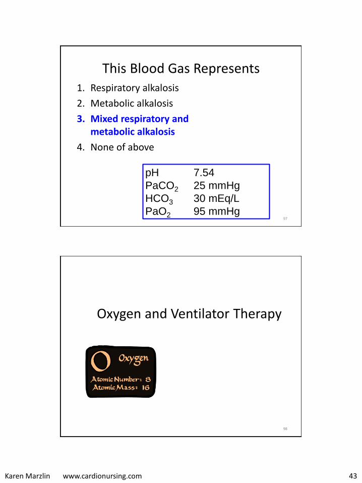
This Blood Gas Represents
96
1. Respiratory alkalosis
2. Metabolic alkalosis
3. Mixed respiratory and metabolic alkalosis
4. None of above
pH 7.54
PaCO2 25 mmHg
HCO3 30 mEq/L
PaO2 95 mmHg
Karen Marzlin www.cardionursing.com 43
This Blood Gas Represents
97
1. Respiratory alkalosis
2. Metabolic alkalosis
3. Mixed respiratory and metabolic alkalosis
4. None of above
pH 7.54
PaCO2 25 mmHg
HCO3 30 mEq/L
PaO2 95 mmHg
98
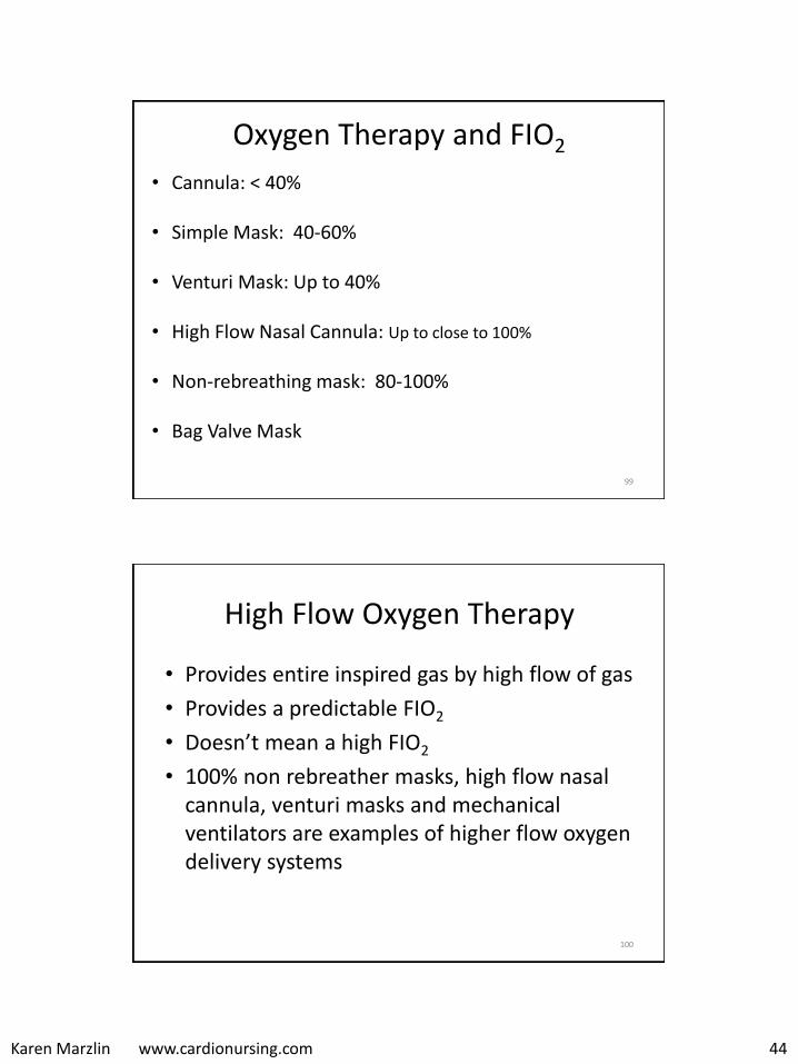
Oxygen and Ventilator Therapy
Karen Marzlin www.cardionursing.com 44
99
Oxygen Therapy and FIO2
• Cannula: < 40%
• Simple Mask: 40-60%
• Venturi Mask: Up to 40%
• High Flow Nasal Cannula: Up to close to 100%
• Non-rebreathing mask: 80-100%
• Bag Valve Mask
100
High Flow Oxygen Therapy
• Provides entire inspired gas by high flow of gas
• Provides a predictable FIO2
• Doesn’t mean a high FIO2
• 100% non rebreather masks, high flow nasal cannula, venturi masks and mechanical ventilators are examples of higher flow oxygen delivery systems
Karen Marzlin www.cardionursing.com 45
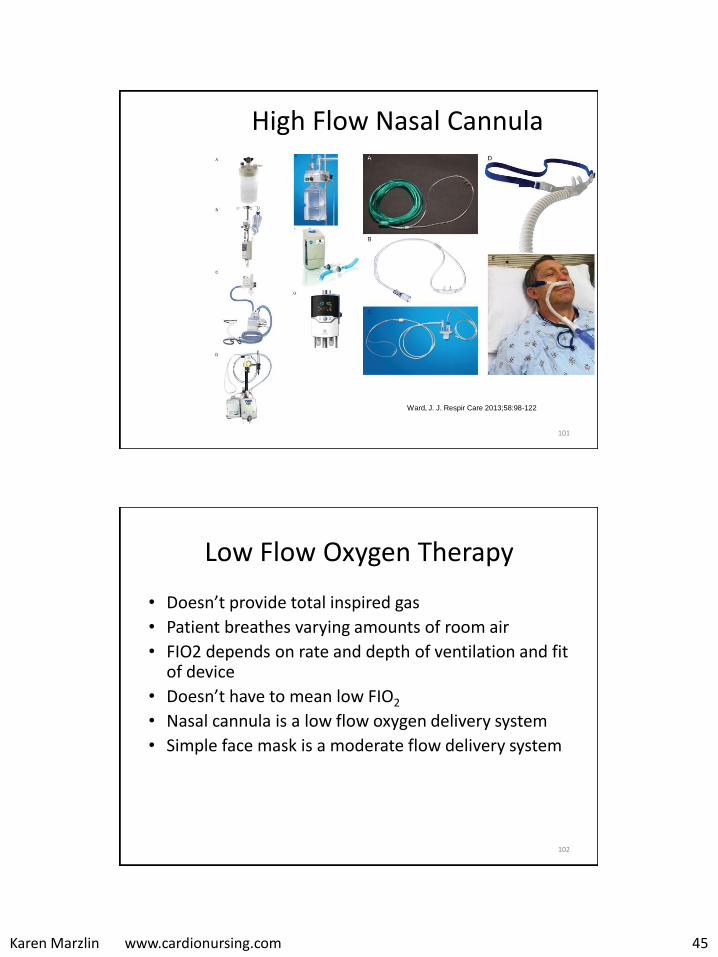
Ward, J. J. Respir Care 2013;58:98-122
High Flow Nasal Cannula
101
102
Low Flow Oxygen Therapy
• Doesn’t provide total inspired gas
• Patient breathes varying amounts of room air
• FIO2 depends on rate and depth of ventilation and fit of device
• Doesn’t have to mean low FIO2
• Nasal cannula is a low flow oxygen delivery system
• Simple face mask is a moderate flow delivery system
Karen Marzlin www.cardionursing.com 46
103
Guidelines for estimating FIO2 with low flow oxygen devices
• 100% O2 flow rate(L)– Nasal Cannula
• 1• 2• 3• 4• 5• 6
– Oxygen Mask• 5-6• 6-7• 7-8
– Mask with Reservoir• 6• 7• 8• 9• 10
• FIO2 (%)• 24• 28• 32• 36• 40• 44
• 40• 50• 60
• 60• 70• 80• 90• 99
104
Oxygen Toxicity
• Complications of O2
– Absorption atelectasis
– Decreased hypoxic drive
• Signs and symptoms of oxygen toxicity – Dyspnea
– Decreased lung compliance
– Retrosternal pain
– Parasthesia in the extremities
• To reduce risk of oxygen toxicity:
• 100% no more than 24 hours
• 60% no more than 2-3 days
• Use 40% for longer term therapy
Karen Marzlin www.cardionursing.com 47
105
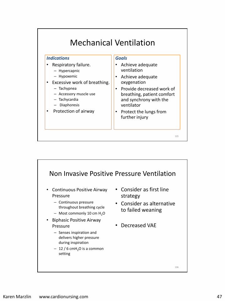
Mechanical Ventilation
Indications
• Respiratory failure.– Hypercapnic
– Hypoxemic
• Excessive work of breathing.– Tachypnea
– Accessory muscle use
– Tachycardia
– Diaphoresis
• Protection of airway
Goals
• Achieve adequate ventilation
• Achieve adequate oxygenation
• Provide decreased work of breathing, patient comfort and synchrony with the ventilator
• Protect the lungs from further injury
106
Non Invasive Positive Pressure Ventilation
• Continuous Positive Airway Pressure – Continuous pressure
throughout breathing cycle
– Most commonly 10 cm H2O
• Biphasic Positive Airway Pressure – Senses inspiration and
delivers higher pressure during inspiration
– 12 / 6 cmH2O is a common setting
• Consider as first line strategy
• Consider as alternative to failed weaning
• Decreased VAE
Karen Marzlin www.cardionursing.com 48
107
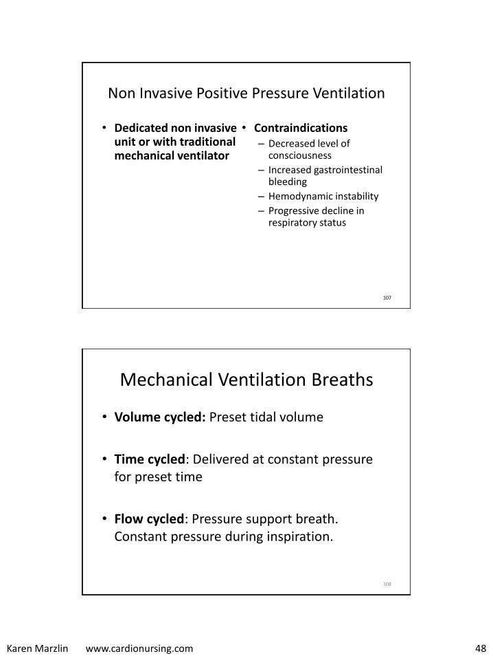
Non Invasive Positive Pressure Ventilation
• Dedicated non invasive unit or with traditional mechanical ventilator
• Contraindications – Decreased level of
consciousness
– Increased gastrointestinal bleeding
– Hemodynamic instability
– Progressive decline in respiratory status
108
Mechanical Ventilation Breaths
• Volume cycled: Preset tidal volume
• Time cycled: Delivered at constant pressure for preset time
• Flow cycled: Pressure support breath. Constant pressure during inspiration.
Karen Marzlin www.cardionursing.com 49
109
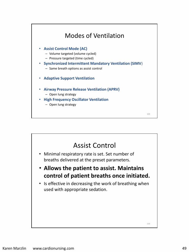
Modes of Ventilation
• Assist Control Mode (AC)– Volume targeted (volume cycled)
– Pressure targeted (time cycled)
• Synchronized Intermittent Mandatory Ventilation (SIMV)– Same breath options as assist control
• Adaptive Support Ventilation
• Airway Pressure Release Ventilation (APRV)– Open lung strategy
• High Frequency Oscillator Ventilation– Open lung strategy
110
Assist Control • Minimal respiratory rate is set. Set number of
breaths delivered at the preset parameters.
• Allows the patient to assist. Maintains control of patient breaths once initiated.
• Is effective in decreasing the work of breathing when used with appropriate sedation.
Karen Marzlin www.cardionursing.com 50
111
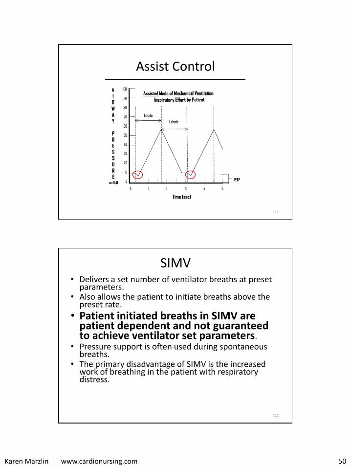
Assist Control
112
SIMV • Delivers a set number of ventilator breaths at preset
parameters. • Also allows the patient to initiate breaths above the
preset rate.
• Patient initiated breaths in SIMV are patient dependent and not guaranteed to achieve ventilator set parameters.
• Pressure support is often used during spontaneous breaths.
• The primary disadvantage of SIMV is the increased work of breathing in the patient with respiratory distress.
Karen Marzlin www.cardionursing.com 51
113
Adaptive Support Ventilation
• Capable of increasing or decreasing support as needed – No spontaneous breathing: Uses AC with time cycled
(pressure controlled) breaths
– Spontaneous breathing below target: Uses SIMV mode with time cycled (pressure controlled) breaths
– Spontaneous breathing above target: the ventilator changes to the pressure support mode using flow cycled breathes
Adaptive Support Ventilation
• Plateau pressure is set and Vt varies breath to breath
• Mandatory breaths are adjusted to assure adequate minute ventilation
• Pressure limits can be adjusted if needed to assure adequate ventilation
• I:E ratio is also adjusted to prevent auto peep
114
Karen Marzlin www.cardionursing.com 52
115
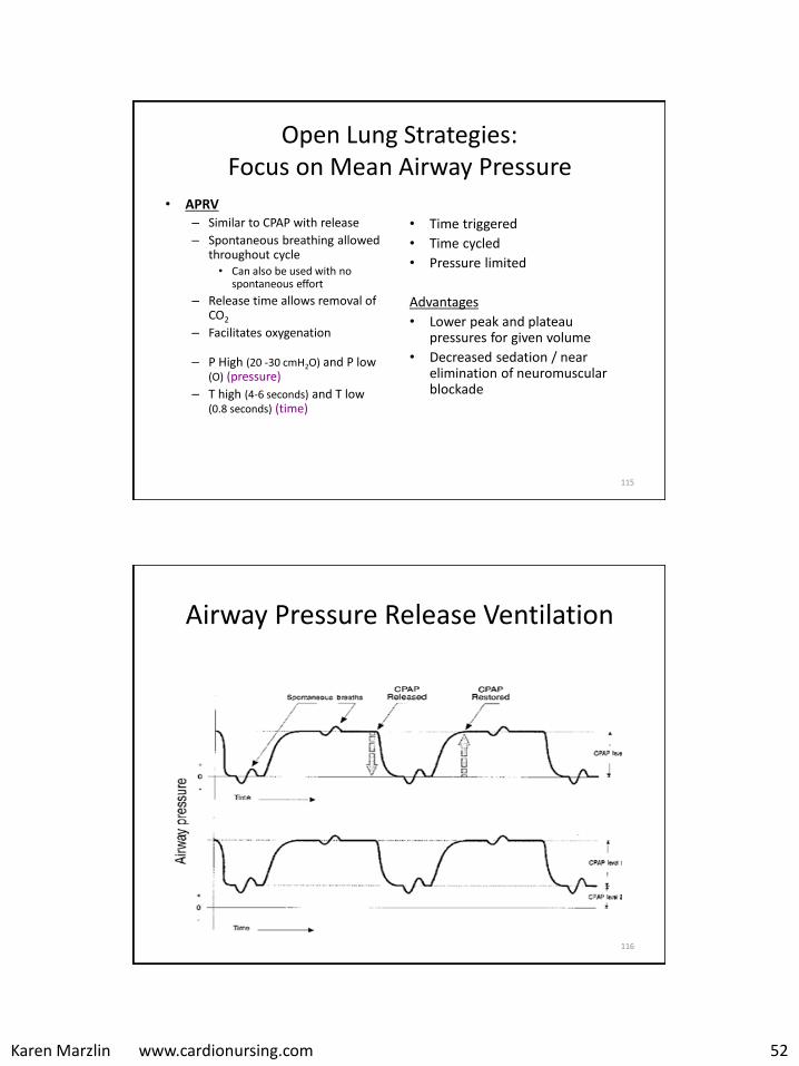
Open Lung Strategies: Focus on Mean Airway Pressure
• APRV – Similar to CPAP with release
– Spontaneous breathing allowed throughout cycle • Can also be used with no
spontaneous effort
– Release time allows removal of CO2
– Facilitates oxygenation
– P High (20 -30 cmH2O) and P low (O) (pressure)
– T high (4-6 seconds) and T low (0.8 seconds) (time)
• Time triggered
• Time cycled
• Pressure limited
Advantages
• Lower peak and plateau pressures for given volume
• Decreased sedation / near elimination of neuromuscular blockade
Airway Pressure Release Ventilation
116
Karen Marzlin www.cardionursing.com 53
117
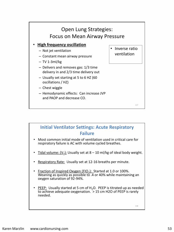
Open Lung Strategies: Focus on Mean Airway Pressure
• High frequency oscillation
– Not jet ventilation
– Constant mean airway pressure
– TV 1-3ml/kg
– Delivers and removes gas: 1/3 time delivery in and 2/3 time delivery out
– Usually set starting at 5 to 6 HZ (60 oscillations / HZ)
– Chest wiggle
– Hemodynamic effects: Can increase JVP and PAOP and decrease CO.
• Inverse ratio ventilation
118
Initial Ventilator Settings: Acute Respiratory Failure
• Most common initial mode of ventilation used in critical care for respiratory failure is AC with volume cycled breathes.
• Tidal volume: (VT): Usually set at 8 – 10 ml/kg of ideal body weight.
• Respiratory Rate: Usually set at 12-16 breaths per minute.
• Fraction of Inspired Oxygen (FIO2): Started at 1.0 or 100%. Weaning as quickly as possible to .4 or 40% while maintaining an oxygen saturation of 92-94%.
• PEEP: Usually started at 5 cm of H2O. PEEP is titrated up as needed to achieve adequate oxygenation. > 15 cm H2O of PEEP is rarely needed.
Karen Marzlin www.cardionursing.com 54
119
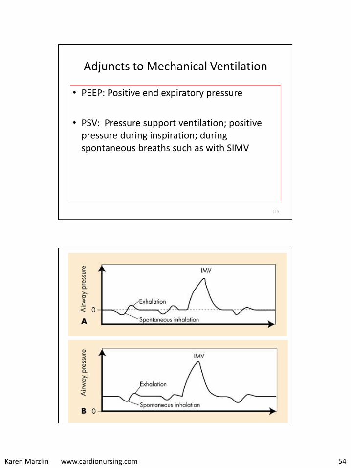
Adjuncts to Mechanical Ventilation
• PEEP: Positive end expiratory pressure
• PSV: Pressure support ventilation; positive pressure during inspiration; during spontaneous breaths such as with SIMV
120
Karen Marzlin www.cardionursing.com 55
121
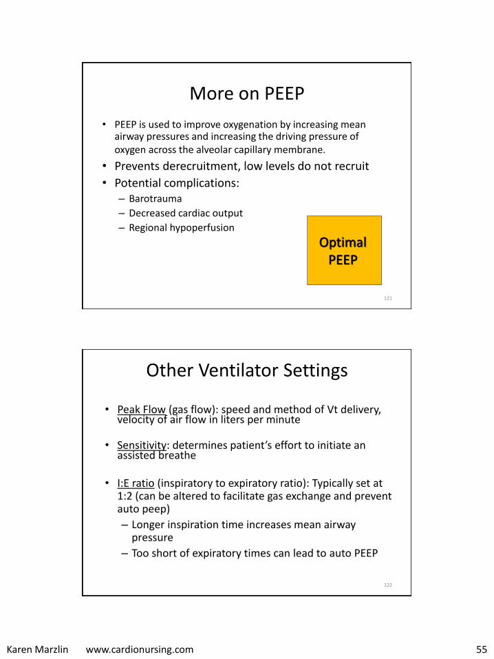
More on PEEP
• PEEP is used to improve oxygenation by increasing mean airway pressures and increasing the driving pressure of oxygen across the alveolar capillary membrane.
• Prevents derecruitment, low levels do not recruit
• Potential complications: – Barotrauma
– Decreased cardiac output
– Regional hypoperfusion
Optimal PEEP
122
Other Ventilator Settings
• Peak Flow (gas flow): speed and method of Vt delivery, velocity of air flow in liters per minute
• Sensitivity: determines patient’s effort to initiate an assisted breathe
• I:E ratio (inspiratory to expiratory ratio): Typically set at 1:2 (can be altered to facilitate gas exchange and prevent auto peep)
– Longer inspiration time increases mean airway pressure
– Too short of expiratory times can lead to auto PEEP
Karen Marzlin www.cardionursing.com 56
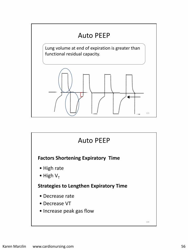
Auto PEEP
123
Lung volume at end of expiration is greater thanfunctional residual capacity.
Auto PEEP
Factors Shortening Expiratory Time
• High rate
• High VT
Strategies to Lengthen Expiratory Time
• Decrease rate
• Decrease VT
• Increase peak gas flow
124
Karen Marzlin www.cardionursing.com 57
125
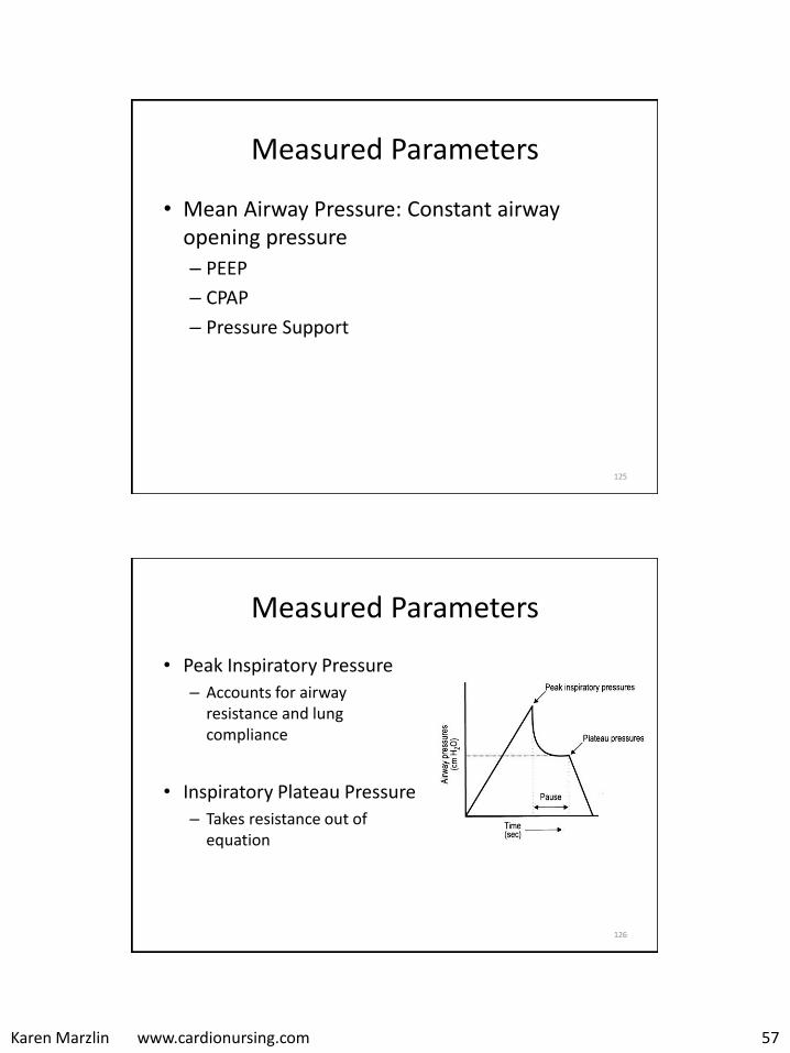
Measured Parameters
• Mean Airway Pressure: Constant airway opening pressure
– PEEP
– CPAP
– Pressure Support
126
Measured Parameters
• Peak Inspiratory Pressure
– Accounts for airway resistance and lung compliance
• Inspiratory Plateau Pressure
– Takes resistance out of equation
Karen Marzlin www.cardionursing.com 58
127
Hemodynamic Effects of Mechanical Ventilation
• Decreased venous return
• Pulmonary capillary compression and increased right ventricular afterload
– Decreased right ventricular stroke volume
• Decreased left ventricular afterload
128
Hypotension with Mechanical Ventilation
• Conversion to positive pressure ventilation / PEEP – Assure adequate circulating fluid volume
• Response to sedation – Titrate sedation
• Development of auto PEEP– Increase expiration time
• Tension Pneumothorax – Chest tube required
Karen Marzlin www.cardionursing.com 59
129
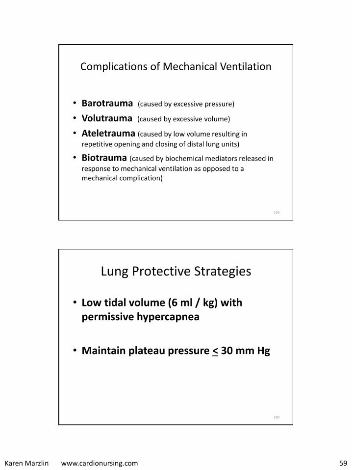
Complications of Mechanical Ventilation
• Barotrauma (caused by excessive pressure)
• Volutrauma (caused by excessive volume)
• Ateletrauma (caused by low volume resulting in
repetitive opening and closing of distal lung units)
• Biotrauma (caused by biochemical mediators released in
response to mechanical ventilation as opposed to a mechanical complication)
130
Lung Protective Strategies
• Low tidal volume (6 ml / kg) with permissive hypercapnea
• Maintain plateau pressure < 30 mm Hg
Karen Marzlin www.cardionursing.com 60
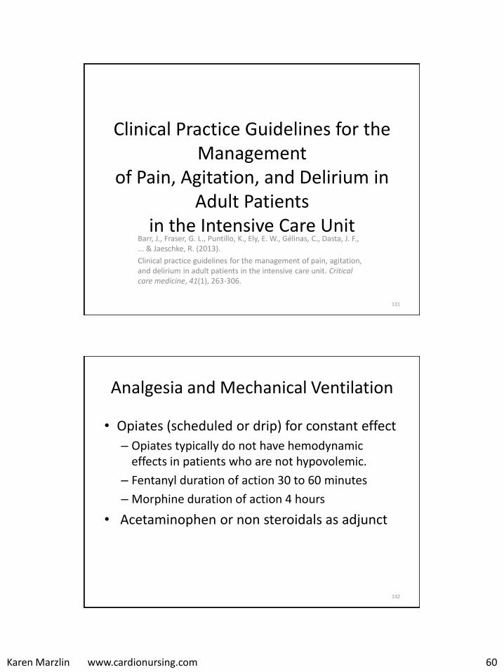
Clinical Practice Guidelines for the Management
of Pain, Agitation, and Delirium in Adult Patients
in the Intensive Care UnitBarr, J., Fraser, G. L., Puntillo, K., Ely, E. W., Gélinas, C., Dasta, J. F., ... & Jaeschke, R. (2013).
Clinical practice guidelines for the management of pain, agitation, and delirium in adult patients in the intensive care unit. Critical care medicine, 41(1), 263-306.
131
Analgesia and Mechanical Ventilation
• Opiates (scheduled or drip) for constant effect
– Opiates typically do not have hemodynamic effects in patients who are not hypovolemic.
– Fentanyl duration of action 30 to 60 minutes
– Morphine duration of action 4 hours
• Acetaminophen or non steroidals as adjunct
132
Karen Marzlin www.cardionursing.com 61
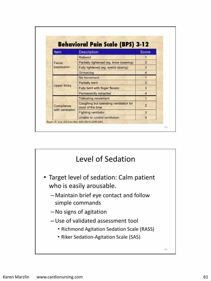
133
Level of Sedation
• Target level of sedation: Calm patient who is easily arousable.
–Maintain brief eye contact and follow simple commands
–No signs of agitation
–Use of validated assessment tool
• Richmond Agitation Sedation Scale (RASS)
• Riker Sedation-Agitation Scale (SAS)
135
Karen Marzlin www.cardionursing.com 62
136
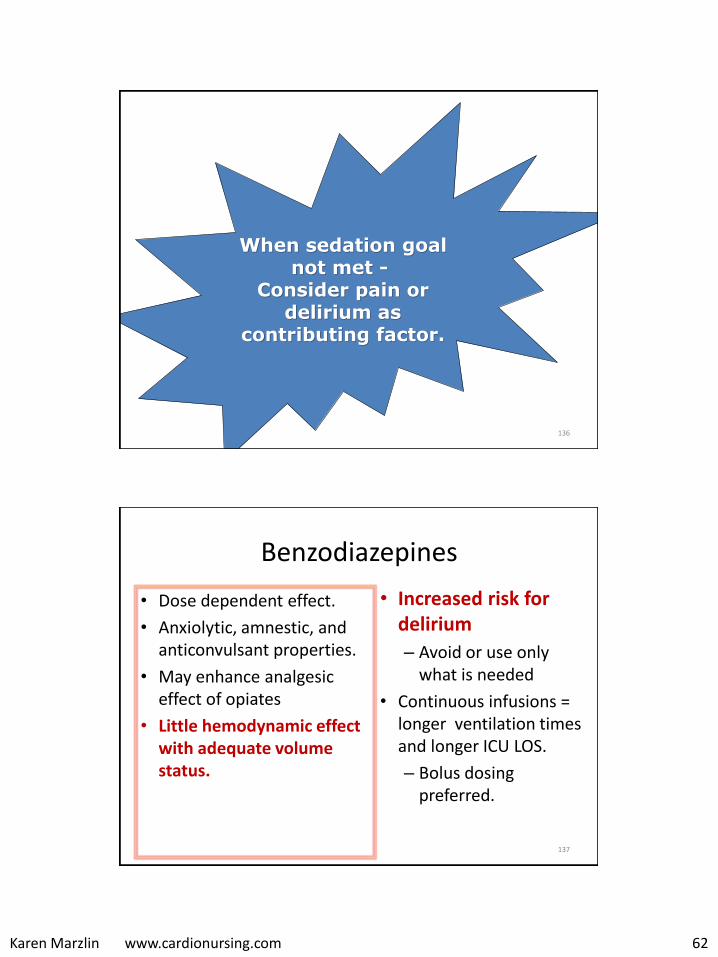
When sedation goal not met -
Consider pain or delirium as
contributing factor.
Benzodiazepines
• Increased risk for delirium
– Avoid or use only what is needed
• Continuous infusions = longer ventilation times and longer ICU LOS.
– Bolus dosing preferred.
• Dose dependent effect.
• Anxiolytic, amnestic, and anticonvulsant properties.
• May enhance analgesic effect of opiates
• Little hemodynamic effect with adequate volume status.
137
Karen Marzlin www.cardionursing.com 63
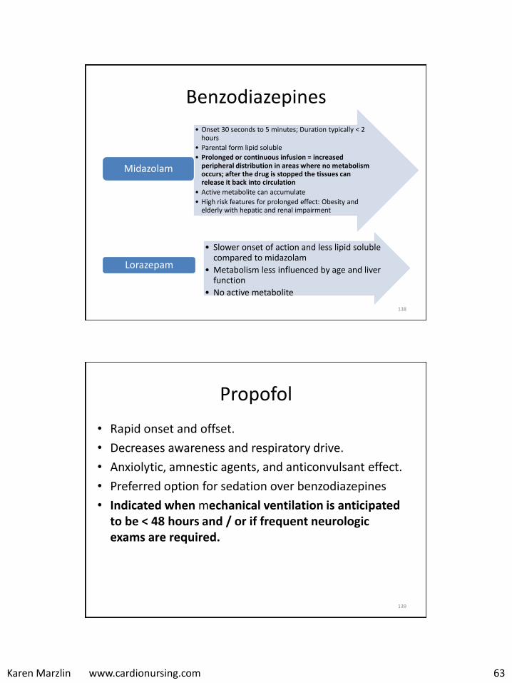
Benzodiazepines
• Onset 30 seconds to 5 minutes; Duration typically < 2 hours
• Parental form lipid soluble
• Prolonged or continuous infusion = increased peripheral distribution in areas where no metabolism occurs; after the drug is stopped the tissues can release it back into circulation
• Active metabolite can accumulate
• High risk features for prolonged effect: Obesity and elderly with hepatic and renal impairment
Midazolam
• Slower onset of action and less lipid soluble compared to midazolam
• Metabolism less influenced by age and liver function
• No active metabolite
Lorazepam
138
Propofol
• Rapid onset and offset.
• Decreases awareness and respiratory drive.
• Anxiolytic, amnestic agents, and anticonvulsant effect.
• Preferred option for sedation over benzodiazepines
• Indicated when mechanical ventilation is anticipated to be < 48 hours and / or if frequent neurologic exams are required.
139
Karen Marzlin www.cardionursing.com 64
Propofol
• Use lowest possible infusion rate for shortest period of time to achieve sedation goal.
• Initiated slowly at a rate of 5 mcg/kg/min. Increase by increments of 5 to 10 mcg/kg/min. Minimum of 5 minutes between dose adjustment.
• Typical maintenance dose of 5 to 50 mcg/kg/min.
• Half life with short term use: 3 to 12 hours
140
Propofol Complications
• Hypotension (common) – Venous vasodilatation and mild
cardiac depressive effect.
– Bolus doing and hypovolemia increase risk.
• Elevated triglycerides, pancreatitis, and infection – High lipid content (1.1 kcal/ml of
fat)
• Propofol-related infusion syndrome.
– Potentially life threatening complication characterized by the onset of metabolic acidosis, dysrhythmias, hyperkalemia, rhabdomyolysis (or elevated CPK levels), and cardiac failure
141
Karen Marzlin www.cardionursing.com 65
Dexmedetomidine
• Selective α2-receptor agonist
– Sedative
– Non opioid analgesic
– Sympatholytic properties
– No anticonvulsant properties
• Patients more easily arousable
• Minimal respiratory depression – can be used in non intubated patient
142
Dexmedetomidine
• Only approved for short-term sedation of ICU patients (< 24 hrs) at a maximal dose of 0.7 μg/kg/hr
• Several studies have demonstrated safety at higher doses and for longer duration of infusion
• Analgesic mechanism: α-2receptors are located in the dorsal region of the spinal cord and in supraspinal sites – however, analgesic effects extend beyond spine; ? Mechanism of action
• Evidence of lower delirium compared to benzodiazepines
143
Karen Marzlin www.cardionursing.com 66
Dexmedetomidine
• Preferred over benzodiazepines to improve clinical outcomes in mechanically ventilated adult ICU patients
• Maintenance dose: 0.2 to 0.7 mcg/kg.hour
• Half life: 1.8 to 3.1 hours
• Side effects: Hypotension and bradycardia
• HTN or hypotension with loading dose
• Loss of oropharyngeal muscle tone – potential obstruction in non intubated patients
144
Daily Interruptions of Sedation
• Allow patients to spend some time awake and interacting
• Time for a thorough neurological assessment
• Opportunity to re-titrate sedatives and analgesics.
• Reduces duration of mechanical ventilation and ICU stay
• Does not increase unplanned extubations.
145
Karen Marzlin www.cardionursing.com 67
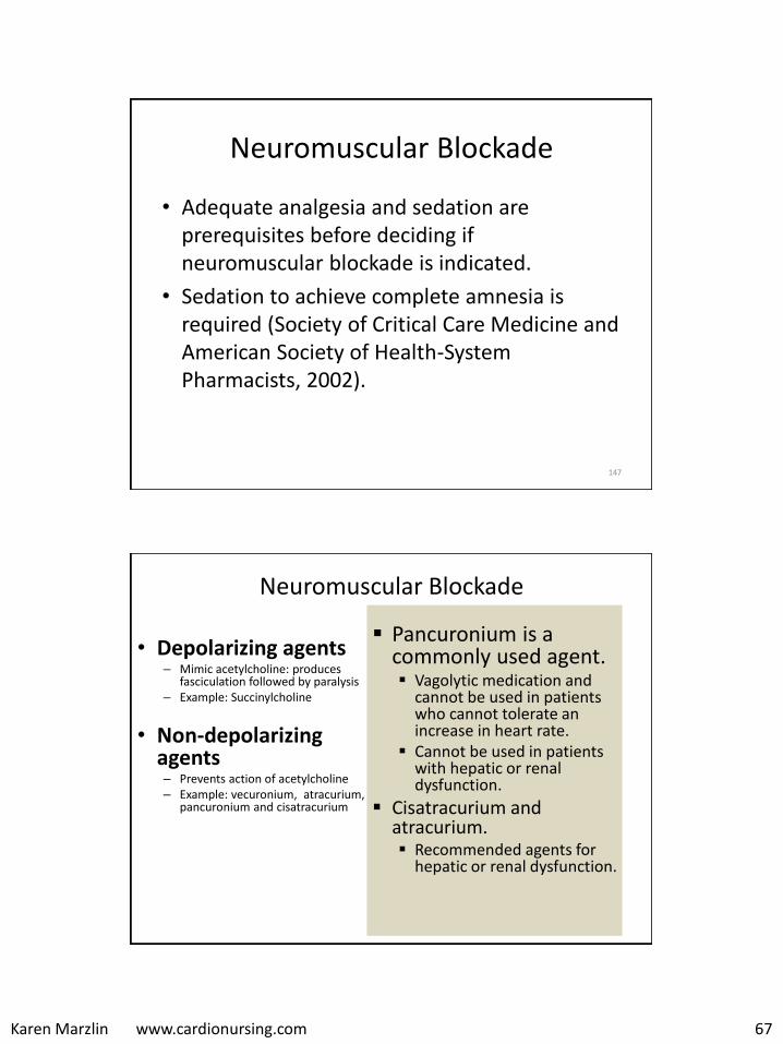
Neuromuscular Blockade
• Adequate analgesia and sedation are prerequisites before deciding if neuromuscular blockade is indicated.
• Sedation to achieve complete amnesia is required (Society of Critical Care Medicine and American Society of Health-System Pharmacists, 2002).
147
148
Neuromuscular Blockade
• Depolarizing agents– Mimic acetylcholine: produces
fasciculation followed by paralysis – Example: Succinylcholine
• Non-depolarizing agents– Prevents action of acetylcholine – Example: vecuronium, atracurium,
pancuronium and cisatracurium
Pancuronium is a commonly used agent. Vagolytic medication and
cannot be used in patients who cannot tolerate an increase in heart rate.
Cannot be used in patients with hepatic or renal dysfunction.
Cisatracurium and atracurium. Recommended agents for
hepatic or renal dysfunction.
Karen Marzlin www.cardionursing.com 68
Neuromuscular Blockade
Assessment
• Objective measures of brain function (i.e.BIS) are recommended
• Peripheral nerve stimulators – The most commonly used
nerve-muscle combination ulnar nerve and adductor pollicus.
– Goal: One or two twitches in response to nerve stimulation.
Complications
• Prolonged recovery from the agents
• Myopathy– With corticosteroids
– Use longer than 1 to 2 days.
• Acute quadriplegic myopathy syndrome – Acute paresis
– Myonecrosis (increased CPK enzymes)
– Abnormal EMG
149
150
Karen Marzlin www.cardionursing.com 69
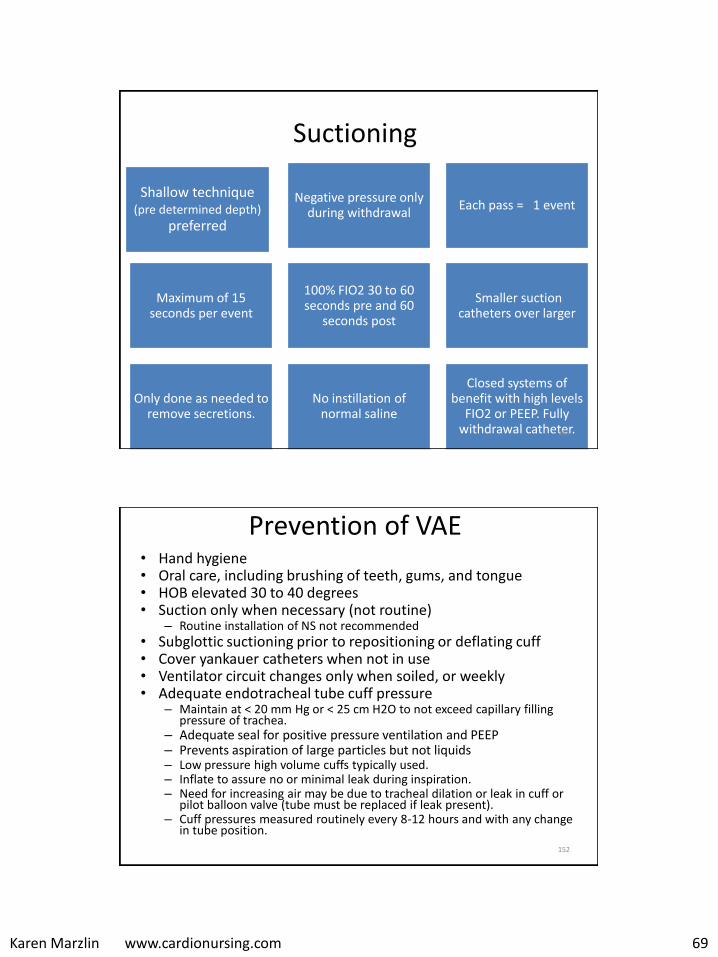
Suctioning
Shallow technique (pre determined depth)
preferred
Negative pressure only during withdrawal
Each pass = 1 event
Maximum of 15 seconds per event
100% FIO2 30 to 60 seconds pre and 60
seconds post
Smaller suction catheters over larger
Only done as needed to remove secretions.
No instillation of normal saline
Closed systems of benefit with high levels
FIO2 or PEEP. Fully withdrawal catheter. 151
152
Prevention of VAE • Hand hygiene• Oral care, including brushing of teeth, gums, and tongue• HOB elevated 30 to 40 degrees• Suction only when necessary (not routine)
– Routine installation of NS not recommended
• Subglottic suctioning prior to repositioning or deflating cuff • Cover yankauer catheters when not in use• Ventilator circuit changes only when soiled, or weekly• Adequate endotracheal tube cuff pressure
– Maintain at < 20 mm Hg or < 25 cm H2O to not exceed capillary filling pressure of trachea.
– Adequate seal for positive pressure ventilation and PEEP – Prevents aspiration of large particles but not liquids – Low pressure high volume cuffs typically used.– Inflate to assure no or minimal leak during inspiration.– Need for increasing air may be due to tracheal dilation or leak in cuff or
pilot balloon valve (tube must be replaced if leak present).– Cuff pressures measured routinely every 8-12 hours and with any change
in tube position.
Karen Marzlin www.cardionursing.com 70
153
Additional Prevention of VAE
• Avoid nasal intubation
• Extubate as soon as possible
• Discontinue NG tubes as soon as possible
• Avoid overuse of antibiotics
Readiness to Wean
• Reversal of the underlying cause of respiratory failure
• Adequate oxygenation
• Hemodynamic stability as defined by no active myocardial ischemia and no clinically significant hypotension
• Patient ability to initiate an inspiratory effort
154
Karen Marzlin www.cardionursing.com 71
155
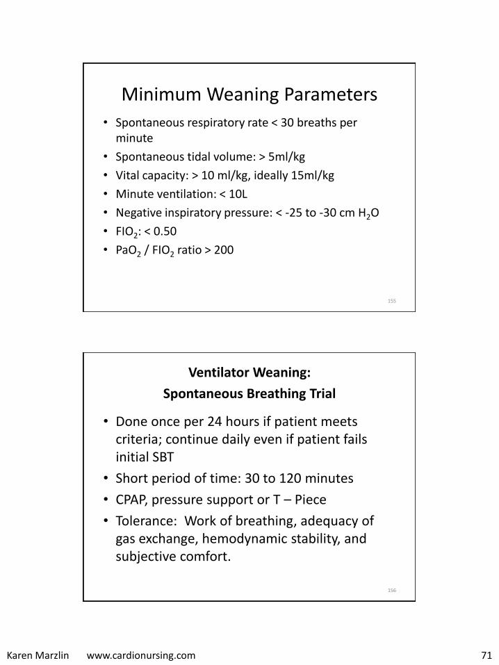
Minimum Weaning Parameters
• Spontaneous respiratory rate < 30 breaths per minute
• Spontaneous tidal volume: > 5ml/kg
• Vital capacity: > 10 ml/kg, ideally 15ml/kg
• Minute ventilation: < 10L
• Negative inspiratory pressure: < -25 to -30 cm H2O
• FIO2: < 0.50
• PaO2 / FIO2 ratio > 200
Ventilator Weaning:
Spontaneous Breathing Trial
• Done once per 24 hours if patient meets criteria; continue daily even if patient fails initial SBT
• Short period of time: 30 to 120 minutes
• CPAP, pressure support or T – Piece
• Tolerance: Work of breathing, adequacy of gas exchange, hemodynamic stability, and subjective comfort.
156
Karen Marzlin www.cardionursing.com 72
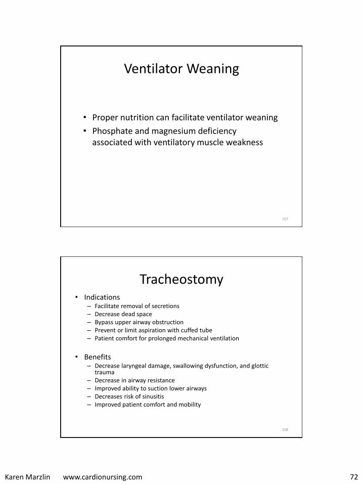
Ventilator Weaning
• Proper nutrition can facilitate ventilator weaning
• Phosphate and magnesium deficiency associated with ventilatory muscle weakness
157
158
Tracheostomy • Indications
– Facilitate removal of secretions– Decrease dead space – Bypass upper airway obstruction – Prevent or limit aspiration with cuffed tube – Patient comfort for prolonged mechanical ventilation
• Benefits – Decrease laryngeal damage, swallowing dysfunction, and glottic
trauma – Decrease in airway resistance – Improved ability to suction lower airways – Decreases risk of sinusitis – Improved patient comfort and mobility
Karen Marzlin www.cardionursing.com 73
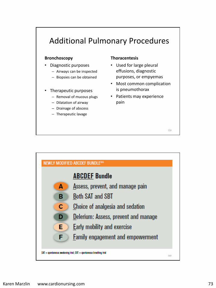
Additional Pulmonary Procedures
Bronchoscopy
• Diagnostic purposes– Airways can be inspected
– Biopsies can be obtained
• Therapeutic purposes – Removal of mucous plugs
– Dilatation of airway
– Drainage of abscess
– Therapeutic lavage
Thoracentesis
• Used for large pleural effusions, diagnostic purposes, or empyemas
• Most common complication is pneumothorax
• Patients may experience pain
159
160
Karen Marzlin www.cardionursing.com 74
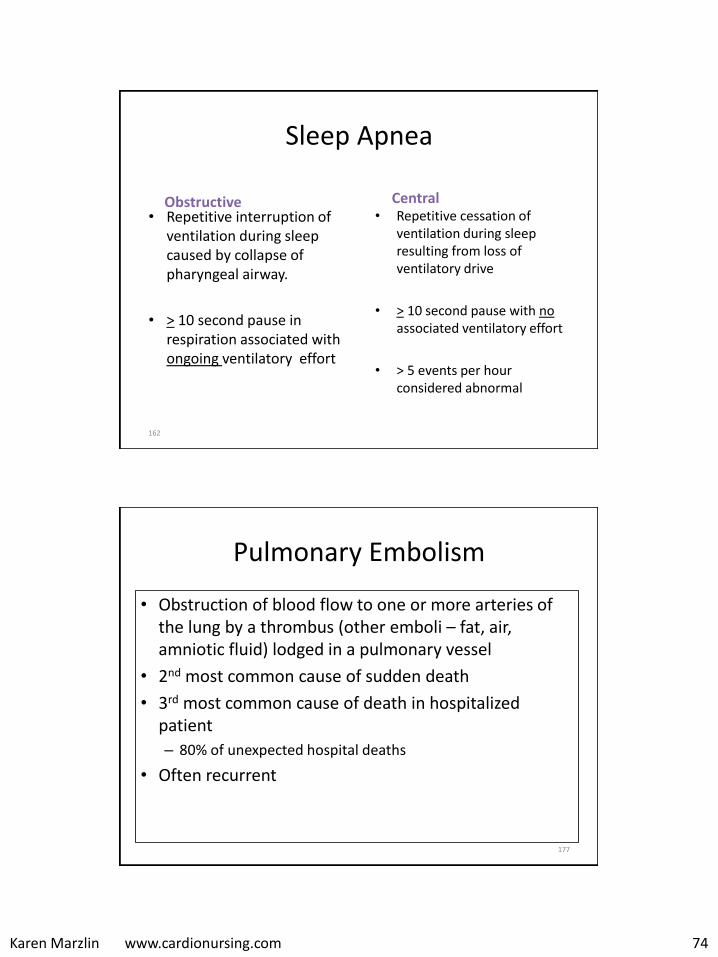
Sleep Apnea
Obstructive Central• Repetitive interruption of
ventilation during sleep caused by collapse of pharyngeal airway.
• > 10 second pause in respiration associated with ongoing ventilatory effort
• Repetitive cessation of ventilation during sleep resulting from loss of ventilatory drive
• > 10 second pause with no associated ventilatory effort
• > 5 events per hour considered abnormal
162
177
Pulmonary Embolism
• Obstruction of blood flow to one or more arteries of the lung by a thrombus (other emboli – fat, air, amniotic fluid) lodged in a pulmonary vessel
• 2nd most common cause of sudden death
• 3rd most common cause of death in hospitalized patient
– 80% of unexpected hospital deaths
• Often recurrent
Karen Marzlin www.cardionursing.com 75
Risk Factors for PE in Hospitalized Patient
• Admitted to the medical intensive care unit
• Admitted with pulmonary disease,
• Post myocardial infarction
• Post cardiopulmonary bypass surgery
(Ouellette, Harrington, & Kamangar, 2013)
178
Pulmonary Embolism
Acute
• Located centrally within the vessel lumen or causes vessel occlusion
• Results in distention of vessel wall
Chronic
Adjoins to vessel wall & reduces vessel diameter by > 50%
Recannulization through thrombus
179
Karen Marzlin www.cardionursing.com 76
Pulmonary Embolism
Central
• Main pulmonary artery, the left and right main pulmonary arteries, the anterior trunk, the right and left interlobar arteries, the left upper lobe trunk, the right middle lobe artery, and the right and left lower lobe arteries
• Can cause massive PE
Peripheral
• Segmental and subsegmental arteries of the three lobes of the right lung, the two lobes of the left lung, and the lingula (a projection of the upper lobe of left lung)
• Pain by initiating inflammation close to the parietal pleura.
180
Massive PE
• Present in less than 5% of patients presenting with PE (Kucher, Rossi, De Rosa, & Goldhaber, 2006).
• Involves both the right and left pulmonary arteries or causes hemodynamic collapse
• Presenting systolic BP of < 90 mmHg
• Mortality rates ange from 30% to 60% and most deaths occur within the first 1 to 2 hours (Ouellette et al., 2013; Wood, 2002).
181
Karen Marzlin www.cardionursing.com 77
182
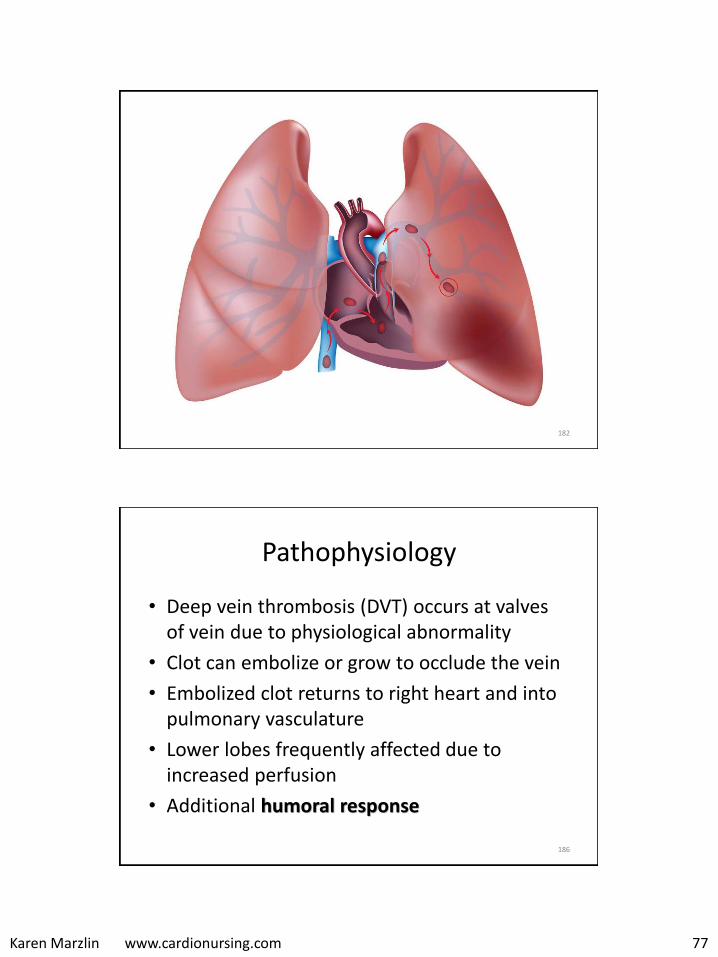
Pathophysiology
• Deep vein thrombosis (DVT) occurs at valves of vein due to physiological abnormality
• Clot can embolize or grow to occlude the vein
• Embolized clot returns to right heart and into pulmonary vasculature
• Lower lobes frequently affected due to increased perfusion
• Additional humoral response
186
Karen Marzlin www.cardionursing.com 78
Pathophysiology
• Increased PVR – Proximal clots
– Substances (thromboxane A and serotonin) released in humoral response also cause vasoconstriction
• PA pressures double to compensate
• Increased work load of RV – Right heart failure
– Leftward shift of septum
– Right coronary branches can be compressed
187
Pathophysiology • Increased V/Q ratio (alveolar dead space)
• Decreased V/Q ratio to other areas due to redistribution of blood flow
• Hypoxemia due to V/Q mismatching
• Increased minute ventilation to compensate for increased dead space – respiratory alkalosis – however, hypercapnea in massive
• Alveolar shrinkage (↓ CO2)– damage Type 2 alveolar cells – loss of surfactant – atelectasis –non cardiac pulmonary edema
• Pulmonary infarction rare due to dual blood supply
188
Karen Marzlin www.cardionursing.com 79
Clinical Presentation
• Pleuritic chest pain, shortness of breath, and hypoxemia is not present in the majority of patients
• May have no respiratory complaint
• Atypical presentation: flank pain, abdominal pain, delirium, syncope, and seizures
• Potential diagnosis in any patient with respiratory symptoms in whom there is not another clear etiology
• Suspect when there is respiratory alkalosis
189
Physical Exam Findings
• The most common physical sign, present in almost everyone with PE, is tachypnea (defined as respiratory rate > 16 per minute)
• Other:
– Dyspnea, rales, cough, hemoptysis
– Accentuated 2nd heart sound, presence of right sided S3 or S4, new systolic murmur of tricuspid regurgitation
– Tachycardia, low grade fever, diaphoresis
– Signs of thrombophlebitis, lower extremity peripheral edema
– Hypoxemia, cyanosis
190
Karen Marzlin www.cardionursing.com 80
More on Assessment
Massive PE
• Shock presentation
Multiple Emboli
• More signs of pulmonary hypertension and corpulmonale
191
Diagnosis
• Cardiac troponins will be elevated in half of patients
with moderate to large PE (Konstantinides, 2008)
• Use of ultrasound to rule out DVT
• Computed tomography angiography (CTA) has become the standard test for the diagnosis of PE
• VQ scan is used as alternative
192
Karen Marzlin www.cardionursing.com 81
ECG in PE
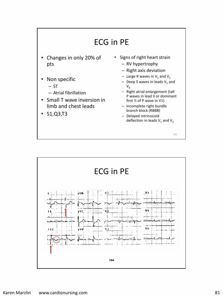
• Changes in only 20% of pts
• Non specific – ST
– Atrial fibrillation
• Small T wave inversion in limb and chest leads
• S1,Q3,T3
• Signs of right heart strain
– RV hypertrophy
– Right axis deviation– Large R waves in V1 and V2
– Deep S waves in leads V5 and V6
– Right atrial enlargement (tall P waves in lead II or dominant first ½ of P wave in V1)
– Incomplete right bundle branch block (RBBB)
– Delayed intrinsicoiddeflection in leads V1 and V2
193
194
ECG in PE
Karen Marzlin www.cardionursing.com 82
Treatment • Treatment with anticoagulation in non-massive PE reduces
mortality to less than 5%
• Full anticoagulation is the priority in any patient with suspected or confirmed PE.– Intravenous unfractionated heparin is the drug of choice in massive
PE, in patients with renal failure, and when there is concern about subcutaneous absorption.• An initial bolus of 80 U/kg followed by an infusion of 18 U/kg/hour
– IV anticoagulation given before dabigatran and edoxaban; overlapped with warfarin
– IV anticoagulation does not need to be given before rivaroxaban or apixaban
– LMWH is preferred in cancer associated thrombus
– Low risk patients can have home treatment or early discharge 195
Treatment
• Fibrinolytic therapy– Hemodynamic compromise as evidenced by
systolic BP < 90 mmHg and no high risk for bleeding
– Deterioration on anticoagulation and low bleeding risk
• Catheter assisted thrombosis removal if high bleeding risk / failed systemic therapy / shock – Surgical pulmonary embolectomy may also be considered
in select patients
196
Karen Marzlin www.cardionursing.com 83
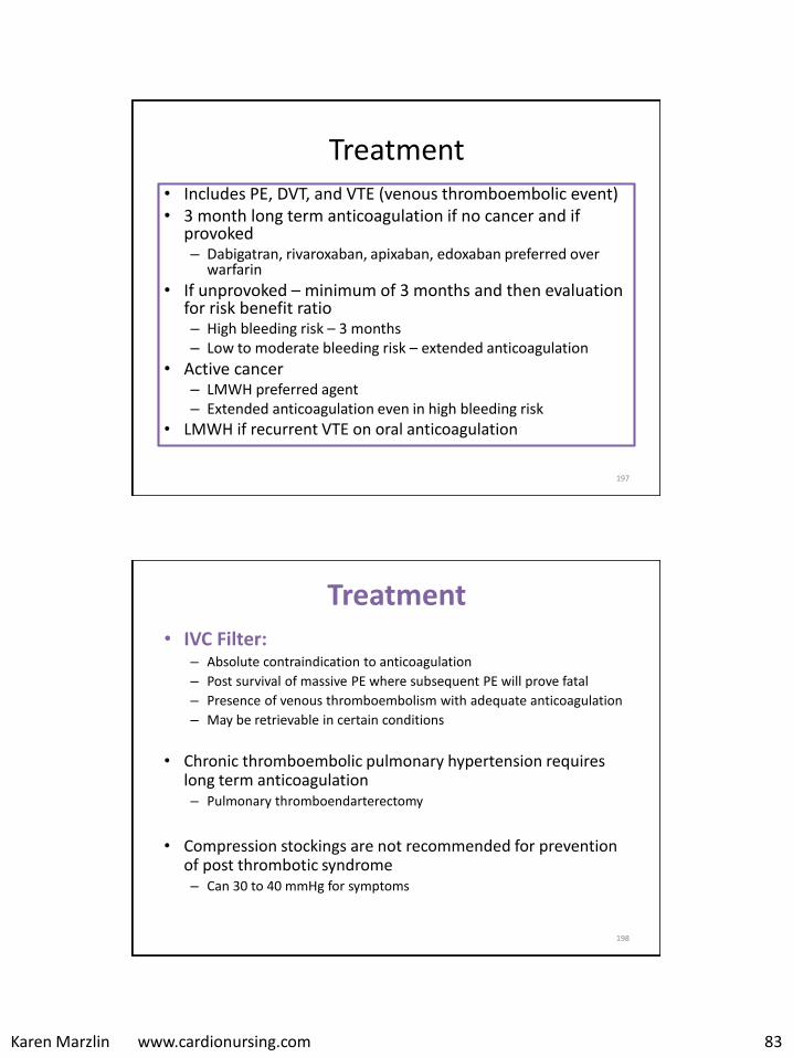
Treatment • Includes PE, DVT, and VTE (venous thromboembolic event) • 3 month long term anticoagulation if no cancer and if
provoked – Dabigatran, rivaroxaban, apixaban, edoxaban preferred over
warfarin
• If unprovoked – minimum of 3 months and then evaluation for risk benefit ratio – High bleeding risk – 3 months – Low to moderate bleeding risk – extended anticoagulation
• Active cancer – LMWH preferred agent – Extended anticoagulation even in high bleeding risk
• LMWH if recurrent VTE on oral anticoagulation
197
Treatment
• IVC Filter: – Absolute contraindication to anticoagulation
– Post survival of massive PE where subsequent PE will prove fatal
– Presence of venous thromboembolism with adequate anticoagulation
– May be retrievable in certain conditions
• Chronic thromboembolic pulmonary hypertension requires long term anticoagulation– Pulmonary thromboendarterectomy
• Compression stockings are not recommended for prevention of post thrombotic syndrome – Can 30 to 40 mmHg for symptoms
198
Karen Marzlin www.cardionursing.com 84
199
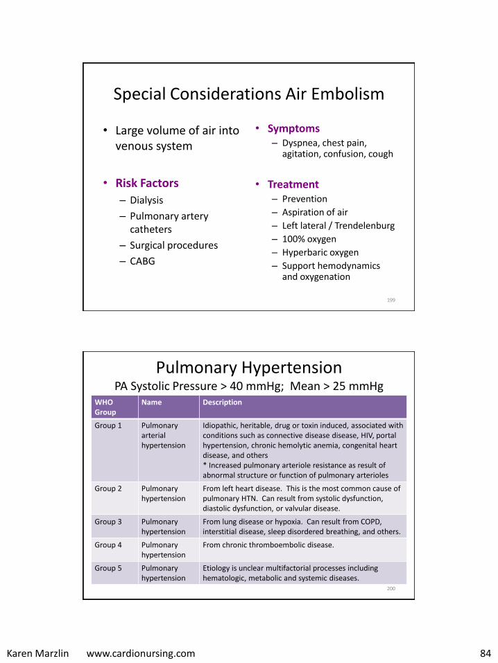
Special Considerations Air Embolism
• Large volume of air into venous system
• Risk Factors
– Dialysis
– Pulmonary artery catheters
– Surgical procedures
– CABG
• Symptoms – Dyspnea, chest pain,
agitation, confusion, cough
• Treatment – Prevention
– Aspiration of air
– Left lateral / Trendelenburg
– 100% oxygen
– Hyperbaric oxygen
– Support hemodynamics and oxygenation
Pulmonary HypertensionPA Systolic Pressure > 40 mmHg; Mean > 25 mmHg
WHO Group
Name Description
Group 1 Pulmonary arterial hypertension
Idiopathic, heritable, drug or toxin induced, associated with conditions such as connective disease disease, HIV, portal hypertension, chronic hemolytic anemia, congenital heart disease, and others * Increased pulmonary arteriole resistance as result of abnormal structure or function of pulmonary arterioles
Group 2 Pulmonary hypertension
From left heart disease. This is the most common cause of pulmonary HTN. Can result from systolic dysfunction,diastolic dysfunction, or valvular disease.
Group 3 Pulmonary hypertension
From lung disease or hypoxia. Can result from COPD, interstitial disease, sleep disordered breathing, and others.
Group 4 Pulmonary hypertension
From chronic thromboembolic disease.
Group 5 Pulmonary hypertension
Etiology is unclear multifactorial processes including hematologic, metabolic and systemic diseases.
200
Karen Marzlin www.cardionursing.com 85
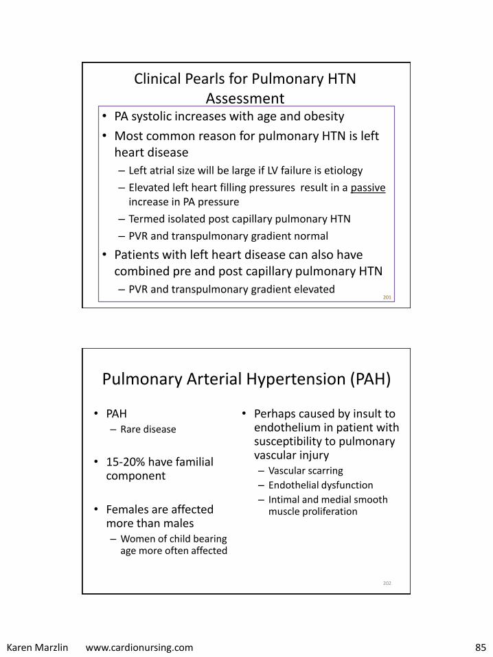
Clinical Pearls for Pulmonary HTN Assessment
• PA systolic increases with age and obesity
• Most common reason for pulmonary HTN is left heart disease
– Left atrial size will be large if LV failure is etiology
– Elevated left heart filling pressures result in a passiveincrease in PA pressure
– Termed isolated post capillary pulmonary HTN
– PVR and transpulmonary gradient normal
• Patients with left heart disease can also have combined pre and post capillary pulmonary HTN
– PVR and transpulmonary gradient elevated 201
202
Pulmonary Arterial Hypertension (PAH)
• PAH – Rare disease
• 15-20% have familial component
• Females are affected more than males – Women of child bearing
age more often affected
• Perhaps caused by insult to endothelium in patient with susceptibility to pulmonary vascular injury – Vascular scarring
– Endothelial dysfunction
– Intimal and medial smooth muscle proliferation
Karen Marzlin www.cardionursing.com 86
203
Associated Conditions
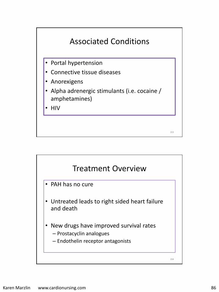
• Portal hypertension
• Connective tissue diseases
• Anorexigens
• Alpha adrenergic stimulants (i.e. cocaine / amphetamines)
• HIV
Treatment Overview
• PAH has no cure
• Untreated leads to right sided heart failure and death
• New drugs have improved survival rates – Prostacyclin analogues
– Endothelin receptor antagonists
204
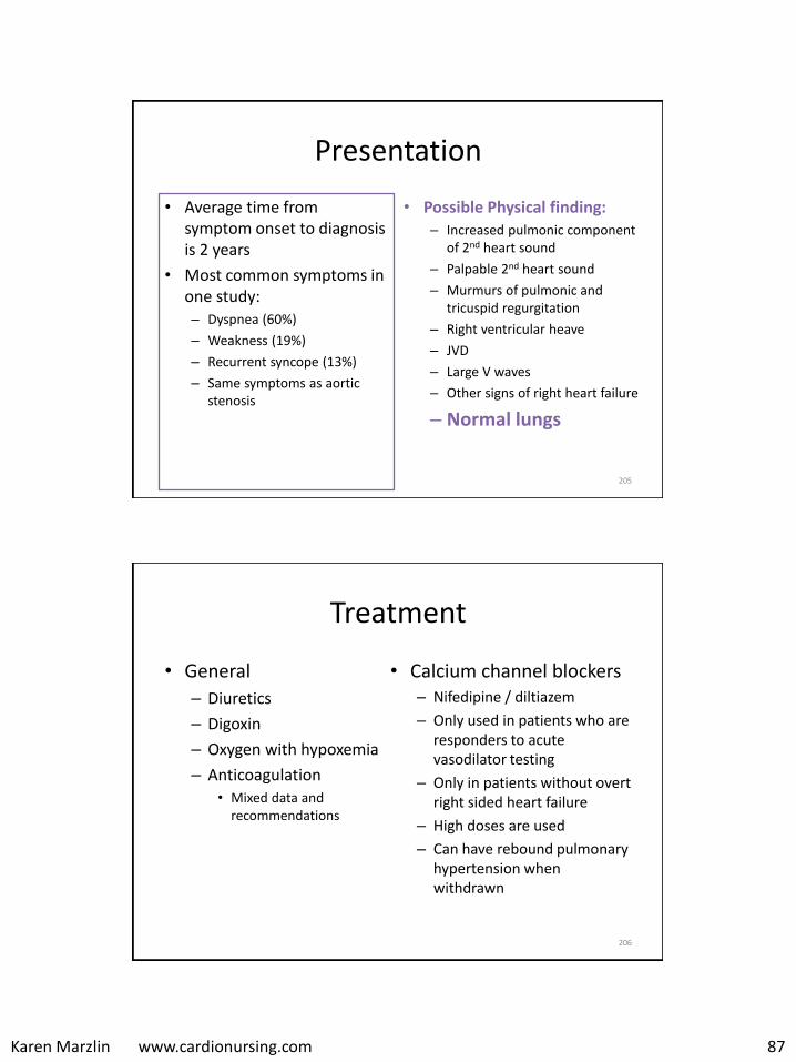
Karen Marzlin www.cardionursing.com 87
205
Presentation
• Average time from symptom onset to diagnosis is 2 years
• Most common symptoms in one study: – Dyspnea (60%)
– Weakness (19%)
– Recurrent syncope (13%)
– Same symptoms as aortic stenosis
• Possible Physical finding: – Increased pulmonic component
of 2nd heart sound
– Palpable 2nd heart sound
– Murmurs of pulmonic and tricuspid regurgitation
– Right ventricular heave
– JVD
– Large V waves
– Other signs of right heart failure
– Normal lungs
206
Treatment
• General
– Diuretics
– Digoxin
– Oxygen with hypoxemia
– Anticoagulation • Mixed data and
recommendations
• Calcium channel blockers – Nifedipine / diltiazem
– Only used in patients who are responders to acute vasodilator testing
– Only in patients without overt right sided heart failure
– High doses are used
– Can have rebound pulmonary hypertension when withdrawn
Karen Marzlin www.cardionursing.com 88
207
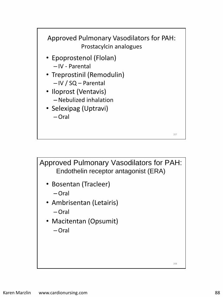
Approved Pulmonary Vasodilators for PAH: Prostacylcin analogues
• Epoprostenol (Flolan) – IV - Parental
• Treprostinil (Remodulin) – IV / SQ – Parental
• Iloprost (Ventavis) –Nebulized inhalation
• Selexipag (Uptravi)–Oral
208
Approved Pulmonary Vasodilators for PAH:Endothelin receptor antagonist (ERA)
• Bosentan (Tracleer) –Oral
• Ambrisentan (Letairis) –Oral
• Macitentan (Opsumit)–Oral
Karen Marzlin www.cardionursing.com 89
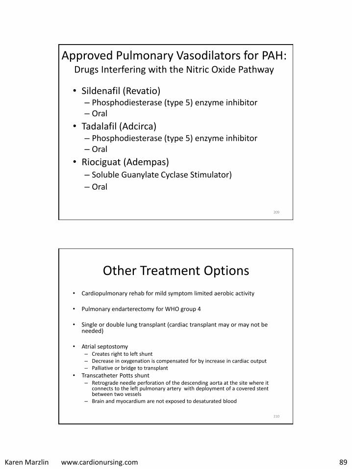
Approved Pulmonary Vasodilators for PAH:Drugs Interfering with the Nitric Oxide Pathway
• Sildenafil (Revatio) – Phosphodiesterase (type 5) enzyme inhibitor– Oral
• Tadalafil (Adcirca)– Phosphodiesterase (type 5) enzyme inhibitor– Oral
• Riociguat (Adempas)– Soluble Guanylate Cyclase Stimulator)
– Oral
209
210
Other Treatment Options
• Cardiopulmonary rehab for mild symptom limited aerobic activity
• Pulmonary endarterectomy for WHO group 4
• Single or double lung transplant (cardiac transplant may or may not be needed)
• Atrial septostomy– Creates right to left shunt – Decrease in oxygenation is compensated for by increase in cardiac output– Palliative or bridge to transplant
• Transcatheter Potts shunt– Retrograde needle perforation of the descending aorta at the site where it
connects to the left pulmonary artery with deployment of a covered stent between two vessels
– Brain and myocardium are not exposed to desaturated blood
Karen Marzlin www.cardionursing.com 90
211
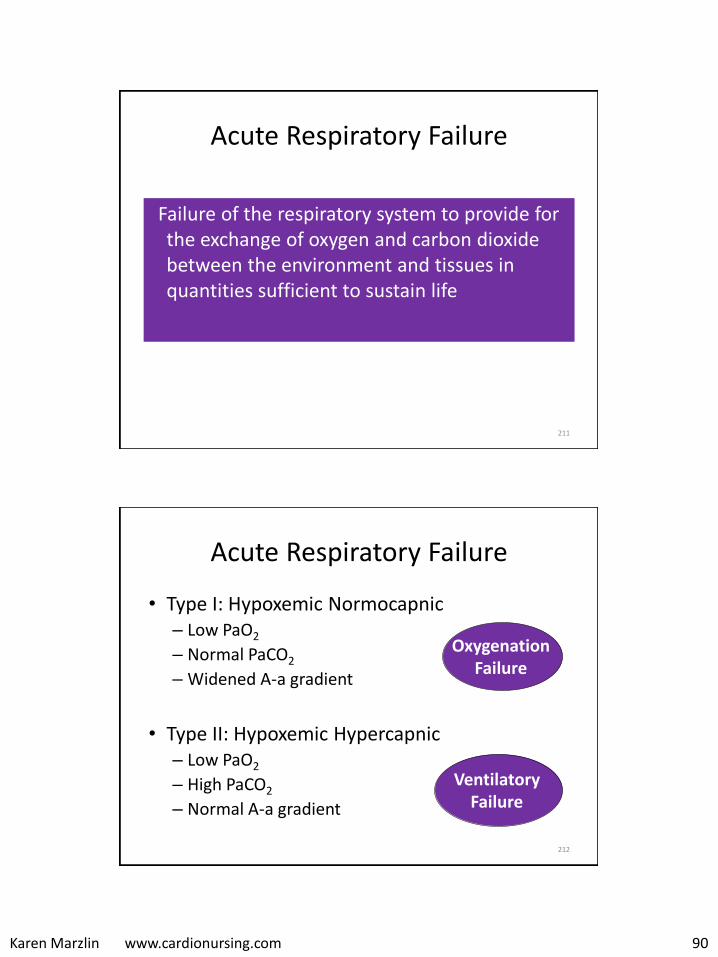
Acute Respiratory Failure
Failure of the respiratory system to provide for the exchange of oxygen and carbon dioxide between the environment and tissues in quantities sufficient to sustain life
212
Acute Respiratory Failure
• Type I: Hypoxemic Normocapnic– Low PaO2
– Normal PaCO2
– Widened A-a gradient
• Type II: Hypoxemic Hypercapnic– Low PaO2
– High PaCO2
– Normal A-a gradient
VentilatoryFailure
Oxygenation Failure
Karen Marzlin www.cardionursing.com 91
213
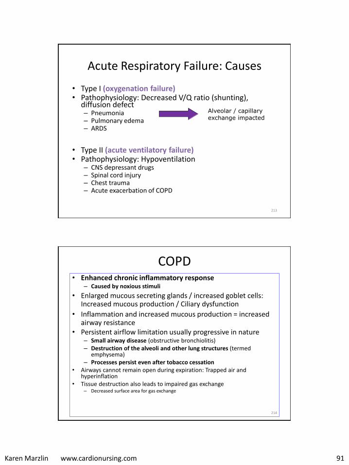
Acute Respiratory Failure: Causes
• Type I (oxygenation failure) • Pathophysiology: Decreased V/Q ratio (shunting),
diffusion defect – Pneumonia – Pulmonary edema – ARDS
• Type II (acute ventilatory failure) • Pathophysiology: Hypoventilation
– CNS depressant drugs – Spinal cord injury – Chest trauma – Acute exacerbation of COPD
Alveolar / capillary exchange impacted
214
COPD • Enhanced chronic inflammatory response
– Caused by noxious stimuli
• Enlarged mucous secreting glands / increased goblet cells: Increased mucous production / Ciliary dysfunction
• Inflammation and increased mucous production = increased airway resistance
• Persistent airflow limitation usually progressive in nature– Small airway disease (obstructive bronchiolitis)– Destruction of the alveoli and other lung structures (termed
emphysema)– Processes persist even after tobacco cessation
• Airways cannot remain open during expiration: Trapped air and hyperinflation
• Tissue destruction also leads to impaired gas exchange– Decreased surface area for gas exchange
Karen Marzlin www.cardionursing.com 92
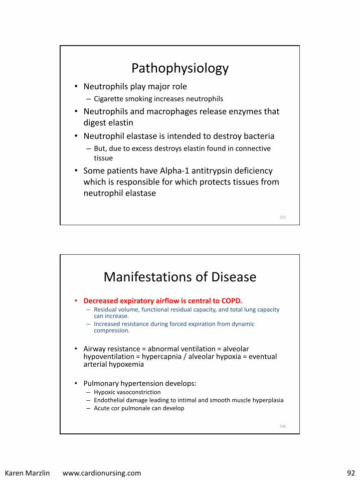
Pathophysiology • Neutrophils play major role
– Cigarette smoking increases neutrophils
• Neutrophils and macrophages release enzymes that digest elastin
• Neutrophil elastase is intended to destroy bacteria
– But, due to excess destroys elastin found in connective tissue
• Some patients have Alpha-1 antitrypsin deficiency which is responsible for which protects tissues from neutrophil elastase
215
Manifestations of Disease
• Decreased expiratory airflow is central to COPD.– Residual volume, functional residual capacity, and total lung capacity
can increase. – Increased resistance during forced expiration from dynamic
compression.
• Airway resistance = abnormal ventilation = alveolar hypoventilation = hypercapnia / alveolar hypoxia = eventual arterial hypoxemia
• Pulmonary hypertension develops:– Hypoxic vasoconstriction – Endothelial damage leading to intimal and smooth muscle hyperplasia – Acute cor pulmonale can develop
216
Karen Marzlin www.cardionursing.com 93
Comparison
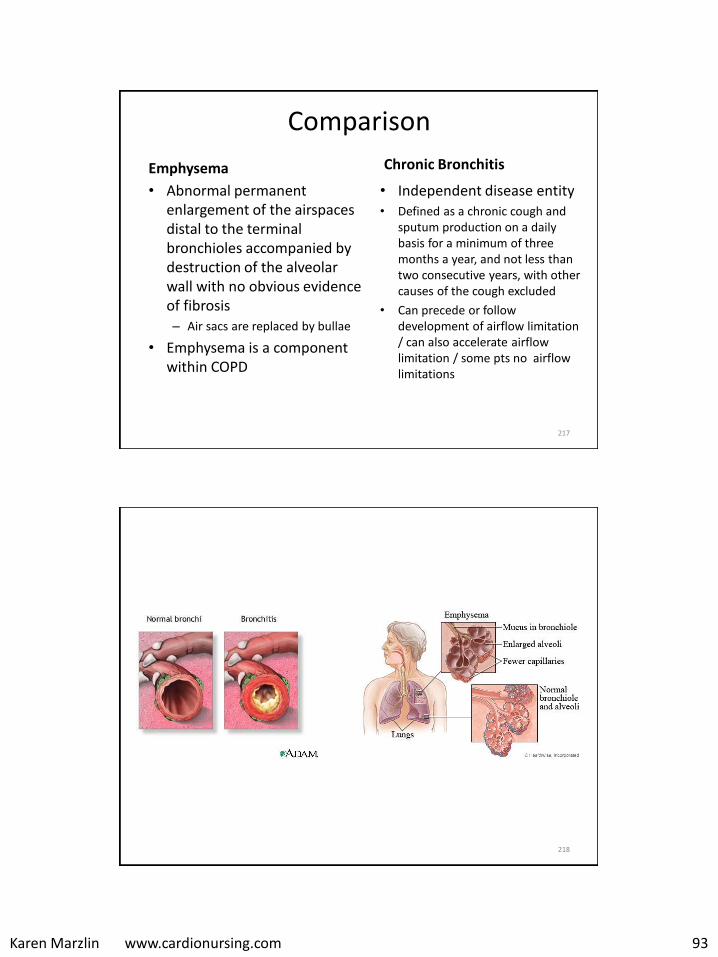
Emphysema
• Abnormal permanent enlargement of the airspaces distal to the terminal bronchioles accompanied by destruction of the alveolar wall with no obvious evidence of fibrosis – Air sacs are replaced by bullae
• Emphysema is a component within COPD
Chronic Bronchitis
• Independent disease entity• Defined as a chronic cough and
sputum production on a daily basis for a minimum of three months a year, and not less than two consecutive years, with other causes of the cough excluded
• Can precede or follow development of airflow limitation / can also accelerate airflow limitation / some pts no airflow limitations
217
218
Karen Marzlin www.cardionursing.com 94
Clinical Presentation • Reduced inspiratory capacity, particularly during
exercise due to hyperinflation
• Hypoxemia and potential hypercapnea due to V/Q mismatching – Increased hypoxemia during sleep
• Increased RBC production in response to chronic hypoxemia– May also have central cyanosis
• Potential for right sided heart failure 219
Diagnosis
• Suspect COPD in patients > 40 years– Dyspnea that is persistent, progressive, and worse with
exercise.
– Chronic cough (often 1st sign) including intermittent and nonproductive coughs.
– Chronic sputum production.
– Exposure to tobacco smoke, smoke from home cooking or heating, and exposure to environmental chemicals.
– Family history of COPD.
* Physical exam findings usually not present until advanced disease.
220
Karen Marzlin www.cardionursing.com 95
Diagnosis
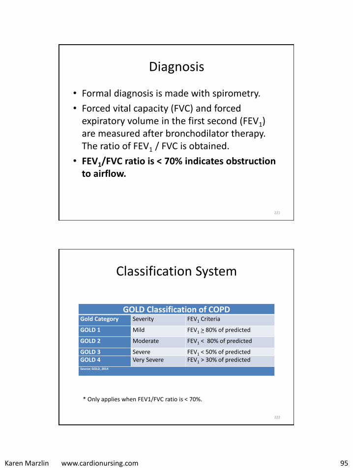
• Formal diagnosis is made with spirometry.
• Forced vital capacity (FVC) and forced expiratory volume in the first second (FEV1) are measured after bronchodilator therapy. The ratio of FEV1 / FVC is obtained.
• FEV1/FVC ratio is < 70% indicates obstruction to airflow.
221
Classification System
222
GOLD Classification of COPDGold Category Severity FEV1 Criteria
GOLD 1 Mild FEV1 > 80% of predicted
GOLD 2 Moderate FEV1 < 80% of predicted
GOLD 3 Severe FEV1 < 50% of predictedGOLD 4 Very Severe FEV1 > 30% of predictedSource: GOLD, 2014
* Only applies when FEV1/FVC ratio is < 70%.
Karen Marzlin www.cardionursing.com 96
Nursing Implications
• QOL related to dyspnea
• Etiology of exacerbations
• Anorexia, weight loss, and fatigue
• Skeletal muscle dysfunction and opportunity for improvement
223
224
COPD: Treatment
• Smoking cessation
• Vaccines
• Treatment of sleep apnea
• Pulmonary Rehab
– Improves perceived breathlessness and increases exercise capacity,
– Enhances effect of long term bronchodilators
– Reduces anxiety and depression and improves health-related quality of life
– Reduces hospitalization and length of stay, enhances recovery after exacerbation, and improves survival
Karen Marzlin www.cardionursing.com 97
COPD: Treatment
• Medications to reduce symptoms, exacerbations and to improve exercise tolerance; no evidence for slowing of disease progression
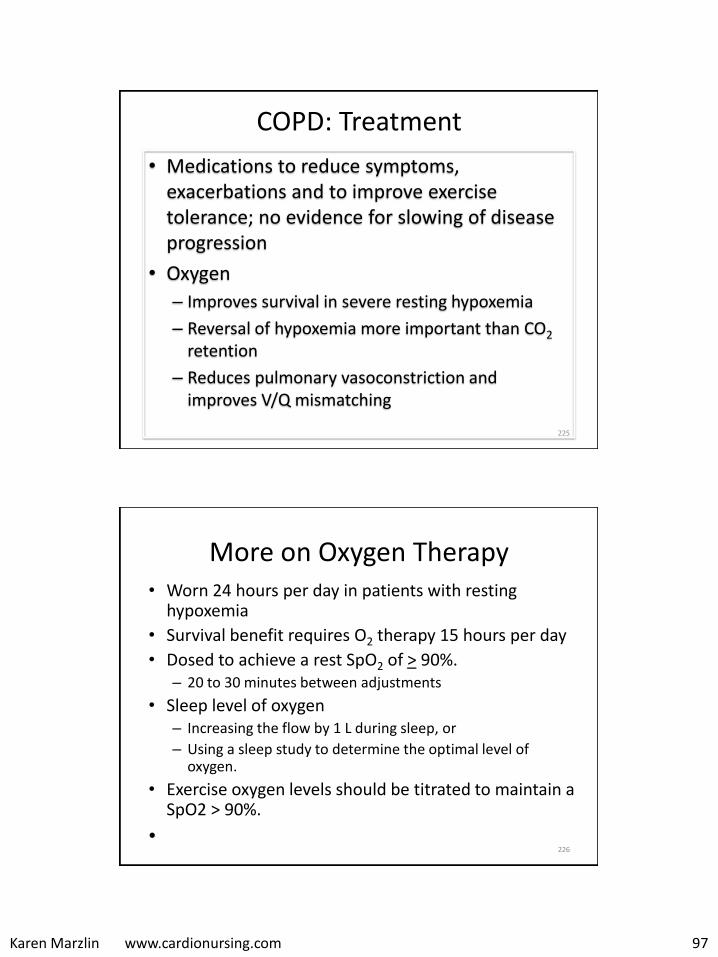
• Oxygen
– Improves survival in severe resting hypoxemia
– Reversal of hypoxemia more important than CO2
retention
– Reduces pulmonary vasoconstriction and improves V/Q mismatching
225
More on Oxygen Therapy • Worn 24 hours per day in patients with resting
hypoxemia
• Survival benefit requires O2 therapy 15 hours per day
• Dosed to achieve a rest SpO2 of > 90%. – 20 to 30 minutes between adjustments
• Sleep level of oxygen – Increasing the flow by 1 L during sleep, or
– Using a sleep study to determine the optimal level of oxygen.
• Exercise oxygen levels should be titrated to maintain a SpO2 > 90%.
•226
Karen Marzlin www.cardionursing.com 98
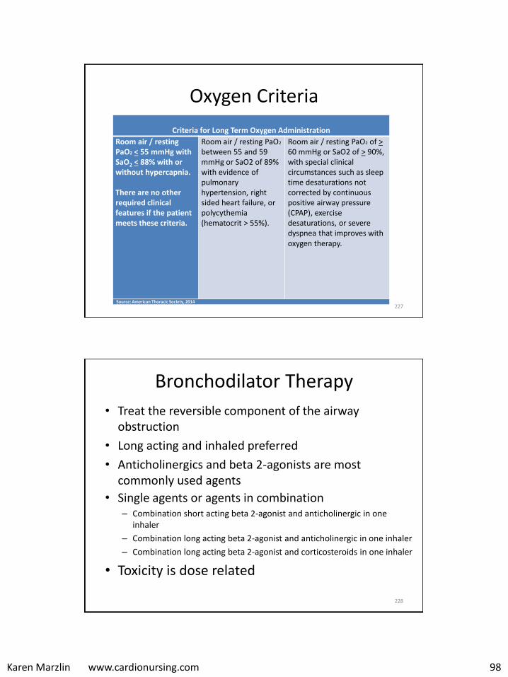
Oxygen Criteria
227
Criteria for Long Term Oxygen Administration
Room air / resting PaO2 < 55 mmHg with SaO2 < 88% with or without hypercapnia.
There are no other required clinical features if the patient meets these criteria.
Room air / resting PaO2
between 55 and 59 mmHg or SaO2 of 89% with evidence of pulmonary hypertension, right sided heart failure, or polycythemia (hematocrit > 55%).
Room air / resting PaO2 of >60 mmHg or SaO2 of > 90%, with special clinical circumstances such as sleep time desaturations not corrected by continuous positive airway pressure (CPAP), exercise desaturations, or severe dyspnea that improves with oxygen therapy.
Source: American Thoracic Society, 2014
Bronchodilator Therapy
• Treat the reversible component of the airway obstruction
• Long acting and inhaled preferred
• Anticholinergics and beta 2-agonists are most commonly used agents
• Single agents or agents in combination – Combination short acting beta 2-agonist and anticholinergic in one
inhaler
– Combination long acting beta 2-agonist and anticholinergic in one inhaler
– Combination long acting beta 2-agonist and corticosteroids in one inhaler
• Toxicity is dose related
228
Karen Marzlin www.cardionursing.com 99
Inhaled Therapy
• Dry powder, metered dose breath activated devices, supplemental spacer devices, nebulizers -often used in exacerbation.
• Incorrect technique: – Increased ED visits,
– Increased hospital admissions
– Increased use of corticosteroids and antibiotics
• Factors affecting correct use: – advanced age
– lower levels of education
– Most significantly, lack of instruction by a health care provider
*(Melani et al., 2011).
232
Acute Exacerbation
• Viral infection of the upper respiratory tract or the tracheobronchial tree
• Other– Bacterial: 3 most common are
Hemophilus influenza, Streptococcus pneumoniae, and Moraxella catarrhalis. Pseudomonas aeruginosa in more advanced COPD.
– Environmental
– Non adherence
– Unknown
• Associated with worsening lung function and increased mortality
• Required hospitalization = poor prognosis
• Several week recovery period
234
Karen Marzlin www.cardionursing.com 100
Clinical Presentation in Acute Exacerbation
• Dyspnea, cough, or sputum production that is a change from baseline.
• Use of accessory muscles when breathing
• Paradoxical chest wall movement
• New or worsened central cyanosis
• Altered mental status
• Evidence of right sided heart failure
235
Specific Treatment Issues in Acute Exacerbation
Oxygen / Ventilation
• Important to know if patient is a chronic CO2 retainer
• If so – drive to ventilate is based on hypoxic drive
• Keep saturation between 88-92% * treatment of hypoxemia is priority
• It is the oxygen saturation level that is important – not the FIO2
• Ventilatory support required if respiratory acidosis or increased work of breathing – prefer BiPAP
Blood Gas Goals • Important to know if patient is a
chronic CO2 retainer
• If so – the key to assessing decompensation is when the pH become abnormal (no longer is this a compensated respiratory acidosis)
• The goal is to return the pH to normal – not to return the PaCO2
to normal
236
Karen Marzlin www.cardionursing.com 101
237
Case Example
• Patient history: COPD (CO2) retainer
• Initial presentation:
– Tachypneic with increased work of breathing
– SpO2 of 78%
The previous patient most likely has what type of acute respiratory failure:
238
1. Oxygenation failure
2. Ventilatory failure
3. This is not acute respiratory failure due to history of CO2 retention
Karen Marzlin www.cardionursing.com 102
Pending blood gas results, the best initial treatment would include:
239
1. CPAP
2. BiPAP
3. FIO2 to achieve saturation 90-92%
4. CPAP and increased FIO2
5. BiPAP and increased FIO2
240
Case Example
• ABG
– 7.29
– PaCO2 60 mmHg
– HCO3 30 mEq/L
– PaO2 48 mmHg

Karen Marzlin www.cardionursing.com 103
Blood gas goals include:
241
1. Normalization of pH
2. Normalization of PaCO2
3. Both of the above
4. Neither of the above
242
Pneumonia
• Acute infection of the lung parenchyma, including alveolar spaces and interstitial space
• Causes: – Bacteria (Community acquired versus Hospital acquired)
– Virus
– Fungi
– Parasites
– Mycoplasma
Karen Marzlin www.cardionursing.com 104
243
Risk Factors for Bacterial Pneumonia
• Previous viral respiratory infection
• Gastro esophageal reflux disease (GERD)
• Chronic alcohol abuse
• Cigarette smoking
• Decreased level of consciousness
• Anesthesia
• Intubation
• Lung disease
• Diabetes mellitus
• Use of corticorsteroids
• Elderly
244
Pneumonia: Pathophysiology
• Causative agent is inhaled or enters pharynx via direct contact
• Alveoli become inflamed
• Alveolar spaces fill with exudate and consolidate
• Diffusion of O2 obstructed
– Hypoxemia.
• Goblet cells are stimulated to increase mucous
– Increased airway resistance and work of breathing
Karen Marzlin www.cardionursing.com 105
245
Pneumonia: Causative Agents • Common agents in community-acquired pneumonia (younger
and healthier population) – Streptococcus pneumoniae (most common agent in community acquired
pneumonia).– Mycoplasma pneumoniae.– Chlamydia pneumoniae– Viral.
• Haemophilus influenza common among smokers • Klebsiella pneumoniae in patients with chronic alcoholism• Agents in the older population commonly include gram negative
bacilli– Moraxella catarrhalis (particularly common in patients with chronic
bronchitis).– Staphylococcus aureus (in the setting of post viral influenza).
• Methicillin-resistant Staphylococcus aureus (MRSA) also as a cause of community-acquired pneumonia
246
Hospital Acquired Pneumonia
Causative agents • Aerobic gram negative rods
– Klebsiella sp.– Psuedomonas sp.– Enterobacter sp.– Escherichia coli.– Proteus sp– Serratia sp.– Enterococci.
• Staphylococcus aureus (including methicillin-resistant Staphylococcus aureus [MRSA])
• Group B streptococci
• Nosocomial pneumonia is typically caused by bacterial agents that are more resistant to antibiotic therapy.
• Sources– Contamination of pharynx and
perhaps stomach with bacteria – Repeated small aspirations of
oral pharyngeal secretions.– Retrograde contamination from
GI tract.
Karen Marzlin www.cardionursing.com 106
Bacterial Pneumonia: Presentation
• Hyperthermia (fever, typically >38°C) or hypothermia (< 35°C)
• Tachypnea (>18 respirations/min)
• Use of accessory respiratory muscles
• Tachycardia (>100 bpm) or bradycardia (< 60 bpm)
• Central cyanosis
• Altered mental status
247
Potential Physical Exam Findings
• Adventitious breath sounds, such as rales/crackles, rhonchi, or wheezes
• Decreased intensity of breath sounds
• Egophony
• Whispering pectoriloquy
• Dullness to percussion
• Tracheal deviation
• Lymphadenopathy
• Pleural friction rub
248
Karen Marzlin www.cardionursing.com 107
249
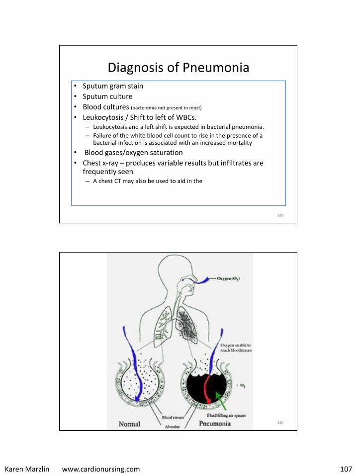
Diagnosis of Pneumonia • Sputum gram stain
• Sputum culture
• Blood cultures (bacteremia not present in most)
• Leukocytosis / Shift to left of WBCs.– Leukocytosis and a left shift is expected in bacterial pneumonia.
– Failure of the white blood cell count to rise in the presence of a bacterial infection is associated with an increased mortality
• Blood gases/oxygen saturation
• Chest x-ray – produces variable results but infiltrates are frequently seen – A chest CT may also be used to aid in the
250
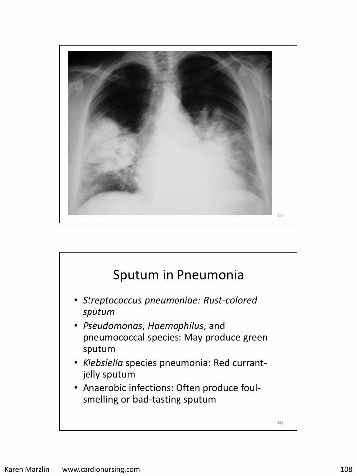
Karen Marzlin www.cardionursing.com 108
251
Sputum in Pneumonia
• Streptococcus pneumoniae: Rust-colored sputum
• Pseudomonas, Haemophilus, and pneumococcal species: May produce green sputum
• Klebsiella species pneumonia: Red currant-jelly sputum
• Anaerobic infections: Often produce foul-smelling or bad-tasting sputum
252
Karen Marzlin www.cardionursing.com 109
254
Complications of Pneumonia • Abscesses may form and rupture into pleural space leading to
pneumothorax and/or empyema– Video assisted thoracoscopy with debridement is a treatment option for
empyema in the early organizing phase– Full thoracotomy with decortication may be necessary in later organizing
phases
• Pleural Effusion• Acute respiratory failure• ARDS• Sepsis
Mortality rates for nosocomial or hospital-acquired pneumonia are higher than those for community acquired pneumonia (particularly in the elderly)
255
Pneumonia: Treatment • Decision to admit
– Pneumonia severity index (PSI) score as a guide for inpatient care and mortality risk.
• Prevent nosocomial infections
• Timely Antibiotics– Cover pneumococcus
• Hydration (Electrolyte Monitoring)
• Deep breathing / incentive spirometry
• Bronchodilators, expectorants, mucolytics
• Avoid: sedatives and antitussives
• Early activity and mobility (DVT Prophylaxis)
Karen Marzlin www.cardionursing.com 110
259
Aspiration
• Vomiting or regurgitation
• Large particles – airway obstruction
• pH of liquid determines injury
– pH<2.5 or large volume
– Chemical burns destroy type II cells
– May induce bronchospasm
– Increase alveolar capillary membrane permeability • Decrease compliance
• Decrease V/Q ratio
260
Aspiration • Non acidic aspiration
– More transient
• Food stuff / small particles
– Inflammatory reaction
– Hemorrhagic pneumonia within 6 hours
• Contaminated material with bacteria can be fatal
• Patients with severe periodontal disease, putrid sputum, or a history of alcoholism may be at greater risk of anaerobic infection.
Karen Marzlin www.cardionursing.com 111
261
Aspiration: Possible Prevention Strategies
• Avoiding sedation.
• Resting prior to meal time.
• Eating slowly.
• Flexing the head slightly to the “chin down” position.
• Determining food viscosity best tolerated (thickening liquids will improve swallowing in some patients).
262
Acute Respiratory Distress Syndrome
A syndrome of acute respiratory failure characterized by non-cardiac pulmonary edema and manifested by refractory hypoxemia. ARDS does not include mild or early acute lung injury, but rather involves severe and diffused lung injury.
Karen Marzlin www.cardionursing.com 112
263
Risk Factors in ARDS
• Sepsis (most common)
• Transfusion
• Aspiration
• Trauma
• Massive transfusion
• Pancreatitis
264
Acute Respiratory Distress Syndrome: Etiology
• Direct lung injury
– Chest trauma
– Near drowning
– Smoke inhalation
– Pneumonia
– Pulmonary embolism
– Or: Change in pulmonary vascular pressure
• Indirect lung injury
– Sepsis
– Shock
– Multi system trauma
– Burns
– CABG
– Head injury
Time from injury of alveolar capillary
membrane to onset of symptoms is 12-48 hours.
Karen Marzlin www.cardionursing.com 113
265
ARDS: Pathophysiology • Stimulation of inflammatory and immune systems• Release of toxic substances, causing micro vascular injury• Pulmonary capillary membranes are damaged
– Increase in capillary permeability.
• Cells and fluids leak into interstitium and alveolar spaces– Pulmonary Edema
• Impaired production and dysfunction of surfactant– Alveolar collapse and massive atelectasis.
• Intrapulmonary shunting• Hypoxic vasoconstriction• Decreased the compliance of lung
– High-peak inspiratory pressures to ventilate the lungs.
• Potential development of pulmonary fibrosis in chronic phase.– Endothelium, epithelium, interstitial space expand.– Protein exudate inside the alveoli produces a hyaline membrane.
266
Acute Respiratory Distress Syndrome: Diagnosis
• Predisposing condition
• PaO2/FIO2 ratio < 200
• Chest x-ray: Diffuse bilateral infiltrates
(Chest CT may also be used)
• Decreased static compliance of lungs
• PAOP < 18 mmHg or no evidence of
increased left-atrial pressure
• No evidence of COPD
• No other explanation for above
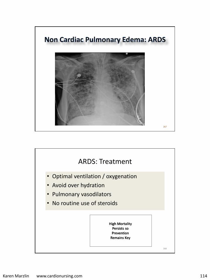
Karen Marzlin www.cardionursing.com 114
267
268
ARDS: Treatment
• Optimal ventilation / oxygenation
• Avoid over hydration
• Pulmonary vasodilators
• No routine use of steroids
High Mortality Persists so Prevention
Remains Key
Karen Marzlin www.cardionursing.com 115
269
Drugs Used to Decrease Right Sided Afterload / Treat Pulmonary
Hypertension
• Oxygen
• Pulmonary vasodilators
• NTG
• Sodium nitroprusside
• Inhaled nitric oxide
• See medications used in the treatment of pulmonary arterial hypertension
Mechanical Ventilator Management Strategies for ARDS
• Lower tidal volume ventilation – Permissive hypercapnia
• Maintain plateau pressure < 30 mmHg
• Uninterrupted PEEP • Avoidance of auto PEEP
• Open Lung / Recruitment Modes of Ventilation – Airway pressure release
ventilation – High frequency
ventilation (Oscillatory)
270
Karen Marzlin www.cardionursing.com 116
Additional Options in ARDS
Independent lung ventilation
– If degree of alveolar collapse is not uniform
– ECMO (extracorporeal membrane oxygenation)
– Venous – venous: facilitates gas exchange but does not provide for hemodynamic support because the blood is returned to the right side of the heart before it enters pulmonary circulation.
– Used as an alternative strategy in adults with ARDS to rest the lungs and avoid insult of mechanical ventilation.
– Hemorrhage and infection are the two most common complications of ECMO.
271
Partial Liquid Ventilation
• Lung is partially filled with perfluorocarbons– To level of functional residual capacity (40% of lung
capacity) – Properties similar to surfactant
• Patient ventilated conventionally with Vt• Improvement in compliance:
– ? Recruitment of alveoli – Possible direct effect on surface tension
• Other possible benefits– Protective effect from infection – Wash out of inflammatory debris
272
Karen Marzlin www.cardionursing.com 117
Prone Position in Severe ARDS • Multicenter prospective randomized trial.
• Randomized to 16 hours of the prone position or to usual care of the standard supine position.
• The 28-day mortality rate was 16.0% in the prone group and 32.8% in the supine group (statistically significant). Also associated with a reduction in 90-day mortality.
• There were no significant adverse effects with the proning intervention. Patients in the supine group had a higher rate of cardiac arrest (Guérin et al., 2013).
273
274
Case Example
• 65 year old female; 85 kg
• Post witnessed cardiac arrest
• Initial PaO2 / FIO2 ratio 102
Karen Marzlin www.cardionursing.com 118
Based on the PaO2/FIO2 ratio, the initial diagnosis in the previous patient is:
275
1. ARDS
2. Acute lung injury
276
Case Example
• Ventilator settings:– Assist control
– Rate 12
– Vt 700 ml
– FIO2 .8
– PEEP 5 cmH2O
• 2nd ABG – pH: 7.33
– PaCO2: 40 mmHg
– HCO3: 14 mEq/L
– PaO2: 92 mmHg
Karen Marzlin www.cardionursing.com 119
Most probable anticipated ventilator changes include:
277
1. Increased rate
2. Increased Vt
3. Increased FIO2
4. Increased PEEP
Ventilator settings:Assist control Rate 12Vt 700 ml FIO2 .8 PEEP 5 cmH2O
2nd ABG pH: 7.33 PaCO2: 40 mmHg HCO3: 14 mEq/LPaO2: 92 mmHg
If this patient develops ARDS, what potential open lung ventilation mode might be
considered:
278
1. Controlled mandatory ventilation
2. Synchronized intermittent mandatory ventilation
3. Airway pressure release ventilation
4. Spontaneous breathing trial
Karen Marzlin www.cardionursing.com 120
Blunt Trauma
• Etiology –Motor vehicle crash: Responsible for 70 to
80% of blunt trauma
• Chest wall injury (ribs)
• Direct lung injury (pulmonary contusion)
• Direct injury to the heart and great vessels 279
Diagnosis
• CXR is initial diagnostic study– Tension pneumothorax
does not require a CXR for treatment
– May not always be necessary in stable patients
• CT scan is more sensitive (chest, abdomen, and cervical)
• TEE to assess rupture of thoracic aorta
• Transthoracic Echo for tamponde
• Fiberoptic or rigid bronchooscopy for tracheobronchial injuries– Endotracheal tube
can be loaded on scope
280
Karen Marzlin www.cardionursing.com 121
Treatment: Immediate Surgery
• Loss of chest wall integrity
• Blunt diaphragmatic injuries
• Massive air leak after chest tube insertion
• Massive hemothorax (1500 ml of blood on CT insertion)
• Continued high blood loss after CT insertion (250 ml/hour for 3 consecutive hours)
• Confirmed tracheal, major bronchial, or esophageal injury
• GI contents in chest tube 281
Treatment: Immediate Surgery
• Cardiac tamponade
• Great vessel injury
• Embolism (pulmonary artery or heart)
282
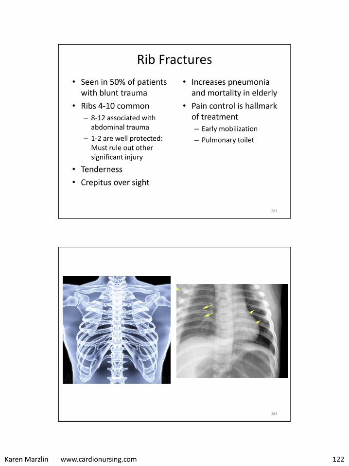
Karen Marzlin www.cardionursing.com 122
Rib Fractures
• Seen in 50% of patients with blunt trauma
• Ribs 4-10 common
– 8-12 associated with abdominal trauma
– 1-2 are well protected: Must rule out other significant injury
• Tenderness
• Crepitus over sight
• Increases pneumonia and mortality in elderly
• Pain control is hallmark of treatment
– Early mobilization
– Pulmonary toilet
283
284
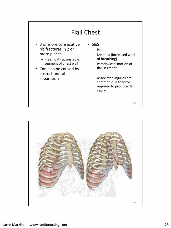
Karen Marzlin www.cardionursing.com 123
Flail Chest
• 3 or more consecutive rib fractures in 2 or more places – Free floating, unstable
segment of chest wall
• Can also be caused by costochondralseparation
• S&S– Pain
– Dyspnea (increased work of breathing)
– Paradoxicaal motion of flail segment
– Associated injuries are common due to force required to produce flail injury
285
286
Karen Marzlin www.cardionursing.com 124
Treatment of Flail Chest
• Same as for fractured ribs
• Caution with fluids – respiratory failure can occur especially when there is co-existing pulmonary contusion
• Surgery is not superior to supportive care
• However, surgical intervention for stabilization if thoracotomy is indicated for another reason
287
Sternal Fracture
• Upper and middle 1/3 most commonly affected
• Inspiratory pain
• 55-70% of patients will have associated injuries
• Blunt cardiac injury present in < 20% of patients
– Begin workup with ECG
288
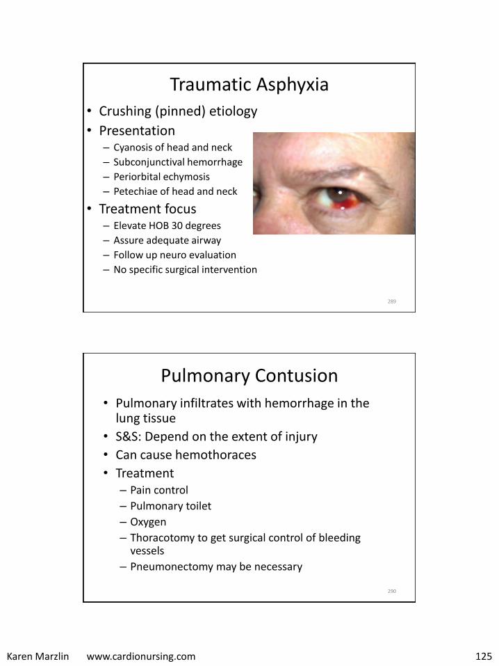
Karen Marzlin www.cardionursing.com 125
Traumatic Asphyxia • Crushing (pinned) etiology
• Presentation – Cyanosis of head and neck
– Subconjunctival hemorrhage
– Periorbital echymosis
– Petechiae of head and neck
• Treatment focus – Elevate HOB 30 degrees
– Assure adequate airway
– Follow up neuro evaluation
– No specific surgical intervention
289
Pulmonary Contusion
• Pulmonary infiltrates with hemorrhage in the lung tissue
• S&S: Depend on the extent of injury
• Can cause hemothoraces
• Treatment – Pain control
– Pulmonary toilet
– Oxygen
– Thoracotomy to get surgical control of bleeding vessels
– Pneumonectomy may be necessary
290
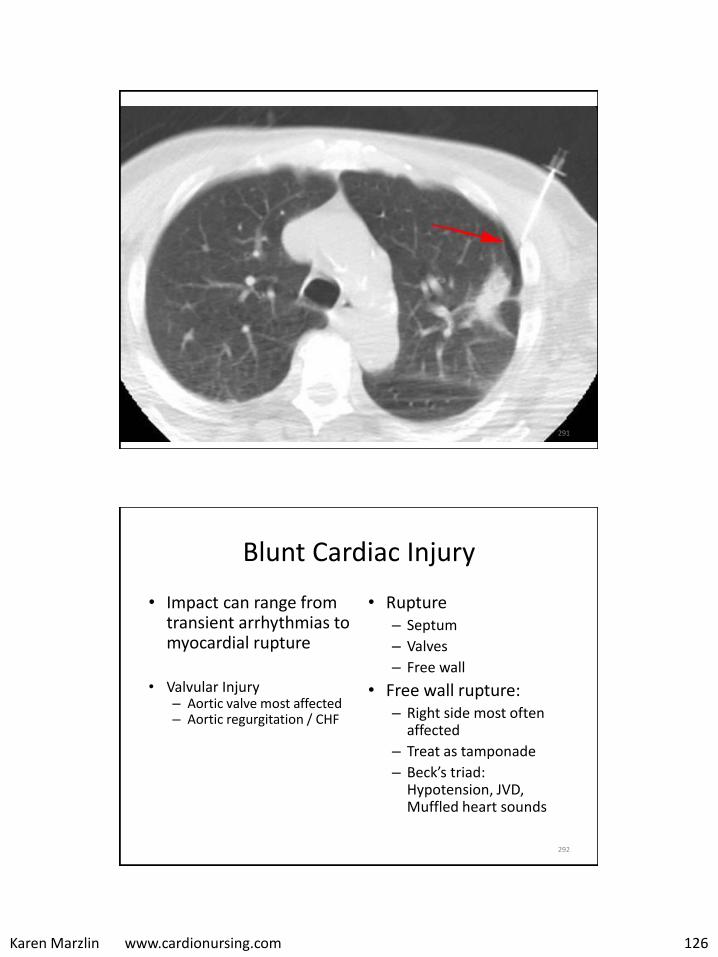
Karen Marzlin www.cardionursing.com 126
291
Blunt Cardiac Injury
• Impact can range from transient arrhythmias to myocardial rupture
• Valvular Injury – Aortic valve most affected – Aortic regurgitation / CHF
• Rupture – Septum
– Valves
– Free wall
• Free wall rupture: – Right side most often
affected
– Treat as tamponade
– Beck’s triad: Hypotension, JVD, Muffled heart sounds
292
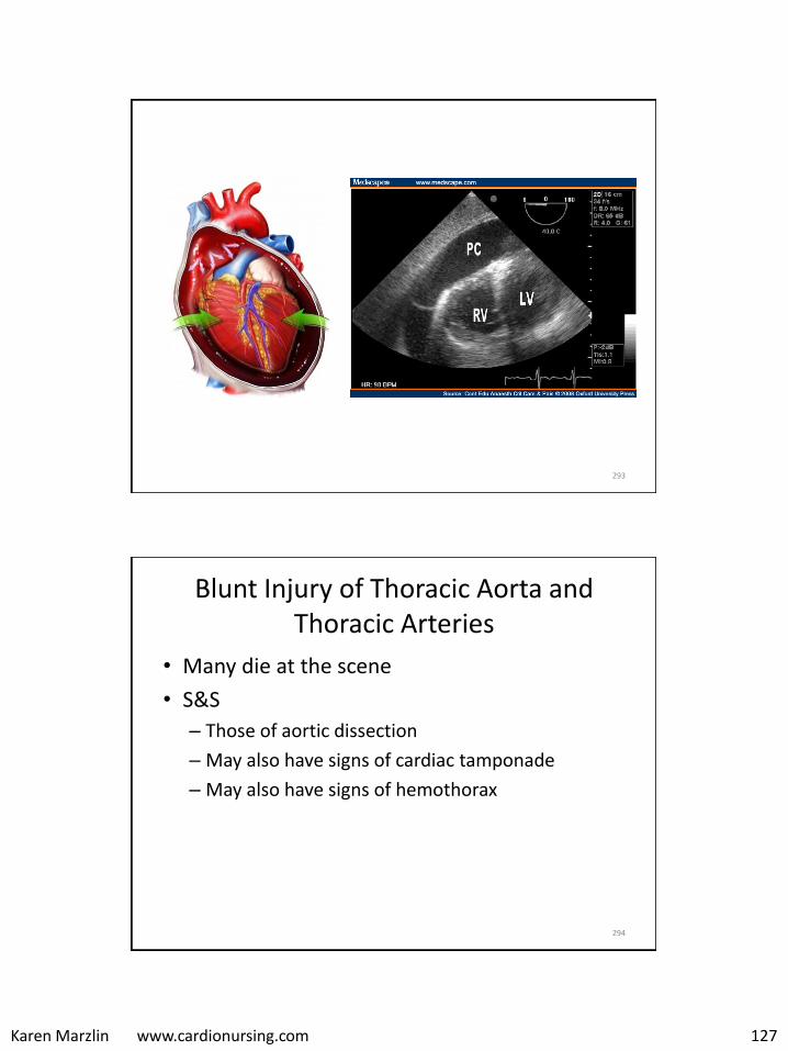
Karen Marzlin www.cardionursing.com 127
293
Blunt Injury of Thoracic Aorta and Thoracic Arteries
• Many die at the scene
• S&S
– Those of aortic dissection
– May also have signs of cardiac tamponade
– May also have signs of hemothorax
294
Karen Marzlin www.cardionursing.com 128
Blunt Trauma Summary
• > 80% of patients require no invasive therapy or only a tube thoracostomy
• High mortality associated with cardiac tamponade and great vessel rupture.
• Future:
– Increased use of thoroscopy for diagnosis and treatment.
– Ultrasound to diagnose tamponde and hemothorax.
– Spiral CT for major vascular lesions.
– Endovascular repair for great vessels.
295
296
Closed (Simple) Pneumothorax
• Air enters the intra pleural space through the lung causing partial or total collapse of the lung – Between visceral and parietal pleura
– If associated with hemorrhage: Hemopneumothorax
• Types / Etiology– Primary (no underlying lung disease)
• Blebs / bullae
• Smoking
– Secondary (underlying lung disease) • Air enters through damaged aveoli
• COPD
– Blunt trauma (lung laceration by rib fracture)
– Iatrogenic – from medical procedure
– Positive pressure ventilation (rupture of weak alveoli, bleb or bullous)
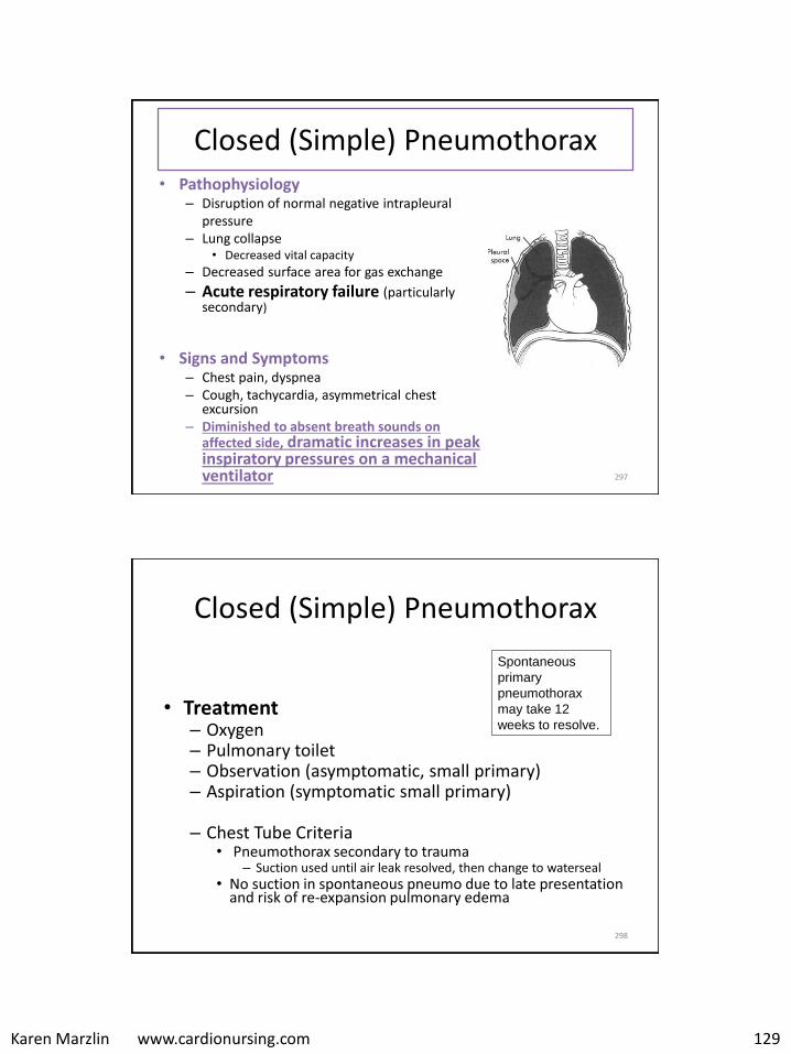
Karen Marzlin www.cardionursing.com 129
297
Closed (Simple) Pneumothorax• Pathophysiology
– Disruption of normal negative intrapleuralpressure
– Lung collapse • Decreased vital capacity
– Decreased surface area for gas exchange
– Acute respiratory failure (particularly secondary)
• Signs and Symptoms – Chest pain, dyspnea– Cough, tachycardia, asymmetrical chest
excursion– Diminished to absent breath sounds on
affected side, dramatic increases in peak inspiratory pressures on a mechanical ventilator
Closed (Simple) Pneumothorax
• Treatment – Oxygen – Pulmonary toilet– Observation (asymptomatic, small primary) – Aspiration (symptomatic small primary)
– Chest Tube Criteria • Pneumothorax secondary to trauma
– Suction used until air leak resolved, then change to waterseal• No suction in spontaneous pneumo due to late presentation
and risk of re-expansion pulmonary edema
298
Spontaneous
primary
pneumothorax
may take 12
weeks to resolve.
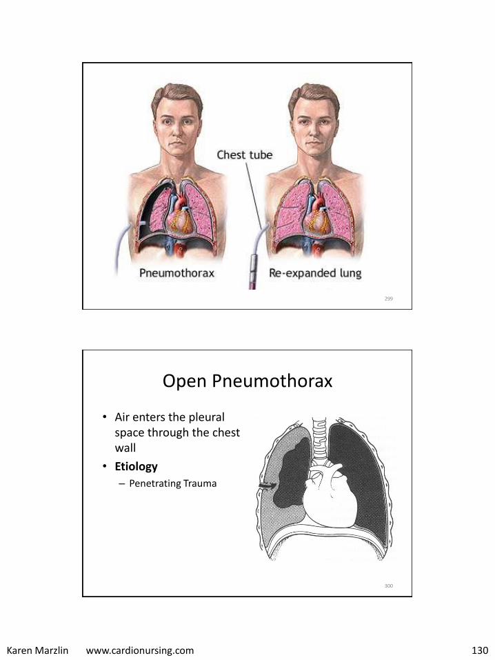
Karen Marzlin www.cardionursing.com 130
299
300
Open Pneumothorax
• Air enters the pleural space through the chest wall
• Etiology
– Penetrating Trauma
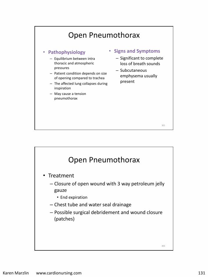
Karen Marzlin www.cardionursing.com 131
301
Open Pneumothorax
• Pathophysiology – Equilibrium between intra
thoracic and atmospheric pressures
– Patient condition depends on size of opening compared to trachea
– The affected lung collapses during inspiration
– May cause a tension pneumothorax
• Signs and Symptoms
– Significant to complete loss of breath sounds
– Subcutaneous emphysema usually present
Open Pneumothorax
• Treatment
– Closure of open wound with 3 way petroleum jelly gauze
• End expiration
– Chest tube and water seal drainage
– Possible surgical debridement and wound closure (patches)
302
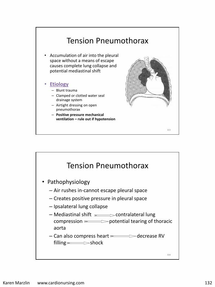
Karen Marzlin www.cardionursing.com 132
303
Tension Pneumothorax
• Accumulation of air into the pleural space without a means of escape causes complete lung collapse and potential mediastinal shift
• Etiology – Blunt trauma
– Clamped or clotted water seal drainage system
– Airtight dressing on open pneumothorax
– Positive pressure mechanical ventilation – rule out if hypotension
304
Tension Pneumothorax
• Pathophysiology
– Air rushes in-cannot escape pleural space
– Creates positive pressure in pleural space
– Ipsalateral lung collapse
– Mediastinal shift contralateral lung compression potential tearing of thoracic aorta
– Can also compress heart decrease RV filling shock
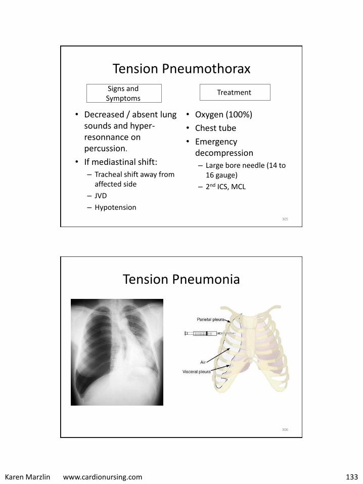
Karen Marzlin www.cardionursing.com 133
305
Tension Pneumothorax
• Decreased / absent lung sounds and hyper-resonnance on percussion.
• If mediastinal shift:
– Tracheal shift away from affected side
– JVD
– Hypotension
• Oxygen (100%)
• Chest tube
• Emergency decompression
– Large bore needle (14 to 16 gauge)
– 2nd ICS, MCL
Signs and Symptoms
Treatment
Tension Pneumonia
306
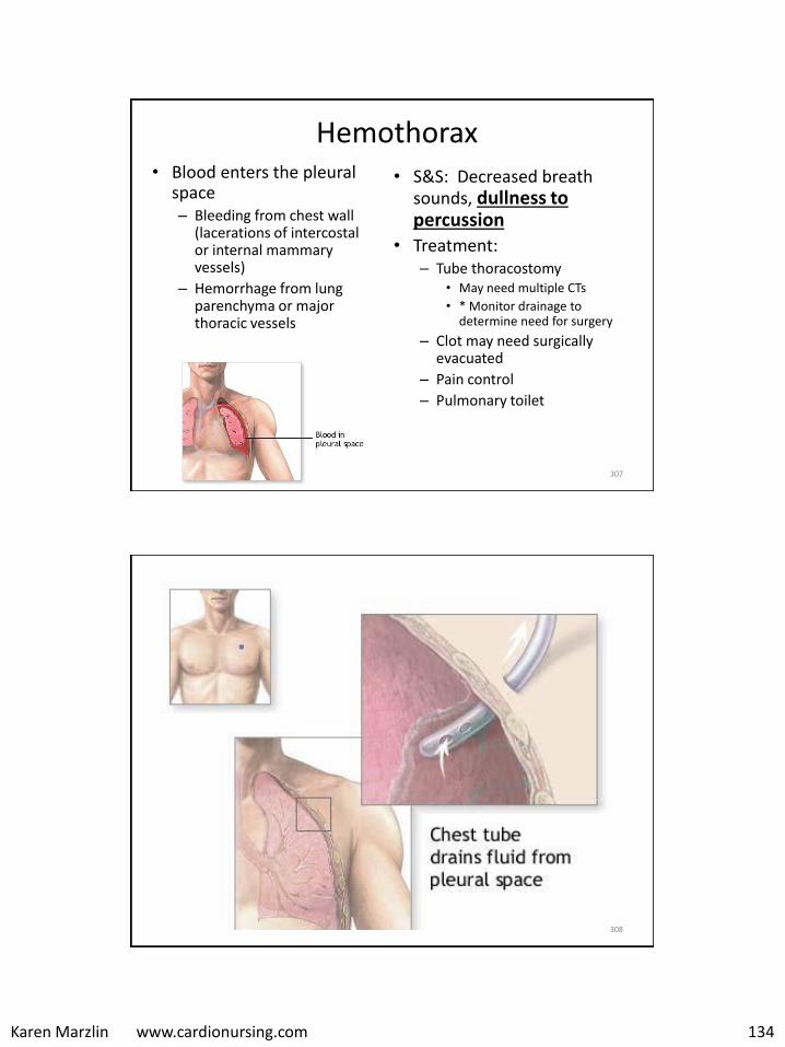
Karen Marzlin www.cardionursing.com 134
Hemothorax• Blood enters the pleural
space – Bleeding from chest wall
(lacerations of intercostal or internal mammary vessels)
– Hemorrhage from lung parenchyma or major thoracic vessels
• S&S: Decreased breath sounds, dullness to percussion
• Treatment: – Tube thoracostomy
• May need multiple CTs
• * Monitor drainage to determine need for surgery
– Clot may need surgically evacuated
– Pain control
– Pulmonary toilet
307
308
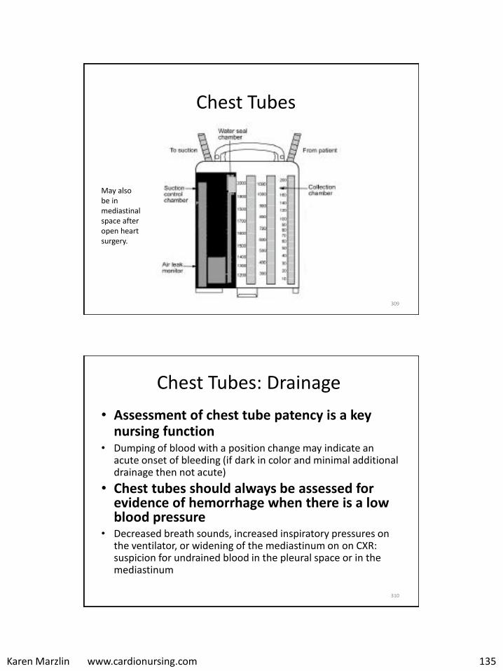
Karen Marzlin www.cardionursing.com 135
Chest Tubes
May also be in mediastinal space after open heart surgery.
309
Chest Tubes: Drainage
• Assessment of chest tube patency is a key nursing function
• Dumping of blood with a position change may indicate an acute onset of bleeding (if dark in color and minimal additional drainage then not acute)
• Chest tubes should always be assessed for evidence of hemorrhage when there is a low blood pressure
• Decreased breath sounds, increased inspiratory pressures on the ventilator, or widening of the mediastinum on on CXR: suspicion for undrained blood in the pleural space or in the mediastinum
310
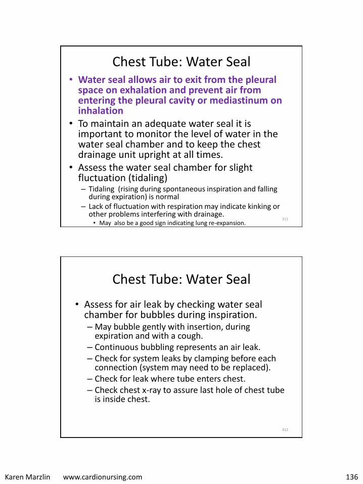
Karen Marzlin www.cardionursing.com 136
Chest Tube: Water Seal • Water seal allows air to exit from the pleural
space on exhalation and prevent air from entering the pleural cavity or mediastinum on inhalation
• To maintain an adequate water seal it is important to monitor the level of water in the water seal chamber and to keep the chest drainage unit upright at all times.
• Assess the water seal chamber for slight fluctuation (tidaling) – Tidaling (rising during spontaneous inspiration and falling
during expiration) is normal– Lack of fluctuation with respiration may indicate kinking or
other problems interfering with drainage. • May also be a good sign indicating lung re-expansion.
311
Chest Tube: Water Seal
• Assess for air leak by checking water seal chamber for bubbles during inspiration. – May bubble gently with insertion, during
expiration and with a cough. – Continuous bubbling represents an air leak.– Check for system leaks by clamping before each
connection (system may need to be replaced).– Check for leak where tube enters chest.– Check chest x-ray to assure last hole of chest tube
is inside chest.
312
Karen Marzlin www.cardionursing.com 137
Chest Tube: Other Nursing Considerations
• Assess the insertion site for subcutaneous emphysema.
• Keep unit below the level of the patient’s chest
• Do not clamp chest tube for transport (can cause
tension pneumothorax with pleural chest tubes or tamponade with mediastinal chest tubes)
– Use portable suction or transport on gravity drainage with tubing from suction chamber open to air
313
Chest Tubes
• Rare but serious complication is the development of unilateral pulmonary edema in response to rapid re-expansion or rapid evacuation of pleural fluid. – Capillary permeability can increase as result of
rapid treatment, resulting in pulmonary edema.
– Strategies for prevention: • Avoiding suction with the initial drainage of a large
pleural effusion or the expansion of a large pneumothorax
• Clamping after the 2 liters of initial drainage
314
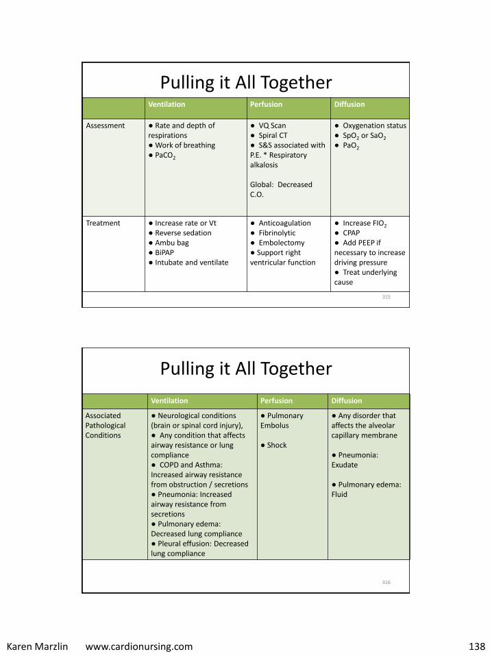
Karen Marzlin www.cardionursing.com 138
Pulling it All Together Ventilation Perfusion Diffusion
Assessment ● Rate and depth of respirations ● Work of breathing ● PaCO2
● VQ Scan ● Spiral CT ● S&S associated with P.E. * Respiratory alkalosis
Global: Decreased C.O.
● Oxygenation status ● SpO2 or SaO2
● PaO2
Treatment ● Increase rate or Vt● Reverse sedation● Ambu bag● BiPAP● Intubate and ventilate
● Anticoagulation ● Fibrinolytic● Embolectomy● Support right ventricular function
● Increase FIO2
● CPAP● Add PEEP if necessary to increase driving pressure ● Treat underlying cause
315
Pulling it All Together
Ventilation Perfusion Diffusion
Associated Pathological Conditions
● Neurological conditions (brain or spinal cord injury), ● Any condition that affects airway resistance or lung compliance ● COPD and Asthma: Increased airway resistancefrom obstruction / secretions● Pneumonia: Increased airway resistance from secretions ● Pulmonary edema: Decreased lung compliance ● Pleural effusion: Decreased lung compliance
● Pulmonary Embolus
● Shock
● Any disorder that affects the alveolar capillary membrane
● Pneumonia: Exudate
● Pulmonary edema: Fluid
316