Embed Size (px)
Citation preview

Visualization of Macromolecular
Structures
Present by: Qihang Li
orig. author: O’Donoghue, et al.
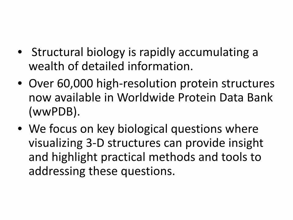
• Structural biology is rapidly accumulating a wealth of detailed information.
• Over 60,000 high-resolution protein structures now available in Worldwide Protein Data Bank (wwPDB).
• We focus on key biological questions where visualizing 3-D structures can provide insight and highlight practical methods and tools to addressing these questions.

Outline
• Protein structures• Ligand binding sites• RNA structures• Molecular motion• Larger macromolecular assemblies• Future perspectives

Protein structures
Finding 3-D structuresThis task is considerably simplified by consolidating 3D structures are into a single data repository, the Worldwide Protein Data Bank (wwPDB).3 entries: RSCB PDB, PDB Europe, PDB Japan.PDB also mrrored at other sites.Becoming one seamless step for most users.
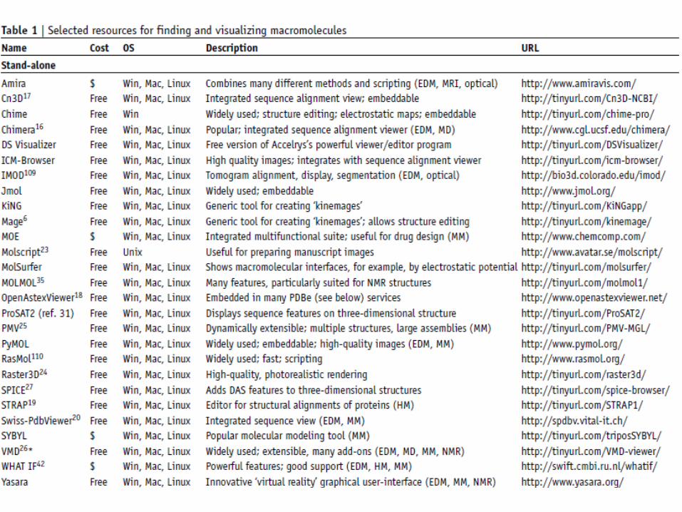


Protein structures (cont.)Finding structures from sequence
Several websites (RCSB PDB) allow the user to find structures using a sequence identifier or BLAST search.Entrez Structure & SRS 3D7 allow the sequence to be aligned to any related 3D structure.Several websites (Swiss-Model) provide comparative models for finding similar protein in PDB.These comparative models can be accessed at a single consolidated website, the Protein Model Portal (PMP).……The original PDB templates also include information on experimental conditions, ligands and cofactors.
Protein structures (cont.)
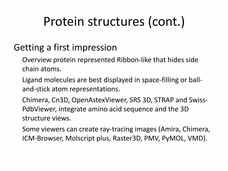
Getting a first impressionOverview protein represented Ribbon-like that hides side chain atoms.Ligand molecules are best displayed in space-filling or ball-and-stick atom representations.Chimera, Cn3D, OpenAstexViewer, SRS 3D, STRAP and Swiss-PdbViewer, integrate amino acid sequence and the 3D structure views.Some viewers can create ray-tracing images (Amira, Chimera, ICM-Browser, Molscript plus, Raster3D, PMV, PyMOL, VMD).
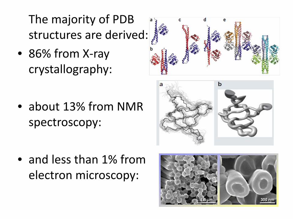
The majority of PDB structures are derived:
• 86% from X-ray crystallography:
• about 13% from NMR spectroscopy:
• and less than 1% from electron microscopy:
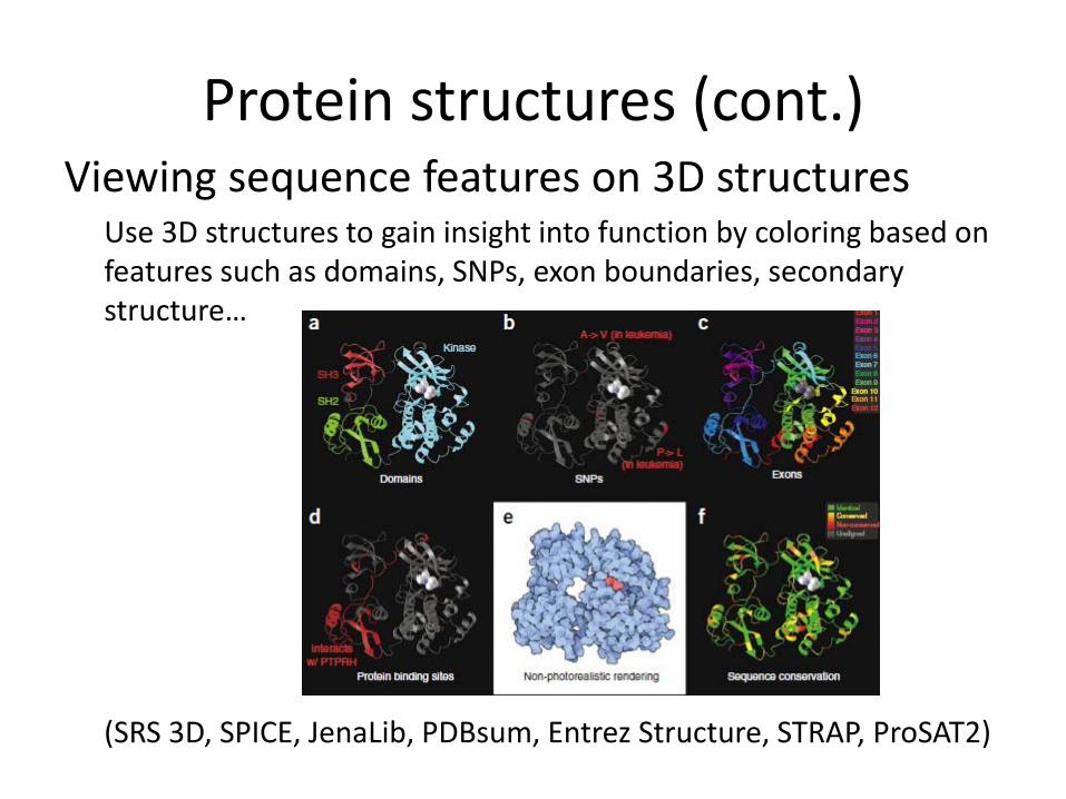
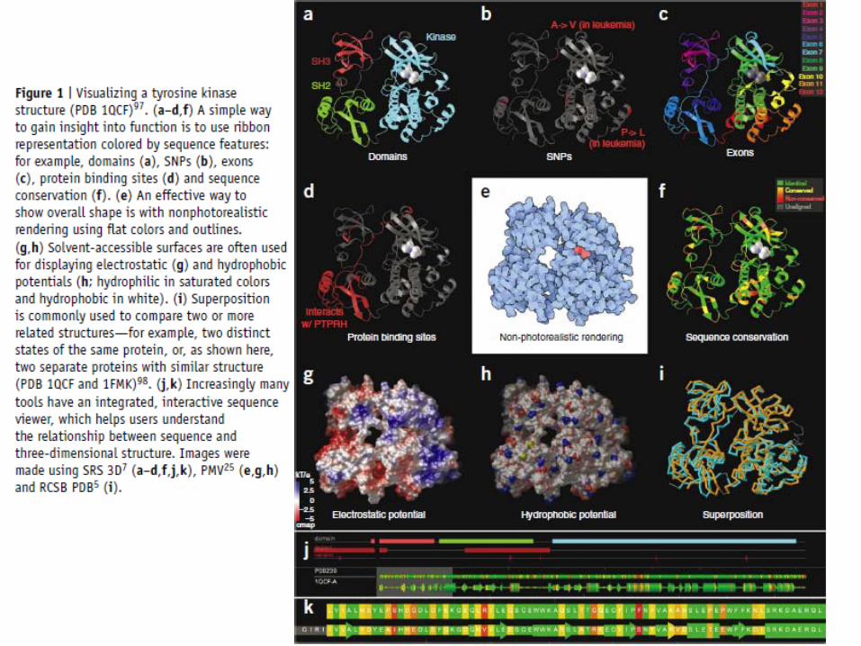
Protein structures (cont.)Viewing sequence features on 3D structures
Use 3D structures to gain insight into function by coloring based on features such as domains, SNPs, exon boundaries, secondary structure…
(SRS 3D, SPICE, JenaLib, PDBsum, Entrez Structure, STRAP, ProSAT2)

Protein structures (cont.)
Protein-protein binding sitesA protein will bind to several other proteins through large but flat binding surfaces.A large percentage of PDB entries contain not just a single protein chain but several.Arrangement of subunits, subunit-subunit contacts interface residues. (PDBsum, MolSurfer)

Protein structures (cont.)
Comparing related structures– two states of the same molecule– two proteins with homologous sequences– two structural homologs found by structural
comparison tools– results dependent on the regions chosen– identifies a more-or-less rigid core of the molecule
and superimposes this region using a subset of the atoms
(MOLMOL, MOE, PyMOL, VMD, STAMP, STRAP, THESEUS)
Protein structures (cont.)
Molecular surface & electrostatic potentialsMany tools can generate molecular surfaces (aka Connolly surf., solvent-excluded surf.)Wide variety of properties:– residue conservation scores– hydrophobicity– depth-cue information– mean-force potentials– Electrostatics(MSMS)
Outline
• Protein structures• Ligand binding sites• RNA structures• Molecular motion• Larger macromolecular assemblies• Future perspectives

Ligand binding sites
• Interactions between macromolecules and small molecules often occur in buried active sites.
• The PDB at present contains over 37,000 binding sites involving about 10,000 different types of ligand molecules.
• A range of methods are available to characterize and visualize these sites, depending on the questions from the end users.
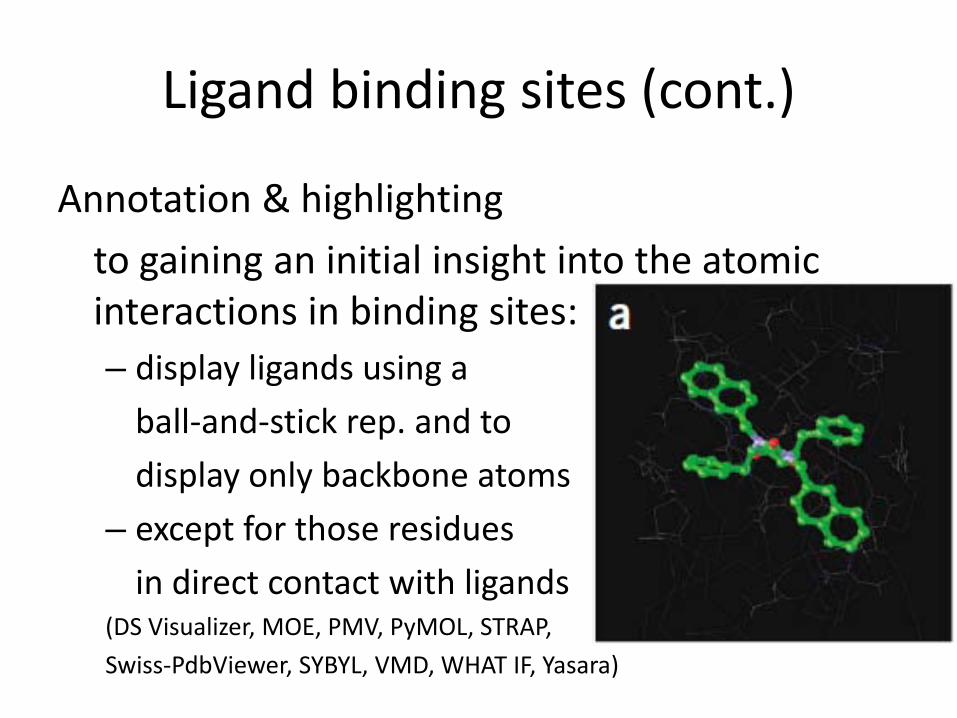
Ligand binding sites (cont.)
Annotation & highlightingto gaining an initial insight into the atomic interactions in binding sites:– display ligands using a
ball-and-stick rep. and to display only backbone atoms
– except for those residues in direct contact with ligands
(DS Visualizer, MOE, PMV, PyMOL, STRAP, Swiss-PdbViewer, SYBYL, VMD, WHAT IF, Yasara)
Ligand binding sites (cont.)What kinds of small molecules may bind
to a given binding site?Surface-based approach:
colored the surface by local properties to allow exploration of chemical complementarily
Volume-based approach:analyze the space around the target molecule, highlighting regions that may form strong interactions with small molecules (AutoLigand )
Sequence-profile approach:uses multiple sequence alignments mapped 3D structures, based on the observation that binding site residues tend to be more conserved than other positions (TraceSuite, ETV)useful when little is known about a protein.

Ligand binding sites (cont.)
Multiple ligands– Same protein but different structures exist with
different ligands , explore the range of conformations available.
– Comparisons can highlight interactions common to all known binding partners, search for further possible binding partners
– Docking tools
(FlexX, AutoDock, Relibase, Superligands)
Ligand binding sites (cont.)
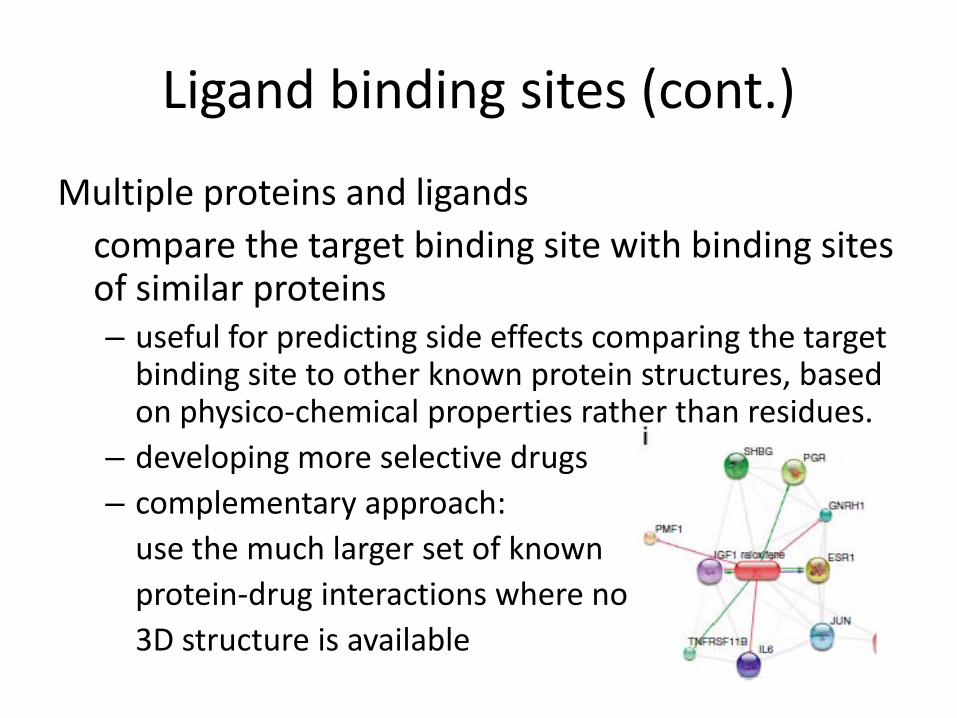
Multiple proteins and ligandscompare the target binding site with binding sites of similar proteins– useful for predicting side effects comparing the target
binding site to other known protein structures, based on physico-chemical properties rather than residues.
– developing more selective drugs – complementary approach:
use the much larger set of known protein-drug interactions where no 3D structure is available

Ligand binding sites (cont.)
Schematic illustrations show the ligand and interacting protein side chains ‘flattened’ in a plane, and indicating relevant hydrogen bonds, covalent bonds, unbonded contacts and water-mediated hydrogen bonds
(LIGPLOT, PoseView)
Outline
• Protein structures• Ligand binding sites• RNA structures• Molecular motion• Larger macromolecular assemblies• Future perspectives
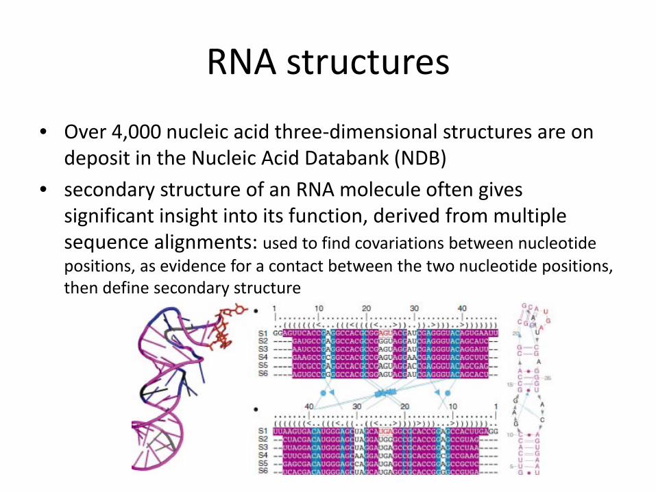
RNA structures
• Over 4,000 nucleic acid three-dimensional structures are on deposit in the Nucleic Acid Databank (NDB)
• secondary structure of an RNA molecule often gives significant insight into its function, derived from multiple sequence alignments: used to find covariations between nucleotide positions, as evidence for a contact between the two nucleotide positions, then define secondary structure

RNA structures (cont.)
• two of the main challenges in RNA visualization: – RNA often adopts multiple structures depending
on experimental conditions, and none of the available tools can deal with this properly
– in vivo usually occurs in complex with proteins, however the RNA-specific tools cannot yet manage such complexes
• researchers can use standard molecular graphics tools to view such complexes, but of course this means losing RNA-specific features and capabilities.
Outline
• Protein structures• Ligand binding sites• RNA structures• Molecular motion• Larger macromolecular assemblies• Future perspectives

Molecular motion
• Biomacromolecules are dynamic entities, and motion is usually essential to function.
• Several vis. tools allow quick and easy exploration of dynamic transitions between two known states of a molecule by animation(Yale Morph Server, Moviemaker)
Molecular motion (cont.)
• For large-amplitude, low-frequency motions, such as protein domain flexing:– normal mode analysis and elastic network models methods
(NOMAD, ANM)
• At higher level of complexity, several programs allow users to:– generate conformational ensembles and trajectories using constraint-
based methods(tCONCOORD, FIRST/FRODA, VMD)
– molecular dynamics simulations, but too CPU-intensive to be provided as a free service…….(DSMM, MoDEL, Dynameomics)
Molecular motion (cont.)
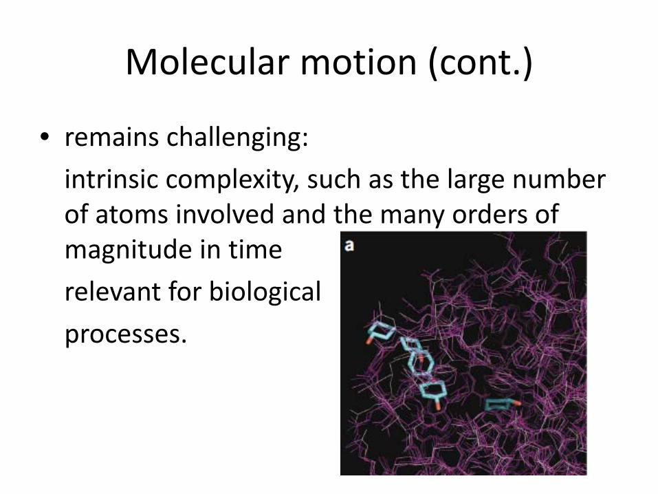
• remains challenging: intrinsic complexity, such as the large number of atoms involved and the many orders of magnitude in timerelevant for biological processes.
Outline
• Protein structures• Ligand binding sites• RNA structures• Molecular motion• Larger macromolecular assemblies• Future perspectives
Large macromolecular assemblies
• X-ray crystallography is being used to solve the structures of larger and more complex systems, electron microscopy is catching up
• These data on large-scale assemblies that integrate data from X-ray crystallography, NMR spectroscopy, electron microscopy and even light microscopy pose many new challenges for visualization:– many of these data are not at atomic detail, so other
representations must be used– the systems can be very large, and there are often
issues with computational and graphics performance
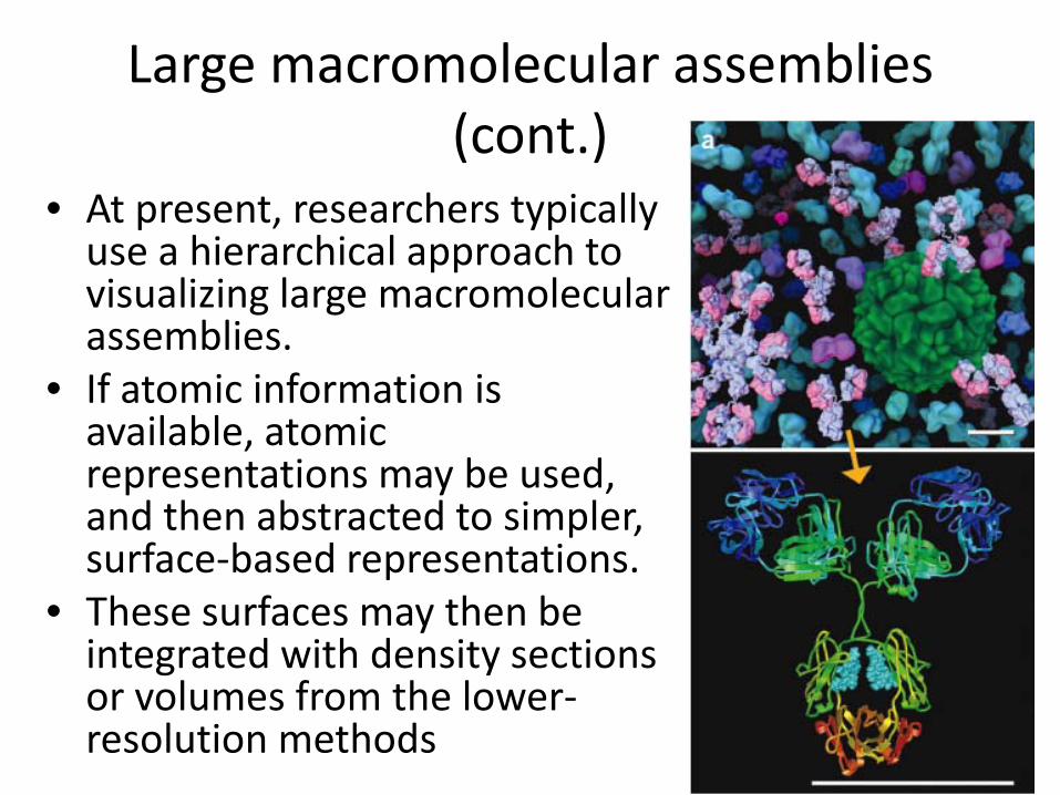
Large macromolecular assemblies (cont.)
• At present, researchers typically use a hierarchical approach to visualizing large macromolecular assemblies.
• If atomic information is available, atomic representations may be used, and then abstracted to simpler, surface-based representations.
• These surfaces may then be integrated with density sections or volumes from the lower-resolution methods

Outline
• Protein structures• Ligand binding sites• RNA structures• Molecular motion• Larger macromolecular assemblies• Future perspectives

Future perspectives
• we can expect more effective computational approaches for representing, analyzing and synthesizing ever-more-complex molecular systems.
• we expect that most of the advances in molecular visualization will come in the areas of computer interfaces, user interaction and new ways to represent and visualize non-spatial information.
• we also expect that collaborative community editing of structure-related data sources will change how scientists relate to structural data, and to each other.

Questions?

Thanks!