Embed Size (px)
Citation preview

Visual field examinationin children with brain disorders
Yvonne Koenraads2016

Visual field examination in children with brain disorders PhD thesis, Utrecht University, the Netherlands
Cover design Yvonne Koenraads & Fotografie UMC Utrecht, afdeling oogheelkundeLayout Yvonne KoenraadsPrinted by Proefschriftmaken.nl & Uitgeverij BOXPressISBN 978-90-393-6545-8
© 2016 Yvonne KoenraadsAll rights reserved. No parts of this publication may be reproduced or transmitted in any form or by any means, electronical or mechanical, including photocopy, recording, or any informa-tion storage or retrieval system, without permission in writing from the author. The copyright of the articles that have been published had been transferred to the respective journals.
CorrespondenceYvonne Koenraads, University Medical Center Utrecht, Department of Ophthalmology, P.O. Box 85500, 3508 GA, Utrecht. E-mail: [email protected]

Visual field examinationin children with brain disorders
Gezichtsveldonderzoek bij kinderen met hersenaandoeningen
(met een samenvatting in het Nederlands)
Proefschriftter verkrijging van de graad van doctor aan de Universiteit Utrecht
op gezag van de rector magnificus, prof.dr. G.J. van der Zwaan, ingevolge het besluit van het college voor promoties in het openbaar te verdedigen
op dinsdag 14 juni 2016 des ochtends te 10.30 uur
door
Yvonne Koenraadsgeboren op 31 december 1986
te Eindhoven

Promotoren Prof.dr. K.P.J. Braun Prof.dr. S.M. Imhof Copromotor Dr. G.L. Porro
The research described in this thesis was financially supported byDr. F.P. Fischer Stichting (through UitZicht), ODAS, Rotterdamse Stichting Blindenbelangen, Stichting Nederlands Oogheelkundig Onderzoek & Stichting Vrienden UMC Utrecht.
Publication of this thesis was kindly supported byRotterdamse Stichting Blindenbelangen, Stichting Blindenhulp, Stichting voor Ooglijders, Landelijke Stichting voor Blinden en Slechtzienden, Allergan, Bayer, BCRM, Chipsoft, Medical Workshop, Procornea, Rockmed, Thea Pharma, Tramedico, UCB Pharma & Visser contactlenzen.

Aan mijn nichtje & neefje±03.06.2016 & ±21.08.2016

Contents
Chapter 1 General introduction
Chapter 2 Perimetry in young and neurologically impaired children: the Behavioral Visual Field (BEFIE) Screening Test revisited JAMA Ophthalmology 2015 Mar;133(3):319-25
Chapter 3 Prediction of visual field defects in newborn infants with perinatal arterial ischemic stroke using early MRI and DTI-based tractography of the optic radiation European Journal of Paediatric Neurology 2016 Mar;20(2):309-18
Chapter 4 Visual field findings in children with suspicion of increased intracranial pressure Submitted
Chapter 5 Visual outcome in Sturge-Weber syndrome: a systematic review and Dutch multicenter cohort Accepted for publication in Acta Ophthalmologica
9
27
47
73
101

Chapter 6 Visual function and compensatory mechanisms for hemianopia after hemispherectomy in children Epilepsia 2014 Jun;55(6):909-17
Chapter 7 Summary and general discussion Nederlandse samenvatting
Chapter 8 List of abbreviations Contributors Assessment committee Dankwoord Curriculum vitae List of publications
127
151 177
182184187188196198


1General introduction

Chapter 1
10
1INTRODUCTION TO THIS THESIS
Cerebral visual impairment (CVI, see Box 1) is nowadays the leading cause of child blindness in developed countries.1–5 Beside a decrease in the prevalence of treatable and preventable vision impairing disorders, the increased survival rate of premature and low birth weight infants is a major cause of the growing proportion of visual impairment due to brain injury.6 However, the true prevalence of CVI is probably even higher than currently estimated, because better diagnostic techniques are being developed to examine children with visual impairment due to brain disorders.2 While other sequelae of cerebral injury, such as epilepsy, motor dysfunction and mental retardation are clinically evident, impairment of visual functions, such as visual acuity (VA) and visual field (VF), may easily escape identification.7,8
The assessment of visual function, especially VF examination, is essential in children with brain disorders for several reasons. For instance, early recognition of visual problems is important for correct interpretation of a child’s behavior, parent counseling, and for the timely initiation of an appropriate rehabilitation strategy. Besides, assessment of visual function may play a significant role in monitoring the progression of a brain disorder, in determining a child’s prognosis and in evaluating the presence of a preexisting VF defect in children eligible for epilepsy surgery. Furthermore, results of visual examination may also facilitate the diagnosis of cerebral disorders.
Unfortunately, assessment of visual function represents a major challenge in young and neu-rologically impaired children. In addition, available methods, mainly those used to assess the VF in these children, are relatively unknown and not widely used.

General introduction
11
1VISUAL PATHWAYS OF THE BRAIN
Visual information enters the eye and subsequently travels from the neurons of the retina via the optic nerve to the optic chiasm. After crossing of the bilateral nasal fibers in the chiasm, it continues through the optic tracts, and subsequently travels from the lateral geniculate body through the optic radiation (OR) to the primary striate visual cortex located in the occipital lobe, in which basic processing takes place (see Figure 1).9,10 Development of this visual system progresses rapidly in the first years of life by influence of changes in photoreceptor organiza-tion, myelination of the visual pathways and developing neural connectivity.11 These changes are responsible for a rapid development of VA in the first few months of life, followed by a slower development until adult levels are reached by the age of 3-4 years.11 In parallel, the extension of the VF expands with age in the first years of life.12,13 However, the exact course of development of the VF is unknown, since the assessed boundaries of the VF also depend on the examination method used.12,14
The cerebral part of the visual system can be affected in several ways, for instance by genetic disorders, malformations, metabolic diseases, hypoglycemic injury, hypoxic-ischemic injury,
Box 1. Cerebral Visual Impairment Cerebral Visual Impairment (CVI) is defined as “visual malfunction due to retro-chiasmal vi-sual and visual association pathway pathology, which can be isolated or accompany anterior visual pathway dysfunction”.5 It is a heterogeneous disorder, which forms a major cause of visual impairment in children worldwide. CVI is also referred to as cortical visual impairment, however, this term is less adequate, since the neurological pathology causing CVI does not necessarily have to be limited to the visual cortex, but may also comprise the optic radiation, visual association areas or the pathways responsible for higher visual functioning.81 Former-ly, the disorder was known as cortical blindness. This term was abandoned since nearly all patients have a certain degree of vision and are rarely completely blind.81
Chapter 1
12
1infection, elevated intracranial pressure (ICP), epilepsy or traumatic injury.5,15 All these disor-ders may impair development of, or cause damage to, neurons along the visual pathways. As consequence of this neuronal pathology, anterograde – “dying forward”, caused by loss of input – or retrograde transneuronal degeneration – “dying backward”, caused by loss of trophic support from the target – may occur.16
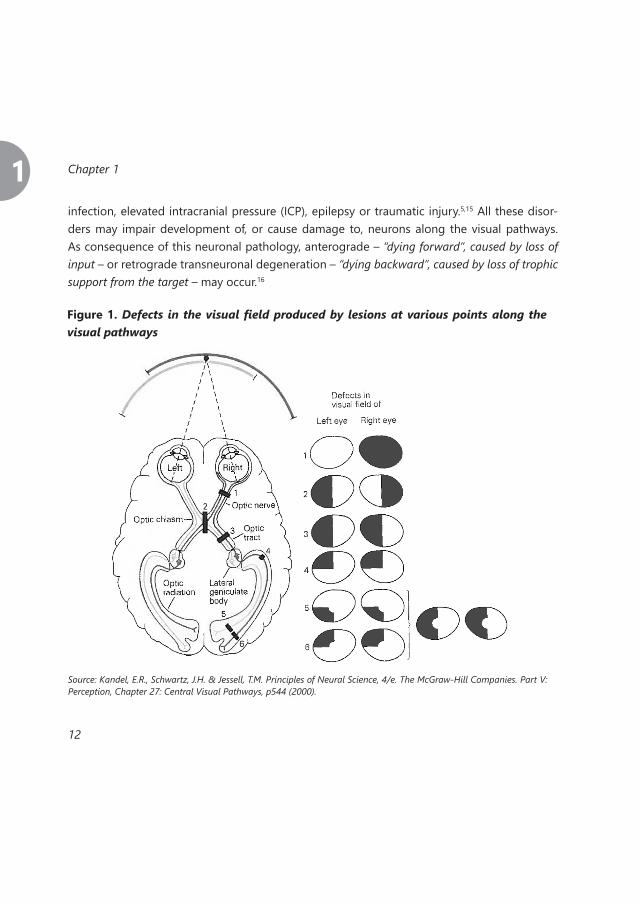
Figure 1. Defects in the visual field produced by lesions at various points along the visual pathways
Source: Kandel, E.R., Schwartz, J.H. & Jessell, T.M. Principles of Neural Science, 4/e. The McGraw-Hill Companies. Part V: Perception, Chapter 27: Central Visual Pathways, p544 (2000).

General introduction
13
1The kind of visual impairment caused by brain disorders mainly depends on the location of in-jury.17 Figure 1 gives a systematic overview on how the VF is generally affected by brain lesions on different locations along the visual pathways.
The visual system is a complex entity, and several additional factors may play a role in the per-ceived (functional) visual impairment. For instance, there are various extrastriate visual associ-ation areas located near the primary striate visual cortex, each responsible for different visual aspects e.g. processing motion, depth perception, color, size estimation, three-dimensional features, visual search or target selection.18 Injury to a selection of these areas may impair specific aspects of vision. In addition, damage of the pathways that serve the higher visual functions, such as the dorsal (“where”) and the ventral (“what”) stream, may impair interpretation of the visual information received by the visual cortex. In particular, damage of the dorsal stream, reaching to the pos-terior parietal lobes, may impair visual search and visually guided movement, while damage of the ventral stream, reaching to the temporal lobes, may impair recognition.5,18 Furthermore, compensation or restoration of visual impairment, optimizing functional visual capacities by means of physical adaptations or neuronal plasticity, might occur.19–21
Regarding physical adaptations, an anomalous head posture (AHP)22–26 and exotropia (XT)23,25,27–
32 contralateral to the side of brain damage may compensate a VF defect in children and adults with early brain injury. In fact, an XT may create a more panoramic view and broaden the func-tional VF, while an AHP may center the VF and enables efficient scanning of the surroundings. In addition, possible neuronal plasticity, mainly present in the developing infant brain, might result in a more favorable outcome than expected when considering a child’s brain dam-age.20,21 Possible explanations of this phenomenon include formation of new interhemispheric connections, reorganization in nearby unaffected cortex or changes in functional interaction between higher-level visual cortical areas and the primary visual areas.33

Chapter 1
14
1EXAMINATION OF VISUAL FUNCTION
Assessment of the visual functions in young or neurologically impaired children requires an alternative approach than the standard conventional perimetry (SCP) methods used in adults. There are several methods available for children in specific functional age ranges according to the expected capabilities of the child at that particular age. These methods are described below, with focus on the techniques that are applied in the studies described in this thesis.
Visual field assessment Table 1 provides an overview of available VF examination methods that can be used in young or neurologically impaired children, separated into SCP,34 confrontational behavioral (see Fig-ure 2 & 3), eye-tracker and multifocal Visual Evoked Potential (mVEP) methods. In general, standard computer assisted perimetry techniques are preferred in both research and patient care, since they are found to be sensitive and reliable. Moreover, they provide quantitative data. Most of these techniques are difficult for children to perform, since they require comprehension, full cooperation, prolonged attention and visual fixation.35,36 Conse-quently, when these characteristics are lacking, such as in young or multi handicapped pa-tients, an appropriate alternative must be chosen with an adequate balance between feasibility and reliability of the method. The standard perimetry of choice performed in our center by older and more cooperative children is the Rodenstock Peritest.37 This technique enables the detection of concentric, hemianopic and quadrantanopic VF defects, as well as enlarged blind spots, widespread de-fects, central scotomas, sensitivity decreases, paracentral scotomas, nasal restrictions (or step) and arcuate defects. SCP methods such as Goldmann34,38, Frequency Doubling Technology (FDT)39,40, SITA Fast34,41 or standard Humphrey perimetry42 were also sometimes used in the children described in this thesis. Although these SCP could be adapted to the needs of chil-dren (i.e. shorter testing time or intervals), they are still often unsuccessful in healthy children younger than 6 years of age, despite extensive explanation and coaching by the examiner.43
General introduction
15
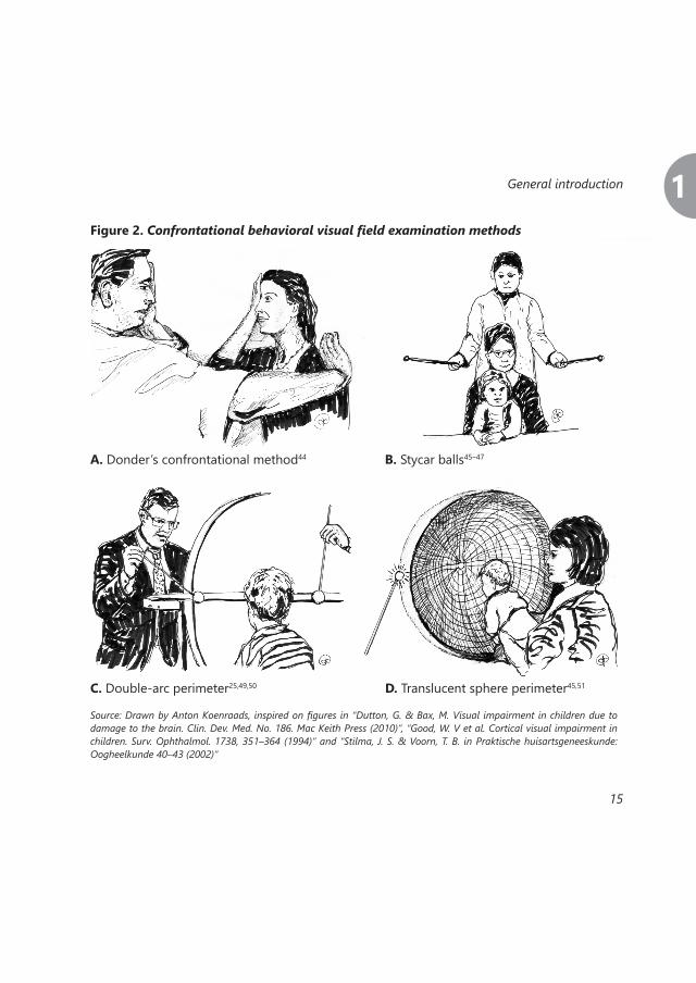
1Figure 2. Confrontational behavioral visual field examination methods
A. Donder’s confrontational method44 B. Stycar balls45–47
C. Double-arc perimeter25,49,50 D. Translucent sphere perimeter45,51
Source: Drawn by Anton Koenraads, inspired on figures in “Dutton, G. & Bax, M. Visual impairment in children due to damage to the brain. Clin. Dev. Med. No. 186. Mac Keith Press (2010)”, “Good, W. V et al. Cortical visual impairment in children. Surv. Ophthalmol. 1738, 351–364 (1994)” and “Stilma, J. S. & Voorn, T. B. in Praktische huisartsgeneeskunde: Oogheelkunde 40–43 (2002)”

Chapter 1
16
1
Tabl
e 1.
Vis
ual fi
eld
exam
inat
ion
met
hods
tha
t ca
n be
use
d in
you
ng o
r ne
urol
ogic
ally
im
pair
ed c
hild
ren
Met
hod
Prin
cipl
eA
dvan
tage
Dis
adva
ntag
eCo
nven
tiona
l (S
CP)
No
freed
om o
f mov
emen
t
Oct
opus
pe
rimet
ry34
,35
Auto
mat
ic st
atic
Shor
t tes
t dur
atio
nPr
elim
inar
y fa
mili
ariza
tion
phas
eRe
quire
s coo
pera
tion
& st
eady
fix
atio
nH
umph
rey
SITA
Fa
st34
,41
Auto
mat
ic st
atic
Shor
t tes
t dur
atio
nRe
quire
s coo
pera
tion
& st
eady
fix
atio
nFD
T pe
rimet
ry39
,40
Auto
mat
ic st
atic
Shor
t tes
t dur
atio
n At
tract
ive
targ
etLe
ss fr
ight
enin
g (n
o bo
wl)
Onl
y ce
ntra
l VF
Requ
ires s
tead
y fix
atio
n
Rode
nsto
ck
Perit
est37
Auto
mat
ic a
nd
sem
i-aut
omat
ic
stat
ic
Can
be a
djus
ted
to
the
child
’s ne
eds
Requ
ires s
tead
y fix
atio
n
Gol
dman
n pe
rimet
ry34
,38
Man
ual k
inet
icM
ost e
ffect
ive
conv
entio
nal
met
hod
in y
oung
ch
ildre
n
Requ
ires a
trai
ned
exam
iner
Requ
ires s
tead
y fix
atio
n
Conf
ront
atio
nal
beha
vior
al
Don
der’s
con
fron
-ta
tiona
l met
hod44
Det
ectio
n of
m
ovin
g fin
gers
of
exa
min
er
No
tool
s are
nee
d-ed
Requ
ires c
oope
ratio
n of
the
child
Very
impr
ecise
ass
essm
ent
Styc
ar b
alls
45–4
7Bl
ack
stic
ks w
ith
whi
te b
alls
(diff
eren
t size
s)
Easy
to p
erfo
rmCa
n be
per
form
ed
from
the
age
of 6
m
onth
s
Onl
y ev
iden
t VF
defe
cts c
an b
e de
tect
ed
Dou
ble-
arc
perim
eter
25,4
9,50
Two
grad
ed a
rcs
with
a w
hite
fix
atio
n ta
rget
in
the
mid
dle
Exam
inat
ion
of
horiz
onta
l, ve
rtic
al
and
diag
onal
me-
ridia
ns
Not
muc
h fre
edom
of m
ovem
ent
The
arc
cons
truct
ion
mig
ht b
e fri
ghte
ning
Onl
y pe
riphe
ral V
F

General introduction
17
1
Tran
sluc
ent s
pher
e pe
rimet
er45
,51
Grad
ed tr
anslu
-ce
nt sp
here
on
whi
ch li
ghts
are
ap
plie
d
Asse
ssm
ent o
f all
poss
ible
loca
tions
in
VF
Fixa
tion
poin
t can
be
turn
ed o
ff
Mig
ht b
e fri
ghte
ning
Not
muc
h fre
edom
of m
ovem
ent
BEFI
E te
st13
,48
Grad
ed a
rc, fi
x-at
ion
targ
et a
nd
posit
ioni
ng st
ick
(see
Fig
ure
3 &
Ch
apte
r 2)
Asse
ssm
ent o
f ho
rizon
tal,
vert
ical
an
d di
agon
al m
e-rid
ians
Child
has
free
dom
of
mov
emen
tIs
cons
ider
ed a
ga
me
Requ
ires t
rain
ed o
bser
ver &
ex-
amin
erO
nly
perip
hera
l VF
Eye-
trac
ker52
–58
SVO
P52,5
3,57
Whi
te d
ots/
pic-
togr
ams a
ppea
r on
the
scre
en
on d
iffer
ent
posit
ions
Rela
tivel
y ob
ject
ive
Requ
ires c
once
ntra
tion
& c
oop-
erat
ion
Mod
erat
e fre
edom
of m
ovem
ent
Onl
y ce
ntra
l VF
Not
pos
sible
with
nys
tagm
usBa
rtim
éus
perim
etry
56,5
8M
ovin
g fix
atio
n ta
rget
(pic
to-
gram
), st
imul
us
appe
ars o
n po
sitio
n re
lativ
e to
it
Rela
tivel
y ob
ject
ive
Mov
ing
fixat
ion
targ
et is
attr
activ
e to
chi
ldre
nCo
mpa
rabl
e to
Go
ldm
ann
V4/II
I
Mod
erat
e fre
edom
of m
ovem
ent
Mea
sure
s onl
y m
id-p
erip
hera
l VF
(20
to 4
0 de
gree
s)
Pref
eren
tial
Look
ing
Perim
eter
54
Scre
en w
ith
web
cam
, car
-to
on w
ith so
und
sudd
enly
mov
es
to a
noth
er p
lace
Cart
oon
with
so
und
is at
tract
ive
to c
hild
ren
Trac
king
of e
ye-m
ovem
ents
by
obse
rvat
or →
less
obj
ectiv
eO
nly
cent
ral V
F
Mul
tifoc
al V
EP59
–6O
bjec
tive
Impr
ecise
mea
sure
men
tM
easu
res o
nly
prim
ary
visu
al
cort
ex

Chapter 1
18
1To solve this problem, confrontational behavioral methods have been developed to assess the VF of children who are unable to perform SCP (see Figure 2). While Donder’s confrontation-al method44 and ‘binocular directional preference’ using two Stycar balls on a stick45–47 were used in few children described in this thesis, the preferred type of confrontational behavioral method that is frequently used in our center is the BEhavioral visual FIEld (BEFIE) screening test13,48 (see Figure 3 & Chapter 2). This device was developed in 1995 by Porro et al.13 in order
Figure 3. BEhavioral visual FIEld (BEFIE) screening test
Source: Porro, G., et al. A new behavioural visual field test for clinical use in pediatric neuro-ophthalmology. Neuro-Oph-thalmology 19, 205–214 (1998).

General introduction
19
1to measure the peripheral VF in young and neurologically impaired children. With the BEFIE test, concentric, hemianopic and quadrantanopic VF defects can be easily detected from the age of four months onwards, even in children with brain disorders. Although not yet widely in use elsewhere, in our experience the BEFIE test can play an important role in the examination of VFs in young children with (suspected) brain disorders.
Visual acuity assessmentThere are several methods to assess VA in young or neurologically impaired children. VA ex-amination methods used in our center and described in this thesis, from easiest to most dif-ficult to perform, include the Teller Acuity Carts (TAC)45,65, the Cardiff Acuity Test66,67, the Kays Picture Chart68–70 and Snellen Charts.71 The most appropriate method is individually chosen depending on the age and cooperation of the child.The TAC45,65 and the Cardiff Acuity Test66,67 both can be performed from very young age on-wards. These techniques make use of the ‘forced preferential looking’ principle, that is based on the observation that infants have a greater tendency to look at a pattern stimulus than at a homogeneous field. Both techniques measure resolution acuity, which is the smallest separa-tion between bars in a grating (or dots) that can be resolved. When a child is able to recognize and name pictograms, the Kays Picture Chart68–70 can be performed, which measures recogni-tion acuity. Recognition acuity is based on the detail in the smallest pictogram (or number or letter) that can be recognized. In older children and adults, Snellen Charts71 such as Landolt rings and E-hooks – measuring resolution acuity – or numbers and letters – measuring recog-nition acuity – are being used.72
Since the results obtained with these various techniques have different underlying principles, they are very difficult to compare. Therefore, the best way to present these heterogeneous results together is after transformation to Logarithm of the Minimum Angle of Resolution (LogMAR).73 On the LogMAR-scale, 0.0 LogMAR represents normal vision, low vision is defined as a VA worse than 0.5 LogMAR and blindness as worse than 1.3 LogMAR, established by the World Health Organization.74

Chapter 1
20
1If quantitative VA assessment cannot be performed, observation of visual fixation may provide some information about the presence or absence of visual perception.
Optic disc assessmentThe optic disc can be assessed by means of dilated fundoscopy. However, uncooperative chil-dren, especially those who are neurologically impaired, may be difficult to examine. As they refuse fundoscopy or only permit a short observation, an experienced examiner is often re-quired. If successful, observation of the optic disc through fundoscopy may provide informa-tion on the existence of e.g. elevated ICP (papilledema or atrophy), retrograde transneuronal degeneration (atrophy) or glaucoma (excavation). In addition to fundoscopy, optic disc abnor-malities can be objectified by ancillary investigations such as Ultrasonography (USG), Fluores-cein Angiography (FA) or Optical Coherence Tomography (OCT).
Orthoptic exam and postural observationsOcular alignment and eye movement of the child can be assessed by an orthoptist by means of light reflexes, the covertest and pursuit movements.75 This examination may e.g. reveal stra-bismus, such as an exodeviation (exotropia, XT) or esodeviation (esotropia, ET), which may be intermittent or continuous. Postural observation may reveal a head turn or torticollis, sometimes used by children to op-timize visual functioning.
Neuroimaging of the visual pathwaysTo assess the integrity of the visual pathway, neuroimaging can be performed using Mag-netic Resonance Imaging (MRI). Advanced MRI techniques such as Diffusion Tensor Imaging (DTI)76–78 and functional MRI (fMRI)79 are used, mainly in investigational settings, to gain more information about the integrity of visual tracts, and localization of visual functions. The DTI technique relies on the fact that diffusion of water molecules in brain tissue is not free but restricted, reflecting the interaction with e.g. macromolecules, fibers and membranes.78

General introduction
21
1By measuring the directionality of water diffusion in white matter of the brain, the probable topographic location and integrity of white matter tracts (such as the OR) can be assessed noninvasively. With tractography, probable white matter structures that connect two parts in the brain can be visualized three-dimensionally. In addition, for quantitative analysis, measures of anisotropy – such as fractional anisotropy (FA), axial diffusivity (λ1), radial diffusivity (λ23) – and of overall diffusion – mean diffusivity (MD) – can be determined. The fMRI technique can be used to gain functional information of cortical representation of visual functions.80

Chapter 1
22
1AIM AND CONTENT OF THIS THESIS
The general aim of the studies described in this thesis was to gain more insight in the diagnos-tic and prognostic implications of visual field (VF) examination in children with brain disorders. With this work, we hope to create more awareness of cerebral visual impairment (CVI) and of the possibilities to assess the VFs of young and neurologically impaired children. The specific aims of this thesis were:
► To evaluate all VF examinations that were performed with the BEhavioral visual FIEld (BEFIE) screening test, a device developed in 1995 by Porro et al.13 in order to measure the peripheral VF in young and neurologically impaired children (Chapter 2). We aimed to assess its applica-bility, reliability, consistency of findings across time, and potential diagnostic value compared with standard conventional perimetry (SCP)
► To explore the possibility to predict clinical VF defects based on neuroimaging abnormali-ties at the age of 3 months in infants after perinatal arterial ischemic stroke (PAIS) (Chapter 3)
► To evaluate the VF defects found in children suspected of intracranial pressure (ICP) eleva-tion, and to describe long-term visual outcome in those with increased ICP who underwent repeated testing (Chapter 4)
► To present a systematic review of previous literature on visual outcome of patients with Sturge-Weber syndrome (SWS), and report on the visual acuity (VA) and VF examination find-ings in a Dutch multicenter cohort of patients with SWS (Chapter 5)
► To investigate the visual outcome and the prevalence of compensatory mechanisms (CMs) for homonymous hemianopia (HH) after hemispherectomy in children (Chapter 6)

General introduction
23
1REFERENCES
1. Chong, C. & Dai, S. Cross-sectional study on childhood cerebral visual impairment in New Zealand. J. AAPOS 18, 71–74 (2014).2. Boonstra, N. et al. Changes in causes of low vision between 1988 and 2009 in a Dutch population of children. Acta Ophthalmol. 90, 277–286 (2012).3. Kong, L. An update on progress and the changing epidemiology of causes of childhood blindness worldwide. J. AAPOS 16, 501–507 (2012).4. Durnian, J. M. et al. Childhood sight impair-ment: a 10-year picture. Eye (Lond). 24, 112–117 (2010).5. Philip, S. S. & Dutton, G. N. Identifying and characterising cerebral visual impairment in children: a re-view. Clin. Exp. Optom. 97, 196–208 (2014).6. Good, W. V, Jan, J. E., Burden, S. K., Skoczenski, A. & Candy, R. Recent advances in cortical visual impair-ment. Dev. Med. Child Neurol. 43, 56–60 (2001).7. Dutton, G. & Bax, M. Visual impairment in chil-dren due to damage to the brain. Clin. Dev. Med. No. 186. Mac Keith Press (2010).8. Harbert, M. J., Yeh-Nayre, L. a, O’Halloran, H. S., Levy, M. L. & Crawford, J. R. Unrecognized visual field defi-cits in children with primary central nervous system brain tumors. J. Neurooncol. 107, 545–9 (2012).9. De Moraes, C. G. Anatomy of the visual path-ways. J. Glaucoma 22 Suppl 5, S2–7 (2013).10. Goodale, M. A. The functional organization of the central visual pathways. Chapter 1 in: Visual impair-ment in children due to damage to the brain (Edited by G.N. Dutton & M. Bax). Clin. Dev. Med. No. 186. Mac Keith Press (2010).11. The Eye M.D. Association. Pediatric Ophthal-mology and Strabismus. American Academy of Ophthal-mology.
12. Delaney, S. M. et al. Stimulus Motion Increases Measured Visual Field Extent in Children 3.5 to 30 Months of Age. Optom. Vis. Sci. 77, 82–89 (2000).13. Porro, G., et al. A new behavioural visual field test for clinical use in pediatric neuro-ophthalmology. Neuro-Ophthalmology 19, 205–214 (1998).14. Mercuri, E. et al. Neonatal cerebral infarction and visual function at school age. Arch Dis Child 88, F487–F491 (2003).15. Soul, J. & Matsuba, C. Common aetiologies of cerebral visual impairment. Chapter 2.1 in: Visual impair-ment in children due to damage to the brain (Edited by G.N. Dutton & M. Bax). Clin. Dev. Med. No. 186. Mac Keith Press (2010).16. Beatty, R., Sadun, A., Smith, L., Vonsattel, J. & Richardson, E. Direct demonstration of transsynaptic de-generation in the human visual system: a comparison of retrograde and anterograde changes. J. Neurol. Neurosurg. Psychiatry 45, 143–146 (1982).17. Kandel, E. R., Schwartz, J. H. & Jessell, T. M. Principles of Neural Science, 4/e. The McGraw-Hill Com-panies. Part V: Perception, Chapter 27: Central Visual Path-ways. 544 (2000).18. Suter, P. S. & Harvey, L. H. Vision Rehabilitation: Multidisciplinary Care of the Patient Following Brain Injury. Taylor & Francis Group, LLC. CRC Press. (2011).19. Koenraads, Y. et al. Visual function and com-pensatory mechanisms for hemianopia after hemispherec-tomy in children. Epilepsia 55, 909–917 (2014).20. Guzzetta, A. et al. Plasticity of the visual sys-tem after early brain damage. Dev. Med. Child Neurol. 52, 891–900 (2010).21. Guzzetta, A., Fiori, S., Scelfo, D., Conti, E. & Ban-cale, A. Reorganization of visual fields after periventricular haemorrhagic infarction: potentials and limitations. Dev. Med. Child Neurol. 55 Suppl 4, 23–6 (2013).22. Paysse, E. A. & Coats, D. K. Anomalous Head Posture With Early-Onset. J AAPOS 1, 209–13 (1997).

Chapter 1
24
123. Donahue, S. P. & Haun, A. K. Exotropia and face turn in children with homonymous hemianopia. J. Neu-roophthalmol. 27, 304–7 (2007).24. Zangemeister, W. H., Meienberg, O., Stark, L. & Hoyt, W. F. Eye-head coordination in homonymous hemi-anopia. J. Neurol. 226, 243–54 (1982).25. Good, W. V et al. Cortical visual impairment in children. Surv. Ophthalmol. 1738, 351–364 (1994).26. Porro, G., van der Linden, D., van Nieuwen-huizen, O. & Wittebol-Post, D. Role of visual dysfunction in postural control in children with cerebral palsy. Neural Plast. 12, 205–10; discussion 263–72 (2005).27. Jacobson, L., Lennartsson, F., Pansell, T., Oqvist Seimyr, G. & Martin, L. Mechanisms compensating for vi-sual field restriction in adolescents with damage to the retro-geniculate visual system. Eye (Lond). 26, 1437–45 (2012).28. Herzau, V., Bleher, I. & Joos-kratsch, E. Infantile exotropia with homonymous hemianopia : a rare contrain-dication for strabismus surgery. Graefe ’ s Arch. Ophthal-mol. 1, 148–149 (1988).29. Levy, Y., Turetz, J., Krakowski, D., Hartmann, B. & Nemet, P. Development of compensating exotropia with anomalous retinal correspondence after early infancy in congenital homonymous hemianopia. J Pediat Ophthalmol Strabismus 3, 236–238 (1995).30. Saleh, G., Sivaprasad, S. & Hammond, C. Hom-onymous hemianopia and exotropia: an important man-agement issue. Eye (Lond). 20, 1402–1404 (2006).31. Gote, H., Gregersen, E. & Rindziunski, E. Exo-tropia and panoramic vision compensating for an occult congenital homonymous hemianopia. A case report. Binoc Vis. Eye Muscle Surg Q 8, 129–132 (1993).32. Gamio, S. & Melek, N. When the patient says no. Management of exotropia with hemianopic visual field defects. Binocul Vis Strabismus Q 18, 167–170 (2003).33. Urbanski, M., Coubard, O. a & Bourlon, C. Visu-alizing the blind brain: brain imaging of visual field defects
from early recovery to rehabilitation techniques. Front. In-tegr. Neurosci. 8, 74 (2014).34. Patel, D. E. et al. Study of optimal perimetric testing in children (OPTIC): Feasibility, reliability and re-peatability of perimetry in children. PLoS One 10, 1–12 (2015).35. Safran, a B. et al. Feasibility of automated visu-al field examination in children between 5 and 8 years of age. Br J Ophthalmol 80, 515–518 (1996).36. Tschopp, C. et al. Automated visual field ex-amination in children aged 5-8 years. Part I: Experimental validation of a testing procedure. Vision Res. 38, 2203–10 (1998).37. Greve, E. L., Dannheim, F. & Bakker, D. The Peritest, a new automatic and semi-automatic perimeter. Int. Ophthalmol. 5, 201–14 (1982).38. Kolling, G. H. & Wabbels, B. Symposium Pro-ceedings Part II Kinetic perimetry in neuroophthalmologi-cal practice. 8, (2000).39. Blumenthal, E. Z., Haddad, A., Horani, A. & An-teby, I. The reliability of frequency-doubling perimetry in young children. Ophthalmology 111, 435–9 (2004).40. Quinn, L. M., Gardiner, S. K., Wheeler, D. T., Newkirk, M. & Johnson, C. a. Frequency doubling technol-ogy perimetry in normal children. Am. J. Ophthalmol. 142, 983–9 (2006).41. Donahue, S. P. & Porter, a. SITA visual field test-ing in children. J. AAPOS 5, 114–7 (2001).42. Beck RW, Bergstrom TJ, L. P. A clinical compari-son of visual field testing with a new automated perimeter, the Humphrey Field Analyzer, and the Goldmann perime-ter. Ophthalmology. 92, 77–82 (1985).43. Porro, G. L. & Wittebol-Post, D. Impairment of peripheral vision and its measurement. Chapter 5 in: Visual impairment in children due to damage to the brain (Edited by G.N. Dutton & M. Bax). Clin. Dev. Med. No. 186. Mac Keith Press (2010).

General introduction
25
144. Stilma, J. S. & Voorn, T. B. in Praktische huisarts-geneeskunde: Oogheelkunde 40–43 (2002).45. Mohn, G. & Van Hof-van Duin, J. Behavioural and electrophysiological measures of visual functions in children with neurological disorders. Behav. Brain Res. 10, 177–87 (1983).46. Hermans, A., Van Hof-van Duin, J. & Oude-sluys-Murphy, A. Visual outcome of low-birth-weight infants (1500-2500 g) at one year of corrected age. Acta Paediatr. 83, 402–7 (1994).47. Sheridan MD. The STYCAR graded-balls vision test. Dev Med Child Neurol. 15, 423–32 (1973).48. Koenraads, Y., Braun, K. P. J., van der Linden, D. C. P., Imhof, S. M. & Porro, G. L. Perimetry in Young and Neurologically Impaired Children: The Behavioral Visual Field (BEFIE) Screening Test Revisited. JAMA Ophthalmol. 133, 319–25 (2015).49. Quinn, G. E. et al. Visual Fields Measured with Double-arc Perimetry in Eyes with Threshold Retinopathy of Prematurity from the Cryotherapy for Retinopathy of Prematurity Trial. Ophthalmology 103, 1432–1437 (1996).50. Dobson, V., Brown, a M., Harvey, E. M. & Nar-ter, D. B. Visual field extent in children 3.5-30 months of age tested with a double-arc LED perimeter. Vision Res. 38, 2743–60 (1998).51. Mayer, D. L., Fulton, a B. & Cummings, M. F. Vi-sual fields of infants assessed with a new perimetric tech-nique. Invest. Ophthalmol. Vis. Sci. 29, 452–9 (1988).52. Murray, I. C. et al. Feasibility of saccadic vec-tor optokinetic perimetry: a method of automated static perimetry for children using eye tracking. Ophthalmology 116, 2017–26 (2009).53. Murray, I. et al. Saccadic Vector Optokinetic Perimetry (SVOP): A novel technique for automated static perimetry in children using eye tracking. Conf Proc IEEE Eng Med Biol Soc. 2013, 3186–9 (2013).54. Allen, L. E., Slater, M. E., Proffitt, R. V, Quarton, E. & Pelah, A. A new perimeter using the preferential look-
ing response to assess peripheral visual fields in young and developmentally delayed children. J. AAPOS 16, 261–5 (2012).55. Pel, J. J. M., van Beijsterveld, M. C. M., Thepass, G. & van der Steen, J. Validity and Repeatability of Saccadic Response Times Across the Visual Field in Eye Movement Perimetry. Transl. Vis. Sci. Technol. 2, 3 (2013).56. De Wit, G. Bartimeus eyetracker project. at <https://www.bartimeus.nl/expertise/projecten/projec-tresultaten/project-eyetracker>57. Tailor, V. et al. Saccadic vector optokinetic pe-rimetry in neuro-disability or isolated visual pathway le-sions: observational cohort study. Under Rev. 1–6 (2016). 58. Wit, G. C. de, Hoeben, F. & Genderen, M. van. Objectieve gezichtsveldbepaling bij verdenking conver-sie met behulp van eye-tracking. NOG Congr. Groningen (2015).59. Harding, G. F. a, Spencer, E. L., Wild, J. M., Con-way, M. & Bohn, R. L. Field-specific visual-evoked poten-tials: identifying field defects in vigabatrin-treated chil-dren. Neurology 58, 1261–5 (2002).60. Kim, Y.-J., Yukawa, E., Kawasaki, K., Nakase, H. & Sakaki, T. Use of multifocal visual evoked potential tests in the objective evaluation of the visual field in pediatric epilepsy surgery. J. Neurosurg. 104, 160–5 (2006).61. Klistorner, a I., Graham, S. L., Grigg, J. & Balachandran, C. Objective perimetry using the multifocal visual evoked potential in central visual pathway lesions. Br. J. Ophthalmol. 89, 739–44 (2005).62. Spencer, E. L. & Harding, G. F. a. Examining vi-sual field defects in the paediatric population exposed to vigabatrin. Doc. Ophthalmol. 107, 281–7 (2003).63. Yukawa, E., Kim, Y.-J., Kawasaki, K., Taketani, F. & Hara, Y. A child with epilepsy in whom multifocal VEPs facilitated the objective measurement of the visual field. Epilepsia 46, 577–9 (2005).64. Yukawa, E., Matsuura, T., Kim, Y.-J., Taketani, F. & Hara, Y. Usefulness of multifocal VEP in a child requiring

perimetry. Pediatr. Neurol. 38, 360–2 (2008).65. Teller, D., McDonald, M., Preston, K., SL, S. S. & Dobson, V. Assessment of visual acuity in infants and children: the acuity card procedure. Dev Med Child Neurol. 28, 779–89 (1986).66. Adoh, T., Woodhouse, J. & Oduwaiye, K. The Cardiff Test: A New Visual Acuity Test for Toddlers and Children with Intellectual Impaiment. A Preliminary Report. Optom. Vis. Sci. 69, 427–432 (1992).67. Adoh, T. O. & Woodhouse, J. M. The Cardiff acuity test used for measuring visual acuity development in toddlers. Vision Res. 34, 555–560 (1994).68. Jones, D., Westall, C., Averbeck, K. & Abdolell, M. Visual acuity assessment: A comparison of two tests for measuring children’s vision. Ophthalmic Physiol. Opt. 23, 541–546 (2003).69. Kay, H. New method of assessing visual acuity with pictures. Br. J. Ophthalmol. 67, 131–133 (1983).70. Elliott, M. C. & Firth, a Y. The logMAR Kay pic-ture test and the logMAR acuity test: a comparative study. Eye (Lond). 23, 85–88 (2009).71. Hetherington, R. The Snellen chart as a test of visual acuity. Psychol Forsch. 24, 349–57 (1954).72. Pointer, J. S. Recognition versus Resolution: a Comparison of Visual Acuity Results Using Two Alternative Test Chart Optotype. J. Optom. 1, 65–70 (2008).73. Rosser, D. a, Laidlaw, D. a & Murdoch, I. E. The development of a ‘reduced logMAR’ visual acuity chart for use in routine clinical practice. Br. J. Ophthalmol. 85, 432–436 (2001).74. World Health Organization. International Sta-tistical Classification of Diseases and Related Health Prob-lems 10th Revision. ICD-10 Version. (2010). at <http://apps.who.int/classifications/icd10/browse/2010/en>75. Blaikie, a. J. & Dutton, G. N. How to assess eyes and vision in infants and preschool children. Bmj 350, h1716–h1716 (2015).
76. Feldman, H. M., Yeatman, J. D., Lee, E. S., Barde, L. H. F. & Gaman-bean, S. Diffusion Tensor Imaging : A Re-view for Pediatric Researchers and Clinicians.77. Ramenghi, L. a et al. Neonatal neuroimaging: going beyond the pictures. Early Hum. Dev. 85, S75–7 (2009).78. Basser, P. J., Mattiello, J. & LeBihan, D. MR Dif-fusion Tensor Spectroscopy and Imaging. Biophys. J. 66, 259–267 (1994).79. Ogawa, S., Lee, T., Nayak, A. & Glynn, P. Oxy-genation-sensitive contrast in magnetic resonance image of rodent brain at high magnetic fields. Magn Reson Med. 14, 68–78 (1990).80. McFadzean, R., Condon, B. & Barr, D. Function-al magnetic resonance imaging in the visual system. J Neu-roophthalmol. 19, 186–200 (1999).81. Ospina, L. Cortical visual impairment. Pediatr Rev. 30, e81–90 (2009).

2Perimetry in young and neurologically impaired children:
The Behavioral Visual Field (BEFIE) Screening Test revisited
Yvonne KoenraadsKees P.J. Braun
Denise C.P. van der LindenSaskia M. ImhofGiorgio L. Porro
JAMA Ophthalmology 2015 Mar;133(3):319-25

Chapter 2
28
2ABSTRACT
ImportanceVisual field examination in young or neurologically impaired children is a challenge. As a result, the BEhavioral visual FIEld (BEFIE) screening test was developed in 1995.
ObjectivesTo evaluate the applicability of the BEFIE test in a large population of young or neurologically impaired children, its reliability and consistency of findings across time, and its potential diag-nostic value compared with standard conventional perimetry.
Design, setting, and participantsThe BEFIE tests were performed at an academic tertiary center and measured the peripheral vi-sual field extension in degrees by observing an individual’s response to a stimulus on a graded arc that moved from the periphery to the center of the visual field along different meridians. Patient files from all children who underwent this test were retrospectively analyzed. In total, 1788 BEFIE tests were performed in 835 children (median age, 3.4 years).
Main outcomes and measuresReliability and results of all tests were longitudinally evaluated. The diagnostic value of the BEFIE test was assessed by comparing monocular BEFIE test results with those of standard conventional perimetry in children who underwent both.
ResultsOf 1788 tests, 74% (95% CI, 72%-76%) were considered reliable from the age of 4 months and older, with increasing success with higher ages; 56%reliable in children younger than 1 year; 71%reliable in children between 1 and 2 years; and more than 75%reliable in children 2 years and older (Spearman r=0.506; P=0.11). Peripheral visual field defects were found in 28% (95%

Perimetry in young and neurologically impaired children
29
2CI, 25%-31%) of all first reliable tests. In 75%of children who underwent serial testing, results were consistent and there were good explanations in the case of discrepancies. Comparison of monocular BEFIE tests with standard conventional perimetry results in 147 eyes yielded a positive predictive value of 98% (95% CI, 94%-100%), negative predictive value of 66% (95% CI, 56%-75%), specificity of 98% (95% CI, 95%-100%), sensitivity of 60% (95% CI, 50%-71%), and superior sensitivity of 80% (95% CI, 70%-91%) when only absolute peripheral visual field defects at standard conventional perimetry were accounted for.
Conclusions and relevanceThese data suggest that the BEFIE test is a valuable tool to detect peripheral visual field de-fects when standard conventional perimetry cannot be performed in young or neurologically impaired children.

Chapter 2
30
2INTRODUCTION
Visual field (VF) examination in young or neurologically impaired children is a challenge.1–3 Standard conventional perimetry (SCP) requires full cooperation. Even the simpler versions, such as the Goldmann perimeter,4 Peritest,5 Humphrey Swedish Interactive Thresholding Algo-rithm Fast VF Analyzer,6 or frequency-doubling technology perimetry,7,8 are often unsuccessful in children younger than 6 years.1 There is a need for an easy, reliable method to assess the VFs of young children in a clinical setting. In the past, attempts to create an adequate VF test have included techniques that used eye movement observations and eye tracking systems.9–12 Although these methods have the potential to examine complete VFs in children, it remains a challenge to keep the child focused and attentive, especially when they are too young or disabled. For the same reasons, multifocal visual evoked potential13 proved not suitable for this population. The known techniques designed to measure VFs in young or neurologically im-paired children consist of behavioral methods such as confrontational methods,3,14 binocular directional preference,15 kinetic double-arc perimetry,2,16,17 and translucent sphere perimetry.3,18 While the simpler behavioral methods only provide a global impression of the peripheral VF, the more sophisticated ones that measure the VF extension in degrees are often insufficient in gaining the cooperation of the child and are difficult to integrate in a consulting room. In 1995, we modified the arc perimeter into a simple behavioral kinetic perimetry device to satisfy the needs of the target population and created the BEhavioral visual FIEld (BEFIE) screening test.19 This method can be applied to children who are preverbal ages and older. It is based on a graded semicircular arc with a stimulus at the end introduced from behind the VF of the child from the periphery to the center (see Figure 1). The individual’s visual (or verbal) response to the stimulus is reported by an observer. This technique combines the advantages of all other behavioral methods; because it quantifies VF extension in degrees, it is easy to perform in clinical practice and is well accepted because the interaction between the observer and the child is considered a game.

Perimetry in young and neurologically impaired children
31
2Figure 1. The BEhavioral visual FIeld (BEFIE) screening test
A Equipment: (1) graded semicircular black metal arc with a stimulus at the end, (2) fixation target on a stick and (3) stick with a level attached to it, used for positioning. B Typical example of the performance: The arc is rotated by an examiner from behind around the head of the subject in such a way that the white ball moves from the periphery towards the center of the VF, were the fixation target is positioned at 35cm distance by an observer who is facing the child that sits in a (wheel)chair or on its parents lap. When fixation of the child is steadily captured, the ball is introduced into the VF and the observer reports the subject’s response (i.e. eye (and head or hand) movements or any verbal answer related to the peripheral stimulus). At the moment of response, the degrees of VF extension along the corresponding half-meridian is measured on the semicircular arc by the examiner. The peripheral stimulus is presented at random along one of the four quadrants and along the horizontal axis (half-meridian at 0, 45, 135, 180, 225, 315 degrees). Each half-meridian is tested three times, of which the mean is noted as the result of that meridian. Before the pro-cedure starts, the examiner familiarizes the child with the stimulus. Subsequently, a binocular test is performed. If a child allows occlusion of an eye with an orthoptic patch, a monocular test is performed.

Chapter 2
32
2Since its development, we have extensively applied the BEFIE test. The aim of this study was to retrospectively evaluate its applicability in a large population of young or neurologically impaired children, its reliability and consistency of findings across time, and its potential diag-nostic value compared with SCP.
Figure 2. Age-dependent pathological peripheral visual field limits of the diagonal meridians for the BEFIE test

Perimetry in young and neurologically impaired children
33
2METHODS
Patient SelectionAll children (<18 years at the first examination) who underwent the BEFIE test between Feb-ruary 1, 1995, and December 31, 2013,were included. The study was approved by the institu-tional ethical committee of the University Medical Center Utrecht. Written informed consent was obtained, authorizing publication of the child pictured in the photograph in Figure 1. For the other 834 children in the study, our institutional ethical committee decided that written informed consent was not needed.
Data CollectionPatient files were retrospectively analyzed. Demographic and clinical characteristics that were collected included sex, type, location of (suspected) pathology (defined as postchiasmal or prechiasmal), and age at examination. The BEFIE test was performed with a graded semicircular arc with a white ball (stimulus) at the end, with a fixation target and a stick with a level attached to it used for positioning (see Figure 1).19 All BEFIE tests were taken by a senior orthoptist (observer) and a pediatric neuro-ophthalmologist (examiner). The methods and test procedure are described more extensively in another study by Porro et al.19 The test is available at the Medical Workshop (http://www.medicalworkshop.nl/International).
BEFIE Test ReliabilityBased on the descriptions of the test results noted at the time of assessment, reliability was rated as unsuccessful, doubtful, or reliable by an unblinded assessor, according to predefined criteria. An unsuccessful test included unsuccessful attempts with incomplete final results or results obtained with alternative confrontational approaches such as using toys. A test was rated doubtful when there was an annotation of slow reactions, discordance of the 3 consec-utive measurements, more than 3 spontaneous looks, comments of poor cooperation (such

Chapter 2
34
2as lack of interest or crying), or absence of a definite conclusion with examiners expressing their uncertainty in the report (using terms such as possible or perhaps). A test was considered reliable when annotations on good cooperation were made or if none of the criteria listed here indicating impaired reliability were present.
Analysis of BEFIE Test Results Results of the BEFIE test (binocular and/or monocular) were categorized as normal when the extension was 40° or more nasally and 70° or more temporally, corresponding to the maxi-mum measurable VF with the Peritest method or when the peripheral borders on the diagonal meridians exceeded the age-dependent pathological limits (see Figure 2).19 For subclassifica-tion of abnormal peripheral VF (PVF) defects, see Box 1.
To assess the consistency of results, a longitudinal analysis was done in all children who under-went more than 1 reliable test. We described whether results remained stable, deteriorated, or improved over time and explored possible causal factors for alterations.
Box 1. Subclassification of peripheral visual field defects Results of the BEFIE tests (bin- and/or monocular) were dichotomized as ‘normal’ or ‘abnor-mal’. A PVF was considered ‘normal’ when it extended ≥40 degrees nasally and ≥70 degrees temporally, corresponding to the maximum measurable VF with the Peritest method. The PVF of children under five years of age was considered ‘normal’ if the peripheral borders on the diagonal meridians exceeded the age-dependent pathological limits (see Figure 2).19 A PVF was considered ‘abnormal’ in all other situations, with a subclassification into (1) symmetric (concentric) PVF defects and (2) asymmetric or homonymous (hemianopic and quadrantanopic) PVF defects. ‘Symmetric’ and ‘asymmetric’ PVF defects were further clas-sified in ‘severe concentric’ or ‘complete homonymous’ if the peripheral limits reached <20 degrees nasally or <30 degrees temporally. The other PVF defects were considered ‘incom-plete homonymous’ or ‘moderate concentric’ or ‘mild concentric’ (reaching ≥30 degrees nasally and ≥60 degrees temporally). Scotomas were not included in this classification since the test only examines the PVF.

Perimetry in young and neurologically impaired children
35
2Comparison With SCPThe diagnostic value of the BEFIE test was assessed by comparing its results with those of SCP (reference test). For this purpose, we included all children who underwent a reliable monoc-ular BEFIE test and SCP of the entire VF (including periphery) on the same day or at some time after the BEFIE test. Children with proven progressive underlying disease that could have caused discordance between both results were excluded. When multiple reliably performed BEFIE tests were taken, the one closest in time to the reference test was selected. Different types of SCP used in our center included manual kinetic testing on the Goldmann perime-ter, semiautomatic-static testing on the Peritest, or automatic-static testing on the Humphrey Field Analyzer.5,20 Any of these tests used could be included as reference tests for the analysis. When different SCPs were performed, the Goldmann perimeter was preferred as a reference because its manual kinetic testing was best comparable with the BEFIE test. Measurements on the Humphrey Field Analyzer were least preferred because it was often difficult to perform in children from our cohort. Furthermore, if multiple tests of the same method were present, the first test with the least VF defects was selected to reduce the chance of false-positive VF defects in the reference test itself. The results of monocular VF measurements of all separate eyes were presented in a frequency table. When static (instead of kinetic) perimetry was used as reference test, the test result was considered normal if fewer than 3 stimuli were missed during the measurement. Visual field measurements not meeting these criteria were considered abnormal. Abnormal VF defects measured with the reference test were further dichotomized into absolute PVF defects and absolute scotomas or relative VF defects. Absolute scotomas included holes in the VF that did not extend to the peripheral borders of the VF or were too small to contain 1 of the half-me-ridians at 0°, 45°, 135°, 180°, 225°, or 315°. Relative VF defects comprised defects on static perimetry that were not totally missed but only seen at an increased intensity compared with the rest of the VF. All other defects were rated as absolute PVF defects. Positive predictive val-ue, negative predictive value, additional value, specificity, and sensitivity of the BEFIE test were calculated. Possible causes for false-positive or false-negative results were explored.

Chapter 2
36
2Statistical AnalysisData were analyzed using IBM SPSS Statistics 21 and 95% CI’s were calculated for proportions and diagnostic values. The changes in percentages across time were assessed by calculating Spearman correlation coefficients.
Figure 3. Reliability of all performed BEFIE tests at different ages

Perimetry in young and neurologically impaired children
37
2RESULTS
A total of 1788 BEFIE tests were performed in 835 patients (468 male) at a median age of 3.4 years (range, 0.3-27.1 years). The location of (suspected) pathology was postchiasmal in 512 patients.
BEFIE Test ReliabilityThe first tests (both binocular and monocular) were performed at a median age of 3.1 years (range, 0.3-17.9 years) and were rated as reliable in 69% (95% CI, 66%-72%). Of the 697 chil-dren who underwent at least 1 reliable test, the first was performed at the median age of 3.2 years (range, 0.4-17.8 years). The overall performance was reliable in 74% (95% CI, 72%-76%), doubtful in 14% (95% CI, 12%-16%), and unsuccessful in 12% (95% CI, 10%-14%). The percentage of reliably performed tests increased with age from 56% in children younger than 1 year to 71% in children between 1 and 2 years and more than 75% in children from the age of 2 years (Spearman r=0.506; P=0.11; see Figure 3). In children 10 years or older, reliability tended to drop. Of all 1330 re-liable tests, the percentage of examinations that could be performed monocularly increased with age (Spearman r=0.882; P=0.001); stabilizing at approximately 80% in children at 6 years (see eFigure).
BEFIE Test ResultsOf all 697 first reliable tests (52% monocular), results were normal in 72% (n=500; 95% CI, 69%-75%). The abnormal results in the remaining tests included 6% mild concentric, 7% moderate concentric, 5% severe concentric, 11% incomplete hemianopic, 48% complete hemianopic, 13% incomplete quadrantanopic, and 10% complete quadrantanopic PVF defects. Of the first reliable tests, 431 were performed in patients with (suspected) postchiasmal pa-thology. Of these tests, 35% (95% CI, 31%-40%) were abnormal (11%, concentric; 64%, hemi-anopic; and 25%, quadrantanopic) compared with 17% (95% CI, 13%-22%) of the 266 first

Chapter 2
38
2reliable tests performed in patients with (suspected) prechiasmal pathology (41%, concentric; 43%, hemianopic; and 16%, quadrantanopic).
Longitudinal Results After Repeated TestingOf the 697 children who underwent at least 1 reliable BEFIE test, 304 had multiple reliable tests (median, 2; range, 2-14) during a median follow-up duration of 1.8 years (range, 0.01-11.4 years). Of these children, 189 had a normal PVF at first examination. In 90% of those, the final measurement still showed normal results. However, 5 children (3%) had 1 abnormal test result during their follow-up. In 19 children (10%), a deterioration of the PVF was seen after the first (normal) test, 8 of whom had developed complete hemianopia after epilepsy surgery during follow-up. Repeated BEFIE tests revealed new concentric PVF abnormalities in 2 children who used vigabatrin and in 3 children with a possibly progressive dis-ease (Alström syndrome, ele-vated intraocular pressure, and Leber congenital amaurosis). In the remaining 6 children who all had perinatal ischemic ce-rebral injury, a suboptimal PVF was already detected at the first examination but did not exceed the age dependent pathological limit and, therefore, was initially scored as normal. In 115 of the children (38%) with multiple reliable tests, an abnormal PVF was present at the first examination. During a
eFigure. Percentage of reliable BEFIE tests in which mon-ocular examination was feasible as a function of age

Perimetry in young and neurologically impaired children
39
2median follow-up of 3 measurements (range, 2-14) in 1.8 years (range, 0.1-9.5 years), 50% had a stable PVF defect, 8% had progressive abnormalities, and 42% revealed improvement of PVF defects. Among the 9 children with deterioration over time, 3 showed a difference of 20° or less while 1 showed homonymous hemianopia secondary to hemispherec-tomy. Peripheral VF measurements of the other 5 children deteriorated at the transition of a binocular to a monocular measurement. In some children, the first binocular PVF measurements may have been influenced by compensatory stra-bismus, which was documented in 3 of these children.21 Of the 48 children whose PVF improved with longitudinal BEFIE testing, 21 had a normal measurement at the end of follow-up. In 22 children, improvement was 20° or less. Furthermore, in 30children, improvement was seen during binocular measurements or at the transition of a binocular to monocular measurement or vice versa, 12 of whom had documen-tation of either convergent or divergent strabismus that could have influenced the binocular measurement.21 In the remaining 9 children, the cause of PVF improvement was unclear. Learn-ing effects, expansion with age,22–24 varying attention shifts,25 and incorrect measurement or neuronal plasticity26–28 might have played a role. In Figure 4, the alterations in results of reliable BEFIE tests after multiple examinations in single individuals are summarized by BEFIE test numbers.
Figure 4. Alterations in results of reliable BEFIE tests after multiple examinations in single in-dividuals after a certain number of BEFIE tests

Chapter 2
40
2Comparison With SCPIn total, 147 eyes of 79 children without proven progressive underlying disease underwent both a reliable monocular BEFIE test and, after a median period of 1.0 year (range, 0.0-10.7 year), the SCP of the entire VF (61, Goldmann; 79, Peritest; and 7, Humphrey Field Analyzer) at a median age of 8.4 years (range, 5.2-17.5 years). Table 1 shows the results of monocular VF measurements of separate eyes. The positive predictive value was 98%, with a prior probability of an abnormal VF of 56%. The negative predictive value was 66%. Specificity and sensitivity were 98% and 60%, respectively. The BEFIE method a priori did not allow the detection of relative VF defects or absolute scoto-mas. Therefore, we recalculated the sensitivity of the BEFIE test when only absolute PVF defects at SCP were taken into account, which was 80% (see Table 1).There was only 1 false-positive BEFIE test result. In this child, a difference of 25° was found between the BEFIE and Goldmann tests. Sixty-seven percent of false-negative BEFIE tests showed limited VF defects (either absolute scotomas or relative defects) at SCP. The 11 false-negative BEFIE tests with absolute reference PVF defects included 4 mild concentric defects (2 with vigabatrin use between BEFIE and ref-erence test), 2 moderate concentric defects with a maximal difference of 30° (1 suspected of a central scotoma), 3 incomplete quadrantanopias with a maximal difference of 30°, and 2 complete hemianopias, of which the discrepancy with the BEFIE test remained unexplained. Of all true-positive VF defects, 80% of BEFIE test results were similar to those of the reference test (with a maximal difference of 20°). Three of the 9 monocular BEFIE tests in which the extent of abnormalities did not completely correspond with the reference test results were performed in children in whom underlying disease may theoretically have progressed during follow-up, although not documented as such (2 with elevated intracranial pressure and 1 with optic path-way glioma). The remaining 6 measurements were performed in children with perinatal brain injury and differences with reference tests included alterations from incomplete hemianopia to complete hemianopia (2 patients), incomplete hemianopia to incomplete quadrantanopia (1 patient), incomplete quadrantanopia to complete quadrantanopia (2 patient) and moderate concentric PVF defect to quadrantanopia (1 patient).

Perimetry in young and neurologically impaired children
41
2
1
Standard conventional perimetry (reference test) Normal Abnormal
Total
‘Absolute PVF defects’
‘Absolute scotomas’ or ‘relative VF defects’
BEFI
E te
st Normal 63
G(19) P(44) H(0) 11
G(8) P(3) H(0) 22
G(1) P(14) H(7) 96
Abnormal 1 G(1) P(0) H(0)
45 G(32) P(13) H(0)
5 G(0) P(5) H(0)
51
Total 56 27 64 83 147
PVF, peripheral visual field; VF, visual field; G, Goldmann perimeter (n); P, Peritest (n); H, Humphrey Field Analyzer (n) Absolute scotomas: ‘holes’ in the VF that (1) did not extend to the peripheral borders of the VF or (2) were too small to contain one of the half-meridians at 0, 45, 135, 180, 225, 315 degrees. Relative VF defects: defects on static perimetry that were not totally missed, but only seen at an increased intensity compared to the rest of the VF. Absolute PVF defects: all other defects. Diagnostic value: Prior probability of an abnormal VF*: 83/147 = 56% (95% CI 48-64%) Posterior probability of an abnormal VF* (positive predictive value): probability of an
abnormal VF* given an abnormal BEFIE test: (45+5)/51 = 98% (95% CI 94-100%) Prior probability of a normal VF*: 64/147 = 44% (95% CI 36-52%) Posterior probability of a normal VF* (negative predictive value): probability of a normal VF*
given a normal BEFIE test: 63/96 = 66% (95% CI 56-75%) Specificity: probability of a normal BEFIE test given the presence of a normal VF*: 63/64 = 98%
(95% CI 95-100%) Sensitivity: (1) probability of an abnormal BEFIE test given the presence of an abnormal VF*:
(45+5)/83 = 60% (95% CI 50-71%). (2) probability of an abnormal BEFIE test given the presence of an absolute PVF defect*: 45/56 = 80% (95% CI 70-91%)
* according to the reference test
Table 1. Frequency table of the monocular VF measurements of all separate eyes

Chapter 2
42
2DISCUSSION
The data of this large single-center reappraisal of the BEhavioral visual FIEld (BEFIE) screening test suggest that the test may be a valuable tool to detect PVF defects when SCP cannot be performed in very young or neurologically impaired children. A limitation of this study was that the pediatric neuro-ophthalmologist who performed all BEFIE tests was aware of the child’s clinical background and (suspected) pathology. In addition, the assessor who rated the reliability according to predefined criteria was not blinded because the test results were retrieved from the patient files. Although the results might have been influenced inherent to the retrospective study design, the test proved to aid in the determina-tion of PVF defects in a clinical setting from the age of 4 months onwards in this considerable cohort collected during the previous 19 years. These PVF defects would otherwise have re-mained unnoticed because there was no alternative in children who were not able to perform SCP. When possible, SCP remains the first choice of VF examination. However, even the simplest SCP methods, such as Goldmann perimetry, are often unsuccessful in healthy children younger than 6 years.1 In accordance, the first successful SCP in our cohort was performed at the age of 5.2 years. Neurologically impaired children may remain incapable to perform SCP, while VF examination is often indicated in this group. This also explains the relatively small proportion of our cohort that was able to perform SCP during their follow-up.Although the BEFIE test requires some investment of time, material, and personnel, it is easy to implement in a routine clinical setting and is, with an average duration of 5 minutes, much faster than SCP to perform. In the development of the test, we searched for a balance to test less-cooperative children and obtain an objective measure as PVF expressed in degrees. This test can be performed in a standard consulting room and needs 1 examiner and observer. It is recommended to train 1 or 2 BEFIE experts in each clinic because test characteristics may prove less robust in the hands of less experienced examiners owing to inter- and intra-exam-iner variability in the speed and extent of movement of the handheld peripheral stimulus.
Perimetry in young and neurologically impaired children
43
2Use of the BEFIE test both in nonacademic and academic settings could prevent diagnostic de-lays in diseases such as craniopharyngiomas and optic pathway gliomas, or following stroke. It is helpful in the early diagnosis of hemianopias and quadrantanopias, of which parents should be aware to have a correct interpretation of their child’s behavior. The overall learning or aging effect in the performance of the BEFIE test was demonstrated by the positive correlation between age and reliability and the possibility to perform a monocular test. The sudden decrease of its reliability in the pooled group of children 10 years and old-er was probably biased toward the most severely handicapped children because most other children were able to perform SCP at that age. Most children (75%) who underwent multiple BEFIE tests had consistent results. If there were discrepancies in longitudinal test findings, most could be explained. When comparing monocular BEFIE tests with SCPs performed later on, positive predictive value (98%) and specificity (98%) were high. Therefore, we concluded that the BEFIE test was able to detect rather than exclude VF defects. The BEFIE test proved less sensitive than specific mainly owing to undetected absolute scoto-mas and relative VF defects for which it is expected the BEFIE test is not suitable. In some chil-dren, possible undetected or undocumented progressive underlying disease may have played a role. In addition, the lower sensitivity may be partly explained by a differential verification, such as comparison with different reference SCPs, given their different underlying principles and varying difficulty. In infants and toddlers, it was described that the VF extent may vary with stimulus flicker rate29,30 and may be larger for moving targets than for static targets.17,31 Finally, the phenomenon of blindsight, such as the perception of movement in a visually blind field,32 may underlie the finding in 1 of our patients, who had a complete homonymous hemianopia at the Peritest reference test that was missed at the monocular BEFIE tests.

Chapter 2
44
2CONCLUSIONS
This study shows that the BEFIE test can be reliably performed in most children who are too young or neurologically impaired to perform SCP. The test had particularly high positive pre-dictive value and specificity in children who were able to perform both BEFIE and SCP exam-inations. These data suggest that the BEFIE test may be a valuable tool to detect PVF defects when SCP cannot be performed in children with (suspected) postchiasmal or prechiasmal pathology. This test can be taken from a very young age, is easy to implement in everyday clinical practice, and allows early detection and quantification of PVF abnormalities that would otherwise remain unnoticed.
Perimetry in young and neurologically impaired children
45
2REFERENCES
1. Porro, G. L. & Wittebol-Post, D. Impairment of peripheral vision and its measurement. Chapter 5 in: Visual impairment in children due to damage to the brain (Edited by G.N. Dutton & M. Bax). Clin. Dev. Med. No. 186. Mac Keith Press (2010).2. Good, W. V et al. Cortical visual impairment in children. Surv. Ophthalmol. 1738, 351–364 (1994).3. Mohn, G. & Van Hof-van Duin, J. Behavioural and electrophysiological measures of visual functions in children with neurological disorders. Behav. Brain Res. 10, 177–87 (1983).4. Kolling, G. H. & Wabbels, B. Symposium Proceedings Part II Kinetic perimetry in neuroophthalmo-logical practice. 8, (2000).5. Greve, E. L., Dannheim, F. & Bakker, D. The Peritest, a new automatic and semi-automatic perimeter. Int. Ophthalmol. 5, 201–14 (1982).6. Donahue, S. P. & Porter, a. SITA visual field testing in children. J. AAPOS 5, 114–7 (2001).7. Blumenthal, E. Z., Haddad, A., Horani, A. & Anteby, I. The reliability of frequency-doubling perimetry in young children. Ophthalmology 111, 435–9 (2004).8. Quinn, L. M., Gardiner, S. K., Wheeler, D. T., Newkirk, M. & Johnson, C. a. Frequency doubling tech-nology perimetry in normal children. Am. J. Ophthalmol. 142, 983–9 (2006).9. Murray, I. C. et al. Feasibility of saccadic vec-tor optokinetic perimetry: a method of automated static perimetry for children using eye tracking. Ophthalmology 116, 2017–26 (2009).10. Murray, I. et al. Saccadic Vector Optokinetic Perimetry (SVOP): A novel technique for automated static perimetry in children using eye tracking. Conf Proc IEEE Eng Med Biol Soc. 2013, 3186–9 (2013).11. Allen, L. E., Slater, M. E., Proffitt, R. V, Quarton,
E. & Pelah, A. A new perimeter using the preferential looking response to assess peripheral visual fields in young and developmentally delayed children. J. AAPOS 16, 261–5 (2012).12. Pel, J. J. M., van Beijsterveld, M. C. M., The-pass, G. & van der Steen, J. Validity and Repeatability of Saccadic Response Times Across the Visual Field in Eye Movement Perimetry. Transl. Vis. Sci. Technol. 2, 3 (2013).13. Harding, G. F. a, Spencer, E. L., Wild, J. M., Conway, M. & Bohn, R. L. Field-specific visual-evoked potentials: identifying field defects in vigabatrin-treated children. Neurology 58, 1261–5 (2002).14. Sheridan MD. The STYCAR graded-balls vision test. Dev Med Child Neurol. 15, 423–32 (1973).15. Hermans, A., Van Hof-van Duin, J. & Oude-sluys-Murphy, A. Visual outcome of low-birth-weight infants (1500-2500 g) at one year of corrected age. Acta Paediatr. 83, 402–7 (1994).16. Quinn, G. E. et al. Visual Fields Measured with Double-arc Perimetry in Eyes with Threshold Retinopathy of Prematurity from the Cryotherapy for Retinopathy of Prematurity Trial. Ophthalmology 103, 1432–1437 (1996).17. Dobson, V., Brown, a M., Harvey, E. M. & Narter, D. B. Visual field extent in children 3.5-30 months of age tested with a double-arc LED perimeter. Vision Res. 38, 2743–60 (1998).18. Mayer, D. L., Fulton, a B. & Cummings, M. F. Visual fields of infants assessed with a new perimetric technique. Invest. Ophthalmol. Vis. Sci. 29, 452–9 (1988).19. Porro, G., et al. A new behavioural visual field test for clinical use in pediatric neuro-ophthalmology. Neuro-Ophthalmology 19, 205–214 (1998).20. Beck RW, Bergstrom TJ, L. P. A clinical compar-ison of visual field testing with a new automated perim-eter, the Humphrey Field Analyzer, and the Goldmann perimeter. Ophthalmology. 92, 77–82 (1985).21. Koenraads, Y. et al. Visual function and compensatory mechanisms for hemianopia after hemi-

spherectomy in children. Epilepsia 55, 909–917 (2014).22. Lewis, T. L. & Maurer, D. The development of the temporal and nasal visual fields during infancy. Vision Res. 32, 903–11 (1992).23. Tschopp, C. et al. Does visual sensitivity improve between 5 and 8 years? A study of automated visual field examination. Vision Res. 39, 1107–19 (1999).24. Allen, D., Tyler, C. W. & Norcia, a M. Devel-opment of grating acuity and contrast sensitivity in the central and peripheral visual field of the human infant. Vision Res. 36, 1945–53 (1996).25. Clohessy, a B., Posner, M. I. & Rothbart, M. K. Development of the functional visual field. Acta Psychol. (Amst). 106, 51–68 (2001).26. Seghier, M. L. et al. Visual recovery after peri-natal stroke evidenced by functional and diffusion MRI: case report. BMC Neurol. 5, 17 (2005).27. Van der Aa, N. E. et al. Neonatal posterior cerebral artery stroke: clinical presentation, MRI findings, and outcome. Dev. Med. Child Neurol. 55, 283–90 (2013).28. Guzzetta, A. et al. Plasticity of the visual sys-tem after early brain damage. Dev. Med. Child Neurol. 52, 891–900 (2010).29. Delaney, S. M., Dobson, V. & Mohan, K. M. Measured visual field extent varies with peripheral stimu-lus flicker rate in very young children. Optom. Vis. Sci. 82, 800–6 (2005).30. Mohan, K., Dobson, V., Harvey, E., Delaney, S. & Leber, N. Does rate of stimulus presentation affect measured visual field extent in infants and toddlers? Optom Vis Sci. 76, 234–40 (1999).31. Delaney, S. M. et al. Stimulus Motion Increases Measured Visual Field Extent in Children 3.5 to 30 Months of Age. Optom. Vis. Sci. 77, 82–89 (2000).32. Boyle, N. J., Jones, D. H., Hamilton, R., Spowart, K. M. & Dutton, G. N. Blindsight in children: does it exist and can it be used to help the child? Observations on a case series. Dev. Med. Child Neurol. 47, 699–702 (2005).

3Prediction of visual field defects in newborn infants
with perinatal arterial ischemic stroke using early MRI and DTI-based tractography of the optic radiation
Yvonne Koenraads Giorgio L. Porro
Kees P.J. BraunFloris Groenendaal
Linda S. de VriesNiek E. van der Aa
European Journal of Paediatric Neurology 2016 Mar;20(2):309-18

Chapter 3
48
3
ABSTRACT
PurposeVisual field (VF) defects are common sequelae of perinatal arterial ischemic stroke (PAIS). The aim of this study was to investigate the predictive value of magnetic resonance imaging (MRI) and diffusion tensor imaging (DTI) for VF defects following PAIS.
MethodsNineteen infants with unilateral PAIS, who underwent conventional MRI (T1/T2) and DTI at three months of age and a VF examination later in life (median age 3.2 yrs) were included. Conventional T1-weighted MRI was used to assess asymmetry of the optic radiation (OR). DTI-based tractography of the bilateral OR was performed, and the average fractional anisotropy (FA), axial (λ1), radial (λ23) and mean diffusivity (MD) were extracted. Asymmetry of the OR on MRI and DTI was used as a predictor of VF defects using receiver operating characteristic (ROC) analysis.
ResultsOf the 19 infants, nine had a normal VF, eight had a VF defect (six hemianopia and two qua-drantanopia), and two had an inconclusive VF test. The presence or absence of a VF defect could be correctly predicted using conventional MRI assessment in the majority of the infants, with an area under the curve (AUC) of 0.90 (95% CI 0.66-0.99). Prediction based on DTI param-eter asymmetry indices showed an AUC of 0.96 (95% CI 0.74-1.00), 0.78 (95% CI 0.52-0.94), 0.93 (95% CI 0.70-1.00) and 0.90 (95% CI 0.66-0.99) for FA, λ1, λ23 and MD, respectively.
ConclusionsVF defects following PAIS can be reliably predicted by assessment of asymmetry of the OR at three months on conventional MRI and DTI-based tractography with comparable predictive values. Conventional T1-weighted MRI can be used in clinical practice.

Prediction of visual field defects using MRI and DTI
49
3
INTRODUCTION
Perinatal arterial ischemic stroke (PAIS) is not uncommon, with an incidence of one out of 1600-5000 live births.1 Patients may present in the neonatal period with seizures or encepha-lopathy. Alternatively, they may be first diagnosed at a few months of age, when hemiparesis becomes overt and are then referred to as ‘presumed’ perinatal stroke.2 Later in life, patients may suffer from motor deficits, epilepsy and problems with language, cognition and vision, depending on the extent and location of brain injury.2 Current literature is mainly focused on motor problems and there are few studies on visual functioning following PAIS. The studies that describe visual outcome reported visual impairments in 15-28% of all cases3,4 with a high prevalence of visual field (VF) defects (13-53%).3,5–8 A visual impairment, including VF defects, in the developing child can have serious conse-quences on a child’s motor, emotional, social and psychological development.9 Furthermore it may affect education and future social and economic prospects. If VF defects are detected early, a rehabilitation program, including an adjusted approach at daycare or school, can be initiated in order to fulfill their needs and stimulate the child’s development.10,11
However, identification of children at risk for development of a VF defect may be challeng-ing, especially in young, noncooperative infants. Examination of their VF requires specialized techniques, which are not yet widely used in everyday clinical practice, e.g. simple behavioral kinetic perimetry such as the BEFIE test (acronym for ‘BEhavioral visual FIEld screening test’).12 MRI has been shown to be an early predictor of visual outcome in infants at risk for cerebral visual impairments.9,13 Following PAIS, Mercuri et al. found that only half of the infants with involvement of the optic radiation (OR) on conventional MRI developed a VF defect.3 In a pre-vious study we were able to demonstrate a clear correlation between asymmetry of the optic radiation at three months and development of VF defects among ten infants with a posterior cerebral artery (PCA) stroke.6 Besides conventional MRI, diffusion tensor imaging (DTI) is in-creasingly being used in neonatal imaging and provides insight in microstructural develop-ment and injury.14 In preterm born infants, a relation was found between visual function and

Chapter 3
50
3
early fractional anisotropy (FA) values of the OR in preterm infants.13 In addition, in a cohort of term born infants who suffered from PAIS, we previously reported that FA values of the OR at three months of age were lower in infants who did develop VF defects compared to those with a normal VF on follow up.15 The predictive value of early MRI and DTI for the presence of VF defects and a comparison between qualitative and quantitative neuroimaging techniques has not been investigated be-fore. Therefore, the aim of this study was to investigate in more detail the predictive value of conventional MRI assessment and DTI-based tractography of the OR at three months of age in the prediction of VF defects after PAIS.

Prediction of visual field defects using MRI and DTI
51
3
MATERIALS AND METHODS
Patient selectionInfants diagnosed with MRI-confirmed unilateral PAIS, and admitted to the neonatal intensive care unit of the Wilhelmina Children’s Hospital between September 2006 and December 2013, were eligible for this study. Inclusion criteria required an MRI scan including DTI obtained at the age of three months and a VF examination later in infancy or childhood. No permission was required from the hospital’s medical ethics committee for retrospective, anonymous data analysis.
Data collectionMagnetic resonance imagingAt the age of three months, MRI scans were acquired on a 1.5-Tesla Philips Gyroscan (Philips Medical Systems, Best, the Netherlands). Infant movement was limited by the use of a vac-uum pillow (Med-Tec, Orange City, IA) and sedation using a combination of chlorpromazine (0.5 mg/kg), pethidine (2 mg/kg), and promethazine (0.5 mg/kg) intramuscularly. Minimuffs (Natus Medical Inc, San Carlos, CA) as well as Earmuffs 4 kids (Culver City, CA) were used for hearing protection. Heart rate and transcutaneous oxygen saturation were monitored by pulse oximetry (Nonin, Minneapolis, MN) as well as respiration rate (Philips, ACS-NT, Best, the Netherlands).Conventional MRI data, including a T2-weighted turbo spin echo (TR/TE 5860/150 ms) and a T1 inversion recovery (TR/TI/TE 4000/600/30 ms), were acquired using 50 2 mm slices with 256 x 256 matrix and a field of view of 180 x 180 mm2. DTI data were acquired using a single-shot echoplanar imaging sequence (50 slices with thickness 2 mm without gap, echoplanar imaging factor 41, TR/TE 6817/87 ms, field of view 190 x 190 mm2, acquisition matrix 96 x 96 and recon-struction matrix 128 x 128). Images were acquired in the axial plane with a single b=0 s/mm2 image and diffusion gradients applied in 32 noncollinear directions with a b-value of 800 s/mm2. In five infants a different DTI sequence was acquired on a 3.0-Tesla Philips Achieva scan-

Chapter 3
52
3
ner, using 45 non-collinear diffusion weighted images (50 slices, 2 mm thickness, echoplanar imaging factor 61, TR/TE 6500/80 ms, field of view 160 x 160 mm, acquisition matrix 80 x 80).
PostprocessingDTI data were analyzed with ExploreDTI (http://www.exploredti.com). To correct for eddy cur-rent induced geometric distortions and subject motion diffusion weighted images were re-aligned to the b0-image. In this procedure, the diffusion tensor was fitted for each voxel after adjusting the diffusion gradients with the proper b-matrix rotation.16 To correct for any asym-metry in the axial plane due to the scan angulation, all DTI scans were rigidly transformed to a neonatal DTI template during this processing step.17 After these procedures, the transformed DWIs were exported, along with the new b-matrix and a brain mask. FSL v5.0 was used for all further analyses. Fractional anisotropy (FA) maps were generated by fitting the diffusion tensor model to the data. Tractography of the bilateral OR was performed as previously described by Counsell et al.18 This was done by a single observer (NvdA), who was unaware of the outcome of the VF tests. Standardized seed masks (9 voxels) were posi-tioned in the white matter lateral to the lateral geniculate nuclei. These were identified in the axial plane at the level of the transition from the posterior limb of the internal capsule to the cerebral peduncle. Waypoints were defined in the distal ORs. Exclusion masks in the sagittal and coronal plan were used to restrict the pathway to the region ipsilateral to the seed mask and posterior to the third ventricle.19 Connectivity distributions were generated from every voxel in the seed mask passing through the waypoint and were thresholded to include only those pathways with a probability of ≥10%. Previous studies have shown that this type of analysis results in a high inter- and intra-observer agreement.18,20 The mean FA, mean diffusivity (MD), axial diffusivity (λ1) and radial diffusivity (λ23) of the con-nectivity distributions were determined. Subsequently, for each infant, an asymmetry index (%) was calculated with the following formula: 100 * (DTI parameter ischemic hemisphere – DTI parameter healthy hemisphere)/DTI parameter healthy hemisphere.

Prediction of visual field defects using MRI and DTI
53
3
Assessment of conventional MRIThe inversion recovery sequence, obtained at three months of age was used to score the sym-metry - in terms of presence, integrity and myelination - of the bilateral OR. This was done in-dependently by two experienced neonatologists (LdV and FG) who were blinded for both the results of the VF examination and the outcome of the DTI parameters. Discrepancies between both assessments were resolved by consensus discussion. The ORs were scored as ‘symmetri-cal’, ‘mildly asymmetrical’ or ‘severely asymmetrical’, examples of which are shown in Figure 1.
Assessment of visual field defectsVF measurements were performed with the BEFIE test12 (‘BEhavioral visual FIEld screening test’) from preverbal age onwards and with conventional (semi-)automatic static perimetry on the Peritest in older infants (≥six years of age). The BEFIE test is a simple behavioral kinetic perim-etry method that is based on a graded semicircular arc with a stimulus at the end that is intro-duced from behind in the VF of the infant, from the periphery to the center. The subject’s visual (or verbal) response to the stimulus is reported by an observer. Results of the VF examination were categorized as ‘normal’, ‘homonymous quadrantanopia’ or ‘homonymous hemianopia’. Infants with inconclusive results in longitudinal follow-up of VF examinations, i.e. VF results that changed over time, were described but were excluded from the prediction analysis.
In addition, we collected information on visual fixation, visual acuity, ocular alignment, eye movement and optic discs. Visual fixation was rated as ‘continuously present’, ‘intermittent’ or ‘absent’. Visual acuity measurements were performed with the Cardiff Acuity Test, the Kays Picture Chart or the Snellen Chart, corresponding to the age and cooperation of the infant. Best corrected visual acuity at the highest applicable test was documented. Values were trans-formed to LogMAR (Logarithm of the Minimum Angle of Resolution), in which -0.2 to 0.1 LogMAR is classified as a normal vision, 0.2-0.5 LogMAR as mild visual impairment (VI), 0.6-0.9 LogMAR as moderate VI, 1.0-1.3 LogMAR as severe VI, 1.4-1.7 LogMAR as profound VI, and 1.8-2.0 LogMAR as (near-) blindness, according to the International Classification of Diseases.21

Chapter 3
54
3
Figure 1. Example of symmetrical, mildly asymmetrical or severely asymmetrical appearance of the optic radiations
The upper row shows illustrative T1 inversion recovery MRI images of the three optic radiation (OR) scores: A. Symmetrical OR: both ORs are present with a similar volume and myelination. B. Mildly asymmetrical OR: though both ORs are present, the affected OR has a smaller volume and the distal part of the affected OR is less well myelinated. C. Severely asymmetrical OR: hardly any OR present in the affected hemisphere. The lower row illustrates the corresponding results of the DTI-based tractography of these infants.
Prediction of visual field defects using MRI and DTI
55
3
Ocular alignment and eye movement were assessed by an experienced orthoptist by means of light reflexes, the covertest and pursuit movements. Optic discs were assessed by means of dilated fundoscopy by an experienced pediatric ophthalmologist.
Statistical analysisStatistical analysis was performed using SPSS 21.0 (IBM SPSS statistics, Armonk, New York, USA). Differences between groups were assessed using the Fischer exact test or the Mann-Whitney U test where appropriate. Results were corrected for multiple testing using a Bonferroni-Holm correction. A p-value below 0.05 was considered statistically significant. Finally, receiver operator characteristic (ROC) curves were created with MedCalc Version 14.10.2 (MedCalc Software, Ostend, Belgium) to determine the area’s under the curve (AUC) and cal-culate the optimal cutoff values for determining the sensitivity, specificity, positive (PPV) and negative predictive value (NPV) of MRI and DTI in the prediction of VF defects. Differences be-tween the AUCs of the individual predictors were tested with a pairwise comparison according to the DeLong et al. methodology.22

Chapter 3
56
3
RESULTS
PatientsNineteen infants (14 male) with unilateral PAIS who were born at term were included in this study. The clinical characteristics and outcome of some of these infants have previously been reported.6,15 Demographic and clinical characteristics of the infants in the current study are shown in Table 1.
Visual fieldThe patients underwent one (n=6) or multiple (n=13) VF measurements (median 4, range 2-7). BEFIE tests were performed in all infants between the age of four months and 6.3 years (medi-an last examination 3.2, range 0.5-6.3 years). Three infants were also tested using conventional (semi-)automatic static perimetry on the Peritest (6.3, 7.4 and 8.0 years of age).
Complete MCA Cortical MCA branch PCA p-
valu
e
n=10
n (%) or median [range]
n=3
n (%) or median [range]
n=6
n (%) or median [range] Gender, male 8 (80) 1 (33) 5 (83) NS Gestational age, wk 39+5 [37+1 – 41+0] 39+0 [38+4 – 40+3] 39+6.5 [39+0 – 41+3] NS Birth weight, g 3357.5 [2450 – 3572] 3170 [3000 – 4040] 3143.5 [2145 – 3880] NS VF defect Hemianopia Quadrantanopia Inconclusive
4 (40) 2 (20) 1 (10)
0 (0) 0 (0) 0 (0)
2 (33) 0 (0) 1 (17)
NS
USCP 10 (100) 1 (33) 1 (17) 0.005 Epilepsy 1 (10) 0 (0) 1 (17) NS
Table 1. Patient characteristics
MCA, middle cerebral artery; PCA, posterior cerebral artery; VF, visual field; NS, not significant; USCP, unilateral spastic cerebral palsy

Prediction of visual field defects using MRI and DTI
57
3
During the first VF measurement nine infants had a normal VF (median age 0.9, range 0.4-5.3 years) and ten a VF defect (median age 0.6, range 0.5-2.1 years), including six hemianopias and four quadrantanopias. After a median follow-up of 2.9 (range 0.6-7.3) years, in two of the four infants with a quadrantanopia (that was assessed at multiple VF examinations before the age of three years) the VF defect could no longer be confirmed; both infants showed a normal VF that was first observed at the age of three years. These two children (one right PCA and one right MCA PAIS) were excluded from the prediction analysis because of their inconclusive VF results. All other infants who underwent multiple VF measurements had consistent VF findings over time.
Conventional MRI and visual fieldThe MRI scans were obtained at a median age of 97 days (range 80-127). In six infants the OR was scored symmetrical, in five mildly asymmetrical and in eight severely asymmetrical. All six infants whose MRI scan was scored as symmetrical had a normal VF (see Table 2). Among the five infants with mildly asymmetrical ORs, two had a VF defect, two had a normal VF and one had inconclusive results. Six of the eight infants assessed with a severely asymmetrical OR had a VF defect, one was normal and one had an inconclusive VF assessment. ROC analysis of the 17 infants with a conclusive VF examination, revealed an AUC of 0.90 (95% CI 0.66-0.99). There was a high inter-observer agreement. In 17 out of 19 infants, asymmetry of the ORs was scored identically by both observers. The MRIs of the remaining two infants were each scored as severely asymmetrical by one of the observers (LdV and FG) while the other observer scored it as mildly asymmetrical (FG and LdV). After reviewing the MRIs again, consensus was reached and both MRIs were scored as mildly asymmetrical.
DTI and visual fieldThe ORs could be tracked in all patients. In Figure 1, examples of tractography results in in-fants with a symmetrical, mildly asymmetrical and severely asymmetrical OR on conventional MRI are shown. The asymmetry indices of the DTI parameters of the OR are presented in

Chapter 3
58
3
OR DTI parameter asymmetry index FA λ1 λ23 MD
> -20.3% ≤ -20.3% ≤ 7.2% > 7.2% ≤ 19.0% > 19.0% ≤ 13.2% > 13.2%
OR
asse
ssm
ent o
n M
RI Sy
mm
etric
al
Mild
ly
asym
met
rical
Seve
rely
as
ymm
etric
al
Table 2. Optic radiation asymmetry on conventional MRI versus DTI parameter asymmetry
The relation between the visually scored asymmetry of the OR and the asymmetry of the DTI parameters of the OR are given for 17 infants with a stroke in the territory of the middle cere-bral artery (/) or posterior cerebral artery (/). Infants with a normal VF are depicted as open symbols, whilst those with a VF defect are depicted as a filled symbol. Results of the two infants with an inconclusive VF are also given (). Cut off values for the asymmetry indices of the DTI parameters were determined using ROC analysis.
DTI, diffusion tensor imaging; MRI, magnetic resonance imaging; VF, visual field; OR, optic radi-ation; FA, fractional anisotropy; λ1, axial diffusivity; λ23, radial diffusivity; MD, mean diffusivity;

Prediction of visual field defects using MRI and DTI
59
3
Figure 2. Infants with a VF defect showed a larger asymmetry, with median differences of 29% (FA, p=0.002), 14% (λ1, p=0.059), 30% (λ23, p=0.005) and 23% (MD, p=0.007), for the different parameters (see Table 3). ROC curve analysis for predicting VF defects (of the 17 infants with conclusive VF measurements) revealed high AUCs, especially for the FA (AUC=0.96) and λ23 (AUC=0.93) asymmetry indices. However, these AUCs were not significantly different from the
Figure 2. Asymmetry of DTI parameters of the optic radiation
The asymmetry indices of the DTI parameters of the optic radiation of all individual infants are plotted. Infants with a normal VF are depicted as open symbols (), infants with a VF defect as a filled symbol () and the two infants with an inconclusive VF are each depicted as a star (*).
DTI, diffusion tensor imaging; VF, visual field; FA, fractional anisotropy; λ1, axial diffusivity; λ23, radial diffusivity; MD, mean diffusivity;

Chapter 3
60
3
AUC of the prediction based on conventional MRI assessment (p=0.250 and p=0.686). Table 2 provides a complete overview of both MRI and DTI assessments in infants with and without a VF defect. The correlation between the asymmetry of DTI parameters and the sever-ity of the VF defects can be found in the eFigure.
n=17 As
ymm
etry
on
MRI
p-va
lue
or 9
5%CI
FA a
sym
met
ry in
dex
(%)
p-va
lue
or 9
5%CI
λ1 a
sym
met
ry in
dex
(%)
p-va
lue
or 9
5%CI
Median (range) *Normal VF (n=9) *VF defect (n=8)
na na
-5.4 (-25.0, 6.4) -34.1 (-58.0, -20.3)
0.002
2.0 (-3.2, 7.2) 15.7 (-2.7, 121.4)
0.059
ROC curve *AUC *Cut-off value *Sensitivity *Specificity *PPV *NPV
0.903 100% 66.7% 72.7% 100%
0.000 (0.661-0.991) (63.1-100) (29.9-92.5) (39.0-94) (54.1-100)
0.958 ≤ -20.3 100% 77.8% 80.0% 100%
0.000 (0.738-1.000) (63.1-100) (40.0-97.2) (44.4-97.5) (59.0-100)
0.778 >7.2 75.0% 100% 100% 81.8%
0.061 (0.515-0.939) (34.9-96.8) (66.4-100) (54.1-100) (48.2-97.7)
λ23
asym
met
ry in
dex
(%)
p-va
lue
or 9
5%CI
MD
asy
mm
etry
inde
x (%
)
p-va
lue
or 9
5%CI
5.4 (-1.6, 19.0) 35.1 (8.4, 155.9)
0.005
3.5 (-1.7, 13.2) 26.3 (4.0, 140.7)
0.007
0.931 >19.0 75.0% 100% 100% 81.8%
0.000 (0.698-0.997) (34.9-96.8) (66.4-100) (54.1-100) (48.2-97.7)
0.903 >13.2 75.0% 100% 100% 81.8%
0.000 (0.661-0.991) (34.9-96.8) (66.4-100) (54.1-100) (48.2-97.7)
Table 3. Diagnostic values of MRI and DTI parameter asymmetry indices to predict
MRI, magnetic resonance imaging; DTI, diffusion tensor imaging; FA, fractional anisotropy; λ1, diffusivity; VF, visual field; ROC, receiver operator characteristic; AUC, area under the curve; PPV,value;

Prediction of visual field defects using MRI and DTI
61
3
Secondary outcomesThe median last ophthalmological visit was at 3.2 (range 0.5-7.7) years of age, with two infants still younger than one year of age. All 19 infants had a good, continuously present visual fixation. Visual acuity was tested in 13 infants and was performed with the Cardiff Acuity Test (n=1), the Kays Picture Chart (n=7) or the Snellen Chart (n=5). Visual acuity was normal (median 0.0, range -0.1 to 0.1 LogMAR) in all but one infant, who had a visual acuity of 0.2 LogMAR (without refractive correc-tion) and was classified as having mild visual impairment. There was no sig-nificant difference in visual acuity be-tween infants with (median 0.0, range -0.1 to 0.0 LogMAR) and without (me-dian 0.0, range 0.0-0.1 LogMAR) a VF defect (p=0.08), neither was there a correlation with one of the DTI pa-rameters. Regarding ocular alignment, which was tested in all 19 infants, three infants had an exotropia (two with and
one without a VF defect) and one had an esotropia (with VF defect). All four deviations were present in the eye contralateral to the PAIS. Eye movements were abnormal in one infant who had a V-pattern due to an overactive inferior oblique muscle. Assessment of the optic disc was abnormal (temporal paleness) in one infant who had hemianopia after a PAIS of the left MCA.
λ23
asym
met
ry in
dex
(%)
p-va
lue
or 9
5%CI
MD
asy
mm
etry
inde
x (%
)
p-va
lue
or 9
5%CI
5.4 (-1.6, 19.0) 35.1 (8.4, 155.9)
0.005
3.5 (-1.7, 13.2) 26.3 (4.0, 140.7)
0.007
0.931 >19.0 75.0% 100% 100% 81.8%
0.000 (0.698-0.997) (34.9-96.8) (66.4-100) (54.1-100) (48.2-97.7)
0.903 >13.2 75.0% 100% 100% 81.8%
0.000 (0.661-0.991) (34.9-96.8) (66.4-100) (54.1-100) (48.2-97.7)
visual field defects
axial diffusivity; λ23, radial diffusivity; MD, mean positive predictive value; NPV, negative predictive

Chapter 3
62
3
eFigure. Correlation between the severity of the visual field defects and optic radiation asymmetry on MRI and DTI

Prediction of visual field defects using MRI and DTI
63
3
DISCUSSION
This study focused on the diagnostic value of neuroimaging of the OR at three months of age in the prediction of VF defects detected in infancy and childhood. In our cohort of infants with unilateral PAIS, we found that both an asymmetry of the OR on conventional MRI and DTI-based tractography can be used to predict VF defects. Although enabling a more exact quantitative assessment of the integrity of the OR, in our relatively small cohort DTI did not significantly better predict VF defects than conventional MRI assessment. Therefore we may conclude that, in everyday clinical practice, assessment of asymmetry of the OR on conven-tional MRI at three months can be used for the prediction of VF defects in children with uni-lateral brain damage.
DTI tractography-based predictionDTI allows assessment of the white matter tracts noninvasively by measuring water diffusion in brain tissue.23 In intact single fiber tracts, the largest diffusivity is measured parallel to the fiber direction and smallest perpendicular to it, presumably due to cohesiveness and com-pactness of the fiber tracts and myelination.23 This is a rather simple model however and the interpretation of changes in diffusion parameters should be performed with care. Many factors influence the diffusion tensor including factors as subject age, crossing fibers, image noise, artifacts and partial volume effects.24 Despite these considerations, DTI is a sensitive marker for neuropathology. In infants with PAIS, proximal axonal or cell body injury occurs, initiating different pathophysio-logical cellular responses that result in anterograde degeneration of axons and myelin sheaths, also known as Wallerian degeneration.25,26 Disruption of the ordered axonal arrangements and the integrity of the myelin sheath causes changes in the diffusivity influencing the DTI param-eters of the involved fiber tract. The correspondence between the histological responses and DTI changers after brain damage was studied in rats, in which particularly a decrease in FA and increase in λ23 were found to correlate with persistent axonal degeneration.27

Chapter 3
64
3
DTI tractography studies in adults extensively studied the exact location of the OR in order to reduce the risk of a postoperative visual field defect following temporal lobe epilepsy sur-gery.28 In patients with iatrogenic VF defects following temporal lobe resection, reduced FA values, increased MD values and visually assessed disruptions of the OR were found.29,30 Also in patients with ophthalmological diseases such as glaucoma and retinitis pigmentosa that impair the VF, decreased FA values and increased diffusivity values of the OR were found com-pared to healthy controls.31,32 A study in adults with stroke performed serial scanning at five time points post stroke and documented decreasing FA values and increasing MD values of the OR over time.25 DTI studies of the OR in infants have mainly focused on premature infants and reported that FA values at term equivalent age were significantly correlated with visual functioning scores based on functions as fixation, motility, tracking and visual acuity.13,33,34 To the best of our knowledge, the prediction of VF defects in infants has not been previously studied. In the cur-rent study in infants with unilateral PAIS, we found that FA was also the best predictor for VF defects with a high AUC in ROC curve analysis.
Conventional MRI-based predictionThe OR is part of the posterior visual pathway that transfers visual input that arrives via the retina, optic nerve, chiasm and optic tract from the lateral geniculate nucleus to the visual cor-tex.35 Myelination of the OR starts around 33-36 gestational weeks and progresses rapidly in the first 2 years of life.36 Using T1-weighted imaging, myelination of the posterior limb of the internal capsule, the middle cerebral peduncle and cerebellar white matter can be visualized at term age.37 Myelination of the OR can be visualized from three months onwards. As a con-sequence, timing of the MRI to assess myelination of the OR is crucial, as lack of myelination on an MRI before the age of three months may be a consequence of either normal, but not yet visible myelination or delayed or absent myelination due to pathologic conditions. Therefore, we chose to assess the MRI at three months instead of the neonatal MRI. It is of interest that prediction based on conventional MRI assessment in our study was com-

Prediction of visual field defects using MRI and DTI
65
3
parable to DTI based prediction. All eight infants with a VF defect showed an asymmetry of the OR. However, two infants with mild and one with severe OR asymmetry on conventional MRI had a normal VF assessment with the BEFIE test (age 3.4, 2.2 and 0.5 years). This is in line with previous studies that described that not all infants with OR injury on neuroimaging develop VF defects.3,6–9 The predictive value of a symmetrical OR for a normal VF in our study was very high, since all infants with a symmetrical T1 appearance of the OR had a normal VF. Previous studies reported VF defects in some infants with PAIS without involvement of the OR.6–8 This discrep-ancy might relate to possible presence of injury in other areas of the visual network that are required for visual functioning.38 Tractography of the OR may assist in predicting outcome in those infants in whom conven-tional MRI is inconclusive. In five infants, conventional MRI was scored as mildly asymmetrical and four of them had a conclusive VF measurement. In two of these infants, all DTI parame-ters were normal, suggesting a normal VF which was indeed confirmed at follow-up. The two infants who developed a VF defect both showed a low FA in the affected OR, while the other DTI parameters were normal in one and abnormal in the other. The number of infants with an inconclusive conventional MRI was small however, and larger cohorts are needed to draw firmer conclusions.
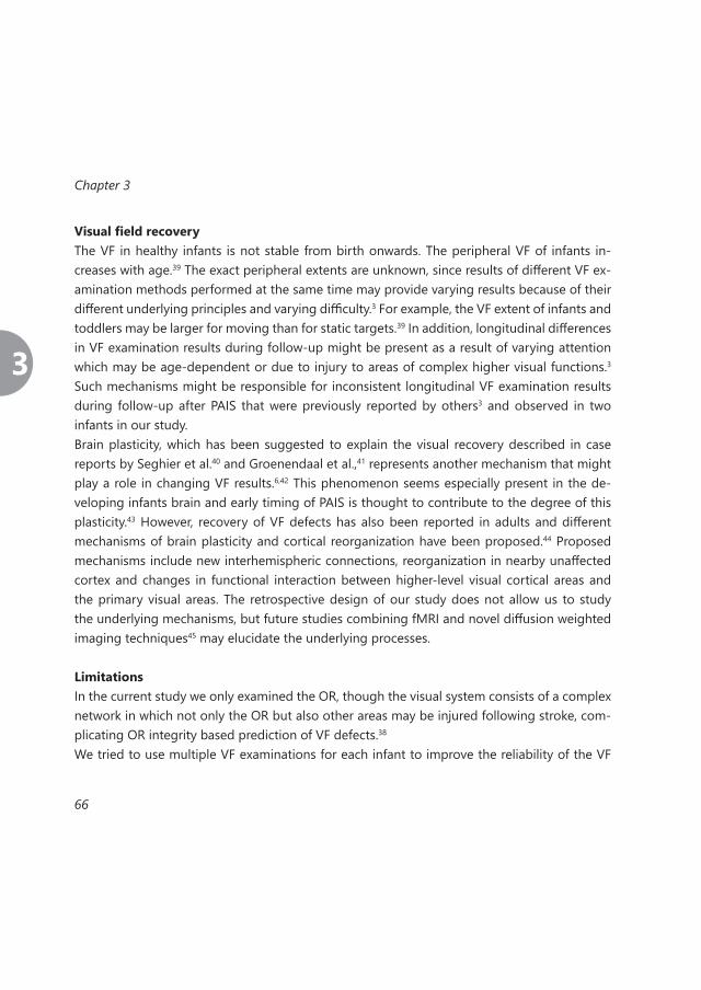
Chapter 3
66
3
Visual field recoveryThe VF in healthy infants is not stable from birth onwards. The peripheral VF of infants in-creases with age.39 The exact peripheral extents are unknown, since results of different VF ex-amination methods performed at the same time may provide varying results because of their different underlying principles and varying difficulty.3 For example, the VF extent of infants and toddlers may be larger for moving than for static targets.39 In addition, longitudinal differences in VF examination results during follow-up might be present as a result of varying attention which may be age-dependent or due to injury to areas of complex higher visual functions.3 Such mechanisms might be responsible for inconsistent longitudinal VF examination results during follow-up after PAIS that were previously reported by others3 and observed in two infants in our study. Brain plasticity, which has been suggested to explain the visual recovery described in case reports by Seghier et al.40 and Groenendaal et al.,41 represents another mechanism that might play a role in changing VF results.6,42 This phenomenon seems especially present in the de-veloping infants brain and early timing of PAIS is thought to contribute to the degree of this plasticity.43 However, recovery of VF defects has also been reported in adults and different mechanisms of brain plasticity and cortical reorganization have been proposed.44 Proposed mechanisms include new interhemispheric connections, reorganization in nearby unaffected cortex and changes in functional interaction between higher-level visual cortical areas and the primary visual areas. The retrospective design of our study does not allow us to study the underlying mechanisms, but future studies combining fMRI and novel diffusion weighted imaging techniques45 may elucidate the underlying processes.
LimitationsIn the current study we only examined the OR, though the visual system consists of a complex network in which not only the OR but also other areas may be injured following stroke, com-plicating OR integrity based prediction of VF defects.38 We tried to use multiple VF examinations for each infant to improve the reliability of the VF

Prediction of visual field defects using MRI and DTI
67
3
outcome. However, some infants were only tested once or twice and it is uncertain whether these infants with a relatively short follow up (under the age of three) would have shown im-provement of their VF on longer follow-up. The use of two different DTI sequences and the wide range in age at scan (ranging from 80 to 127 days) prevented us from using the individual FA values from the ipsilateral OR - rather than the asymmetry index - to predict VF defects. Asymmetry indices are frequently used however, and have been related to clinical outcome before.20 Another explanation for the reduced FA observed in the affected OR is a partial volume effect. It is well known that partial volume effects affect both fiber tractography and the derived DTI parameters.46 In ORs with a smaller volume due to degeneration of axons, the contribution of partial volume contaminated voxels is likely to be higher. It has been suggested to incorporate partial volume effects related factors in DTI analyses, but the sample size of the current study was too small to do so.

Chapter 3
68
3
CONCLUSION
This study shows that assessment of asymmetry of the OR on both conventional MRI and DTI-based tractography at three months of age can be used to predict VF defects after unilateral PAIS with comparable predictive values. Based on our findings, we propose that an accurate follow-up with multiple VF examinations is indicated in children with OR asymmetry on a MRI performed at three months, in order to ensure early diagnosis of VF defects and subsequent start of rehabilitation. Our current data do suggest that there is no need for visual follow-up in the presence of a symmetrical OR on conventional T1-weighted imaging, however, this finding needs to be confirmed by others before applied to clinical practice.

Prediction of visual field defects using MRI and DTI
69
3
REFERENCES
1. Van der Aa, N. E., Benders, M. J. N. L., Groenendaal, F. & de Vries, L. S. Neonatal stroke: a review of the current evidence on epidemiology, pathogenesis, diagnostics and therapeutic options. Acta Paediatr. 103, 356–64 (2014).2. Kirton, A., Deveber, G., Pontigon, A.-M., Macgregor, D. & Shroff, M. Presumed perinatal ischemic stroke: vascular classification predicts outcomes. Ann. Neurol. 63, 436–43 (2008).3. Mercuri, E. et al. Neonatal cerebral infarction and visual function at school age. Arch Dis Child 88, F487–F491 (2003).4. Sreenan, C., Bhargava, R. & Robertson, C. M. Cerebral infarction in the term newborn: clinical presen-tation and long-term outcome. J. Pediatr. 137, 351–5 (2000).5. Trauner, D., Chase, C., Walker, P. & Wulfeck, B. Neurologic Profiles of Infants and Children after Perinatal Stroke. Pediatr Neurol 1993;9:383-6. 9, 383–386 (1993).6. Van der Aa, N. E. et al. Neonatal posterior cerebral artery stroke: clinical presentation, MRI findings, and outcome. Dev. Med. Child Neurol. 55, 283–90 (2013).7. Guzzetta, A. et al. Visual function in children with hemiplegia in the first years of life. Dev. Med. Child Neurol. 43, 321–329 (2001).8. Mercuri, E. et al. Visual outcome in children with congenital hemiplegia: correlation with MRI findings. Neuropediatrics 27, 184–8 (1996).9. Van Hof-van Duin, J., Bertuccelii, B., Fazzi, B. & Maris, S. Visual outcome at 5 years ’ of newborn infants at risk of cerebral visual impairment. Dev. Med. Child Neurol. 40, 302–309 (1998).10. Dale, N. & Salt, A. Early support developmen-tal journal for children with visual impairment: the case for a new developmental framework for early interven-
tion. Child. Care. Health Dev. 33, 684–90 (2007).11. Good, W. V et al. Cortical visual impairment in children. Surv. Ophthalmol. 1738, 351–364 (1994).12. Koenraads, Y., Braun, K. P. J., van der Linden, D. C. P., Imhof, S. M. & Porro, G. L. Perimetry in Young and Neurologically Impaired Children: The Behavioral Visual Field (BEFIE) Screening Test Revisited. JAMA Ophthalmol. 133, 319–25 (2015).13. Bassi, L. et al. Probabilistic diffusion tractogra-phy of the optic radiations and visual function in preterm infants at term equivalent age. Brain 131, 573–82 (2008).14. Ramenghi, L. a et al. Neonatal neuroimaging: going beyond the pictures. Early Hum. Dev. 85, S75–7 (2009).15. Van der Aa, N. E. et al. Quantification of white matter injury following neonatal stroke with serial DTI. Pediatr. Res. 73, 756–62 (2013).16. Leemans, A. & Jones, D. K. The B-matrix must be rotated when correcting for subject motion in DTI data. Magn. Reson. Med. 61, 1336–49 (2009).17. Oishi, K. et al. Multi-contrast human neonatal brain atlas: application to normal neonate development analysis. Neuroimage 56, 8–20 (2011).18. Counsell, S. J. et al. Thalamo-cortical connec-tivity in children born preterm mapped using probabi-listic magnetic resonance tractography. Neuroimage 34, 896–904 (2007).19. Ciccarelli, O. et al. From diffusion tractography to quantitative white matter tract measures: a reproduc-ibility study. Neuroimage 18, 348–359 (2003).20. Van der Aa, N. E. et al. Does diffusion tensor imaging-based tractography at 3 months of age contrib-ute to the prediction of motor outcome after perinatal arterial ischemic stroke? Stroke. 42, 3410–4 (2011).21. World Health Organization. International Statistical Classification of Diseases and Related Health Problems 10th Revision. ICD-10 Version. (2010). at <http://

Chapter 3
70
3
apps.who.int/classifications/icd10/browse/2010/en>22. DeLong, E. R., DeLong, D. M. & Clarke-Pear-son, D. L. Comparing the areas under two or more cor-related receiver operating characteristic curves: a non-parametric approach. Biometrics 44, 837–845 (1988).23. Basser, P. J., Mattiello, J. & LeBihan, D. MR Diffusion Tensor Spectroscopy and Imaging. Biophys. J. 66, 259–267 (1994).24. Alexander, A. L., Lee, J. E., Lazar, M. & Field, A. S. Diffusion tensor imaging of the brain. Neurotherapeu-tics 4, 316–329 (2007).25. Zhang, Y., Wan, S. & Zhang, X. Geniculocal-carine Tract Disintegration after Ischemic Stroke: A Dif-fusion Tensor Imaging Study. AJNR Am J Neuroradiol 34, 1890–94 (2013).26. Groenendaal, F., Benders, M. J. & de Vries, L. S. Pre-wallerian degeneration in the neonatal brain fol-lowing perinatal cerebral hypoxia-ischemia demonstrated with MRI. Semin. Perinatol. 30, 146–50 (2006).27. Tuor, U. I. et al. Cellular correlates of longi-tudinal diffusion tensor imaging of axonal degeneration following hypoxic-ischemic cerebral infarction in neonatal rats. NeuroImage. Clin. 6, 32–42 (2014).28. Lilja, Y. & Nilsson, D. T. Strengths and lim-itations of tractography methods to identify the optic radiation for epilepsy surgery. Quant Imaging Med Surg 5, 288–299 (2015).29. Powell, H. W. R. et al. MR tractography pre-dicts visual field defects following temporal lobe resec-tion. Neurology 65, 596–599 (2005).30. Wieshmann, U. C. et al. Wallerian Degener-ation in the Optic Radiation After Temporal Lobectomy Demonstrated In Vivo with Diffusion Tensor Imaging. Epilepsia 40, 1155–1158 (1999).31. Ohno, N. et al. Alteration of the optic radia-tions using diffusion-tensor MRI in patients with retinitis pigmentosa. Br. J. Ophthalmol. 1–4 (2015).
32. Kaushik, M., Graham, S. L., Wang, C. & Klistorner, A. A topographical relationship between visual field defects and optic radiation changes in glaucoma. Invest. Ophthalmol. Vis. Sci. 55, 5770–5 (2014).33. Groppo, M. et al. Development of the optic radiations and visual function after premature birth. Cortex. 56, 30–7 (2014).34. Berman, J. I. et al. Quantitative fiber tracking analysis of the optic radiation correlated with visual performance in premature newborns. AJNR. Am. J. Neu-roradiol. 30, 120–4 (2009).35. De Moraes, C. G. Anatomy of the visual path-ways. J. Glaucoma 22 Suppl 5, S2–7 (2013).36. Iai, M., Yamamura, T. & Takashima, S. Early Expression of Proteolipid Protein in Human Fetal and Infantile Cerebri. Pediatr Neurol 17, 235–239 (1997).37. Barkovich, A., Kjos, B., Jackson, D. & Norman, D. Normal maturation of the neonatal and infant brain: MR Imaging at 1.5T. Neuroradiology 173–180 (1988).38. Govaert, P. et al. Network injury to pulvinar with neonatal arterial ischemic stroke. Neuroimage 39, 1850–7 (2008).39. Delaney, S. M. et al. Stimulus Motion Increases Measured Visual Field Extent in Children 3.5 to 30 Months of Age. Optom. Vis. Sci. 77, 82–89 (2000).40. Seghier, M. L. et al. Visual recovery after peri-natal stroke evidenced by functional and diffusion MRI: case report. BMC Neurol. 5, 17 (2005).41. Groenendaal, F. & van Hof-van Duin, J. Partial visual recovery in two fullterm infants after perinatal hypoxia. Neuropediatrics 21, 76–8 (1990).42. Guzzetta, A., Fiori, S., Scelfo, D., Conti, E. & Bancale, A. Reorganization of visual fields after periven-tricular haemorrhagic infarction: potentials and limita-tions. Dev. Med. Child Neurol. 55 Suppl 4, 23–6 (2013).43. Nelson, K. B. & Lynch, J. K. Stroke in newborn infants. Lancet Neurol 3, 150–158 (2004).

Prediction of visual field defects using MRI and DTI
71
3
44. Urbanski, M., Coubard, O. a & Bourlon, C. Visualizing the blind brain: brain imaging of visual field defects from early recovery to rehabilitation techniques. Front. Integr. Neurosci. 8, 74 (2014).45. Tuch, D. S. et al. High angular resolution diffusion imaging reveals intravoxel white matter fiber heterogeneity. Magn. Reson. Med. 48, 577–82 (2002).46. Vos, S. B., Jones, D. K., Viergever, M. a & Lee-mans, A. Partial volume effect as a hidden covariate in DTI analyses. Neuroimage 55, 1566–76 (2011).


4Visual field abnormalities in children suspected of
increased intracranial pressure
Yvonne KoenraadsKees P.J. Braun
Saskia M. ImhofGiorgio L. Porro

Chapter 4
74
4
ABSTRACT
PurposeAim of this study was to describe the ophthalmological findings, with emphasis on prevalence and type of visual field defects (VFDs), in children suspected of increased intracranial pressure (ICP).
Methods Patient files of 192 children referred to our tertiary center between August 1997 and Septem-ber 2011 because of suspicion of ICP increase were retrospectively analyzed up to September 2015. Ophthalmological findings were presented for children with and without current ICP ele-vation, differentiating between patients with and those without previous episodes of increased ICP or other pathology. Contribution of clinical characteristics at presentation in demonstrat-ing ICP elevation was explored with receiver operating characteristic analysis. Furthermore, the association between final visual outcome and clinical characteristics at presentation was investigated for children with intracranial hypertension.
ResultsOf the 142 children with normal ICP, 70% had normal VA, 41% normal fundoscopy findings, and 67% normal VF examination, in contrast with 82% , 18% , and 34% of 50 children with increased ICP respectively. Majority (46%) of VFDs in children with increased ICP consisted of blind spot enlargement, which had no diagnostic contribution to funduscopic findings (AUC 85% and 83% respectively). In children with isolated ICP elevation, both VA impairment and VFDs at presentation predicted VFDs at follow-up.

Visual field findings in increased intracranial pressure
75
4
Conclusions VF examination at presentation in children suspected of increased ICP does not differentiate between children with and those without intracranial hypertension. However, in children with ICP elevation, standard conventional perimetry (SCP) could provide arguments to start thera-py and may help to predict final VF outcome.

Chapter 4
76
4
INTRODUCTION
Optic disc swelling or papilledema is often observed at fundoscopy in children with increased intracranial pressure (ICP).1 Although papilledema in increased ICP can be objectified with flu-orescein angiography (FA), optical coherence tomography (OCT),2–6 ultrasound (USG) or orbital CT-scan, it is easily confused with other forms of optic disc swelling that are not caused by in-creased ICP, e.g. papillitis, compressive optic neuropathy or pseudopapilledema.7,8 Symptoms of ICP elevation in children are also non-specific and may mimic migraine or ophthalmological disorders with visual impairment and secondary headache. Visual loss, particularly manifesting as visual field defects (VFD), has been extensively reported as an important ophthalmological consequence of ICP elevation, mainly in idiopathic intra-cranial hypertension (IIH).1–3,6,8–30 Until VF loss is profound, patients may be unaware of these deficits.19,23,31,32 Therefore, most authors state that VF examination is essential for diagnosis and start of treatment in case of suspected ICP increase.8,17,19,26,33,34 However, interpretation of VFDs may be difficult since VFDs also occur in other ophthalmological – some of which accompa-nied with papillary abnormalities – or neurological pathology, or they may have developed during an earlier episode of increased ICP, while pressure at time of presentation is normal.8,35 Moreover, inability of children to understand the test and poor attention span may bias the VF test results with false positive findings.8,19 In addition, simplified methods developed for VF examination in young children, such as the BEFIE test, are not specifically adapted to study the consequences of ICP elevation since they only detect peripheral VFDs.36 The principal aim of this retrospective study was to describe the ophthalmological findings, with emphasis on the prevalence and type of VFDs, in all children suspected of increased ICP, seen in our center. In particular, we correlated ophthalmological findings with the eventual diagnosis – i.e. ICP elevation or not – taking into account the absence or presence of previous episodes of ICP elevation or other pathology that may influence VFs. Finally, we aimed to relate clinical characteristics and ophthalmological findings at presentation with final visual outcome of children with increased ICP.

Visual field findings in increased intracranial pressure
77
4
METHODS
Patient selectionAll children referred to our center between August 1997 and September 2011 because of sus-picion of ICP increase, who underwent standard conventional perimetry (SCP), were consec-utively included. To select these patients, medical files of 1696 children who underwent 3259 SCPs were screened using the following criteria: (1) annotations in the medical files of possibly elevated ICP, or the need to rule out ICP elevation, or (2) symptoms consistent with elevated ICP, such as headache, nausea, vomiting, transient visual obscuration or papilledema37, present around time of SCP. If a child underwent more than one SCP examination in our center, only the first examination was selected. Unreliable SCP measurements or those performed only after treatment, were excluded. The study was approved by our institutional medical ethical committee.
Data collectionDemographic and clinical characteristics were retrospectively collected from the patient files up to September 2015. SCP was performed with the Goldmann perimeter, Rodenstock Peritest38 or Humphrey Field Analyzer. Possible VFDs included enlarged blind spots (EBS) (graded as mild/severe), wide-spread defects (few/extensive), central scotomas (relative/absolute), homonymous defects (incomplete/complete quadrantanopia/hemianopia), concentric defects (mild/severe), overall sensitivity decreases (mild/severe), paracentral scotomas, nasal restrictions (or step), altitudi-nal defects (incomplete/complete), arcuate defects, or bitemporal defects.Visual acuity (VA) measurements were performed with the Kays Picture or Snellen Chart. Best corrected VA was converted to LogMAR and classified as normal (-0.2-0.1), mildly impaired (0.2-0.5), moderately impaired (0.6-0.9), severely impaired (1.0-1.3), profound (1.4-1.7) or (near-)blind (1.8-2.0).39

Chapter 4
78
4
Optic discs were assessed by means of dilated fundoscopy and classified as normal, (suspect) papilledema or optic atrophy or paleness. According to the annotations in the medical files, the appearance of (suspect) papilledema was graded (possible/mild/moderate/severe) based on the Frisén Papilledema Grading Scale.40 Children in whom it was noted in the chart that there was any uncertainty about the optic disc appearance were again examined by a more ex-perienced ophthalmologist or underwent ancillary investigation of the optic disc by means of USG, FA or OCT. Eventual interpretation of the optic disc abnormality, as noted in the medical files, was based on these findings and on additional clinical information, and was classified as ‘(chronic) papilledema’, ‘optic atrophy/paleness’, ‘idiopathic pseudopapilledema’, ‘optic disc dru-sen’, ‘tilted optic disc’, ‘hypoplastic/dysplastic optic disc’ or ‘papillitis’ based on the differential diagnosis of papilledema by Friedman.41 Findings on neuro-imaging were classified as ‘possible clue for increased ICP’, ‘doubtful clue’ or ‘other’, according to the annotations made by the radiologist. ‘Possible’ clues were categorized as ‘new findings’ or ‘abnormalities that were non-progressive over time’ and included a hy-drocephalus, cerebral venous thrombosis or cerebral tumor with mass effect, while ‘doubtful’ clues comprised a small cerebral tumor, slightly dilated ventricular system or small intracranial hemorrhage. Neuro-imaging findings classified as ‘other’ included normal scans and scans with abnormalities not related to ICP increase.
ICP categoriesWe classified patients into two categories, based on the eventual assessment of ICP, as either ‘elevated’ or ‘normal’. Children in whom ICP increase was uncertain were excluded from fur-ther analysis. Within the category ‘elevated ICP’, a subgroup was considered to suffer from ‘proven’ ICP increase, when fulfilling one or more of the following criteria: (1) cerebrospinal fluid pressure (CSFP) measurement above 30cm H2O, (2) CSFP measurement above 25cm H2O and disease course suiting ICP elevation or (3) symptoms that decreased after treatment for elevated ICP. Within the category ‘normal ICP’, children were designated as ‘proven normal’, when (1) CSFP measurement was under 15cm H2O, (2) they had another established diagnosis

Visual field findings in increased intracranial pressure
79
4
explaining symptoms and signs or (3) they had another diagnosis with a follow-up of at least one year without other signs of increased ICP.VFDs at presentation may have also been caused by focal central nervous system (CNS) pa-thology itself, or by ophthalmological diagnoses that led to the suspicion of raised ICP. We therefore presented ophthalmological findings of children with such disorders separately from those without. Furthermore, we differentiated between patients with and without previous episodes of ICP elevation.
Final visual outcomeVisual outcome at the end of follow-up, consisting of VA, optic disc and VF assessment, was collected of children with ICP elevation at presentation or in the past, but no other CNS or ophthalmological pathology. To explore possible predictive factors, the relation between final visual outcome and clinical characteristics at presentation with ICP elevation was investigated.
Statistical analysis Statistical analysis was performed using SPSS 21.0.42 ROC-curves were created to determine the differences in the area’s under the curve (AUC), representing the contribution of clinical characteristics and ophthalmological findings at presentation to establish ICP elevation. As-sociations between possible predictive factors and final visual outcome in children with ICP elevation at first examination in our center were assessed using Fischer’s exact or Mann-Whit-ney U tests.

Chapter 4
80
4
RESULTS
Patients Of all 216 children, selected out of 1696, who performed reliable SCP, 23% (n=50) were retro-spectively categorized as having increased ICP at time of SCP, while 66% (n=142) were con-sidered to have normal ICP. In 11% of children, presence or absence of ICP elevation remained inconclusive, leaving 192 children for further analysis.Diagnoses of children with increased ICP included IIH, hydrocephalus, drain dysfunction, cere-bral venous thrombosis or chronic meningitis. The group without ICP elevation (and without other pathology that may cause VFDs) included children with CSF over-drainage, refractive errors, accommodation problems, postprandial hypoglycemia, simulation disorder, tonsillitis, headache, fatigue, unexplained neurologic symptoms or visual complaints unrelated to ICP elevation, or children who turned out to have normal ICP while being investigated because of suspected papilledema, macrocephaly, ventriculomegaly, craniosynostosis or osteopetrosis. Clinical diagnoses in all separate groups are listed in the eTable.
Findings at presentationClinical symptomsBaseline characteristics of children with and without ICP elevation are presented in Table 1. Ages were comparable between groups. Complaints were relatively least frequent in children with no current nor previous ICP elevation. In most of these children without complaints the suspicion of ICP elevation arose because suspect papilledema was observed at fundosco-py. In children with current ICP elevation and a previous history of intracranial hypertension, complaints were most often (67%) acute (<6 wks), while they were least often (16%) acute in children without current, but with previous ICP elevation. Complaints consisted mainly of headache, except in children without ICP elevation but with other (VF influencing) pathology (41%), in whom visual complaints were more frequent (76%).

Visual field findings in increased intracranial pressure
81
4
ICP elevation No ICP elevation
No other (focal CNS or
ophthalmological) pathology
Benign/idiopathic intracranial hyper-
tension (n=23), Chronic sterile
meningitis (n=1), Hydrocephalus (n=2), Cerebral
venous thrombo-sis (n=3).
Accommodation problems (n=3), Chronic fa-tigue syndrome (n=1), Craniosynostosis with-out elevated ICP (n=2), Physiologic macro-
cephaly (n=4), Physiologic ventriculomegaly (n=1), Idiopathic pseudopapilledema (n=21), Headache not related to elevated ICP (n=33),
Osteopetrosis without elevated ICP (n=1), Postprandial hypoglycemia (n=1), Refractive error (n=3), Unexplained visual complaints
(n=5).
Previous ICP elevation
Benign/idiopathic intracranial hy-
pertension (n=4), Drain dysfunction (n=4), Hydroceph-
alus (n=2).
Fatigue (n=2), Headache not related to el-evated ICP (n=14), Over-drainage (n=2),
Refractive error (n=1), Simulation (n=1), Ton-sillitis (n=1), Unexplained visual complaints
(n=4).
Focal CNS or ophthalmological
pathology
Benign/idiopathic intracranial hy-pertension (also
panuveitis) (n=1), intracranial hem-
orrhage (n=1), Drain dysfunction (also manifesta-tions of tuberous sclerosis in brain and eye) (n=1), cerebral tumor
(n=8).
Check-up after traumatic brain injury (n=1), Cone dysfunction (n=1), Diffuse encephalopa-thy with focal lesions on MRI (n=1),Headache not related to elevated ICP (but also optic disc
atrophy or previous cerebral tumor) (n=5), Hypoplastic/dysplastic optic disc (n=4), Optic atrophy (n=4), Optic disc drusen (n=8), Optic neuritis/papillitis (n=9), Optic pathway glioma (n=1), Refractive error (but also hypoplastic/
dysplastic optic disc, optic disc atrophy or tilted disc) (n=3), Tilted disc (n=1), Unex-
plained neurologic symptoms (but previous intracranial hemorrhage) (n=1), Unexplained visual complaints (but also optic disc atrophy
or cone dysfunction) (n=3).
eTable. Clinical diagnoses of children suspected of ICP increase

Chapter 4
82
4
1
ICP increase (n=50) median [range] or % (n)
Focal CNS or ophthalmological pathology
- (n=39)
+ (n=11)
Previous ICP elevation - (n=29)
+ (n=10)
+/- (n=1/10)
Age at first episode of ICP elevation (yrs)
11.5 [5.4-17.6]
2.4 [0.0-17.1]
12.2 [5.1-16.9]
Sex (male) 38% (11/29) 30% (3/10) 64% (7/11) Age, yrs 11.4 [5.3-17.6] 14.0 [5.2-17.8] 12.2 [5.1-16.9] COMPLAINTS Duration, wks Acute (< 6 wks) Type Headache Visual complaint Nausea/vomiting Cognitive problems Fatigue Other
97% (28/29) 13.8 [0.6-584.2]
43% (12/28)
79% (22/28) 36% (10/28) 39% (11/28) 7% (2/28)
11% (3/28) 29% (8/28)
90% (9/10) 3.8 [0.2-22.3]
67% (6/9)
78% (7/9) 33% (3/9) 33% (3/9) 11% (1/9)
- 22% (2/9)
100% (11) 8.4 [1.4-20.0] 45% (5/11)
73% (8/11) 55% (6/11) 55% (6/11) 18% (2/11)
- 55% (6/11)
NEUROIMAGING CLUES Possible1, new finding
Possible1, non-progressive Doubtful2
21% (6/28) 4% (1/28) 7% (2/28)
50% (5/10) 10% (1/10)
-
18% (2/11) 9% (1/11)
73% (7/11) ICP CLASSIFICATION Proven elevated ICP4
Proven normal ICP5
97% (28/29)
-
100% (9/9)
-
91% (10/11)
-
Table 1. Baseline characteristics of 192 children suspected of ICP increase
ICP, intracranial pressure; CNS, central nervous system; VF, visual field; 1 Possible clue for ICPsystem, cerebral venous thrombosis or cerebral tumor with mass effect; 2 Doubtful clue for ICPmor, slightly dilated ventricular system or intracranial hemorrhage; 3physiologically enlarged surement above 30cm H2O, (2) a CSFP measurement above 25cm H2O and a disease coursecreased after treatment for elevated ICP; 5 Proven normal ICP, (1) CSFP measurement was un-ing symptoms and signs or (3) another diagnosis with a follow-up of at least one year without

Visual field findings in increased intracranial pressure
83
4
Visual acuityVA was normal in 70% (95/135) of children with normal ICP and in 82% (37/45) of children with increased ICP (see Table 2). In the group without ICP elevation but with other CNS or ophthalmological pathol-ogy, VA was impaired most often (49%), corresponding to the higher percentage of visual complaints.
Funduscopic findingsFindings at fundoscopy were normal in 41% (58/142) of children with normal ICP and in 18% (9/49) of children with increased ICP. Suspect papill-edema was present in ~75% of children with ICP elevation (without other pathology), in ~90% of children with in-creased ICP and other pa-thology, and in up to 60% of children who were eventually diagnosed with normal ICP. Presence and severity of (sus-
1
ICP normal (n=142) median [range] or % (n)
- (n=100)
+ (n=42)
- (n=75)
+ (n=25)
+/- (n=6/36)
-
0.3 [0.0-15.7]
n=5 5.2 [0.1-14.3]
32% (24/75) 28% (7/25) 45% (19/42) 11.3 [5.4-18.0] 13.7 [6.8-16.8] 10.7 [6.7-17.6] 73% (55/75)
14.3 [0.0-560.0] 25% (14/55)
80% (44/55) 47% (26/55) 20% (11/55) 7% (4/55)
13% (7/55) 20% (11/55)
100% (25/25) 15.6 [1.1-82.0]
16% (4/25)
84% (21/25) 28% (7/25) 24% (6/25) 4% (1/25)
12% (3/25) 20% (5/25)
88% (37/42) 7.9 [0.0-273.3] 46% (17/37)
41% (15/37) 76% (28/37) 11% (4/37) 5% (2/37) 3% (1/37)
24% (9/37)
9% (3/32)3 6% (2/32)3 3% (1/32)3
4% (1/23)3 9% (2/23)
13% (3/23)
-
3% (1/29) 14% (4/29)
-
71% (53/75)
-
92% (23/25)
-
81% (34/42)
elevation on neuroimaging, such as a dilated ventricular elevation on neuroimaging, such as a small cerebral tu-ventricular system; 4 Proven elevated ICP, (1) a CSFP mea-suiting ICP elevation or (3) symptoms and/or signs that de-der 15cm H2O, (2) another established diagnosis explain-other signs of increased ICP.

Chapter 4
84
4
pect) papilledema in each subcategory are shown in eFigure 1. ICP was assessed as elevated in only 35% (39/112) of children with (suspect) papilledema. In 21% (23/112) of children, the assumed papilledema was eventually – after ancillary investigations or a second look – diag-nosed as optic disc drusen (n=8), tilted optic disc (n=2), hypoplastic/dysplastic optic disc (n=5) or papillitis (n=8). In the remaining 45% (50/112) of children with (suspect) papilledema, ICP was assessed to be normal and the observed papilledema was assumed to be chronic (n=6) or classified as idiopathic pseudopapilledema (n=44). An overview of final optic disc classifi-cations is given in Figure 1.
Visual field examinationSCP was performed with the Rodenstock Peritest (95%), Goldmann perimeter (1%) or Hum-phrey Field Analyzer (5%). Results were normal in 67% (95/142) of children with normal ICP
Table 2. Visual acuity impairment among all children suspected of ICP increaseICP elevation No ICP elevation
No other (focal CNS or
ophthalmological) pathology
14% (4/28)*3 mildly [2]
*1 severely [1]
19% (13/70)*10 mildly [2]
*3 mild and moderately [3]
Previous ICP elevation
22% (2/9)*2 mildly [1]
29% (7/24)*7 mildly [4]
Focal CNS or ophthalmological
pathology
25% (2/8)*1 mildly [1]
*1 nearly blind [0]
49% (20/41)*12 mildly [4]
*8 moderately to blind [6]
Percentage (n) and severity of visual acuity (VA) impairment in each subcategory of children with and without ICP elevation [number of children in which both eyes are affected]ICP, intracranial pressure; CNS, central nervous system.

Visual field findings in increased intracranial pressure
85
4
eFigure 1. Presence and severity of (suspect) papilledema at fundoscopy in all children suspected of ICP increase

Chapter 4
86
4
Figure 1. Optic disc classifications of all children suspected of ICP increase

Visual field findings in increased intracranial pressure
87
4
and in 34% (17/50) of children with increased ICP. In children with a first episode of ICP el-evation, VFDs were present in 72% (see Figure 2). In this group, VFDs mainly consisted of EBS (55%). In some children widespread defects (21%), central scotoma (14%), homonymous defect (3%) or concentric VFD (3%) were reported. In both groups with previous ICP elevation (with and without current ICP elevation) VFDs occurred in approximately one third of children. Remarkably, 17% (13/75) of children with normal ICP without previous ICP elevation and no other pathology (that may cause VFDs) still had VFDs. These children had idiopathic pseudo-papilledema (n=5), headache unrelated to ICP elevation (n=2) or both (n=6), and showed EBS (n=7), widespread defects (n=4), a nasal restriction with a peripheral inferior defect (n=1) and a peripheral superior defect with a homonymous component (n=1) on SCP. The latter was a coincidental finding without presence of any neurologic abnormality on neuroimaging.EBS also occurred in 11 children with papillitis (n=4), optic drusen (n=3), optic atrophy (n=2) or previous ICP elevation (n=2). In addition, other kinds of VFDs were seen in children with these (n=19) and other (n=9) pathologies.
While complaints, VA and VFDs other than EBS were aspecific, the presence and severity of (suspect) papilledema and EBS on SCP were more distinctive for having ICP elevation. To inves-tigate whether the finding of an EBS on SCP contributes to the assessment of the optic disc in the diagnosis of increased ICP, the predicted probabilities of these parameters were combined in a ROC-curve (see eFigure 2). This analysis revealed that adding results of EBS assessment on SCP to the fundoscopy findings did not essentially change the AUC (83% [95%CI 0.75-0.90] and 85% [95%CI 0.78-0.91] respectively).
Visual outcome of children after elevated ICPOf all 64 children with current or previous ICP elevation without other pathology, 51 underwent ophthalmological follow-up (final median age 15.8, range 6.2-28.0 yrs). Of the 50 children with multiple VA and optic disc assessments, 86% had a normal and 12% a mildly impaired final VA. One child with IIH seemed (near-)blind but was suspected of conversion disorder, based on VEP and eye-tracker examinations. Final assessment of the optic discs revealed (possible)
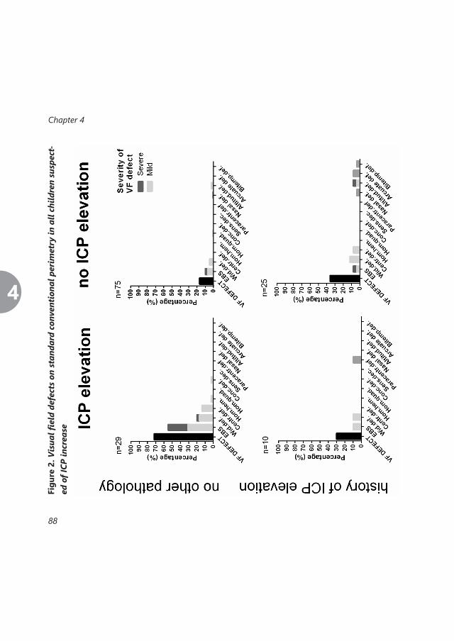
Chapter 4
88
4
Figu
re 2
. Vis
ual fi
eld
defe
cts
on s
tand
ard
conv
enti
onal
per
imet
ry in
all
child
ren
susp
ect-
ed o
f ICP
incr
ease
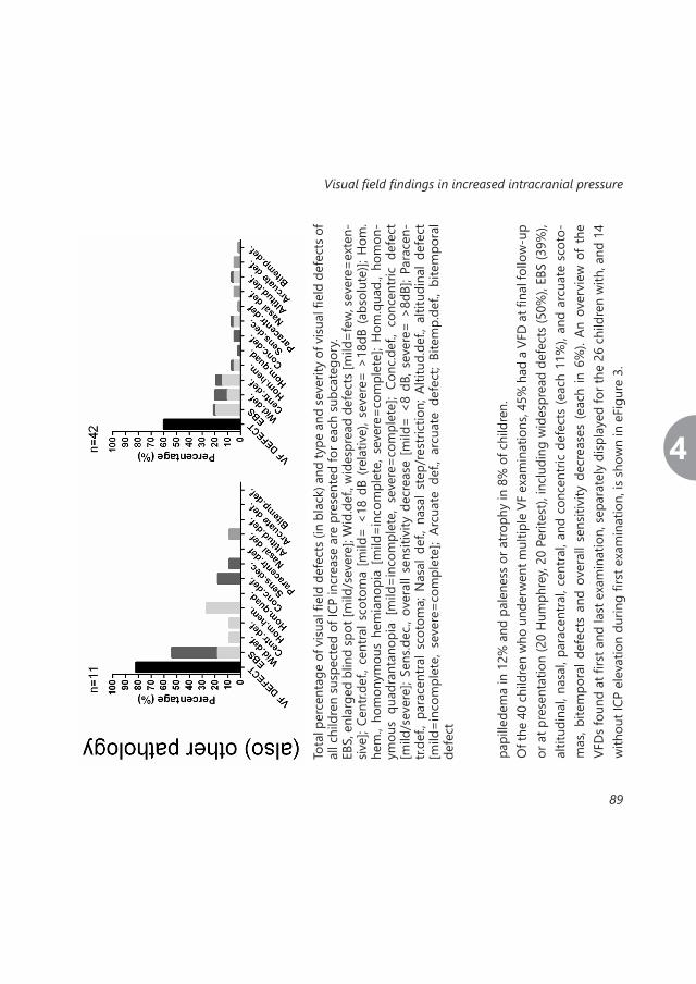
Visual field findings in increased intracranial pressure
89
4pa
pille
dem
a in
12%
and
pal
enes
s or a
troph
y in
8%
of c
hild
ren.
Of t
he 4
0 ch
ildre
n w
ho u
nder
wen
t mul
tiple
VF
exam
inat
ions
, 45%
had
a V
FD a
t fina
l fol
low
-up
or a
t pre
sent
atio
n (2
0 H
umph
rey,
20 P
erite
st),
incl
udin
g w
ides
prea
d de
fect
s (50
%),
EBS
(39%
), al
titud
inal
, nas
al, p
arac
entra
l, ce
ntra
l, an
d co
ncen
tric
defe
cts
(eac
h 11
%),
and
arcu
ate
scot
o-m
as, b
item
pora
l def
ects
and
ove
rall
sens
itivi
ty d
ecre
ases
(ea
ch in
6%
). An
ove
rvie
w o
f th
e VF
Ds f
ound
at fi
rst a
nd la
st e
xam
inat
ion,
sepa
rate
ly d
ispla
yed
for t
he 2
6 ch
ildre
n w
ith, a
nd 1
4 w
ithou
t ICP
ele
vatio
n du
ring
first
exa
min
atio
n, is
show
n in
eFi
gure
3.
Tota
l per
cent
age
of v
isual
fiel
d de
fect
s (in
bla
ck) a
nd ty
pe a
nd se
verit
y of
visu
al fi
eld
defe
cts o
f al
l chi
ldre
n su
spec
ted
of IC
P in
crea
se a
re p
rese
nted
for e
ach
subc
ateg
ory.
EBS,
enl
arge
d bl
ind
spot
[mild
/sev
ere]
; Wid
.def
., wid
espr
ead
defe
cts [
mild
=few
, sev
ere=
exte
n-siv
e]; C
entr.
def.,
cent
ral s
coto
ma
[mild
= <1
8 dB
(rel
ativ
e), s
ever
e= >
18dB
(abs
olut
e)];
Hom
.he
m.,
hom
onym
ous
hem
iano
pia
[mild
=inc
ompl
ete,
sev
ere=
com
plet
e]; H
om.q
uad.
, hom
on-
ymou
s qu
adra
ntan
opia
[m
ild=i
ncom
plet
e, s
ever
e=co
mpl
ete]
; Co
nc.d
ef.,
conc
entri
c de
fect
[m
ild/s
ever
e]; S
ens.d
ec.,
over
all s
ensit
ivity
dec
reas
e [m
ild=
<8 d
B, s
ever
e= >
8dB]
; Par
acen
-tr.
def.,
para
cent
ral s
coto
ma;
Nas
al d
ef.,
nasa
l ste
p/re
stric
tion;
Alti
tud.
def.,
altit
udin
al d
efec
t [m
ild=i
ncom
plet
e, s
ever
e=co
mpl
ete]
; Ar
cuat
e de
f., ar
cuat
e de
fect
; Bi
tem
p.de
f., bi
tem
pora
l de
fect

Chapter 4
90
4
eFig
ure
3. V
F de
fect
s at
firs
t and
last
exa
min
atio
n of
chi
ldre
n w
ith
ICP
elev
atio
n w
itho
ut
othe
r pa
thol
ogy
who
had
a S
CP f
ollo
w-u
p, s
epar
atel
y di
spla
yed
for
child
ren
wit
h, a
nd
thos
e w
itho
ut IC
P el
evat
ion
duri
ng fi
rst e
xam
inat
ion

Visual field findings in increased intracranial pressure
91
4
Tota
l per
cent
age
of v
isual
fiel
d de
fect
s (in
bla
ck) a
nd ty
pe a
nd se
verit
y of
visu
al fi
eld
defe
cts.
EBS,
enl
arge
d bl
ind
spot
[mild
/sev
ere]
; Wid
.def
., wid
espr
ead
defe
cts [
mild
=few
, sev
ere=
exte
n-siv
e]; C
entr.
def.,
cent
ral s
coto
ma
[mild
= <1
8 dB
(rel
ativ
e), s
ever
e= >
18dB
(abs
olut
e)];
Hom
.he
m.,
hom
onym
ous
hem
iano
pia
[mild
=inc
ompl
ete,
sev
ere=
com
plet
e]; H
om.q
uad.
, hom
on-
ymou
s qu
adra
ntan
opia
[m
ild=i
ncom
plet
e, s
ever
e=co
mpl
ete]
; Co
nc.d
ef.,
conc
entri
c de
fect
[m
ild/s
ever
e]; S
ens.d
ec.,
over
all s
ensit
ivity
dec
reas
e [m
ild=
<8 d
B, s
ever
e= >
8dB]
; Par
acen
-tr.
def.,
para
cent
ral s
coto
ma;
Nas
al d
ef.,
nasa
l ste
p/re
stric
tion;
Alti
tud.
def.,
altit
udin
al d
efec
t [m
ild=i
ncom
plet
e, s
ever
e=co
mpl
ete]
; Ar
cuat
e de
f., ar
cuat
e de
fect
; Bi
tem
p.de
f., bi
tem
pora
l de
fect
eFig
ure
2. R
ecei
ver
Ope
rati
ng C
hara
cter
isti
c cu
rve
of t
he c
ontr
ibut
ion
of o
ptic
dis
cas
sess
men
t and
blin
d sp
ot e
nlar
gem
ent o
n SC
P to
dem
onst
rate
ICP
elev
atio
n

Chapter 4
92
4
Table 3. Association between characteristics at presentation and visual outcome at final examination in children with ICP elevation
1
Median [range] in yrs or n (%)
Final VF defect 12/26 children
p-value Final VA impairment 5/30 children
p-value
Sex Male Female
3 (33%) 9 (53%)
0.43
2 (20%) 3 (15%)
1.00
Pathology Hydrocephalus BIH/IIH Other
2 (50%) 8 (42%) 2 (67%)
0.83 2 (33%) 2 (10%) 1 (33%)
0.17
Age at first ICP elevation Whole VF group 10.4 [0.0-17.6] Whole VA group 10.4 [0.0-17.6]
11.4 [0.0-17.6] 0.60 11.9 [0.0-17.6] 0.87
Age at presentation Whole VF group 11.2 [5.2-17.6] Whole VA group 11.5 [5.2-17.8]
11.4 [5.3-17.6] 0.35 12.1 [11.4-17.8] 0.10
VA at presentation Normal Impaired
8 (36%) 4 (100%)
0.03
1 (4%) 4 (67%)
0.003
Optic disc at presentation Normal Abnormal
1 (25%) 11 (50%)
0.60
1 (17%) 4 (17%)
1.00
VF at presentation Normal VF defect
2 (20%) 10 (63%)
0.05 2 (20%) 3 (15%)
1.00
Recurrences No Yes
7 (44%) 5 (50%)
1.00 0 (0%) 5 (45%)
0.003
VF, visual field; VA, visual acuity; VA impairment, worse than 0.1 LogMAR in one or two eyes; BIH/IIH, benign or idiopathic intracranial hypertension; Other, other kinds of underlying pa-thology including cerebral venous thrombosis, cranium deformations, intracranial hemorrhag-es or inflammation; presentation, first examination in our center at the moment of ICP eleva-tion; Recurrences, recurrences between presentation and final visit.

Visual field findings in increased intracranial pressure
93
4
All children with final VA impairment also had VFDs if SCP was performed. The association be-tween characteristics at presentation and the final presence of a VFD or VA impairment was in-vestigated in the children with ICP elevation during first examination (see Table 3). VFDs at final examination were associated with VA impairment and the presence of a VFD at presentation, while VA impairment at final examination was associated with VA impairment and recurrent episodes of increased ICP between first and last examination. Remarkably, ICP recurrences did not seem to influence final presence or absence of a VFD.

Chapter 4
94
4
DISCUSSION
This study describes the ophthalmological findings in 192 children suspected of ICP elevation, with emphasis on the prevalence and type of VFDs. Of 142 patients with normal ICP 41% had normal funduscopic and 67% normal VF findings, in contrast to 18% and 34% of 50 children with increased ICP respectively. Only 35% of children with suspected papilledema were diag-nosed with increased ICP. Eventual follow-up of children with documented intracranial hyper-tension revealed a normal VA in 86%, mildly impaired VA in 12%, and VFDs in 45% of patients. SCP findings were rarely distinctive for ICP elevation, while they might – together with VA findings – predict final VF outcome in children with ICP elevation at presentation. VFDs in children with ICP elevation include deficits secondary to (1) optic nerve compression with axoplasmic flow stasis in papilledema8,19 such as EBS17 and glaucomatous-like nerve fiber layer (NFL) defects,2,8,11,18–28 or (2) compression of the retrochiasmatic pathways (e.g. in hydro-cephalus) which may provoke homonymous VFDs.30 In this study we noticed that in children with ICP elevation VFDs at presentation mainly consisted of EBS while other VFDs were scarce and also occurred in children with other pathology than ICP elevation.EBS presumably arises secondary to a refractive scotoma, caused by displacement of the peri-papillary rods and cones by swollen axons, and could confirm papilledema.2,8,17 However, our ROC analysis revealed that it did not significantly contribute to optic disc assessment in chil-dren suspected of ICP elevation. Corresponding to previous reports, EBS in our study was not present in all children with papilledema, while other children, who were referred for the evaluation of possibly increased ICP because of suspected papilledema but were eventually diagnosed with other papillary abnormalities, could also have an EBS.2,19,27
Glaucomatous-like NFL defects may also occur in an early stage of ICP elevation and probably result from mechanical compression and ischemic damage, which provokes paracentral, nasal, altitudinal and arcuate VFDs.18,20,27 Surprisingly, these VFDs were rarely seen at presentation in patients with ICP elevation while they did occur in children without ICP elevation but with previous ICP elevation, papillitis, cone dysfunction or optic atrophy. Possibly the NFL was not

Visual field findings in increased intracranial pressure
95
4
yet damaged at the moment of presentation in children with ICP elevation. Alternatively the Peritest, used in most children because of feasibility reasons, may have a lower sensitivity to pick up these kind of VFDs compared to Humphrey VF testing. VFDs that were present at presentation in some children with ICP elevation included concen-tric and (relative) central VFDs, known as late findings caused by expanding edema, but also homonymous and widespread defects.2,8,18,19,24,27,29 Corresponding to the literature, all these VFDs were not specific for increased ICP, being also detected in children without ICP elevation.At the end of follow-up most children had a favorable VA but VFDs were regularly present, especially in children who had an impaired visual function (VA or VF defects) at presentation. In contrast, Gospe et al.43 recently showed that mainly the grade of papilledema was highly predictive for (permanent) VA or VF loss in 31 children with IIH.In contrast to the finding of Pollak et al.44 that adult IIH patients with recurrent ICP elevation showed more VFDs than those without, we found that (1) recurrent episodes of ICP increase seemed not associated with the presence of VFDs at the end of follow-up, and (2) VFDs at presentation in children with increased ICP occurred less often in children with previous epi-sodes of ICP elevation, compared to those who had their first episode. A possible explanation, supported by the short duration of symptoms in most children with previous ICP increase, may be that an increased awareness in these patients has resulted in earlier diagnosis of their recurrent ICP elevation.Our study mainly focused on VF measurements, as perimetry is one of the oldest methods used in the assessment of ICP elevation54 and VFDs are considered one of the most important consequences of ICP elevation.1–3,6,9–16,19,26,27,46,47 However, in current ophthalmological practice various alternative methods to objectify papilledema, assess the presence of ICP increase, and monitor disease progression, are often used. These methods include assessment of leakage and disc fluorescence on FA, optic nerve sheath diameter on USG48 or CT scan of the orbits and peripapillary and perimacular retinal NFL thickness on OCT.2–6 While USG examination of the optic disc is crucial to rule out optic neuropathies such as optic disc drusen,8 OCT might monitor disease progression in children who cannot reliably perform SCP.6
Chapter 4
96
4
Limitations of our study included a possible selection bias due to exclusion of children with uncertain ICP, unreliable SCPs, or those who performed SCP after start of treatment. In addi-tion, the classification used was based on eventual ICP assessment, which could not guarantee the demonstration or exclusion of actual ICP elevation at time of SCP in all cases. Finally, since it was impossible in the group of children classified with ‘idiopathic pseudopapilledema’ to distinguish between false positive or true pseudopapilledema, some children with pseudo-papilledema and secondary VFDs might have been mistakenly classified in the group without other pathology.

Visual field findings in increased intracranial pressure
97
4
CONCLUSIONS
SCP findings in children rarely contribute to the diagnostic work-up at moment of first evalu-ation in case of suspected ICP elevation. In our series, EBS had no additional diagnostic value to the presence of (suspect) papilledema, while other VFDs were scarce and should not be confused with those attributable to other or former pathology. The current study shows that SCP should not necessarily be used for diagnostic purposes in daily ophthalmological practice when increased ICP is suspected in children. The presence of VFDs in children with ICP ele-vation may, however, provide arguments to initiate therapy and may help to predict a child’s final VF outcome.

Chapter 4
98
4
REFERENCES
1. Soiberman, U. et al. Idiopathic intracranial hypertension in children: visual outcome and risk of recurrence. Childs. Nerv. Syst. 27, 1913–8 (2011).2. Kosmorsky, G. S. Idiopathic Intracranial Hyper-tension : Pseudotumor Cerebri. Headache Curr. 389–393 (2014).3. Dagi, L. R. et al. Using spectral-domain op-tical coherence tomography to detect optic neuropathy in patients with craniosynostosis. J. Am. Assoc. Pediatr. Ophthalmol. Strabismus 18, 543–549 (2014).4. Marzoli, S. B. et al. Quantitative analysis of op-tic nerve damage in idiopathic intracranial hypertension (IIH) at diagnosis. Neurol. Sci. 34 Suppl 1, S143–5 (2013).5. Kuper-Smith, M. J. OCT Sub-Study Commit-tee for the NORDIC Idiopathic Intracranial Hypertension Study Group. Baseline OCT Measurements in the Idio-pathic Intracranial Hypertension Treatment Trial, Part I: Quality Control, Comparisons, and Variability. Invest. Ophthalmol. Vis. Sci. 55, 8180–8188 (2014).6. Chern, J. J., Tubbs, R. S., Gordon, A. S., Don-nithorne, K. J. & Oakes, W. J. Management of pediatric patients with pseudotumor cerebri. Childs. Nerv. Syst. 28, 575–8 (2012).7. Jung, J. J., Baek, S.-H. & Kim, U. S. Analysis of the causes of optic disc swelling. Korean J. Ophthalmol. 25, 33–6 (2011).8. Friedman, D. I. Papilledema and Idiopathic Intracranial Hypertension. 857–876 (2014).9. Lessell S. Pediatric pseudotumor cerebri (id-iopathic intracranial hypertension). Surv Ophthalmol. 37, 155–66 (1992).10. Ko, M. W. & Liu, G. T. Pediatric idiopathic intracranial hypertension (pseudotumor cerebri). Horm. Res. pædiatrics 74, 381–9 (2010).11. Cinciripini, G. S., Donahue, S. & Borchert, M. S.
Idiopathic intracranial hypertension in prepubertal pe-diatric patients: characteristics, treatment, and outcome. Am. J. Ophthalmol. 127, 178–82 (1999).12. Salman, M. S., Kirkham, F. J. & Macgregor, D. L. Idiopathic ‘Benign’ Intracranial Hypertension: Case Series and Review. J Child Neurol. 16, 465–470 (2001).13. Kesler, a. & Fattal-Valevski, a. Idiopathic Intra-cranial Hypertension in the Pediatric Population. J. Child Neurol. 17, 745–748 (2002).14. Phillips, P. H., Repka, M. X. & Lambert, S. R. Pseudotumor cerebri in children. J. AAPOS 2, 33–8 (1998).15. Ravid, S., Shahar, E., Schif, A. & Yehudian, S. Visual Outcome and Recurrence Rate in Children With Idiopathic Intracranial Hypertension. J. Child Neurol. 5–9 (2015). 16. Wolf, A. & Hutcheson, K. a. Advances in evalu-ation and management of pediatric idiopathic intracranial hypertension. Curr. Opin. Ophthalmol. 19, 391–7 (2008).17. Wakerley, B. R., Tan, M. H. & Ting, E. Y. Idio-pathic intracranial hypertension. Cephalalgia 35, 248–61 (2015).18. Sergott, R. C. Headaches associated with papilledema. Curr. Pain Headache Rep. 16, 354–8 (2012).19. Wall, M. The importance of visual field testing in idiopathic intracranial hypertension. Continuum (N. Y). 20, 1067–74 (2014).20. Keltner, J. L., Johnson, C. a, Cello, K. E. & Wall, M. Baseline Visual Field Findings in the Idiopathic Intra-cranial Hypertension Treatment Trial (IIHTT). Invest. Oph-thalmol. Vis. Sci. 55, 3200–7 (2014).21. Weisberg, L. A. Benign Intracranial Hyperten-sion. Med. (Baltimore). 54, 197–207 (1975).22. Lysak, W. R. & Svien, H. J. Long-term fol-low-up on patients with diagnosis of pseudotumor cere-bri. J. Neurosurg. 25, 284–287 (1966).23. Bruce, B. B., Biousse, V. & Newman, N. J. Update on idiopathic intracranial hypertension. Am. J. Ophthalmol. 152, 163–169 (2011).

Visual field findings in increased intracranial pressure
99
4
24. Rangwala, L. M. & Liu, G. T. Pediatric idio-pathic intracranial hypertension. Surv. Ophthalmol. 52, 597–617 (2007).25. Wall, M. et al. The Idiopathic Intracranial Hypertension Treatment Trial. JAMA Neurol. 1091, 1–9 (2014).26. Thurtell, M. J. & Wall, M. Idiopathic intracra-nial hypertension (Pseudotumor Cerebri): Recognition, treatment, and ongoing management. Curr. Treat. Options Neurol. 15, 1–12 (2013).27. Rowe, F. J. Assessment of visual function in idiopathic intracranial hypertension. Br. J. Neurosurg. 25, 45–54 (2011).28. Digre, K. B. et al. A comparison of idiopathic intracranial hypertension with and without papilledema. Headache 49, 185–193 (2009).29. Rudolph, D. et al. Visual field constriction in children with shunt-treated hydrocephalus. J. Neurosurg. Pediatr. 6, 481–5 (2010).30. Andersson, S. et al. Vision in children with hydrocephalus. Dev. Med. Child Neurol. 48, 836–41 (2006).31. Yri, H. M., Wegener, M., Sander, B. & Jensen, R. Idiopathic intracranial hypertension is not benign: a long-term outcome study. J. Neurol. 259, 886–94 (2012).32. Standridge, S. M. Idiopathic intracranial hy-pertension in children: a review and algorithm. Pediatr. Neurol. 43, 377–90 (2010).33. Mukherjee, N. & Bhatti, M. T. Update on the surgical management of idiopathic intracranial hyperten-sion. Curr. Neurol. Neurosci. Rep. 14, 438 (2014).34. Jirásková, N. & Rozsíval, P. Idiopathic intracra-nial hypertension in pediatric patients. Clin. Ophthalmol. 2, 723–726 (2008).35. Tibussek, D. et al. Clinical spectrum of the pseudotumor cerebri complex in children. Childs. Nerv. Syst. 26, 313–21 (2010).36. Koenraads, Y., Braun, K. P. J., van der Linden,
D. C. P., Imhof, S. M. & Porro, G. L. Perimetry in Young and Neurologically Impaired Children: The Behavioral Visual Field (BEFIE) Screening Test Revisited. JAMA Ophthalmol. 133, 319–25 (2015).37. Smith, J. L. Whence Pseudotumor Cerebri? J. Clin. Neuro-ophthalmol. 5, 55–56 (1985).38. Greve, E. L., Dannheim, F. & Bakker, D. The Peritest, a new automatic and semi-automatic perimeter. Int. Ophthalmol. 5, 201–14 (1982).39. World Health Organization. International Statistical Classification of Diseases and Related Health Problems 10th Revision. ICD-10 Version. (2010). at <http://apps.who.int/classifications/icd10/browse/2010/en>40. Frisén, L. Swelling of the optic nerve head: a staging scheme. J. Neurol. Neurosurg. Psychiatry 45, 13–18 (1982).41. Olson, J. Walsh and Hoyt’s Clinical Neu-ro-Ophthalmology. Br. J. Ophthalmol. 83, 1096 (1999).42. IBM Corp. Released 2012. IBM SPSS Statistics for Windows, Version 21.0. Armonk, NY: IBM Corp.43. Gospe, S. M., Bhatti, M. T. & El-Dairi, M. a. Anatomic and visual function outcomes in paediatric idio-pathic intracranial hypertension. Br. J. Ophthalmol. [Epub ahead of print] (2015). 44. Pollak, L., Zohar, E., Glovinsky, Y. & Huna-Bar-on, R. Reevaluation of presentation and course of idio-pathic intracranial hypertension--a large cohort compre-hensive study. Acta Neurol. Scand. 127, 406–12 (2013).45. Baker, R. S., Baumann, R. J. & Buncic, J. R. Idio-pathic intracranial hypertension (pseudotumor cerebri) in pediatric patients. Pediatr. Neurol. 5, 5–11 (1989).46. Babikian P, Corbett J, B. W. Idiopathic intracra-nial hypertension in children: the Iowa experience. J Child Neurol. 9, 144– (1994).47. Rowe, F. J. & Sarkies, N. J. Assessment of visual function in idiopathic intracranial hypertension: a pro-spective study. Eye (Lond). 12 ( Pt 1), 111–8 (1998).
48. Newman, W. D., Hollman, a S., Dutton, G. N. & Carachi, R. Measurement of optic nerve sheath diameter by ultrasound: a means of detecting acute raised intra-cranial pressure in hydrocephalus. Br. J. Ophthalmol. 86, 1109–13 (2002).

5Visual outcome in Sturge-Weber syndrome:
a systematic review and Dutch multicenter cohort
Yvonne KoenraadsMichelle B. van Egmond-Ebbeling
Joke H. de BoerSaskia M. Imhof
Kees P.J. BraunGiorgio L. Porro
on behalf of the SWS study group:Nomdo M. Jansonius
Carlien A. BennebroekNicoline E. Schalij-Delfos
Chris C.M. NielsenMirjan van Dijk
Wouter B.H.J. Vehmeijer
Accepted for publication in Acta Ophthalmologica

Chapter 5
102
5
ABSTRACT
Purpose Visual functions in Sturge-Weber syndrome (SWS) may be impaired by glaucoma, diffuse cho-roidal hemangioma (DCH) or leptomeningeal angioma. Aim of this study was to gain better insight in the visual deficits of SWS patients.
Methods A systematic literature search using Pubmed and Embase medical databases was performed to identify articles describing visual acuity (VA) and/or visual field (VF) findings in SWS patients. In addition, a Dutch multicenter cohort with 33 SWS patients was collected and the combined results of VA and VF findings are presented.
Results VA results of 25 studies and VF results of 12 studies were suitable for data-extraction. Descrip-tion of the combination of both VA and VF findings was scarce. Homonymous hemianopia (HH) was present in 42% of SWS patients. Seventy per cent of eyes had a (near)normal vision, while VA of eyes with glaucoma or DCH was severely impaired in 28% and 67%, respectively. In the Dutch cohort only 18% (6/33) of patients had (near)normal findings of both visual pa-rameters. In addition, half of the patients with glaucoma suffered from a combination of a HH and VA impairment.
Conclusions Although SWS patients are exposed to severe functional visual impairment due to the possible cumulative consequences of glaucoma, DCH and cerebral injury, description of the combina-tion of both VA and VF results is scarce in the literature. Particularly the combination of visual impairment due to glaucoma or DCH, and HH might be invalidating.

Visual outcome in Sturge-Weber syndrome
103
5
INTRODUCTION
The Sturge-Weber syndrome (SWS) is a rare congenital disorder with angiomatous vascular malformations in the meninges, dermis and eye, affecting one in 20,000-50,000 people.1,2 In the literature, three types of SWS are described.3 SWS type I, on which this study is mainly focused, includes a facial port-wine stain and leptomeningeal angioma with possible ocular abnormalities such as glaucoma. SWS type II comprises a facial port-wine stain (with possible glaucoma) without evident cerebral involvement, while SWS type III consists of an exclusive leptomeningeal angioma. The most visible sign of SWS is the port-wine birthmark which typically affects the ophthalmic division of the trigeminal nerve.1 The leptomeningeal angioma, mainly unilateral and located in the occipital or posterior parietal lobe, may initially be unnoticed.1,4,5 However, during the first years of life, impaired venous outflow may lead to progressive ischemic cerebral damage, atrophy and calcification in the brain region below the angioma.1,6 These cerebral manifes-tations, which can be confirmed by neuroimaging (CT or MRI), regularly cause neurological problems such as seizures (72-97%), motor deficits, cognitive decline and visual field (VF) defects.7,8
Ocular vascular abnormalities include hemangioma of the conjunctiva, episclera, iris (heter-ochromia), ciliary body, choroid and retina.2,9,10 A diffuse choroidal hemangioma develops in approximately one-third of SWS patients and may cause visual loss from hyperopia, amblyo-pia, secondary exudative retinal detachment or cystoid macular edema.11–13 The most common eye disease in SWS is uni- or bilateral glaucoma which occurs in 30-70% of patients.2,14–16 It may present around birth (60%) secondary to trabecular dysgenesis inducing buphthalmos, or develop in child- or adulthood (40%) due to increased episcleral venous pressure, and may result in VF and visual acuity (VA) impairment. 2,5,14,16–19
Most studies on SWS are reported from a strictly ophthalmological perspective, focusing on diffuse choroidal hemangioma (DCH) or glaucoma and mostly reporting only data of VA out-comes, or a neurological perspective, focusing on homonymous VF defects secondary to ce-

Chapter 5
104
5
rebral involvement. However, accurate description of the consequences of SWS-related com-bined ocular and neurological disorders, such as glaucoma and leptomeningeal angioma, for VF and VA is lacking in the current literature. Collection of information on visual functioning of SWS patients may encounter difficulties, since patients are often young and sometimes mentally retarded. Especially examination of the VFs, which may be impaired both in case of glaucoma and occipital leptomeningeal angioma, is very challenging in this patient population and therefore may have to be omitted.The aim of the present study is to gain better insight in the visual prognosis of SWS patients, first by providing a systematic review of visual outcome, as previously described in the litera-ture, and second by presenting a Dutch multicenter cohort with focus on the combination of VA and VF findings.
Search terms used in Pubmed and EmbaseDomain ((((((“sturge weber”[Title/Abstract]) OR “encephalotrigeminal angio-
matosis”[Title/Abstract]) OR “encephalo trigeminal angiomatosis”[Ti-tle/Abstract]) OR “sturge kalischer weber”[Title/Abstract]) OR “menin-go oculo facial”[Title/Abstract])) AND
Outcome ((((((((((((((((((((((((((((((Visual) OR Ophthalmology) OR Ophthalmologic) OR Ophthalmologics) OR Ophthalmological) OR Ophthalmological-ly) OR Ophthalmologist) OR Ophthalmologists) OR Ophthalmic) OR Ophthalmics) OR Ophthalmical) OR Ophthalmically) OR Visually) OR Eye) OR Eyes) OR Vision) OR Ocular) OR Optic) OR Optics) OR Opti-cal) OR Optically) OR Perimetry) OR Perimetric) OR Perimetrics) OR Perimetrical) OR Perimetrically) OR Octopus) OR Goldmann) OR Hum-phrey) OR Rodenstock)
eTable 1. Syntax for search on visual outcome in Sturge-Weber syndrome in medical databases

Visual outcome in Sturge-Weber syndrome
105
5
METHODS
Systematic review of the literatureSearch strategy and selectionOn June 19th 2015, a structured literature search was performed using Pubmed and Embase medical databases. A syntax was designed to search for visual outcome in SWS by combining a series of synonyms (see eTable 1). After eliminating duplicates, titles and abstracts were inde-pendently screened by two authors (ME and YK) on predefined inclusion and exclusion criteria (see Figure 1). All articles that included SWS patients and might describe visual function were included. Exclusion criteria were (1) population without SWS patients, (2) description of visual outcome highly improbable, (3) non-original research (reviews) or abstracts of a conference, (4) case reports or series of less than 4 patients or (5) language other than English, Dutch, German, French, Spanish, Portuguese, Italian or Russian.Subsequently, of all selected articles, full text was screened on the presence of a description of VA or VF examination in patients with SWS. Doubtful cases were discussed until consensus was reached by the two authors.
Quality assessment and data extractionThe selected articles were independently appraised by the two authors on different aspects of validity for the purpose of this study (see eTable 2). Regarding the domain, neuroimaging confirmation of the diagnosis (type I or III SWS) and way of selection of patients with SWS was assessed. Regarding the VA or VF outcome, description of the method of examination, description of the outcome itself and percentage of missing data were assessed. Discrepancies were resolved by consensus discussion. Studies in which (1) the validity of the outcome was assessed as ‘poor’ (red) on all three items, (2) only a mean score for the outcome was given, or (3) less than four VF or VA results were reported, were not used for the data-extraction. VA results of all reported eyes were presented of the studies that described general SWS popu-lations. In addition, VA results of eyes affected with glaucoma or a DCH were described of all

Chapter 5
106
5
Figure 1. Flowchart of search strategy
Search strategy: 'Sturge-Weber Syndrome' AND 'Visual outcome'
Pubmed
n = 380
Embase
n = 530
Total
n = 910
After deduplication
n = 584
After screening title/abstract
n = 97
After screening full text
Screening full text Records excluded (56) * Other domain (12)
* Other outcome (44)
n = 41
Screening title/abstract Records excluded (487)
* Other domain (56) * Other outcome (62)
* Non-original research (70) * Case reports/series (278)
* Language (21)

Visual outcome in Sturge-Weber syndrome
107
5
studies that also – or solely – reported results of children with these disorders separately. In the presentation of the VF results, studies that used standard perimetry and confrontational methods were separated because standard perimetry is more sensitive for glaucomatous de-fects then confrontational methods.
Dutch multicenter cohortSubjectsSummer 2015, we approached all ophthalmologists of the Dutch Pediatric Ophthalmology Society to cooperate in this study to collect data from as many as possible Dutch SWS patients. All available neuroimaging-confirmed SWS patients who visited a society member Dutch oph-thalmologist were included.The study was approved by the institutional ethical committee of the University Medical Cen-ter Utrecht, which concluded that the Dutch Medical Research Involving Human Subjects Act did not apply and written informed consent was not needed, since the study was confined to a retrospective, anonymized data collection.
Data collectionAll patient files were retrospectively analyzed. Demographic and clinical characteristics col-lected consisted of gender, presence of a facial port-wine stain, location of leptomeningeal vascular malformation, age of last ophthalmological examination, final VA, findings on final VF, presence of ophthalmological morbidity and treatment, presence of epilepsy and its treat-ment, developmental delay, hemiparesis or other relevant comorbidity.The use of a formal definition of glaucoma, based on optic disc appearance and glaucomatous VF defects, would result in a high percentage of underdiagnosis or misclassification in this group of patients. Therefore, we categorized patients as having glaucoma if they had, either medical or surgical, glaucoma treatment. VA measurements were performed with the Kays Picture or Snellen Chart, depending on the age and cooperation of the child. Best corrected VA was transformed to LogMAR (Logarithm of the Minimum Angle of Resolution) and cate-

Chapter 5
108
5
eTab
le 2
. Cri
tica
l app
rais
al o
f art
icle
s on
vis
ual o
utco
me
in S
turg
e-W
eber
syn
drom
e

Visual outcome in Sturge-Weber syndrome
109
5

Chapter 5
110
5
gorized as (near)normal (≤0.30 LogMAR), moderately impaired (>0.30 - <1.00 LogMAR) or severely impaired (≥1.0 LogMAR). VF measurements were performed by means of confrontational measurement during neuro-logical examination, behavioral confrontational peripheral VF testing with Stycar balls or the BEFIE test20, automatic static VF testing with Frequency Doubling Technology (FDT) perimetry, manual kinetic VF testing on the Goldmann perimeter, (semi-)automatic static VF testing on the Rodenstock Peritest or automatic static VF testing on the Humphrey Field Analyzer. VF defects were categorized as normal, homonymous quadrantanopia or hemianopia, glaucoma-tous defects, concentric defect, sensitivity decrease or a combination of defects.

Visual outcome in Sturge-Weber syndrome
111
5
RESULTS
Systematic review of the literatureIn total, 41 articles were retrieved that described VA (n=20)2,9,11–13,17,19,21–33, VF (n=12)7,8,34–43 or both (n=9)4,5,44–50 assessments in patients with SWS (see eTable 2). Articles reported general SWS populations (n=18)4,5,8,21,34–36,38,39,41–47,49,50 or only selected cohorts of patients who suffered from a specific SWS-related disorder, such as glaucoma (n=12),2,9,17,19,22,23,25,28,29,32,33,48 epilep-sy (n=3)7,37,40 or a diffuse choroidal hemangioma (DCH) (n=8).11–13,24,26,27,30,31 In addition, from 12 of the ‘general SWS’ articles, outcomes of patients with glaucoma (n=3),45,46,49 epilepsy (n=4)4,34,35,42 or from both subcategories (n=5)21,36,38,39,44 could be retrieved separately.Many studies, particularly those on SWS-related glaucoma or DCH, included patients who underwent a specific treatment or diagnostic examination. Ten articles mentioned confirma-tion of the SWS diagnosis by means of neuroimaging (see eTable 2). Methods for VA (n=10) or VF (n=3) assessment were properly described in 13 articles and were derivable from the VA (n=16) or VF (n=2) result in 16 articles. The quality of the description of VA and VF results itself and the percentage of missing data in the outcome varied strongly between studies.Results of VA (n=3)4,5,44 or VF (n=3)5,40,48 measurements of five studies were not used for da-ta-extraction since validity of the visual outcome for the purpose of the systematic review – i.e. method, description, missing data – were all assessed as ‘poor’ (red). The VF results of one article could not be further evaluated since solely an overall mean score was given.7 Five studies were withdrawn from data-extraction since from less than four patients the VA results (n=1) 47 or VF results (n=4)47,41,49,43,50 were reported. Therefore, VA results of 25 stud-ies2,9,11–13,17,19,21–33,45,46,48–50 (see Figure 2) and VF results of 12 studies4,8,34–39,42,44–46 (see Table 1) were suitable for data-extraction.Of the four articles that described VA results from general SWS populations,21,45,49,50 the pooled percentage of all reported eyes with a (near)normal VA was 70% (see Figure 2). The presence of moderate and severely impaired VA could be pooled from three articles and were found to occur in 12% and 14% of eyes, respectively.49,50,21 VA of eyes affected by glaucoma or DCH was severely impaired in 28% and 67%, respectively.

Chapter 5
112
5
Figu
re 2
. Vis
ual a
cuit
y ou
tcom
e of
stu
dies
on
Stur
ge-W
eber
syn
drom
e
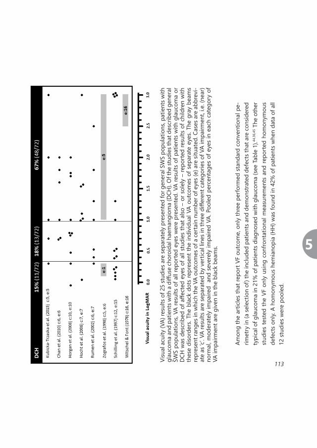
Visual outcome in Sturge-Weber syndrome
113
5Am
ong
the
artic
les
that
repo
rt V
F ou
tcom
e, o
nly
thre
e pe
rform
ed s
tand
ard
conv
entio
nal p
e-rim
etry
in (a
sele
ctio
n of
) the
incl
uded
pat
ient
s and
dem
onst
rate
d de
fect
s tha
t are
con
sider
ed
typi
cal o
f gla
ucom
a in
21%
of p
atie
nts d
iagn
osed
with
gla
ucom
a (s
ee Ta
ble
1).44
,36,
45 T
he o
ther
st
udie
s te
sted
the
VF o
nly
usin
g co
nfro
ntat
iona
l mea
sure
men
ts a
nd re
port
ed h
omon
ymou
s de
fect
s on
ly. A
hom
onym
ous
hem
iano
pia
(HH
) was
foun
d in
42%
of p
atie
nts
whe
n da
ta o
f all
12 st
udie
s wer
e po
oled
.
Visu
al a
cuity
(VA)
resu
lts o
f 25
stud
ies a
re se
para
tely
pre
sent
ed fo
r gen
eral
SW
S po
pula
tions
, pat
ient
s with
gl
auco
ma
and
patie
nts w
ith a
diff
use
chor
oida
l hae
man
giom
a (D
CH).
Of t
he st
udie
s tha
t des
crib
ed g
ener
al
SWS
popu
latio
ns, V
A re
sults
of a
ll re
port
ed e
yes
wer
e pr
esen
ted.
VA
resu
lts o
f pat
ient
s w
ith g
lauc
oma
or
DCH
was
des
crib
ed o
f affe
cted
eye
s of
all
stud
ies
that
also
– o
r sol
ely
– re
port
ed re
sults
of c
hild
ren
with
th
ese
diso
rder
s. Th
e bl
ack
dots
repr
esen
t the
indi
vidu
al V
A ou
tcom
es o
f sep
arat
e ey
es. T
he g
ray
beam
s re
pres
ent r
ange
s in
whi
ch th
e VA
out
com
es o
f a c
erta
in n
umbe
r of e
yes (
e) a
re si
tuat
ed. C
ases
are
abb
revi
-at
e as
‘c’. V
A re
sults
are
sepa
rate
d by
ver
tical
line
s in
thre
e di
ffere
nt c
ateg
orie
s of V
A im
pairm
ent,
i.e. (
near
)no
rmal
, mod
erat
ely
impa
ired
and
sev
erel
y im
paire
d VA
. Poo
led
perc
enta
ges
of e
yes
in e
ach
cate
gory
of
VA im
pairm
ent a
re g
iven
in th
e bl
ack
beam
s.

Chapter 5
114
5
Table 1. Visual field outcome of studies on Sturge-Weber syndrome
a, number of complete hemianopias are given in brackets for all articles that made distinction between incomplete and complete homonymous hemianopias; b, not sure if all patients were tested with standard perimetry; c, all patients tested with Goldmann perimetry; d, patients were tested with a simple behavioral kinetic method; e, in 4 patients the presence of a VF defect was uncertain; f, uncertain whether patients were tested confrontatively or with standard perimetry.
Article
Cases (glaucoma)
Homonymous hemianopia (complete)a
Glaucomatous defect
Arcu
ate
defe
ct
Nas
al fi
eld
loss
Enla
rged
bl
ind
spot
Standard perimetry
Jagtap et al. (2013)b 10 (9) 2 < ---------- 3 ---------- > Parsa (2013)b 14 (5) 7 1 Sharan et al. (2009)c 14 (14) 5 1 1
Pooled (glaucoma patients)
38 (28)
21% (6/28) Confrontational
Jeong et al. (2015) 10 7 Lance et al. (2014) 12 6 Lance et al. (2013) 35 10 (3) Zabel et al. (2010) 4 (2) 2 (1) Batista et al. (2007)d 17 (6) 12e Hatfield et al. (2007) 14 (4) 9 (7) Kelley et al. (2005) 18 (7) 8 Van Emelen et al. (2000)f 19 (8) 1 Uram & Zubillaga (1982)f 25 (11) 11
Pooled (all patients)
192
42% (80/192)

Visual outcome in Sturge-Weber syndrome
115
5
From five articles, the relation between the location of the leptomeningeal angioma and the presence of a VF defect could be retrieved. In total, 16 of 47 (34%) patients reported in these studies had a normal VF despite occipital involvement.4,8,36–38
A history of epilepsy surgery was reported in 3-27% of patients in five (other) studies.2,44,48,45,42 The authors of the study by Jagtap et al.44 mentioned that the VF defects in patients after ep-ilepsy surgery were pre-existent in the two patients that could be tested. However, the study by Arzimanoglou et al.40, describing a SWS cohort in which solely patients who underwent epilepsy surgery were included, reported that 3 of 14 patients who underwent a resection of the angiomatous cortex acquired a VF defect. Of note, the occipital angiomatous lesion in one patient was not removed to avoid a VF defect.
Only few authors described the combination of VA and VF impairments that may occur in SWS. Jeong et al.4 reported that one child (age 3 yrs) had a homonymous left field deficit in combi-nation with impaired right eye VA due to glaucoma. Awad et al.47 and Stokes et al.49 reported in total three patients with glaucomatous arcuate scotomas, of which two had a normal VA and one an unilaterally impaired VA of 1.3 LogMAR. Jagtap et al.44 mentioned that five patients suf-fered from impairment of both VA and VF. Batista et al.37 correlated homonymous field defects to the visual cortex glucose metabolism in a neuroradiological study and reported six patients affected by glaucoma without a severely impaired VA.
Dutch multicenter cohort In total, data from 33 SWS patients with neuroimaging confirmation (type I or III) could be collected from our (n=19) and other centers (n=14) in the Netherlands. Baseline characteristics of patients are listed in Table 2.Visual outcome results including both VA and VF are presented in Table 3. VA was (near)normal in 72%, moderately impaired in 20% and severely impaired in 8% of the 50 eyes that under-went a VA assessment. VA was moderately or severely impaired in 44% (7/16) and 19% (3/16) of glaucomatous eyes that were tested, and in 50% (4/8) and 25% (2/8) of eyes with a DCH, respectively. A retinal detachment occurred in 38% (3/8) of the eyes in which a DCH was re-

Chapter 5
116
5
ported. Of the 15 patients who un-derwent one or multiple surgical interventions for glaucoma, none had an intraoperative or postoper-ative suprachoroidal hemorrhage.VF examination was performed with the Humphrey Field Analyz-er in 17% (5/30), the Rodenstock Peritest in 23% (7/30), the Gold-mann perimeter in 7% (2/30), FDT in 3% (1/30), the BEFIE test in 33% (10/30) 20, the Stycar balls in 3% (1/30) and confrontational meth-ods during neurological exam-ination in 13% (4/30) of patients. In three patients VF findings were unknown. Possible glaucomatous VF de-fects [arcuate scotoma on Perit-est (n=1), few inferior defects on FDT (n=1), superior constriction on Goldmann (n=1), VF defect exceeding the vertical meridian of the HH on Humphrey (n=1), partial defect of the nasal inferi-or quadrant on Humphrey (n=1) VF testing] were found in 5 of 14 (36%) patients with glaucoma who were tested with Humphrey (n=2),
Table 2. Baseline characteristics
total (n), number of patients of whom data was available; FU, follow-up; DCH, diffuse choroidal hemangioma.
total (n)
n (%) or median [range]
Gender, male 33 16 (48) Center -Utrecht -Groningen -Leiden -Amsterdam -Nijmegen
33 19 (58) 7 (21) 3 (9) 3 (9) 1 (3)
Age last FU, yrs 33 9 [3-26] Facial port-wine stain -unilateral -bilateral
32 26 (81) 21 (81) 5 (19)
DCH -unilateral -bilateral
30 6 (20) 4 (67) 2 (33)
Glaucoma -unilateral -bilateral
29 16 (55) 12 (75) 4 (25)
Leptomeningeal angioma -bilateral -left -right Number of lobes affected -frontal -temporal -parietal -occipital
33
30
33 (100) 3 (9)
12 (36) 18 (55) 2 [1-4] 14 (47) 14 (47) 17 (57) 22 (73)
Epilepsy 32 30 (94) Neurosurgery -hemispherectomy -focal resection
30 11 (37) 7 (64) 4 (36)
Developmental delay 29 21 (72) Hemiparesis 30 16 (53)

Visual outcome in Sturge-Weber syndrome
117
5
Table 3. Visual acuity and visual field outcome of patients with Sturge-Weber syndrome
Best eye
≥0.3 <1.0 - >0.3 ≤1.0 no VA available
Wor
st e
ye
≥0.
3
39% (13/33)
<1.
0 -
>0.
3 18% (6/33)
3% (1/33)
≤1.
0
9% (3/33)
3% (1/33)
no V
A
avai
labl
e 3% (1/33)
3% (1/33)
21% (7/33)
VA, visual acuity in LogMAR; VF, visual field; , normal VF; , few VF defects; , arcuate
scotoma; , superior constriction; , unilateral inferior sensitivity decrease; ,
homonymous quadrantanopia; , homonymous hemianopia (HH); , HH and unilateral VF
defect exceeding the vertical meridian; , HH and unilateral partial defect of the nasal inferior
quadrant; , HH and unilateral concentric restriction; , bilateral concentric VF; , no VF
assessment; DCH, diffuse choroidal haemangioma; blue, glaucoma patient; red, patient with DCH;
purple, patient with glaucoma and DCH.

Chapter 5
118
5
Peritest (n=2), Goldmann (n=2), FDT (n=1), BEFIE (n=3), Stycar (n=1) or confrontational (n=3) VF examination. However, the latter three defects might also have been a result of the DCH present in these eyes, while the superior constriction also could be explained by the ptosis on that side. In addition, in one of the other three patients with DCH (without glaucoma), an inferior sensitivity decrease was found on Humphrey VF testing.A HH was present in 57% (17/30) and quadrantanopia in 3% (1/30) of patients. These 18 patients with homonymous VF defects underwent hemispherectomy (n=7) or suffered from an occipital involvement of the leptomeningeal angioma (n=10) or an occipital cyst (n=1). However, 38% (8/21) of all patients with occipital involvement who underwent VF examination did not show any homonymous defect. In six of the seven patients who underwent epilepsy surgery and performed a pre-operative VF assessment, the VF defect was already present before surgery. One patient had a normal VF before undergoing a hemispherectomy which caused a HH.In two patients who suffered from hydrocephalus and were treated with a ventriculo-perito-neal shunt, no VF defects other than a HH were found. These children, however, underwent BEFIE and confrontational VF testing only, which are not sensitive enough for the detection of VF defects resulting from elevated intracranial pressure.Of the seven patients who used the antiepileptic drug Vigabatrin, one had a bilateral concen-tric defect on BEFIE VF testing. Finally, an unilateral concentric restriction on Goldmann VF testing was found in a patient with DCH and amblyopia in that eye.
In total, half (12/24) of patients who underwent VA assessment of both the right and the left eye, had a bilaterally (near)normal VA. Half of these 12 patients had a homonymous VF defect. Patients with a bilaterally impaired or unilaterally severely impaired VA all had glaucoma in combination with a HH (if VF testing was performed). These six patients represent 46% (6/13) of the total number of glaucoma patients who underwent VA assessment.One patient (8 yrs) with congenital glaucoma and a HH in whom VA assessment was not pos-sible due to developmental delay, was dyskinetic, confined to a wheelchair and only able to communicate using an eye-controlled computer.

Visual outcome in Sturge-Weber syndrome
119
5
DISCUSSION
This study describes the visual outcome of patients with SWS, who regularly suffer from a combination of vision impairing disorders such as glaucoma, DCH and cerebral injury. The study comprises first a systematic review, with the aim to extract data from articles describing visual acuity (VA) and/or visual field (VF) findings in SWS patients, and second an analysis of a Dutch multicenter cohort of SWS patients with collected and combined results of VA and VF findings. The systematic review of previous studies revealed that 1) a homonymous hemiano-pia (HH) was present in 42% of SWS patients, 2) 70% of eyes had a (near)normal vision, while 3) VA of eyes with glaucoma or DCH was severely impaired in 28% and 67%, respectively. In addition, when focusing on the combination of VA and VF findings in the Dutch cohort, we found that only 18% (6/33) of patients had (near)normal findings of both parameters, while half of the patients with glaucoma suffered from a combination of a HH and VA impairment.Description of the combination of VA and VF findings was scarce in the articles included in the systematic review. Ophthalmological studies mainly reported data on VA while neurological studies mainly reported data on VF as part of the neurological examination. In addition, visual parameters such as VA and VF were frequently not the primary outcome, which might partly explain the poor description of VA or VF methods and outcome in several studies. However, as demonstrated in the Dutch cohort, the combination of VA and VF impairment due to mul-tifactorial pathology including glaucoma, DCH and a leptomeningeal angioma in the occipital cortex could be expected in SWS patients and may be very invalidating. In addition, because SWS children often suffer from multiple other disabilities, such as hemiplegia and intellectual disability, vision might be very important for them.The pathophysiological mechanisms that underlie the SWS-related visual disorder determine its clinical approach, including potential treatment, with the aim to preserve visual function. Glaucoma is related to both the presence of a DCH and a port-wine stain on the eyelids and has a bimodal onset.2,5,48 Bilateral and contralateral glaucoma with unilateral facial port-wine stain has been reported.3 Congenital glaucoma may be treated with trabeculotomy or goniot-

Chapter 5
120
5
omy, while older children are controlled with medications or filtering procedures.16,17,51 Khitri et al.22 found that VA outcome of glaucoma related to SWS was more favorable than that of other secondary glaucoma diagnoses. Regarding complications of glaucoma surgery, most of the articles included in the systematic review reported no intraoperative complications, while rele-vant intra- and postoperative complications such as suprachoroidal hemorrhages (0-13%) 19,47, retinal detachment (10-12%) 2,19,25, cataract (7-11%) 47,25, or late endophthalmitis (2%) 2 were reported in only few studies. Although in the Dutch cohort no suprachoroidal hemorrhages occurred during surgery and this type of complications was rarely reported in the studies included in the systematic review, the risk of serious complications after glaucoma surgery in SWS patients, especially when a DCH is present, should not be ignored. Symptoms from the DCH itself usually appear in the second decade of life.26 DCH can be treat-ed using laser photocoagulation, transpupillary thermotherapy, cryotherapy, radiation therapy or photodynamic therapy.12,31 DCH particularly impairs VA, but may also provoke VF defects, metamorphopsia, floaters and progressive hypermetropia.26 A leptomeningeal angioma may cause a progressive VF defect due to a chronic impairment of cerebral bloodflow, leading to atrophy and calcifications, which is exacerbated by seizures.52
In patients with SWS, visual function is often difficult to determine, which may explain the high percentage of missing data in most of the studies included in the systematic review. Pascual-Castroviejo et al.5, Awad et al.47, Taylor et al.48 and Rosenbaum et al.41 explicitly state that reliable testing of particularly VF, but also VA, was difficult or impossible in many patients because of various problems, such as young ages, low VA, photophobia or cognitive impair-ment. This explains why seven different methods were used to assess the VF field in the Dutch cohort. The use of different VF examination methods in our cohort, but also in the studies included in the systematic review, makes it particularly difficult to demonstrate glaucomatous VF defects, since these are often more subtle and therefore require standard conventional perimetry to be detected.

Visual outcome in Sturge-Weber syndrome
121
5
Beside the high percentage of missing data and the different VF examination techniques used, two other important factors might have caused bias in the results of the systematic review. First, many patients, mainly included in the studies on glaucoma and DCH, were selected for a specific treatment, which might have resulted in a selection towards the more severe cases with possibly worse outcomes. Second, the type of SWS was frequently unknown due to the lack of information on neuroimaging and cerebral manifestations. This, and the uncertainty regarding the number of patients that underwent VF examination in several studies, probably resulted in an underestimation of the overall percentage of VF defects in the systematic review. The higher proportion of patients with homonymous VF defects in the Dutch cohort (60%), compared to the systematic review cohort (42%), may not only be explained by a more con-sistent use of VF examination, but also by a selection bias towards SWS patients affected more often by cerebral involvement with epilepsy, particularly those undergoing epilepsy surgery. This is explained by the fact that our center serves as a national tertiary referral center for children with refractory epilepsy and epilepsy surgery. Findings of VA examinations, however, were remarkably similar between the systematic review and the Dutch multicenter patient cohorts.
The studies included in the systematic review and the findings of the Dutch cohort reveal that VF defects are detected in two-third of patients with occipital involvement. This finding underlines the necessity of VF examination especially when an occipital leptomeningeal an-gioma is present. Moreover, in patients who are possible candidates for epilepsy surgery it is essential to evaluate the pre-existence of a VF defect. However, in most of the SWS patients who underwent epilepsy surgery a VF defect already existed before surgery. The functional consequences of a possible deterioration of VF following epilepsy surgery in SWS, especially when also glaucoma is present, should be carefully considered when counselling patients and caregivers who face surgery.

Chapter 5
122
5
CONCLUSIONS
Description of VA and VF abnormalities – and particularly the combination thereof – in SWS patients is scarce in the literature. SWS patients may be exposed to severe functional visual impairment due to the possible combination of glaucoma, DCH and cerebral injury. Although VA is generally preserved in SWS patients at presentation, both glaucoma and DCH are asso-ciated with worse VA. In addition, the combination with a VF defect, mainly a HH, which was found to be present in about half of SWS patients, might be invalidating. Visual outcome in SWS is heterogeneous, with some patients having a favorable outcome while others suffer from severe visual impairment by a combination of VA and VF deficits. Since the progressive character of glaucoma, DCH, and brain pathology may deteriorate visual function during life-time, we recommend an adequate visual follow-up of these patients, with VF examinations as early as possible, at least with a confrontational method, alongside the regular ophthalmolog-ical follow-up of glaucoma and DCH. Furthermore, the presence or absence of homonymous defects should be re-examined when standard conventional perimetry becomes possible with ageing, or in case of suspicion of progressive cerebral injury based on neuro-imaging findings or symptoms.
AcknowledgementsWe thank Jan van Trier and dr. Nienke Soeters for acquiring all articles that were not retrievable via our institutional library.

Visual outcome in Sturge-Weber syndrome
123
5
REFERENCES
1. Thomas-Sohl, K. a, Vaslow, D. F. & Maria, B. L. Sturge-Weber syndrome: A review. Pediatr. Neurol. 30, 303–310 (2004).2. Sullivan, T., Clarke, M. & Morin, J. The ocular manifestations of the Sturge-Weber syndrome. J Pediatr Ophthalmol Strabismus 29, 349–356 (1992).3. Hoyt, C. S. & Taylor, D. Pediatric Ophthalmolo-gy and Strabismus. Fouth Edition. (2013).4. Jeong, J.-W., Tiwari, V. N., Shin, J., Chugani, H. T. & Juhász, C. Assessment of brain damage and plasticity in the visual system due to early occipital lesion: Compar-ison of FDG-PET with diffusion MRI tractography. J. Magn. Reson. Imaging 41, 431–8 (2015).5. Pascual-Castroviejo, I., Pascual-Pascual, S., Velazquez-Fragua, R. & Viaño, J. Sturge-Weber syndrome: study of 55 patients. Can J Neurol Sci. 35, 301–7 (2008).6. Martí-Bonmatí, L., Menor, F. & Mulas, F. The Sturge-Weber syndrome: correlation between the clinical status and radiological CT and MRI findings. Childs Nerv Syst. 9, 107–109 (1993).7. Kossoff, E. H., Ferenc, L. & Comi, A. M. An in-fantile-onset, severe, yet sporadic seizure pattern is com-mon in Sturge-Weber syndrome. Epilepsia 50, 2154–7 (2009).8. Zabel, T. A. et al. Neuropsychological features and risk factors in children with Sturge-Weber syndrome: four case reports. Clin. Neuropsychol. 24, 841–59 (2010).9. Wagner, R. S., Caputo, A. R., Negro, R. G. del & Neigel, J. Trabeculectomy with Cyclocryotherapy for Infantile Glaucoma in the Sturge-Weber Syndrome. Ann Ophthalmol 20, 289–295 (1988).10. Basler, L. & Sowka, J. Sturge-Weber syndrome and glaucoma. Optometry 82, 306–309 (2011).11. Kubicka-Trząska, A., Karska-Basta, I., Oleksy, P. & Romanowska-Dixon, B. Management of diffuse choroidal hemangioma in Sturge-Weber syndrome with
Ruthenium-106 plaque radiotherapy. Graefes Arch Clin Exp Ophthalmol 253, 2015–9 (2015).12. Chan, R. V. P. et al. Proton beam irradiation using a light-field technique for the treatment of choroi-dal hemangiomas. Ophthalmologica. 224, 209–16 (2010).13. Witschel, H. & Font, R. L. Hemangioma of the Choroid. A Clinicopathologic Study of 71 Cases and a review of the Literature. Surv Ophthalmol. 20, 415–431 (1976).14. Font, R. & Ferry, A. The phakomatoses. Int Ophthalmol Clin. 12, 1–50 (1972).15. Sujansky, E. & Conradi, S. Sturge-Weber syndrome: age of onset of seizures and glaucoma and the prognosis for affected children. J Child Neurol. 10, 49–58 (1995).16. Iwach, A., Hoskins, H., Hetherington, J. & Shaffer, R. Analysis of surgical and medical management of glaucoma in Sturge-Weber syndrome. Ophthalmology. 97, 904–9 (1990).17. Mandal, A. K. Primary combined Trabecu-lotomy – Trabeculectomy for Early- onset Glaucoma in Sturge-Weber Syndrome. Ophthalmology 106, 1621–1627 (1999).18. Phelps, C. D. The pathogenesis of glaucoma in Sturge-Weber syndrome. Ophthalmology 85, 276–286 (1978).19. Budenz, D. L., Sakamoto, D., Eliezer, R., Varma, R. & Heuer, D. K. Two-staged Baerveldt Glaucoma Implant for Childhood Glaucoma Associated with Sturge-Weber Syndrome. Ophthalmology 107, 2105–2110 (2000).20. Koenraads, Y., Braun, K. P. J., van der Linden, D. C. P., Imhof, S. M. & Porro, G. L. Perimetry in Young and Neurologically Impaired Children: The Behavioral Visual Field (BEFIE) Screening Test Revisited. JAMA Ophthalmol. 133, 319–25 (2015).21. Conway, M. & Hosking, S. L. Investigation of Ocular Hemodynamics in Sturge-Weber Syndrome. 89, 922–928 (2012).

Chapter 5
124
5
22. Khitri, M. R., Mills, M. D., Ying, G.-S., Davidson, S. L. & Quinn, G. E. Visual acuity outcomes in pediatric glaucomas. J. AAPOS 16, 376–81 (2012).23. Saltzmann, R. M., Reinecke, S., Lin, X., Dwight Cavanagh, H. & Whitson, J. T. Long-term outcomes of a pseudo 360-degree trabeculotomy ab externo technique for congenital glaucoma at children ’ s medical center. Clin. Ophthalmol. 6, 689–698 (2012).24. Horgan, N., O’Keefe, M., McLoone, E. & Lani-gan, B. Fundus Fluorescein Angiographic Characterization of Diffuse Choroidal Hemangiomas. J Pediatr Ophthalmol Strabismus 45, 26–30 (2008).25. Amini, H., Razeghinejad, M. R. & Esfandiar-pour, B. Primary single-plate Molteno tube implantation for management of glaucoma in children with Sturge-We-ber syndrome. Int. Ophthalmol. 27, 345–50 (2007).26. Höcht, S. et al. Proton or photon irradiation for hemangiomas of the choroid? A retrospective com-parison. Int. J. Radiat. Oncol. Biol. Phys. 66, 345–51 (2006).27. Rumen, F. et al. [Sturge-Weber syndrome: medical management of choroidal hemangiomas]. J Fr Ophtalmol. 25, 399–403 (2002).28. Irkeç, M., Kiratli, H. & Bilgiç, S. Results of trabeculotomy and guarded filtration procedure for glaucoma associated with Sturge-Weber syndrome. Eur. J. Ophthalmol. 9, 99–102 (1999).29. Olsen, K. E., Huang, A. S. & Wright, M. M. The Efficacy of Goniotomy/Trabeculotomy in Early-Onset Glaucoma Associated With the Sturge-Weber Syndrome. J AAPOS 2, 365–8 (1998).30. Zografos, L., Egger, E., Bercher, L., Chamot, L. & Munkel, G. Proton Beam Irradiation of Choroidal Hemangiomas. Am. J. Ophthalmol. 126, 261–268 (1998).31. Schilling, H. et al. Long term results after low dose ocular irradiation for choroidal haemangiomas. Br J Ophthalmol 81, 267–273 (1997).32. Agarwal, H. C., Sandramouli, S., Sihota, R. & Sood, N. N. Sturge-Weber syndrome: management of
glaucoma with combined trabeculotomy-trabeculectomy. Ophthalmic Surg. 24, 399–402 (1993).33. Ali, M. A., Fahmy, I. A. & Spaeth, G. L. Trabe-culectomy for glaucoma associated with Sturge-Weber syndrome. Ophthalmic Surg. 21, 352–355 (1990).34. Lance, E. I., Lanier, K. E., Zabel, T. A. & Comi, A. M. Stimulant use in patients with sturge-weber syndrome: safety and efficacy. Pediatr. Neurol. 51, 675–80 (2014).35. Lance, E. I., Sreenivasan, A. K., Zabel, T. A., Kossoff, E. H. & Comi, A. M. Aspirin use in Sturge-Weber syndrome: side effects and clinical outcomes. J. Child Neurol. 28, 213–8 (2013).36. Parsa, C. F. Focal venous hypertension as a pathophysiologic mechanism for tissue hypertony, port-wine stains, the Sturge-Weber syndrome, and related disorders. Trans Am Ophthalmol Soc 111, 180–215 (2013).37. Batista, C. E. A., Juhasz, C., Muzik, O., Chun-gani, D. & Chungani, H. Increased visual cortex glucose metabolism contralateral to angioma in children with Sturge – Weber syndrome. Dev. Med. Child Neurol. 49, 567–573 (2007).38. Hatfield, L. a et al. Quantitative EEG asym-metry correlates with clinical severity in unilateral Sturge-Weber syndrome. Epilepsia 48, 191–5 (2007).39. Kelley, T. M., Hatfield, L. A., Lin, D. D. M. & Comi, A. M. Quantitative Analysis of Cerebral Cortical Atrophy and Correlation With Clinical Severity in Unilat-eral Sturge-Weber Syndrome. J Child Neurol. 20, 867–870 (2005).40. Arzimanoglou, A. A. et al. Sturge-Weber syn-drome: Indications and results of surgery in 20 patients. Neurology 55, 1472–1479 (2000).41. Rosenbaum, L. J. Glaucoma in Sturge-Weber syndrome. Birth Defects Orig.Artic.Ser. 18, 645–649 (1982).42. Uram, M. & Zubillaga, C. The Cutaneous Manifestations of Sturge-Weber Syndrome. J. Clin. Neu-ro-ophthalmol. 2, 245–248 (1982).

Visual outcome in Sturge-Weber syndrome
125
5
43. Feng, Y. & Yang, Y. Sturge-Weber Syndrome. A report of 22 cases. Chin.Med.J.(Engl) 93, 697–708 (1980).44. Jagtap, S., Srinivas, G., Harsha, K. J., Rad-hakrishnan, N. & Radhakrishnan, A. Sturge-Weber syn-drome: clinical spectrum, disease course, and outcome of 30 patients. J. Child Neurol. 28, 725–31 (2013).45. Sharan, S. et al. Port-wine vascular malforma-tions and glaucoma risk in Sturge-Weber syndrome. J. AAPOS 13, 374–8 (2009).46. Van Emelen, C., Goethals, M., Dralands, L. & Casteels, I. Treatment of glaucoma in children with Sturge-Weber syndrome. J Pediatr Ophthalmol Strabis-mus. 37, 29–34 (2000).47. Awad, A. H., Mullaney, P. B., Al-mesfer, S. & Zwaan, J. T. Glaucoma in Sturge-Weber Syndrome. J AAPOS 3, 40–45 (1999).48. Taylor, R. H., Ainsworth, J. R., Evans, A. R. & Levin, A. V. The Epidemiology of Pediatric Glaucoma : The Toronto Experience. J AAPOS 3, 308–315 (1999).49. Stokes, J. The Ocular Manifestations of the Sturge-Weber Syndrome. South Med J 50, 82–9 (1957).50. Celebi, S., Alagöz, G. & Aykan, U. Ocular findings in Sturge-Weber syndrome. Eur J Ophthalmol. 10, 239–43 (2000).51. Audren, F. et al. Non-penetrating deep sclerectomy for glaucoma associated with Sturge-Weber syndrome. Acta Ophthalmol. Scand. 84, 656–660 (2006).52. Comi, A. M. et al. Increased fibronectin ex-pression in Sturge-Weber syndrome fibroblasts and brain tissue. Pediatr. Res. 53, 762–769 (2003).


6Visual function and compensatory mechanisms for
hemianopia after hemispherectomy in children
Yvonne KoenraadsDenise C.P. van der Linden
Monique M.J. van SchooneveldSaskia M. Imhof
Peter H. GosselaarGiorgio L. Porro
Kees P.J. Braun
Epilepsia 2014 Jun;55(6):909-17

Chapter 6
128
6
ABSTRACT
ObjectiveLittle is known about the functional visual outcome of children after hemispherectomy. Several case reports have described an anomalous head posture (AHP) and exotropia (XT) contra-lateral to the side of early brain damage, as possible compensatory mechanisms (CMs) for homonymous hemianopia (HH). The aim of this study was to determine visual outcome and the prevalence of such CMs in hemispherectomized children.
MethodsPatient files from all children who underwent hemispherectomy and had a postoperative oph-thalmologic examination in the University Medical Center (UMC) Utrecht up to October 2012 were retrospectively reviewed. Preoperative and postoperative clinical information on visual fixation, visual acuity, visual fields, optic discs, head posturing, ocular alignment, and cognitive development was collected. Clinical characteristics were compared between children who de-veloped CMs and those who did not.
ResultsForty-five children (21 male) underwent a hemispherectomy (22 right) at a median age of 2.1 years. Median ophthalmologic follow-up was 2.3 years. After hemispherectomy, visual fixation was present in all children, and 87% of the examined children had a normal visual acuity or a mild visual impairment. All children who underwent a visual field measurement had an HH. Anomalous head posturing and continuous or intermittent XT contralateral to the side of hemispherectomy were found in 53% and 38% of children, respectively. Children with CMs had more frequently rightsided surgery and earlier onset of epilepsy, and they tended to be younger when they underwent hemispherectomy than children without.

Compensatory mechanisms for hemianopia
129
6
SignificanceDespite HH, the majority of children who undergo hemispherectomy have a good visual out-come. Furthermore, they frequently develop AHP and continuous or intermittent XT contra-lateral to the hemispherectomy as part of a coping strategy to optimize the functional visual field.

Chapter 6
130
6
INTRODUCTION
Hemispherectomy is an effective treatment for children with pharmacoresistant epilepsy orig-inating from one hemisphere.1 Long-term seizure freedom is reached in 63% of children,2 and mental development often improves after hemispherectomy.3,4 Postoperatively, all chil-dren have homonymous hemianopia (HH), although the majority of patients had already been diagnosed with partial or complete hemianopic visual field defects due to the underlying epileptogenic hemispheric disorder prior to surgery. Until now, three studies have reported on the frequency of visual field defects before and after hemispherectomy in children. Hemiano-pia was described in 57–79% of patients preoperatively and in all children postoperatively.5–7 The actual impact of living with HH is unclear.8 Some children seem to develop coping strate-gies to maximize the functional visual field. Several case reports and case series have demon-strated that an anomalous head posture (AHP)9–13 and exotropia (XT)10,12,14–19 contralateral to the side of brain damage may compensate a hemianopic defect in children and adults with early onset HH. XT contralateral to the brain damage, that is, toward the hemianopic side, may create a more panoramic view and broaden the functional visual field. This XT could be considered a compensatory mechanism (CM), which may be permanently present (continuous XT), or the blind field may be explored by scanning movements (intermittent XT). By AHP to the contralateral side of the brain damage, the visual field will be centered, since the blind field is moved to the side12 and scanning movements are efficient from this position.9,11 We do not know how frequently these CMs occur in children who undergo hemispherectomy and which children are prone to develop them. Furthermore, there are no studies that described visual fixation and visual acuity in hemi-spherectomized children, and little is known about visual outcome in this cohort of patients. When counseling parents of children who will undergo hemispherectomy, it is important to inform about the expectations of all aspects of visual outcome, including visual fixation, visual acuity, visual field and the possible functional strategies children may develop to compensate for the HH. The aim of this study was to determine visual outcome and the prevalence of CMs,

Compensatory mechanisms for hemianopia
131
6
eVideo 1. Movie AHP & eVideo 2. Movie XT Scan the code using a QR scan application on your smartphone or tablet, or go to:http://onlinelibrary.wiley.com/doi/10.1111/epi.12615/suppinfo
such as AHP and XT contralateral to the side of the brain damage, in children with HH after hemispherectomy, and to compare children who developed these CMs with those who did not.

Chapter 6
132
6
METHODS
SubjectsAll children who were younger than 18 years at the time of surgery and underwent a hemi-spherectomy in our center between January 1994 and October 2012 were included in the study, provided that they underwent at least one postoperative ophthalmologic examination.The study was approved by the institutional ethical committee of the University Medical Cen-ter (UMC) Utrecht, which concluded that the Dutch Medical Research Involving Human Sub-jects Act did not apply and written informed consent was not needed.
Data collectionPatient files were retrospectively analyzed. Demographic and clinical characteristics collected consisted of gender, etiology of epilepsy, age of onset of epilepsy, seizure type and frequency, age at hemispherectomy, duration of epilepsy up to hemispherectomy, side of hemispherec-tomy, postoperative complications, seizure freedom, and number of follow-up investigations. Etiology of epilepsy was classified as developmental, stable acquired, or progressive.5,20 Post-operative seizure freedom was recorded up to the final ophthalmologic consultation. Preoperative and postoperative clinical information on developmental status, visual function, head posturing, and ocular alignment was collected. Intelligence quotient (IQ) or mental de-velopmental index (MDI) scores were retrieved from neuropsychological investigations prior to and 1 or 2 years after hemispherectomy. Corresponding to their age and cognitive level, children were assessed using the Dutch versions of the Bayley Scales of Infant and Toddler De-velopment, McCarthy Scales of Children’s Abilities, the Groningen developmental scale, Stut-sman intelligence test for infants, Snijders-Oomen Nonverbal Intelligence Test, or Wechsler Intelligence Scales. If mental development could not be determined more precisely than “be-low the mental development index provided by the manual,” a developmental quotient (DQ) was calculated.20

Compensatory mechanisms for hemianopia
133
6
Visual functionVisual outcome included information on visual fixation, visual acuity, visual fields, and optic discs. Best presurgical and postsurgical visual fixation was noted. Visual acuity measurements reflect central vision, provided by the approximately 2-degree wide center of the macular region of the retina, also known as the fovea. These measurements were performed with the Teller Acuity Carts, the Cardiff Acuity Test, the Kays/Amsterdam Picture Chart, or the Snellen Chart, corresponding to the age and cooperation of the child. Preoperative and postoperative best corrected visual acuity at the highest applicable test was documented in children older than 3 years of age. Assessments of children with younger age were excluded, since in this specific group of braindamaged children, in our experience, lack of cooperation results in very low visual acuity values, which was expected to strongly influence the reliability of the results. Values were converted into LogMAR (Logarithm of the Minimum Angle of Resolution), in which 0.2 to 0.1 LogMAR represents normal vision, 0.2–0.5 LogMAR mild visual impairment (VI), 0.6–0.9 LogMAR moderate VI, 1.0–1.3 LogMAR severe VI, 1.4–1.7 LogMAR profound VI, and 1.8–2.0 LogMAR (near-)blindness, according to the 10th revision of the International Clas-sification of Diseases.21 Presence of a continuously present visual fixation and a (near-)normal vision, that is, normal vision or mild visual impairment, was considered a good visual outcome. Preoperative and postoperative visual field measurements were, depending on the age and participation of the child, done by means of the behavioral visual field test (BEFIE test)22 from preverbal age onward, or with standard conventional static (Peritest/Humphrey Field Analyzer) or kinetic (Goldmann perimeter) visual field tests in the older children. Optic discs were as-sessed by means of dilated funduscopy by an experienced ophthalmologist.
Head posturingWe noted whether AHP was documented during the course of the ophthalmologic follow-up, at what time AHP was first described, and to what direction the head was turned. We defined AHP as a head turn or torticollis contralateral to the hemispherectomy, that is, toward the hemianopic field.

Chapter 6
134
6
Ocular alignmentOcular alignment was assessed by an experienced orthoptist by means of light reflexes and the cover test. Continuous or intermittent XT could either be ipsilateral or contralateral to the hemispherectomy. We defined potential compensatory XT as an exodeviation of the contra-lateral eye, that is, to the side of the hemianopic field. We noted the first examination during which this contralateral XT was documented. Other ocular alignment abnormalities, for ex-ample, esotropia and XT of the ipsilateral eye to the hemispherectomy, were also described.Detailed descriptions of AHP and ocular alignment were often lacking in the retrospective review of the reports of ophthalmologic investigations. To further elucidate the origin and function of the possible CMs in more detail, we paid specific attention on duration and time of occurrence of AHP, and on the intermittent character of contralateral XT in the eight children that visited our outpatient clinical after July 30, 2012.
AnalysisProportions and median values of all clinical data were calculated, and the prevalence of the possible CMs (AHP and contralateral XT) was determined, as was the time interval between surgery and first description of AHP or contralateral XT. Clinical characteristics were com-pared between patients who did and those who did not develop CMs. To account for the potential effect of limited follow-up time, children with absent CMs were included only if follow-up duration was considered sufficient to have developed them, that is, at least 2 years after surgery. p-Values of differences between the patient groups were calculated with t-tests and Mann-Whitney U tests for continuous data, depending on the normality of the data. Chi-square tests were used for categorical data. The Fisher exact test was applied if ≥25% of cells had the expected count <5. p≤0.05 was considered statistically significant.

Compensatory mechanisms for hemianopia
135
6
RESULTS
Forty-five children (21 male) who all underwent a functional hemispherectomy were included in this study. Demographic and clinical characteristics of the patients are shown in Table 1. Etiology of epilepsy was developmental in 20 children (44%: 10 with hemimegalencephaly [including one with tuberous sclerosis complex], eight with multilobar or hemispheric cortical dysplasia [including one with tuberous sclerosis complex], and two with multidevelopmental anomalies). In 18 children (40%), etiology was considered stable and acquired early in life (10 with perinatal ischemic lesions, 6 with a porencephalic cyst, one with perinatally acquired uni-lateral traumatic injury, and one with hemiconvulsion hemiplegia epilepsy syndrome). The oth-er seven children (16%) had progressive pathology (three had Sturge-Weber syndrome with progressive atrophy or neuro-logic deficits, and four had Ras-mussen encephalitis). Epilepsy emerged between birth and the age of 10.2 years (median 0.4 years). Seventeen of the children had epileptic spasms at some point during their ac-tive epilepsy prior to surgery. In total, only three children had <20 seizures per month. The other 43 children all had multi-ple seizures a day, 17 of whom had >20 seizures a day or had (repeated) status epilepticus. Hemispherectomy was per-formed at a median age of 2.1
n, number of patients investigated; %, percentage of pa-tients; HS, hemispherectomy; FU, follow-up.
Table 1. Demographic and clinical characteristics
1
Characteristics (n=45)
n (%) or median [range]
Gender Male
21 (47)
Etiology of epilepsy Developmental Stable acquired Progressive
20 (44) 18 (40) 7 (16)
Age of onset of epilepsy (years) 0.4 [0.0-10.2] Age at HS (years) 2.1 [0.2-14.3] Duration of epilepsy up to HS (years) 1.6 [0.1-10.9] Side of HS Right
22 (49)
Seizure freedom at the end of ophthalmological FU
35 (78)
Longest ophthalmological FU after HS (years) 2.3 [0.03-14.7] Number of FU investigations 2 [1-13]
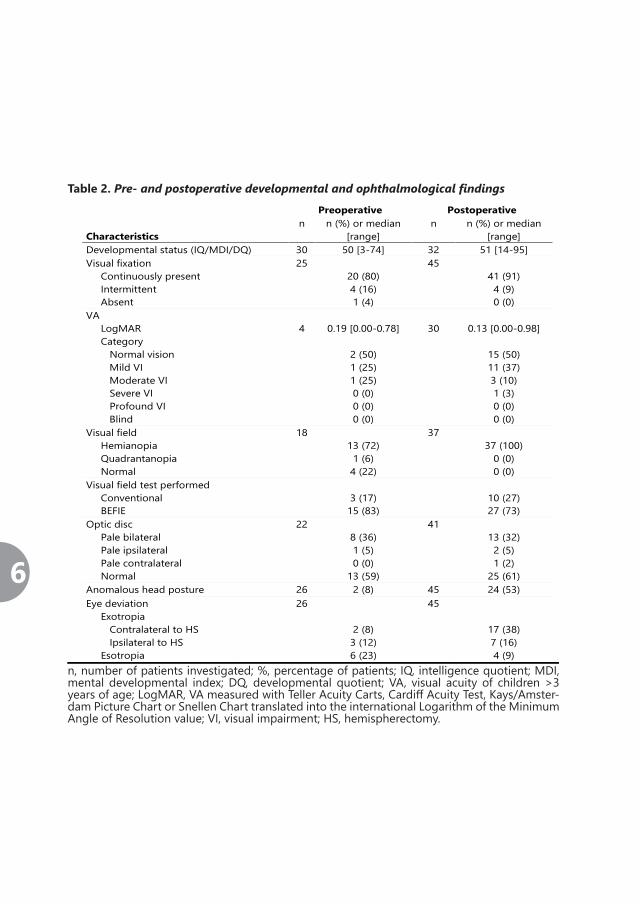
Chapter 6
136
6
Table 2. Pre- and postoperative developmental and ophthalmological findings
n, number of patients investigated; %, percentage of patients; IQ, intelligence quotient; MDI, mental developmental index; DQ, developmental quotient; VA, visual acuity of children >3 years of age; LogMAR, VA measured with Teller Acuity Carts, Cardiff Acuity Test, Kays/Amster-dam Picture Chart or Snellen Chart translated into the international Logarithm of the Minimum Angle of Resolution value; VI, visual impairment; HS, hemispherectomy.
1
Preoperative Postoperative Characteristics
n n (%) or median [range]
n n (%) or median [range]
Developmental status (IQ/MDI/DQ) 30 50 [3-74] 32 51 [14-95] Visual fixation Continuously present Intermittent Absent
25 20 (80) 4 (16) 1 (4)
45 41 (91) 4 (9) 0 (0)
VA LogMAR Category Normal vision Mild VI Moderate VI Severe VI Profound VI Blind
4
0.19 [0.00-0.78]
2 (50) 1 (25) 1 (25) 0 (0) 0 (0) 0 (0)
30
0.13 [0.00-0.98]
15 (50) 11 (37) 3 (10) 1 (3) 0 (0) 0 (0)
Visual field Hemianopia Quadrantanopia Normal
18
13 (72) 1 (6) 4 (22)
37
37 (100)
0 (0) 0 (0)
Visual field test performed Conventional BEFIE
3 (17)
15 (83)
10 (27) 27 (73)
Optic disc Pale bilateral Pale ipsilateral Pale contralateral Normal
22
8 (36) 1 (5) 0 (0)
13 (59)
41
13 (32) 2 (5) 1 (2)
25 (61) Anomalous head posture 26 2 (8) 45 24 (53) Eye deviation Exotropia Contralateral to HS Ipsilateral to HS Esotropia
26
2 (8) 3 (12) 6 (23)
45
17 (38) 7 (16) 4 (9)

Compensatory mechanisms for hemianopia
137
6
years (range 0.2–14.3 years) after a median epilepsy duration of 1.6 years (range 0.1–10.9 years). Five children had a postoperative complication that required surgical or other invasive treatment in the interval between surgery and last ophthalmologic evaluation. In total, 35 children (78%) were seizure free after a median postoperative follow-up duration of 2.3 years (range 0.03–14.7 years). None of the hemispherectomized children underwent specific visual therapy or rehabilitation. Patients underwent 1–13 follow-up visual investigations (median 2). Preoperative and post-operative developmental and ophthalmologic characteristics are listed in Table 2. Among the children who underwent neuropsychological assessments, preoperative and postoperative median IQ, MDI, or DQ scores were similar, with median values of 50 and 51, respectively.
Visual outcomeAfter hemispherectomy, all children showed visual fixation behavior, which was continuously present in 41 patients (91%). Visual acuity varied between 0.00 and 0.98 LogMAR (median 0.13 LogMAR) in postoperative assessments of patients older than 3 years of age. Of these 30 chil-dren, 15 (50%) had normal vision. A mild, moderate, severe, and profound visual impairment was present in 11 (37%), 3 (10%), one (3%), and zero (0%) children, respectively. None of the children was blind. In five children, visual acuity was not measurable because of lack of atten-tion or cooperation (median age 2.7 years, range 0.8–5.9 years); four children did not undergo visual acuity measurement (all younger than 3 years of age), and visual acuity measurements of six children were excluded because of age younger than 3 years. Preoperative visual acuity could be measured in only four children older than 3 years of age and varied between 0.00 and 0.78 LogMAR (median 0.19 LogMAR). After hemispherectomy, visual acuity of these four chil-dren (two with normal vision; two with mild visual impairment) remained stable. Among the 18 children who had a preoperative visual field measurement, 13 children (72%) already were diagnosed with HH before surgery. After hemispherectomy all children who underwent visual field measurements revealed contralateral HH. Preoperative and postoperative assessment of the optic disc by means of dilated funduscopy showed paleness in 9 (41%) and 16 (39%) of the examined children, respectively.

Chapter 6
138
6
n, number of patients investigated; %, percentage of patients; HS, hemispherectomy; FU, fol-velopmental index; DQ, developmental quotient; VA, visual acuity of children >3 years of age;diff Acuity Test, Kays/Amsterdam Picture Chart or Snellen Chart translated into the internation-value; VI, visual impairment. a, children who did not demonstrate compensatory mechanismshemispherectomy; *, p ≤0.05; #, not all children were tested, see aberrant n-value in n-column;1
Anomalous head posture Characteristics
n ______Yes______ n ______Noa______ 24 n (%) or median
[range] 10 n (%) or median
[range] Anomalous head posture - - Contralateral exotropia 10 (42) 5 (50) Gender Male
10 (42)
5 (50)
Etiology of epilepsy Developmental Stable acquired Progressive
13 (54) 10 (42) 1 (4)
3 (30) 5 (50) 2 (20)
Age of onset of epilepsy (years) 0.3 [0.0-2.7]* 1.2 [0.1-10.2] Age at HS (years) 2.1 [0.2-8.1] 5.1 [0.4-12.4] Duration of epilepsy up to HS (years) 1.6 [0.2-6.3] 1.6 [0.1-10.9] Side of HS Right
15 (63)*
2 (20)
Seizure freedom at the end of ophthalmological FU
21 (88) 6 (60)
Longest ophthalmological FU after HS (years) 2.4 [0.3-10.3]* 5.7 [2.6-14.7] Number of FU investigations 3.5 [1-11] 4 [1-13] Developmental status (IQ/MDI/DQ) # 21 50 [14-95] 6 60 [16-85] Visual fixation Continuously present Intermittent Absent
24 (100)
0 (0) 0 (0)
9 (90) 1 (10) 0 (0)
VA # LogMAR
19
0.10 [0.00-0.98]
8
0.24 [0.00-0.62]
Optic disc Pale bilateral Pale unilateral Normal
8 (33) 2 (8)
14 (58)
2 (20) 1 (10) 7 (70)
Table 3. Clinical characteristics of children who did and those who did not develop
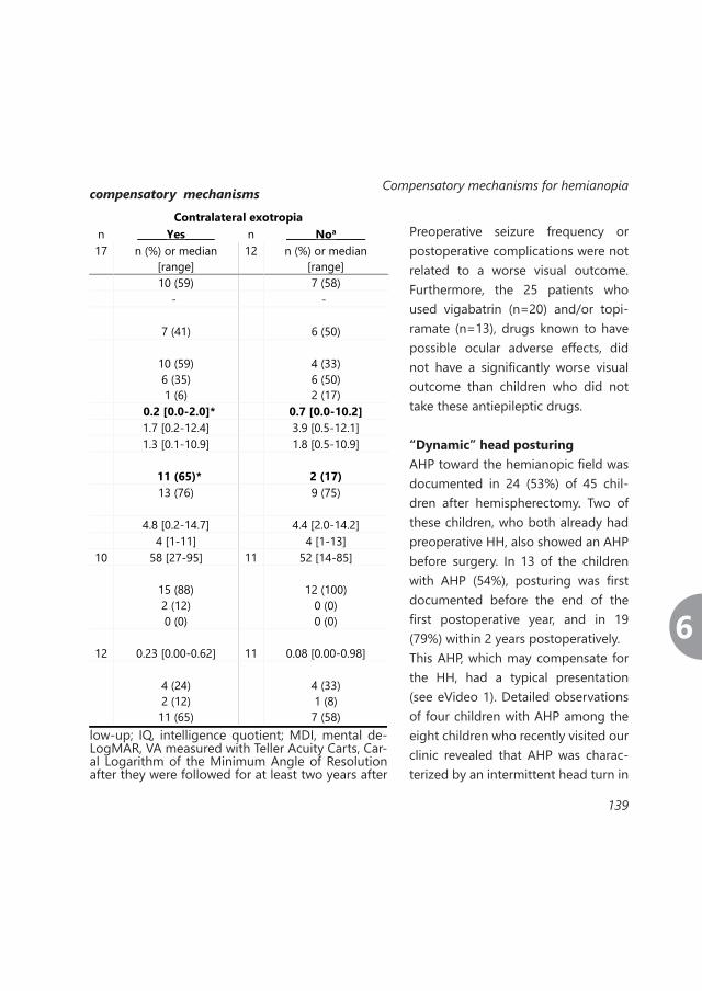
Compensatory mechanisms for hemianopia
139
6
Preoperative seizure frequency or postoperative complications were not related to a worse visual outcome. Furthermore, the 25 patients who used vigabatrin (n=20) and/or topi-ramate (n=13), drugs known to have possible ocular adverse effects, did not have a significantly worse visual outcome than children who did not take these antiepileptic drugs.
“Dynamic” head posturingAHP toward the hemianopic field was documented in 24 (53%) of 45 chil-dren after hemispherectomy. Two of these children, who both already had preoperative HH, also showed an AHP before surgery. In 13 of the children with AHP (54%), posturing was first documented before the end of the first postoperative year, and in 19 (79%) within 2 years postoperatively.This AHP, which may compensate for the HH, had a typical presentation (see eVideo 1). Detailed observations of four children with AHP among the eight children who recently visited our clinic revealed that AHP was charac-terized by an intermittent head turn in
compensatory mechanisms
1
Contralateral exotropia n ______Yes______ n ______Noa______ 17 n (%) or median
[range] 12 n (%) or median
[range] 10 (59) 7 (58) - -
7 (41)
6 (50)
10 (59) 6 (35) 1 (6)
4 (33) 6 (50) 2 (17)
0.2 [0.0-2.0]* 0.7 [0.0-10.2] 1.7 [0.2-12.4] 3.9 [0.5-12.1] 1.3 [0.1-10.9] 1.8 [0.5-10.9]
11 (65)*
2 (17)
13 (76)
9 (75)
4.8 [0.2-14.7] 4.4 [2.0-14.2] 4 [1-11] 4 [1-13]
10 58 [27-95] 11 52 [14-85]
15 (88) 2 (12) 0 (0)
12 (100)
0 (0) 0 (0)
12
0.23 [0.00-0.62]
11
0.08 [0.00-0.98]
4 (24) 2 (12)
11 (65)
4 (33) 1 (8) 7 (58)
low-up; IQ, intelligence quotient; MDI, mental de-LogMAR, VA measured with Teller Acuity Carts, Car-al Logarithm of the Minimum Angle of Resolutionafter they were followed for at least two years after
Chapter 6
140
6
the horizontal plane to the side of the HH, persisting for several seconds each time when it was seen, and occurring especially during tasks that required proper visual fixation, such as visual acuity measurements in the office room. In contrast to torticollis, the presence of this AHP was variably present and activated especially when scanning the environment for targets positioned far away. During this AHP, the eyes were pointed back to the front, that is, ipsilateral to the hemispherectomy.
Compensatory exotropiaExotropia contralateral to the hemispherectomy, either continuously or intermittently present (see eVideo 2), was documented in 17 (38%) of 45 children postoperatively, compared to doc-umentation in only two (8%) of 26 children preoperatively. Contralateral XT was first described before the end of the first postoperative year in 9 children (53%), and within 2 years post-operatively in 13 children (76%). Its presentation was intermittent in 11 of 17 children (65%). Postoperative esotropia or XT ipsilateral to the hemispherectomy was seen in only four (9%) and seven (16%) children, respectively. During observation of the XT that was present in two of the eight children who recently visited our clinic, we could not detect a consistent pattern in the use of contralateral XT.
PredispositionWe compared clinical characteristics of children who developed CMs with those of children who did not demonstrate CMs but were followed for at least 2 years after hemispherectomy (see Table 3). The distribution of these groups is shown in the Venn diagram in Figure 1. Of all 31 children who developed CMs, 10 showed both AHP and contralateral XT. Only 5 of 26 children who were followed for at least 2 years did not develop any of the two CMs. In the group of patients who developed CMs, the percentage of children who underwent right-sided hemispherectomy was higher (AHP: 63%, contralateral XT: 65%) compared to those who did not develop CMs (AHP: 20%, contralateral XT: 17%). This difference reached statistical significance for both AHP (chi-square test: p=0.024) as well as for contralateral XT

Compensatory mechanisms for hemianopia
141
6
(chi-square test: p=0.010). Furthermore, of the 10 children who developed both CMs, 9 un-derwent right-sided hemispherectomy compared to none of the five children who did not develop any CM after a follow-up of at least 2 years (Fisher exact test: p=0.002). Children who developed CMs were significantly younger at onset of epilepsy (AHP: median 0.3 years, contralateral XT: median 0.2 years) compared to patients who did not develop these CMs (AHP: median 1.2 years, contralateral XT: median 0.7 years. Mann-Whitney U test: AHP p=0.008, contralateral XT p=0.026). This difference in age at onset of epilepsy was even larger when the 10 children who developed both CMs (median 0.0 years, range 0.0–0.5 years) were compared to children who did not develop any CM after a follow-up of at least 2 years (medi-an 2.0 years, range 0.3–10.2 years, Mann-Whitney U test: p=0.004). In addition, children who developed CMs tended to be younger when they underwent surgery (AHP: median 2.1 years, contralateral XT: median 1.7 years) than children who did not develop these CMs (AHP: me-dian 5.1 years, contra-lateral XT: median 3.9 years). There were no signifi-cant differences in gen-der, etiology of epilepsy, duration of epilepsy, postoperative seizure freedom, developmen-tal status, visual fixation, visual acuity, and assess-ment of optic discs be-tween the two groups.
Figure 1. Venn diagram showing the distribution of children who did and those who did not develop compensatory mechanisms
XT, exotropia; AHP, anomalous head posture; FU, longest fol-low-up. Nine children with a FU of <2 years did not develop compensatory mechanisms.

Chapter 6
142
6
DISCUSSION
This study focused on visual outcome and the prevalence of compensatory mechanisms (CMs) for homonymous hemianopia (HH) in children who underwent hemispherectomy. In our co-hort of 45 hemispherectomized children, we found that anomalous head posture (AHP) and exotropia (XT) contralateral to the hemispherectomy occurred in 24 (53%) and 17 (38%) pa-tients, respectively. We propose that both function as CMs for the visual field defect, since they enable functional visual field optimization.
Visual outcomeLiterature about visual outcome after hemispherectomy is scarce. In our series all children showed visual fixation behavior after hemispherectomy. Normal visual acuity or only mild visual impairment was found in most children (87%) who were able to undergo visual acuity measurements. In five children of the hemispherectomized cohort, visual acuity could not be quantified because of lack of attention or cooperation. In this group, even the Teller Acuity Cart or the Cardiff Acuity Test could not be performed, which are both widely used in preverbal children, since they are based on resolution acuity using pattern preferences.23 Nevertheless, based on the 30 children in whom visual function could be reliably assessed, visual acuity, reflecting central vision, probably remains intact when one hemisphere is eliminated. This might be explained by sparing of the fovea, the main structure to obtain high visual acuity.24 However, whether the fovea has a bilateral or split representation is still a matter of debate,25 which cannot be resolved by the findings of our study. Parents of young children visiting our pediatric neuroophthalmologic outpatient clinic are generally highly positive about their child’s postoperative visual development. They are often amazed by the amelioration from an isolated child whose visual functioning is lacking or limit-ed, to an alert and attentive child who fixates, follows moving objects and makes eye contact. These developments, probably based on visual, attentional, and cognitive profits of the hemi-spherectomy, are essential for social-emotional and further cognitive development and may

Compensatory mechanisms for hemianopia
143
6
be crucial in the parent-child relationship. All children whose visual fields could be reliably examined had postoperative HH. The ma-jority of them (72%) had already been diagnosed with HH before surgery. To the best of our knowledge, only one child has been reported in the literature with remarkably preserved visual fields after hemispherectomy. Werth et al.26 described a normal visual field extent in a child with a cerebral malformation, 5.7 years after a complete hemispherectomy at 0.3 years of age. Whether this finding is a consequence of a systematic error in the method of visual field examination, or may be explained by neuronal rerouting and cortical reorganization still has to be elucidated.26,27 In our cohort, none of the children showed a recovery of the hemianopic visual field. Nevertheless, according to Moosa et al.,7 daily activities are not disturbed by visual symptoms in up to 46% of children. Paleness of the optic disc was observed in 16 children (39%) and may be explained by retro-grade transsynaptic neuronal degeneration.28 This retrograde degeneration has been inves-tigated in hemispherectomized monkeys and appeared to be inversely correlated with age at time of the lesion.29 Correspondingly, children with paleness of the optic disc in our study had earlier onset of epilepsy and underwent hemispherectomy at an earlier age than children without.
Compensatory mechanisms for hemianopiaAbnormal head posturing in children has various origins. It may arise to stabilize nystagmus or incomitant strabismus (i.e., the strabismus angle depends on gaze direction), or it may arise when a visual field defect is present. If AHP occurs directly after hemispherectomy, it might be interpreted as a compensation strategy for a postoperative gaze preference, due to damage of the frontal motor eye fields. However, such a gaze preference nearly always resolves within several weeks.30,31 Based on previous descriptions of AHP in combination with an (early) visual field defect,9–13 we hypothesize that AHP is a CM to maximize the use of the functional visual field in children with brain injury. Remarkably, we show that 24 of 45 hemispherectomized children (53%) develop AHP. Accord-

Chapter 6
144
6
ing to Good et al.,12 children with HH attempt to move their blind field to the side, resulting in centralization of the residual visual field. Contrarily, it is frequently observed that children point their eyes back to the front. Possibly children use an AHP to search for their target and subsequently point their eyes back to fixate on this target. Whether this hypothetical mech-anism, which might be an extremely rapid movement, is the reason AHP develops could not be established with our observations. In agreement with previous suggestions by Paysse et al.9 and Donahue et al.,10 the phenomenon of AHP in hemispherectomized children may also be explained by its efficient position for exploring the blind field by making saccades to the side of the HH. In addition, we speculate that this position is efficient for using a contralateral XT, since the contralateral eye merely has to deviate back into its neutral position to broaden the visual field. These theories may elucidate why, in our experience, AHP in children with HH is especially ob-served during tasks that require proper visual fixation, and is mostly activated in scanning the environment for targets positioned far away. Because this AHP is distinguished by its variable and intermittent character, we propose the phenomenon should be named “dynamic” head posturing. Equally intriguing is the observation that 17 (38%) of 45 hemispherectomized children develop contralateral XT.10,12,14–19 This phenomenon was first reported by Herzau et al.,15 who described two adult patients with HH and XT, both existing from early childhood, that led to functional enlargement of the binocular visual field, an observation later confirmed by Saleh et al.17 and Jacobson et al.14 In our study, the postoperative shift of ocular alignment to contralateral XT advocates a compensatory origin. However, there is an ongoing debate about the origin of this XT in children with HH. Good et al.12 suggested that this XT arises coincidentally as a con-sequence of neurologic damage, whereas according to several other authors XT is a CM to ex-pand the functional visual field.10 We speculate that these children somehow prefer the expan-sion of the functional visual field by means of contralateral XT above normal ocular alignment. In addition, several authors have suggested that the expected double vision might be avoided when this functional visual field enlargement by XT is accompanied by an anomalous retinal

Compensatory mechanisms for hemianopia
145
6
correspondence,16,18,32,33 resulting in continuous XT, or by using scanning movements into the blind field, observed as intermittent XT.14 Both continuous and intermittent were observed in our hemispherectomized children. These two CMs might explain the relatively good visual outcome in daily activities found by Moosa et al.7 The observation that these mechanisms particularly occur after hemispherecto-my could relate to attentional and cognitive profits of the surgical procedure, enabling children to develop compensatory strategies to cope with the hemianopic field defect. It is important to note that surgical correction of contralateral XT in these children should be avoided,15,16,18,19 since it may constrict the functional visual field. In addition, we cannot rule out a possible compensatory component in ipsilateral XT. If AHP and contralateral XT truly serve as CMs for HH in hemispherectomized children, it is interesting to search for clinical parameters that predispose to the development of compen-sation. Most of the possible preoperative and surgical determinants tested in this study were not significantly related to the occurrence of AHP or contralateral XT. Remarkably, however, there was a correlation between the side of hemispherectomy and the occurrence of AHP and contralateral XT. The majority of children who developed CMs underwent right-sided hemispherectomy, whereas most children who did not develop AHP or contralateral XT had left-sided surgery. It is unlikely that this could be explained by differences in intelligence, since there were no differences in global cognitive development between left and right-sided hemi-spherectomized children.4 Possibly, this finding relates to differences in cerebral specialization between both hemispheres, such as hemispheric lateralization of aspects of the high-level visual processing subsystems, such as visual recognition, navigation, tracking, and imagery.34–36 Furthermore, children with CMs had an earlier onset of epilepsy and there was a tendency toward hemispherectomy at younger age in children with CMs compared to those without. This suggests that timing of the insult may relate to the potency to develop CMs. According to Jacobson et al.,14 timing of the lesion might also differentiate between children who develop continuous versus intermittent or scanning XT, a suggestion we could not confirm with the findings of this study.

Chapter 6
146
6
Limitations of our study were the retrospective design with follow-up that varied in time and number of follow-up investigations. Eleven children had only one and 13 had only two fol-low-up investigations. In addition, follow-up time was sometimes too short to rule out the possible development of CMs later during the time course following surgery. To account for that, we only included children with absent CMs in the comparative analysis who had been followed for at least 2 years. Moreover, by relying on retrospective review of original reports of ophthalmologic assessments, we cannot guarantee that all elements of visual outcome were examined and documented consistently. Possibly, in some patients AHP or XT were seen, but left unrecorded, which may have led to an underestimation of their frequency. Finally, visual acuity may have been overestimated, because results of assessments were available in only 30 children. In the other 15, it was not always clear whether acuity was not documented because of lack of cooperation or because values were immeasurably low.

Compensatory mechanisms for hemianopia
147
6
CONCLUSION
This study shows that the majority of children who undergo hemispherectomy have a good visual outcome. Complete HH is present in all children, and many develop AHP and continu-ous or intermittent XT contralateral to the hemispherectomy. We suggest that both function as CMs for the visual field defect, since they enable functional visual field optimization. It is still unclear which children are prone to develop these CMs, although right-sided surgery and earlier onset of epilepsy and hemispherectomy seem to predispose. When examining children prior to and after hemispherectomy, parents should be informed on the visual acuity and visual field consequences of surgery, and on the possible development of compensation mechanisms that may play a role in a coping strategy to maximize the func-tional visual field. Surgical correction of XT in these children should be avoided.

Chapter 6
148
6
REFERENCES
1. Spencer, S. & Huh, L. Outcomes of epilepsy surgery in adults and children. Lancet Neurol. 7, 525–37 (2008).2. Moosa, A. N. V et al. Longitudinal seizure out-come and prognostic predictors after hemispherectomy in 170 children. Neurology 80, 253–60 (2013).3. Van Schooneveld, M. M. J., Jennekens-Schin-kel, A., van Rijen, P. C., Braun, K. P. J. & van Nieuwenhuizen, O. Hemispherectomy: a basis for mental development in children with epilepsy. Epileptic Disord. 13, 47–55 (2011).4. Van Schooneveld, M. M. J. & Braun, K. P. J. Cognitive outcome after epilepsy surgery in children. Brain Dev. (2013). 5. Devlin, a. M. Clinical outcomes of hemi-spherectomy for epilepsy in childhood and adolescence. Brain 126, 556–566 (2003).6. Basheer, S. N. et al. Hemispheric surgery in children with refractory epilepsy: seizure outcome, com-plications, and adaptive function. Epilepsia 48, 133–40 (2007).7. Moosa, A. N. V et al. Long-term functional outcomes and their predictors after hemispherectomy in 115 children. Epilepsia 54, 1771–9 (2013).8. Hayes, A., Chen, C. S., Clarke, G. & Thompson, A. Functional improvements following the use of the NVT Vision Rehabilitation program for patients with hemi-anopia following stroke. NeuroRehabilitation 31, 19–30 (2012).9. Paysse, E. A. & Coats, D. K. Anomalous Head Posture With Early-Onset. J AAPOS 1, 209–13 (1997).10. Donahue, S. P. & Haun, A. K. Exotropia and face turn in children with homonymous hemianopia. J. Neuroophthalmol. 27, 304–7 (2007).11. Zangemeister, W. H., Meienberg, O., Stark, L. & Hoyt, W. F. Eye-head coordination in homonymous hemianopia. J. Neurol. 226, 243–54 (1982).
12. Good, W. V et al. Cortical visual impairment in children. Surv. Ophthalmol. 1738, 351–364 (1994).13. Porro, G., van der Linden, D., van Nieuwen-huizen, O. & Wittebol-Post, D. Role of visual dysfunction in postural control in children with cerebral palsy. Neural Plast. 12, 205–10; discussion 263–72 (2005).14. Jacobson, L., Lennartsson, F., Pansell, T., Oqvist Seimyr, G. & Martin, L. Mechanisms compensating for visual field restriction in adolescents with damage to the retro-geniculate visual system. Eye (Lond). 26, 1437–45 (2012).15. Herzau, V., Bleher, I. & Joos-kratsch, E. In-fantile exotropia with homonymous hemianopia : a rare contraindication for strabismus surgery. Graefe ’ s Arch. Ophthalmol. 1, 148–149 (1988).16. Levy, Y., Turetz, J., Krakowski, D., Hartmann, B. & Nemet, P. Development of compensating exotropia with anomalous retinal correspondence after early in-fancy in congenital homonymous hemianopia. J Pediat Ophthalmol Strabismus 3, 236–238 (1995).17. Saleh, G., Sivaprasad, S. & Hammond, C. Homonymous hemianopia and exotropia: an important management issue. Eye (Lond). 20, 1402–1404 (2006).18. Gote, H., Gregersen, E. & Rindziunski, E. Exo-tropia and panoramic vision compensating for an occult congenital homonymous hemianopia. A case report. Binoc Vis. Eye Muscle Surg Q 8, 129–132 (1993).19. Gamio, S. & Melek, N. When the patient says no. Management of exotropia with hemianopic visual field defects. Binocul Vis Strabismus Q 18, 167–170 (2003).20. Boshuisen, K. et al. Contralateral MRI abnor-malities affect seizure and cognitive outcome after hemi-spherectomy. Neurology 75, 1623–30 (2010).21. World Health Organization. International Statistical Classification of Diseases and Related Health Problems 10th Revision. ICD-10 Version. (2010). at <http://apps.who.int/classifications/icd10/browse/2010/en>

Compensatory mechanisms for hemianopia
149
6
22. Porro, G., et al. A new behavioural visual field test for clinical use in pediatric neuro-ophthalmology. Neuro-Ophthalmology 19, 205–214 (1998).23. Fantz, R. Pattern vision in young children. Psychol Rec. 8, 43–47 (1958).24. Chalam, K., Ambati, B., Beaver, H. & et al. Basic and Clinical ScienceCourse, Section 2: Fundamentals and Principles of Ophthalmology. Am. Acad. Ophthalmol. Eye M.D. Assoc. 2011-2012
25. Lavidor, M. & Walsh, V. The nature of foveal representation. Nat. Rev. Neurosci. 5, 729–35 (2004).26. Werth, R. Visual functions without the occipi-tal lobe or after cerebral hemispherectomy in infancy. Eur. J. Neurosci. 24, 2932–44 (2006).27. Haak, K. V et al. Abnormal visual field maps in human cortex: A mini-review and a case report. Cortex. 1–12 (2012). 28. Vanburen, J. M. Trans-Synaptic Retrograde Degeneration in the Visual System of Primates. J. Neurol. Neurosurg. Psychiatry 26, 402–9 (1963).29. Herbin, M., Boire, D., Théoret, H. & Ptito, M. Transneuronal degeneration of retinal ganglion cells in early hemispherectomized monkeys. Neuroreport 10, 1447–52 (1999).30. DeJong, R. The neurologic examination. 4th ed. Hagerstown (MD): Harper & Row. (1979).31. Leigh, R. & Zee, D. No TitleThe neurology of eye movements. New York: Oxfort University Press. (2006).32. Van Waveren, M., Jägle, H. & Besch, D. Man-agement of strabismus with hemianopic visual field defects. Graefes Arch. Clin. Exp. Ophthalmol. (2012). 33. Herzau, V. How useful is anomalous corre-spondence? Eye (Lond). 10 ( Pt 2), 266–9 (1996).34. Laeng, B. Lateralization of categorical and coordinate spatial functions: a study of unilateral stroke patients. J. Cogn. Neurosci. 6, 189–203 (1994).
35. Marsolek, C. J. Abstract visual-form repre-sentations in the left cerebral hemisphere. J. Exp. Psychol. Hum. Percept. Perform. 21, 375–86 (1995).36. Kosslyn, S. M. Seeing and imagining in the cerebral hemispheres: a computational approach. Psychol. Rev. 94, 148–75 (1987).


7Summary and general discussion
Nederlandse samenvatting

Chapter 7
152
7
SUMMARY
The aim of this thesis is to gain more insight in the diagnostic and prognostic implications of visual field (VF) examination in children with brain disorders. Several aspects of VF examination in children with brain disorders were evaluated.
As a general introduction, Chapter 1 provides background information on the visual pathways of the brain and on several techniques to examine visual function in young or neurologically impaired children.
In Chapter 2 we evaluated all VF examinations that were performed with the BEhavioral vi-sual FIEld (BEFIE) screening test, a technique developed in 1995 by Porro et al.1 in our center, to measure the peripheral VF of young and neurologically impaired children. We found that 74% of 1788 tests could be reliably performed, and reported increasing success with higher ages. VF defects were found in 28% of the reliably performed tests. Furthermore, results were consistent over time in 75% of the 304 children who underwent multiple reliable tests, with a positive relation between consistency and number of performed tests. When we compared BEFIE test results with standard conventional perimetry (SCP) performed in the same children later in time, a high positive predictive value and specificity of both 98% was found. The neg-ative predictive value was 66% and the sensitivity 60%, which even increased to 80% when only absolute peripheral VF defects were accounted for. We concluded that the BEFIE test is a valuable tool to detect peripheral VF defects when SCP cannot be performed in young or neurologically impaired children.
In Chapter 3 the possibility to predict VF defects based on neuroimaging of the anatomy of the brain at the age of 3 months was explored in infants after perinatal arterial ischemic stroke (PAIS). In 19 infants with unilateral PIAS, of whom eight turned out to have a VF defect at fol-low-up, we found that the presence or absence of the VF defect could be correctly predicted

Summary
153
7
in the majority of infants by both Magnetic Resonance Imaging (MRI) and Diffusion Tensor Im-aging (DTI) of the optic radiation (OR) in the brain. This was objectified by a Receiver Operating Characteristic (ROC) analysis, which revealed an area under the curve (AUC) of 0.90 and 0.96 of MRI and DTI, respectively. Conventional MRI seems most suitable to use in clinical practice, since DTI requires a comprehensive analysis per individual while it added little to conventional MRI assessment. An accurate follow-up with multiple VF examinations is indicated especially in children with asymmetry of the OR.
In Chapter 4 VF defects found in children suspected of intracranial pressure (ICP) elevation were evaluated. Our cohort of 192 children with suspicion of ICP increase revealed that VF defects in children with ICP elevation mainly consisted of blind spot enlargement, which had little additional value to the presence of (suspect) papilledema during fundoscopy performed earlier. Other types of VF defects were scarce in these children at presentation and also oc-curred in children with suspicion of ICP increase but who turned out to suffer from other pa-thology than ICP increase. However, in children with documented ICP elevation, the presence of VF defects or visual acuity (VA) impairment at presentation both related to VF defects at final examination. Therefore, we concluded that VF examination at presentation in children suspected of increased ICP does not differentiate between children with or without intracranial hypertension, but that it could provide arguments to start therapy and may help to predict final VF outcome in children with documented ICP increase.
In Chapter 5 a systematic review of previous literature on the visual outcome of patients with the Sturge-Weber syndrome (SWS) and the results of a Dutch multicenter cohort are present-ed. SWS is a rare congenital disorder with angiomatous vascular malformations in the me-ninges, dermis and eye. SWS patients regularly suffer from a combination of vision impairing disorders such as glaucoma, a diffuse choroidal hemangioma (DCH), and a leptomeningeal angioma, which can lead to neurological deficits and epilepsy. Previous reports have described mainly either strictly ophthalmological or neurological findings, with consequent reporting of

Chapter 7
154
7
either VA or VF outcomes. Our systematic review revealed that a homonymous hemianopia (HH) was present in 42% of SWS patients. Of all reported eyes from a general SWS population, 70% had a (near)normal vision, while VA of eyes affected with glaucoma or DCH was severely impaired in 28% and 67%, respectively. Our cohort of 33 Dutch SWS patients, in whom we spe-cifically focused on the combination of VF and VA findings, revealed that only 18% of patients had (near)normal findings for both parameters, while half of patients with glaucoma suffer from a combination of a HH and VA impairment. We recommend adequate follow-up of both VA and VF in patients with SWS.
In Chapter 6 the visual outcome and the prevalence of compensatory mechanisms (CMs) for HH are described in children who underwent hemispherectomy. Several previous case-reports described an anomalous head posture (AHP) and exotropia (XT) contralateral to the side of early brain damage, as possible CM for HH. In our cohort of 45 hemispherectomized children, we found that visual fixation was present in all children, 87% of the examined children had a normal VA or a mild visual impairment, and all tested children had a HH. Anomalous head pos-turing and continuous or intermittent XT contralateral to the side of hemispherectomy were observed in 53% and 38% of children, respectively. We found that the children with these CMs had more frequently right-sided surgery and earlier onset of epilepsy. Moreover, they were younger when they underwent hemispherectomy than children without CMs. We concluded that the majority of children who undergo hemispherectomy have a good visual outcome and frequently develop CMs which may optimize their functional VF.

General discussion
155
7
GENERAL DISCUSSION
Below, the most important themes of research described in this thesis are discussed and sug-gestions for future research are given.
Visual field assessmentStandard conventional perimetryVisual field (VF) examination in healthy children is possible from the age of 6 years using the Goldmann2,3, Peritest4 and Frequency Doubling Technology (FDT)5,6 perimetry or the fast programs on the Octopus3,7 and Humphrey3,8 VF examiners. If a child cooperates very well, performance of a standard conventional perimetry (SCP) method is preferred, because of its greater accuracy. However, VF examination in children with brain disorders is a major chal-lenge because they are often not able to perform the most simple types of SCP.9 Although the awareness of the importance of testing VF in this group of children among both pediatric neuro-ophthalmologists and neurologists increases and multiple alternative VF examination methods are being developed, all of them have their own limitations.
Development of confrontational behavioral methodsThe well-known Donder’s confrontational method10, often used as a standard part of neuro-logical examination, was modified into a the more objective ‘binocular directional preference’ test, which can be performed using Stycar balls.11–13 The advantage of the observation of the behavioral reaction of the child on the stimuli, brought into the VF from behind, instead of a patient being asked to tell if fingers were observed during Donder’s confrontational testing, makes the ‘binocular directional preference’ test suitable for children, even those with neuro-logical disabilities. The Double-arc14–16 and Translucent sphere perimeter11,17 made VF testing in children more sophisticated and less imprecise, by making use of a half-spherical shape and gradation to measure the peripheral VF extent. However, freedom of movement of the child is limited to perform a reliable test and the observer-child interaction, which is useful to moti-
Chapter 7
156
7
vate a child to cooperate, is minimal. These methods were mostly confined to an experimental setting and rarely implemented in everyday clinical practice.
The BEFIE testThe idea of the BEhavioral visual FIEld (BEFIE) screening test1,18 arose from the existing confron-tational behavioral VF examination methods, in an attempt to solve the limitations associated with those methods and to introduce VF examination of children in a clinical setting.9 In 1998, the inventor Dr. G.L. Porro stated in his thesis “An early assessment of the visual field in neu-ropediatric patients with the BEFIE test is better than no assessment at all”.19 By now, we have better insight in the diagnostic capabilities, the limitations as well as the clinical application of this method.18 Infants seem to react unconsciously to the presented stimulus, while the test is also very well accepted by toddlers and older children, who consider it a play-like game. With the present study, the well-known inability of the BEFIE method to test the central VF or detect VF deficits such as scotomas (holes in the VF) and relative defects was confirmed. In fact, when compared with SCP performed on later age, we found that especially the positive predictive value (98%) and specificity (98%) of the BEFIE test were high. Although this finding might be biased by the probably “less impaired” population in which this comparison was made, this suggests that the test is especially useful to detect rather than to exclude peripheral VF de-fects. In addition, VF results obtained using this test proved consistent over time in the major-ity (75%) of children and deteriorations could generally be explained by progressive disease. The improvements of the VF in time observed in a minority of children might be explained by a previous false positive finding, physiological expansion of the VF, improved cooperation with age, or possible neuronal plasticity. However, the generalization of these results to other clinical settings in other centers might be hampered by the performance of the BEFIE tests by one examiner, resulting in an unknown interobserver variability. In fact, results might be influenced by the learning curve of the exam-iner, since proper performance of the BEFIE method requires an appropriate training of both the examiner and the observer. Proper performance includes a smooth adequate execution,

General discussion
157
7
effective communication between the examiner and the observer, but also a correct inter-pretation of the behavioral responses of the child, such as eye movements, finger pointing, verbal responses or rocking movements.20 For example, the presence of a hemiparesis might be misleading in diagnosing an intact or impaired VF if only the pointing hand gestures are observed, since the child will be unable to point at the target on that side.Regarding the clinical application, the current 21 years of experience illustrate that the BEFIE test, with its strength to detect peripheral VF defects in children from a very young age on-wards, can be implemented in an everyday pediatric neuro-ophthalmological practice. If pos-sible, and when it suits the conditions of the underlying disease, multiple testing is advised, in order to minimize false positive and negative findings with the BEFIE test. After performance of three measurements with intervals of approximately half a year, or shorter when necessary, possible age or mood dependent bias can be excluded and the child can get familiar with the test. This ensures a more secure assessment of VF outcome. When cooperation is sufficient, a VF examination using SCP should always be performed at later age as part of the follow-up, not only to confirm BEFIE results, but also to be informed about the central part of the VF.
Eye-tracker methodsConsidering the need for new and reliable VF examination techniques in children several methods that make use of eye-trackers are under development.21–27 Most of these methods are still being modified to improve their feasibility for application in the target group of young children. Since most eye-tracker-based VF examination methods measure, in contrast to con-frontational behavioral methods, the central VF instead of the peripheral, and are sensitive to scotomas, we strongly believe that the combination of the BEFIE test and eye-tracker VF examination methods would be interesting in order to obtain a reliable impression of the entire spectrum of both central and peripheral VF abnormalities in children. Currently these eye-tracker methods are under development in order to optimize their diagnostic capabilities and to make them attractive enough for young or neurologically impaired children, with great potential for the near future. For instance, at the Bartiméus institute, our collaborating center

Chapter 7
158
7
for diagnostics and rehabilitation of visually impaired patients, an eye-tracker program initially used to detect whether patients suffer from a VF defect as part of a simulation or conversion disorder, can nowadays also be used for examination of VF defects in children.25,27
Multifocal VEPMultifocal Visual Evoked Potential (mVEP) is described in the literature as a method to detect VF defects when SCP is not possible.28 This electrophysiological method has mainly been ap-plied to evaluate preoperative and postoperative VF defects in children undergoing epilepsy surgery,29,30 and in children with epilepsy who use vigabatrin,31,32 an antiepileptic drug which may induce a concentric VF defect as adverse effect. Although we have no experience with mVEP in our center, several articles have described its applicability in children. However, results should be interpreted with caution, since this method appeared not sensitive for VF defects caused by lesions in the extrastriate cortex, and consequently the VF measurement may not correspond to the real VF defect as would be measured with SCP.33
How to choose an appropriate method for VF examination in children?Pediatric neuro-ophthalmologists should consider several items when confronted with the clinical suspicion of a VF defect. Firstly, the child’s mental and physical capabilities to perform standard perimetry should be taken into account. When considered possible, either kinetic or static perimetry can be chosen, based on the required cooperation and requested investiga-tion time of different methods. Secondly, the expected type of VF defects should be considered. For example, in children with brain disorders along the chiasma or the retrochiasmatic visual pathways, as in those suffering from perinatal arterial ischemic stroke (PAIS), Sturge-Weber syndrome (SWS) or candidates for epilepsy surgery (Chapter 3, 5 & 6), hemianopic VF defects can be expected. This type of VF defect can easily be objectified with both SCP or confrontational behavioral VF examination methods such as the BEFIE test, which measure the peripheral VF. On the contrary, for instance in children with (or suspected of) increased intracranial pressure (ICP), enlarged blind spots

General discussion
159
7
may be expected (Chapter 4). This type of VF defect requires SCP to be detected. In this group of children, the BEFIE test is only useful in profound cases in which more severe VF defects are present. A similar problem arises in the VF examination of children with epilepsy who use vigabatrin. For early detection of toxicity of vigabatrin to the retina monocular VF examination is essential, because the VF defect that vigabatrin might cause starts nasally and expands to a concentric defect in later stage.34,35 Unfortunately, young children do generally not easily accept patching for monocular VF investigation. So, it should be considered that binocular confrontational behavioral VF examination in these children is not conclusive, as it may under-estimate of the vigabatrin related VF defect.
The main difference between VF measurement techniques in children lies in a kinetic or static execution. The perception of moving targets has another underlying mechanism than the perception of static targets and requires interpretation by different visual association areas.36 Considering that both patients with cerebral visual impairment (CVI) and young children may show a greater sensitivity to observe moving than static targets, this might result in a larger VF extent observed with kinetic compared to static examination.16,37,38 In addition, other factors, such as a manual execution by an examiner versus automatic computerized execution of the VF examination may influence results. Although manual VF testing is less objective and might introduce bias, it may aid the child’s performance and improve the reliability of the test when a child has difficulties with the examination. Furthermore, varying target size, luminance and test algorithms are factors that may cause inequalities in results assessed with different methods.
When a choice for an appropriate VF examination method is made, it is important to continue the VF assessments in time using the same or a rather similar type of VF examination in order to ensure reliable follow-up of VF defects and to avoid discrepancies between methods. In our experience, the BEFIE method corresponds better with kinetic SCP (Goldmann VF examination) than with static perimetry. Since the ‘reference’ SCP used in Chapter 2 consisted not only of kinetic, but also of static perimetry methods, this may partly explain the differences found in the comparison with the BEFIE test.

Chapter 7
160
7
Influence of cognitive and perceptual visual functions on visual function assessmentAn important yet only partly elucidated aspect of the assessment of visual function (VA and VF) in neuro-ophthalmology, is how cognitive and perceptual visual functioning – enabled by the visual association areas and the higher visual functioning pathways that are responsible for e.g. the different processing of kinetic and static targets – may influence visual outcomes.36,39 Beside possible focal damage to these areas and pathways, in children with cerebral injury, exhaustion, distraction and stress are easily provoked and may impair their cognitive and per-ceptual visual functioning, resulting in problems with e.g. recognition, orientation, handling complex visual scenes, visual guided movement or visual attention.39 This might influence the child’s visual function assessment of both VF and VA, and explains the fluctuating results that are often found when testing visual functions of neurologically impaired children. As an example, crowding, a phenomenon in which a child is unable to see a symbol surrounded by other symbols that crowd the VF, is regularly observed in VA testing in children with brain disorders.40 Regarding the VF, an impairment of the visual association areas and the higher visual functioning pathways might play a role in the not yet fully understood phenomenon of blindsight.41,42 This phenomenon, which is observed in few patients with a VF defect due to brain injury, is defined as unconscious perception of objects in the blind field that cannot be observed consciously.42 Although most children in our studies were tested multiple times and we generally used the best measured outcome in the analyses, it remains unknown to what degree VF measurements were influenced by possibly impaired cognitive and perceptual visual functions.
Prediction of visual field defectsEarly prediction by means of neuroimagingThe alternative perimetry methods for VF assessment in children described above, are not yet widely used. Therefore, early prediction of VF defects that are caused by acquired or develop-mental brain abnormalities by means of neuroimaging could be useful for academic centers in which only SCP can be performed. To visualize cortical areas and the white matter of visual

General discussion
161
7
pathways, conventional T1 and T2 Magnetic Resonance Imaging (MRI) are generally used in daily clinical practice. However, more advanced MRI methods that enable assessment of functional activity or three-dimensional anatomy of the white matter may provide additional valuable information.Diffusion Tensor Imaging (DTI) has many applications and can be used to study the integrity of the white matter globally for the whole brain or of specific tracts on anatomical locations.43 Regarding prediction of visual outcome in infants, DTI literature mainly focused on the associ-ation of diffusion parameters at term age with visual function scores based on functions such as fixation, motility, tracking and VA, in premature infants.44–46 The prediction of VF defects in infants with DTI has not been previously investigated. In this thesis, we explored the possibility to predict VF defects based on three month MRI and DTI scans in infants after unilateral PAIS and we concluded that both techniques are useful in the prediction of their VF defects. However, MRI is most convenient since it is easier to per-form in daily clinical practice. Although not all infants with optic radiation (OR) injury on neu-roimaging develop VF defects, we recommended an accurate VF follow-up of children with OR asymmetry on MRI at three months of age, in order to ensure early diagnosis of VF defects and subsequent start of rehabilitation. In addition, since all infants in our study with a symmetrical OR had a normal VF, we suggested that there is no need for visual follow-up in the presence of a symmetrical OR. Obviously, it should not be overlooked that also injury to other areas in the brain responsible for visual functioning, such as the (extra)striate visual cortexes and pathways that serve higher visual functioning, may provoke VF defects in the presence of an symmetrical intact OR,47–49 and that these children are also entitled to VF examination.
Although we concluded that both MRI and DTI are useful in the prediction of VF defects with comparable predictive values and recommend to use MRI in daily clinical practice, DTI still might play a role in specific cases. For instance, in subjects in whom qualitative conventional MRI assessment of the OR is inconclusive, more sophisticated techniques such as DTI, that pro-vide quantitative data, might assist in predicting VF outcomes. In the few infants in our study

Chapter 7
162
7
who had a mildly asymmetrical OR, the fractional anisotropy (FA) asymmetry index on DTI gave direction toward the final presence or absence of a VF defect. Though, larger cohorts are needed to confirm this finding. Moreover, DTI might be of additional value to MRI assessment in infants with bilateral brain damage. While asymmetry assessment of the OR on conventional MRI in these infants cannot be used, DTI may provide helpful quantitative data of the individu-al ORs. Though, the possible variation in outcomes due to individual differences in myelination of the OR should be taken into account.50 Finally, DTI might be of potential value in predicting what kind of VF defect is present. Although both the qualitative conventional MRI assessment and the quantitative DTI measurements provided too little information to make distinction be-tween incomplete or complete quadrantanopias or hemianopias (HHs) in our study, we found a correlation between the degree of asymmetry and the severity of the VF defect. Separate investigation of specific temporal or parietal parts of the OR with DTI, instead of analyzing the OR as a whole, could be a next step in order to achieve this goal.
Visual outcome in pediatric brain diseasesDifficulties in data collectionVisual outcome in pediatric brain diseases is often incompletely investigated and reported for several reasons. First, children may be difficult to examine due to other deficits caused by their brain injury, such as severe epilepsy, motor dysfunction and developmental delay. Sec-ond, visual outcome might be regarded as relatively less important, compared to motor and cognitive outcome, or might be overlooked in the often complex combination of multiple dis-abilities.51–53 Therefore, a multidisciplinary approach by a team of different specialists, such as a child-neurologist, pediatrician, ophthalmologist, orthoptist and physiotherapist is required, but might take effort to accomplish. This collaboration is especially essential in disorders in which both ocular and cerebral deficits that affect visual functioning are present. For instance, in the systematic review on visual outcome in patients with SWS (Chapter 5), who regularly suffer from a combination of glaucoma, a diffuse choroidal hemangioma (DCH) and cerebral

General discussion
163
7
manifestations, we found that the ophthalmological literature was mainly confined to descrip-tion of VA results, while neurological articles often only described VF outcomes. Nonetheless, it is in particular the combination of visual acuity (VA) and VF impairments that frequently invalidates the child with neuro-ophthalmological disorders. It should always be kept in mind that even in children in whom VA and ophthalmological investigations, including fundoscopy, appear to be normal, VF can be impaired (Chapter 3, 4, 5 & 6) and may cause problems in daily functioning. Despite the growing incidence of neuro-ophthalmological disorders, most of these are still relatively rare, resulting in small research populations. Conduction of systematic reviews is therefore valuable to draw firmer conclusions, though distortion of results due to selection bias might easily occur when combining outcomes of various studies, and must be taken into account.
Influence of visual field defects on functioningEarly detection of visual impairment, including VF defects, is meaningful for optimal rehabil-itation of the child, with an adjusted approach at daycare or school, in order to restrict the serious consequences it can have on the child’s functioning, by affecting motor, emotional, social and psychological development14,54–57 The exact relation between VF defects and func-tioning of the child is difficult to investigate, since they frequently suffer from other associated neurological disabilities, such as motor and cognitive impairment, which also influence their functional outcome. In addition, cognitive impairment can make a reliable assessment of the functional outcome, by means of questionnaires, impossible.58 In fact, although questionnaires can be completed by a proxy when children are too young to do so themselves, the population of children with VF defects is too heterogeneous to be assessed with the same investigative instrument. While the cognitively normal functioning children can be tested with validated questionnaires that are widely used, these instruments are not validated, and therefore less useful, in children with cognitive impairment.

Chapter 7
164
7
Box 1. Quality of life and functioning in children and adults with homonymous hemi-anopia diagnosed in childhood
Yvonne Koenraads, Giorgio L. Porro, Saskia M. Imhof, Kees P.J. Braun
Purpose: Homonymous hemianopia (HH) may have impact on health related quality of life (HRQoL) and functioning, which requires adequate coping. However, literature on the influ-ence of HH on the life of children is lacking. Aim of this study was to investigate the func-tional outcome of children and adults with HH diagnosed in childhood.
Methods: Sixty-one patients (median age 15.0, range 4.3-31.5 yrs), and their caregivers, who were diagnosed with HH before the age of 18 yrs in our center between 1995 and 2014, par-ticipated in this study. Questionnaires measuring HRQoL (children: KIDSCREEN-27, adults: SF-36), participation (children: CASP, adults: D-AI2 mobility), symptoms of cerebral visual impairment (CVI-checklist) and additional open questions about the consequences of HH were evaluated. For the analysis, patients were classified in three groups: one group of pa-tients with normal cognition (IQ ≥70 or following mainstream education) and normal motor function (NCNM, n=9), a second group of patients with normal cognition and an impaired motor function (NCIM, n=15), and a third and largest group of patients with an impaired cognition (IC, n=37).
Results: Nearly all HRQoL domains, of both children and adults with normal cognition, re-vealed median values in the normal range. In children, there were no major differences between the NCNM and NCIM group, while almost all median scores in adults were lower in the NCIM group than in the NCNM group, of which scores on the domains ‘physical functioning’ (p=0.004), ‘general health’ (p=0.05) and the ‘physical component summary’ (p=0.01) were significantly lower (Mann-Whitney U test). Also regarding participation, both children and adults had relatively normal median scores. Although median participation was

General discussion
165
7
During our attempt to investigate the functional outcome of patients with a HH diagnosed in childhood (before the age of 18 years), we were hampered by the fact that a large proportion of hemianopic patients also had a developmental delay or motor deficits (see Box 1). When excluding patients with cognitive impairment, we were able to compare two groups of children with relatively normal cognitive functioning, those with and without motor dysfunction. Al-though the number of patients was too low to draw definite conclusions, most health-related quality of life (HRQoL) and participation scores were in the normal range, while lower scores in physical domains were mostly explained by impaired motor functioning. However, it should be considered that patients with brain disorders, and their caregivers, may fill out the ques-
worse in the NCIM than the NCNM group on almost all domains in both children and adults, differences were not significant. At the CVI-checklist, median scores on both dorsal and ventral stream functioning were approximately equally distributed among all three groups. However, dorsal stream functioning was more impaired than the functions of the ventral stream (p<0.001, Mann-Whitney U test). Disturbing consequences of the HH listed most frequently included unsafe participation in traffic, bumping into objects, scare & anxiety, orientation & overview and reading difficulties.
Discussion: The majority of patients with HH diagnosed in childhood suffer from additional motor or cognitive impairment. This hampers the investigation of the separate impact of HH on functioning. Furthermore, HRQoL and participation questionnaires are not validated for patients with intellectual disability, the largest group in our HH cohort. Although the number of patients with normal cognition was limited, most HRQoL and participation scores were within the normal range, and lower scores in physical domains were mostly explained by impaired motor functioning. There clearly is a need for adequate instruments to investigate HRQoL and participation in children with developmental delay. To establish the independent contribution of HH to the functional outcome of children with or without motor or intellec-tual impairment, studies should compare patients with and those without HH.
Chapter 7
166
7
tionnaires from a different perspective than a normal population.59 Moreover, although the instruments that were applied are properly validated and widely used, maybe they were not sensitive enough for specific disturbances caused by HH.60–67 When looking at the responses obtained, consequences of the HH that were considered most disturbing included unsafe participation in traffic, bumping into objects, scare & anxiety, orientation & overview and reading difficulties. However, some participants stated that there were no impediments of the HH, or that these were considered unimportant compared to other disabilities. Regardless of this consideration, the actual importance of the VF for a mentally disabled, often wheelchair dependent, child is still unknown. The impression that visual functioning is less important in the presence of more evident motor and cognitive disorders does not mean that VF defects in these children deserve no attention. For instance, visual capability of some multi-handicapped children might be the only possible way to communicate.68
Functional visual field, compensation and plasticitySometimes functional visual capacities improve by compensation or restoration of visual im-pairment.69–71 Compensation might be achieved by physical adaptations such as an anomalous head posture (AHP)14,72–75 and exotropia (XT)14,73,76–81 contralateral to the side of brain damage. Although we did not objectify the actual optimization of the VF in this thesis, we found that children who underwent hemispherectomy, and therefore all had a HH, regularly showed such compensatory mechanisms (CMs).69 An AHP can center the VF and enables efficient scanning of the surroundings, while an XT enables a broader ‘panoramic’ view.Several case-reports and series already demonstrated the beneficial use of these physical ad-aptations, but the underlying mechanism of these ‘CMs’ is still unknown. The association with right sided brain injury suggests that there might be a relation to lateralized cognitive func-tions present in one of the two hemispheres, such as visual recognition, navigation, tracking, and imagery.82–84 In addition, the association with an earlier onset of epilepsy and the ten-dency towards early hemispherectomy in children developing CMs, suggests that especially the developing infant brain enables such adaptations.69,85 Preference of use of the peripheral

General discussion
167
7
VF above using central vision (VA) by children with, mostly severe, CVI could also play an im-portant role.14,86 It might explain why the AHP is especially observed during tasks that require proper visual fixation and it is mostly activated while scanning the environment for targets positioned far away.69 Regarding the origin of the XT, several theories have been proposed. It might develop as a consequence of neurologic injury,14,87 or relate to the original function of the nasal and tem-poral retinal functions,88,89 while other authors stated that it might arise as CM to expand the functional VF.73 We speculate that these children somehow prefer the expansion of the func-tional VF by means of contralateral XT above normal ocular alignment, possibly due to their preference for use of the peripheral VF instead of central vision,14,86 similar to what a chame-leon does to improve its survival chances.Regardless of the underlying origin, children with a homonymous VF defect and XT frequently suffer from recurrence of XT after strabismus surgery. Together with the probable beneficial function of the XT for the functional VF, we consider the presence of HH a contraindication for strabismus surgery in these patients.Improvement of functional visual capacities by restoration of visual impairment is also known as neuronal plasticity.70,71 It might play a role in improving VF results,47 as has been suggest-ed in the case-reports by Seghier et al.90 and Groenendaal et al.91 Mechanisms of neuronal plasticity that have been described include formation of new interhemispheric connections, reorganization in nearby unaffected cortex or changes in functional interaction between high-er-level visual cortical areas and the primary visual areas.92 Neuronal plasticity mainly occurs in the developing brain and might therefore especially be present in congenital disorders that have an effect on the VF in very early stage.70,85,93
Using functional MRI (fMRI) in a patient who was born with only one cerebral hemisphere, which is associated with HH, we demonstrated the presence of receptive fields of the bilateral retina in the early visual cortex (see Box 2). This indicates reorganization of intra-cortical wiring of the early visual cortex and confirms neuronal plasticity and reorganization after early brain injury in humans.

Chapter 7
168
7
Box 2. Bilateral population receptive fields in congenital hemihydranencephaly
Alessio Fracasso, Yvonne Koenraads, Giorgio L. Porro, Serge O. DumoulinAccepted for publication in Ophthalmic and Physiological Optics
Purpose: Congenital hemihydranencephaly is a very rare disorder characterized by prena-tal near-complete unilateral loss of the cerebral cortex. We investigated a patient affected by congenital right hemihydranencephaly whose visual field extended significantly into the both visual hemifields, suggesting a reorganization of the remaining left visual hemisphere. We examined the early visual cortex reorganization using functional MRI (7T) and popula-tion receptive field (pRF) modeling.
Methods: Data were acquired by means of a 7T MRI (Phillips, Best, Netherlands) while the patient affected by hemihydranencephaly viewed conventional population receptive field mapping stimuli. Two possible pRF reorganization schemes were evaluated: where every cortical location processed information from either (i) a single region of the visual field or (ii) from two bilateral regions of the visual field.
Results: In the patient affected by hemihydranencephaly, bilateral pRFs in single cortical locations of the remaining hemisphere were found. In addition, using this specific pRF reor-ganization scheme, the biologically known relationship between pRF size and eccentricity was found.
Conclusions: Bilateral pRFs were found in the remaining left hemisphere of the patient affect-ed by hemihydranencephaly, indicating reorganization of intra-cortical wiring of the early visual cortex and confirming brain plasticity and reorganization after an early cerebral dam-age in humans.

General discussion
169
7
Future developmentsDevelopment of visual field examination techniquesInnovative VF examination methods, such as confrontational behavioral and eye-tracker meth-ods, are being developed to suit VF testing in young and neurologically impaired children.94 By combining and modifying these existing techniques, the feasibility and accuracy will improve, while limitations will be covered. Essential in this development is a fast, attractive, accepted, play-like and objective measurement that can be performed without distraction of the child by other factors.Possibly, in addition to these methods, there will be place for objective VF measurements by means of pupillometry or pupillography.95–98 Such a method is currently being developed and investigated in adult patients in several centers.96,98 If this technique proves to be valuable, it may have a potential applicability in (neurologically impaired) children.
Development of neuroimaging of the visual systemIn the meantime, improving neuroimaging techniques that make use of measuring diffusion, functional activation or connective networks in the brain are potentially useful for objective VF examination.99,100 At present, DTI tractography already enables identification of (1) fibers that connect brain areas and (2) retinotopic organization of fibers in the splenium, used in higher visual integration, in adults.99 The methods of measuring diffusion still improve by an increasing spatial resolution of the diffusion-weighted data.101 In addition, the tractography algorithms, that trace connections, become more sophisticated. fMRI already enables retino-topic mapping in adults, which represents the correspondence between the VF and its cortical representation in the brain.99,100 Information on fMRI field maps can also be combined with data from electro-encephalography (EEG), magneto-encephalography (MEG) or implanted subdural grid electrodes (electro-corticography) in order to clarify spatial locations of the neu-ral signals.99 However, since these methods are either time consuming or invasive, they are less suitable for application in children. Although the received signals by the visual cortex cannot confirm actual functional perception, these neuroimaging methods may be more objective

Chapter 7
170
7
than the current standard VF examination methods.Beside using one single technique, especially the combination of several techniques might improve our understanding of the visual system by connecting the various aspects that can be measured. Possibly, in the future these techniques may objectively predict or diagnose not only the presence, but also the location and type of VF defects in children. In addition, though neuronal plasticity might occur in a selection of patients and will complicate accurate predic-tion, these neuroimaging techniques could also be able to reveal the still unknown underlying mechanisms of plasticity.93
Clinical implications of the findings described in this thesisThe BEhavioral visual FIeld (BEFIE) screening test can be reliably performed in most children who are too young or neurologically impaired to perform standard conventional perimetry (SCP), and is particularly valuable in the detection of peripheral visual field (VF) defects. There-fore, the BEFIE test should be performed in young children with (suspected) visual pathway pathology in order to allow early detection and quantification of peripheral VF abnormalities that would otherwise remain unnoticed.
Assessment of asymmetry of the optic radiation (OR) on both conventional MRI and DTI-based tractography at three months of age can be used to predict VF defects after unilateral perinatal arterial ischemic stroke (PAIS) with comparable predictive values.
VF examination by means of SCP in children with the clinical suspicion of increased intracra-nial pressure (ICP) does not differentiate between children with and those without intracranial hypertension. In children with ICP elevation, SCP can provide arguments to start therapy and help to predict final VF outcome.
In Sturge-Weber syndrome (SWS) patients, a homonymous VF defect (42%) and visual acuity (VA) impairment (30%) regularly occurs. The combination of these two visual parameters is

General discussion
171
7
rarely described in the literature. With the Dutch multicenter cohort we demonstrated and emphasized the combined functional impairments, that may especially be invalidating.
Despite having a homonymous hemianopia (HH), the majority of children who undergo hemi-spherectomy have a good visual outcome and frequently develop an anomalous head posture (AHP) and continuous or intermittent exotropia (XT) contralateral to the hemispherectomy, which are considered compensatory mechanisms (CMs) that may optimize their functional VF. The presence of these CMs should discourage surgical correction of XT in children with HH.
The diagnostic and prognostic clinical implications described in this thesis contribute to the awareness of cerebral visual impairment (CVI) and its clinical consequences, such as the impor-tance of VF examination, for children with brain disorders.

Chapter 7
172
7
REFERENCES
1. Porro, G., et al. A new behavioural visual field test for clinical use in pediatric neuro-ophthalmology. Neuro-Ophthalmology 19, 205–214 (1998).2. Kolling, G. H. & Wabbels, B. Symposium Proceedings Part II Kinetic perimetry in neuroophthalmo-logical practice. 8, (2000).3. Patel, D. E. et al. Study of optimal perimetric testing in children (OPTIC): Feasibility, reliability and repeatability of perimetry in children. PLoS One 10, 1–12 (2015).4. Greve, E. L., Dannheim, F. & Bakker, D. The Peritest, a new automatic and semi-automatic perimeter. Int. Ophthalmol. 5, 201–14 (1982).5. Blumenthal, E. Z., Haddad, A., Horani, A. & Anteby, I. The reliability of frequency-doubling perimetry in young children. Ophthalmology 111, 435–9 (2004).6. Quinn, L. M., Gardiner, S. K., Wheeler, D. T., Newkirk, M. & Johnson, C. a. Frequency doubling tech-nology perimetry in normal children. Am. J. Ophthalmol. 142, 983–9 (2006).7. Safran, a B. et al. Feasibility of automated visual field examination in children between 5 and 8 years of age. Br J Ophthalmol 80, 515–518 (1996).8. Donahue, S. P. & Porter, a. SITA visual field testing in children. J. AAPOS 5, 114–7 (2001).9. Porro, G. L. & Wittebol-Post, D. Impairment of peripheral vision and its measurement. Chapter 5 in: Visual impairment in children due to damage to the brain (Edited by G.N. Dutton & M. Bax). Clin. Dev. Med. No. 186. Mac Keith Press (2010).10. Stilma, J. S. & Voorn, T. B. in Praktische huis-artsgeneeskunde: Oogheelkunde 40–43 (2002).11. Mohn, G. & Van Hof-van Duin, J. Behavioural and electrophysiological measures of visual functions in children with neurological disorders. Behav. Brain Res. 10, 177–87 (1983).
12. Hermans, A., Van Hof-van Duin, J. & Oude-sluys-Murphy, A. Visual outcome of low-birth-weight infants (1500-2500 g) at one year of corrected age. Acta Paediatr. 83, 402–7 (1994).13. Sheridan MD. The STYCAR graded-balls vision test. Dev Med Child Neurol. 15, 423–32 (1973).14. Good, W. V et al. Cortical visual impairment in children. Surv. Ophthalmol. 1738, 351–364 (1994).15. Quinn, G. E. et al. Visual Fields Measured with Double-arc Perimetry in Eyes with Threshold Retinopathy of Prematurity from the Cryotherapy for Retinopathy of Prematurity Trial. Ophthalmology 103, 1432–1437 (1996).16. Dobson, V., Brown, a M., Harvey, E. M. & Narter, D. B. Visual field extent in children 3.5-30 months of age tested with a double-arc LED perimeter. Vision Res. 38, 2743–60 (1998).17. Mayer, D. L., Fulton, a B. & Cummings, M. F. Visual fields of infants assessed with a new perimetric technique. Invest. Ophthalmol. Vis. Sci. 29, 452–9 (1988).18. Koenraads, Y., Braun, K. P. J., van der Linden, D. C. P., Imhof, S. M. & Porro, G. L. Perimetry in Young and Neurologically Impaired Children: The Behavioral Visual Field (BEFIE) Screening Test Revisited. JAMA Ophthalmol. 133, 319–25 (2015).19. Porro, G. L. Vision and Visual Behaviour in Responsive and Unresponsive Neurologically Impaired Children. Thesis (1998).20. Porro, G. et al. Visual behaviours of neurolog-ically impaired children with cerebral visual impairment: an ethological study. Br. J. Ophthalmol. 82, 1231–1235 (1998).21. Murray, I. C. et al. Feasibility of saccadic vec-tor optokinetic perimetry: a method of automated static perimetry for children using eye tracking. Ophthalmology 116, 2017–26 (2009).22. Murray, I. et al. Saccadic Vector Optokinetic Perimetry (SVOP): A novel technique for automated static perimetry in children using eye tracking. Conf Proc IEEE Eng Med Biol Soc. 2013, 3186–9 (2013).

General discussion
173
7
23. Allen, L. E., Slater, M. E., Proffitt, R. V, Quarton, E. & Pelah, A. A new perimeter using the preferential looking response to assess peripheral visual fields in young and developmentally delayed children. J. AAPOS 16, 261–5 (2012).24. Pel, J. J. M., van Beijsterveld, M. C. M., The-pass, G. & van der Steen, J. Validity and Repeatability of Saccadic Response Times Across the Visual Field in Eye Movement Perimetry. Transl. Vis. Sci. Technol. 2, 3 (2013).25. De Wit, G. Bartimeus eyetracker project. at <https://www.bartimeus.nl/expertise/projecten/projec-tresultaten/project-eyetracker>26. Tailor, V. et al. Saccadic vector optokinetic perimetry in neuro-disability or isolated visual pathway lesions: observational cohort study. Under Rev. 1–6 (2016). 27. Wit, G. C. de, Hoeben, F. & Genderen, M. van. Objectieve gezichtsveldbepaling bij verdenking conversie met behulp van eye-tracking. NOG Congr. Groningen (2015).28. Yukawa, E., Matsuura, T., Kim, Y.-J., Taketani, F. & Hara, Y. Usefulness of multifocal VEP in a child requir-ing perimetry. Pediatr. Neurol. 38, 360–2 (2008).29. Kim, Y.-J., Yukawa, E., Kawasaki, K., Nakase, H. & Sakaki, T. Use of multifocal visual evoked potential tests in the objective evaluation of the visual field in pediatric epilepsy surgery. J. Neurosurg. 104, 160–5 (2006).30. Yukawa, E., Kim, Y.-J., Kawasaki, K., Taketani, F. & Hara, Y. A child with epilepsy in whom multifocal VEPs facilitated the objective measurement of the visual field. Epilepsia 46, 577–9 (2005).31. Harding, G. F. a, Spencer, E. L., Wild, J. M., Conway, M. & Bohn, R. L. Field-specific visual-evoked potentials: identifying field defects in vigabatrin-treated children. Neurology 58, 1261–5 (2002).32. Spencer, E. L. & Harding, G. F. a. Examining visual field defects in the paediatric population exposed to vigabatrin. Doc. Ophthalmol. 107, 281–7 (2003).33. Klistorner, a I., Graham, S. L., Grigg, J. & Balachandran, C. Objective perimetry using the multifocal
visual evoked potential in central visual pathway lesions. Br. J. Ophthalmol. 89, 739–44 (2005).34. James Willmore, L., Abelson, M. B., Ben-Men-achem, E., Pellock, J. M. & Donald Shields, W. Vigabatrin: 2008 Update. Epilepsia 50, 163–173 (2009).35. Kedar, S., Ghate, D. & Corbett, J. J. Visual fields in neuro-ophthalmology. Indian J. Ophthalmol. 59, 103–9 (2011).36. Suter, P. S. & Harvey, L. H. Vision Rehabilita-tion: Multidisciplinary Care of the Patient Following Brain Injury. Taylor & Francis Group, LLC. CRC Press. (2011).37. Delaney, S. M. et al. Stimulus Motion Increases Measured Visual Field Extent in Children 3.5 to 30 Months of Age. Optom. Vis. Sci. 77, 82–89 (2000).38. Roman-Lantzy, C. Cortical Visual Impairment: An Approach to Assessment and Intervention. AFB Press. Am. Found. Blind. 11 Penn Plaza, Suite 300, New York 23–24 (2007).39. Dutton, G. N., Macdonald, E., Drummond, S. R., Saidkasimova, S. & Mitchell, K. Clinical features of per-ceptual and cognitive visual impairment in children with brain damage of early onset. Chapter 7 in: Visual impair-ment in children due to damage to the brain (Edited by G.N. Dutton & M. Bax). Clin. Dev. Med. No. 186. Mac Keith Press (2010).40. Good, W. V, Jan, J. E., Burden, S. K., Skoczenski, A. & Candy, R. Recent advances in cortical visual impair-ment. Dev. Med. Child Neurol. 43, 56–60 (2001).41. Boyle, N. J., Jones, D. H., Hamilton, R., Spowart, K. M. & Dutton, G. N. Blindsight in children: does it exist and can it be used to help the child? Observations on a case series. Dev. Med. Child Neurol. 47, 699–702 (2005).42. Cooper, S. A. & O’Sullivan, M. Here, there and everywhere: higher visual function and the dorsal visual stream. Pract. Neurol. practneurol–2015–001168 (2016). 43. Feldman, H. M., Yeatman, J. D., Lee, E. S., Barde, L. H. F. & Gaman-bean, S. Diffusion Tensor Imag-ing : A Review for Pediatric Researchers and Clinicians. J Dev Behav Pediatr. 31, 346-56 (2010).

Chapter 7
174
7
44. Bassi, L. et al. Probabilistic diffusion tractogra-phy of the optic radiations and visual function in preterm infants at term equivalent age. Brain 131, 573–82 (2008).45. Groppo, M. et al. Development of the optic radiations and visual function after premature birth. Cortex. 56, 30–7 (2014).46. Berman, J. I. et al. Quantitative fiber tracking analysis of the optic radiation correlated with visual performance in premature newborns. AJNR. Am. J. Neu-roradiol. 30, 120–4 (2009).47. Van der Aa, N. E. et al. Neonatal posterior cerebral artery stroke: clinical presentation, MRI findings, and outcome. Dev. Med. Child Neurol. 55, 283–90 (2013).48. Guzzetta, A. et al. Visual function in children with hemiplegia in the first years of life. Dev. Med. Child Neurol. 43, 321–329 (2001).49. Mercuri, E. et al. Visual outcome in children with congenital hemiplegia: correlation with MRI findings. Neuropediatrics 27, 184–8 (1996).50. Iai, M., Yamamura, T. & Takashima, S. Early Expression of Proteolipid Protein in Human Fetal and Infantile Cerebri. Pediatr Neurol 17, 235–239 (1997).51. Dutton, G. & Bax, M. Visual impairment in children due to damage to the brain. Clin. Dev. Med. No. 186. Mac Keith Press (2010).52. Philip, S. S. & Dutton, G. N. Identifying and characterising cerebral visual impairment in children: a review. Clin. Exp. Optom. 97, 196–208 (2014).53. Harbert, M. J., Yeh-Nayre, L. a, O’Halloran, H. S., Levy, M. L. & Crawford, J. R. Unrecognized visual field deficits in children with primary central nervous system brain tumors. J. Neurooncol. 107, 545–9 (2012).54. Van Hof-van Duin, J., Bertuccelii, B., Fazzi, B. & Maris, S. Visual outcome at 5 years ’ of newborn infants at risk of cerebral visual impairment. Dev. Med. Child Neurol. 40, 302–309 (1998).55. Dale, N. & Salt, A. Early support developmen-tal journal for children with visual impairment: the case
for a new developmental framework for early interven-tion. Child. Care. Health Dev. 33, 684–90 (2007).56. Fazzi, E., Signorini, S. G. & Lanners, J. The effect of impaired vision on development. Chapter 9 in: Visual impairment in children due to damage to the brain (Edited by G.N. Dutton & M. Bax). Clin. Dev. Med. No. 186. Mac Keith Press (2010).57. Buultjens, M., Hyvarinen, L., Walthes, R. & Dutton, G. N. Strategies to support the development and learning of children with cerebral visual impairment at home and at school: communication, orientation, and mobility. Chapter 15 in: Visual impairment in children due to damage to the brain (Edited by G.N. Dutton . Clin. Dev. Med. No. 186. Mac Keith Press (2010).58. Evenhuis, H. M. Children with intellectual disabilities and cerebral visual impairment: Problems with detection and diagnosis. Chapter 13 in: Visual impairment in children due to damage to the brain (Edited by G.N. Dutton & M. Bax). Clin. Dev. Med. No. 186. Mac Keith Press (2010).59. Schooneveld, M. van. Development after epilepsy surgery in children. Thesis (2016).60. Ravens-Sieberer, U. et al. The KIDSCREEN-27 quality of life measure for children and adolescents: psychometric results from a cross-cultural survey in 13 European countries. Qual. Life Res. 16, 1347–1356 (2007).61. Teunissen, M. et al. Quality of life and behav-ioral functioning in Dutch pediatric patients with heredi-tary spherocytosis. Eur. J. Pediatr. 173, 1217–23 (2014).62. Gutter, T., Brouwer, O. F. & de Weerd, a W. Subjective sleep disturbances in children with partial epilepsy and their effects on quality of life. Epilepsy Behav. 28, 481–8 (2013).63. Van der Heijden, P. G. M., van Buuren, S., Fekkes, M., Radder, J. & Verrips, E. Unidimensionality and reliability under Mokken scaling of the Dutch language version of the SF-36. Qual. Life Res. 12, 189–98 (2003).64. Aaronson, N. K. et al. Translation, validation, and norming of the Dutch language version of the SF-36

General discussion
175
7
Health Survey in community and chronic disease popula-tions. J. Clin. Epidemiol. 51, 1055–1068 (1998).65. De Kloet, A. J. et al. Psychometric evaluation of the Dutch language version of the Child and Family Follow-up Survey. Dev. Neurorehabil. 18, 357–64 (2015).66. Bruijning, J. E., van Nispen, R. M. & van Rens, G. H. Feasibility of the Dutch ICF Activity Inventory: a pilot study. BMC Health Serv. Res. 10, 318 (2010).67. Bruijning, J., Rens, G. van, Knol, D. & Nispen, R. van. Psychometric Analyses to Improve the Dutch ICF Activity Inventory. Optom. Vis. Sci. 90, 806–819 (2013).68. Sargent, J., Salt, A. & Dale, N. Children with severe brain damage: Functional assessment for diag-nosis and intervention. Chapter 11 in: Visual impairment in children due to damage to the brain (Edited by G.N. Dutton & M. Bax). Clin. Dev. Med. No. 186. Mac Keith Press (2010).69. Koenraads, Y. et al. Visual function and compensatory mechanisms for hemianopia after hemi-spherectomy in children. Epilepsia 55, 909–917 (2014).70. Guzzetta, A. et al. Plasticity of the visual sys-tem after early brain damage. Dev. Med. Child Neurol. 52, 891–900 (2010).71. Guzzetta, A., Fiori, S., Scelfo, D., Conti, E. & Bancale, A. Reorganization of visual fields after periven-tricular haemorrhagic infarction: potentials and limita-tions. Dev. Med. Child Neurol. 55 Suppl 4, 23–6 (2013).72. Paysse, E. A. & Coats, D. K. Anomalous Head Posture With Early-Onset. J AAPOS 1, 209–13 (1997).73. Donahue, S. P. & Haun, A. K. Exotropia and face turn in children with homonymous hemianopia. J. Neuroophthalmol. 27, 304–7 (2007).74. Zangemeister, W. H., Meienberg, O., Stark, L. & Hoyt, W. F. Eye-head coordination in homonymous hemianopia. J. Neurol. 226, 243–54 (1982).75. Porro, G., van der Linden, D., van Nieuwen-huizen, O. & Wittebol-Post, D. Role of visual dysfunction in postural control in children with cerebral palsy. Neural Plast. 12, 205–10; discussion 263–72 (2005).
76. Jacobson, L., Lennartsson, F., Pansell, T., Oqvist Seimyr, G. & Martin, L. Mechanisms compensating for visual field restriction in adolescents with damage to the retro-geniculate visual system. Eye (Lond). 26, 1437–45 (2012).77. Herzau, V., Bleher, I. & Joos-kratsch, E. In-fantile exotropia with homonymous hemianopia : a rare contraindication for strabismus surgery. Graefe ’ s Arch. Ophthalmol. 1, 148–149 (1988).78. Levy, Y., Turetz, J., Krakowski, D., Hartmann, B. & Nemet, P. Development of compensating exotropia with anomalous retinal correspondence after early in-fancy in congenital homonymous hemianopia. J Pediat Ophthalmol Strabismus 3, 236–238 (1995).79. Saleh, G., Sivaprasad, S. & Hammond, C. Homonymous hemianopia and exotropia: an important management issue. Eye (Lond). 20, 1402–1404 (2006).80. Gote, H., Gregersen, E. & Rindziunski, E. Exo-tropia and panoramic vision compensating for an occult congenital homonymous hemianopia. A case report. Binoc Vis. Eye Muscle Surg Q 8, 129–132 (1993).81. Gamio, S. & Melek, N. When the patient says no. Management of exotropia with hemianopic visual field defects. Binocul Vis Strabismus Q 18, 167–170 (2003).82. Laeng, B. Lateralization of categorical and coordinate spatial functions: a study of unilateral stroke patients. J. Cogn. Neurosci. 6, 189–203 (1994).83. Marsolek, C. J. Abstract visual-form repre-sentations in the left cerebral hemisphere. J. Exp. Psychol. Hum. Percept. Perform. 21, 375–86 (1995).84. Kosslyn, S. M. Seeing and imagining in the cerebral hemispheres: a computational approach. Psychol. Rev. 94, 148–75 (1987).85. Nelson, K. B. & Lynch, J. K. Stroke in newborn infants. Lancet Neurol 3, 150–158 (2004).86. Baker-nobles, L. & Rutherford, A. Understand-ing Cortical Visual Impairment in Children. Am. J. Occup. Ther. 49, 899–903 (1995).

Chapter 7
176
7
87. Ten Tusscher, M. P. A neural model for cyclo-vertical eye movements and their disorders. Strabismus 19, 162–5 (2011).88. Ten Tusscher, M. Darwin en de retina. Nasale en temporale retina functioneel verschillend. Congr. 204e Jaarvergadering NOG. (2010).89. Ten Tusscher, M. P. M. Does dominance of crossing retinal ganglion cells make the eyes cross? The temporal retina in the origin of infantile esotropia – a neuroanatomical and evolutionary analysis. Acta Ophthal-mol. 92, e419–23 (2014).90. Seghier, M. L. et al. Visual recovery after peri-natal stroke evidenced by functional and diffusion MRI: case report. BMC Neurol. 5, 17 (2005).91. Groenendaal, F. & van Hof-van Duin, J. Partial visual recovery in two fullterm infants after perinatal hypoxia. Neuropediatrics 21, 76–8 (1990).92. Urbanski, M., Coubard, O. a & Bourlon, C. Visualizing the blind brain: brain imaging of visual field defects from early recovery to rehabilitation techniques. Front. Integr. Neurosci. 8, 74 (2014).93. Hoffmann, M. B. & Dumoulin, S. O. Congenital visual pathway abnormalities: a window onto cortical stability and plasticity. Trends Neurosci. 38, 55–65 (2015).94. Koenraads, Y. Visual field examination in chil-dren with brain disorders (general introduction). Thesis (2016).95. Carle, C. F., Maddess, T. & James, A. C. Con-traction anisocoria: Segregation, summation, and satu-ration in the pupillary pathway. Investig. Ophthalmol. Vis. Sci. 52, 2365–2371 (2011).96. Carle, C. F., James, A. C., Kolic, M., Loh, Y.-W. & Maddess, T. High-resolution multifocal pupillographic objective perimetry in glaucoma. Invest. Ophthalmol. Vis. Sci. 52, 604–10 (2011).97. Maddess, T. et al. Multifocal pupillographic perimetry with white and colored stimuli. J. Glaucoma 20, 336–343 (2011).
98. Maddess, T., Bedford, S. M., Goh, X. L. & James, A. C. Multifocal pupillographic visual field testing in glaucoma. Clin. Exp. Ophthalmol. 37, 678–686 (2009).99. Wandell, B. a & Winawer, J. Imaging retino-topic maps in the human brain. Vision Res. 51, 718–37 (2011).100. Raz, N. & Levin, N. Cortical and white matter mapping in the visual system-more than meets the eye: on the importance of functional imaging to understand visual system pathologies. Front. Integr. Neurosci. 8, 1–11 (2014).101. Tuch, D. S. et al. High angular resolution diffusion imaging reveals intravoxel white matter fiber heterogeneity. Magn. Reson. Med. 48, 577–82 (2002).

Samenvatting
177
7
SAMENVATTING
Het doel van dit proefschrift was meer inzicht te krijgen in de diagnostische en prognostische implicaties van gezichtsveldonderzoek bij kinderen met hersenaandoeningen. Verschillende aspecten van gezichtsveldonderzoek bij kinderen met hersenaandoeningen werden geëval-ueerd.
Als algemene introductie geeft Hoofdstuk 1 achtergrondinformatie over de visuele banen in de hersenen en over verschillende technieken die gebruikt kunnen worden om de visuele functie te meten in jonge of neurologisch beperkte kinderen.
In Hoofdstuk 2 evalueerden we alle gezichtsveldonderzoeken die werden uitgevoerd met de “BEhavioral visual FIEld” (BEFIE) screening test, een techniek die in 1995 ontwikkeld werd door Porro et al. in ons centrum, met als doel het perifere gezichtsveld van jonge en neurolo-gisch beperkte kinderen te meten. We vonden dat 74% van de 1788 testen betrouwbaar kon worden uitgevoerd en rapporteerden een toenemend slagingspercentage bij hogere leefti-jd. Gezichtsvelddefecten werden gevonden in 28% van de betrouwbaar uitgevoerde testen. Verder waren de resultaten consistent over de tijd bij 75% van de 304 kinderen die meerdere betrouwbare testen ondergingen, met een positieve relatie tussen consistentie en het aantal uitgevoerde testen. Toen we BEFIE test resultaten vergeleken met resultaten gevonden bij standaard conventioneel gezichtsveldonderzoek uitgevoerd bij dezelfde kinderen op een later moment, vonden we een hoge positief voorspellende waarde en specificiteit van beide 98%. De negatief voorspellende waarde was 66% en de sensitiviteit 60%, welke steeg naar 80% als enkel naar absolute perifere gezichtsvelddefecten werd gekeken. We concludeerden dat de BEFIE test een waardevol instrument is om perifere gezichtsvelddefecten te detecteren indien standaard conventioneel gezichtsveldonderzoek niet kan worden uitgevoerd bij jonge of neu-rologisch beperkte kinderen.

Chapter 7
178
7
In Hoofdstuk 3 werd de mogelijkheid onderzocht om gezichtsvelddefecten bij kinderen na een perinataal herseninfarct te voorspellen, gebaseerd op beeldvormend onderzoek van de anatomie van de hersenen op de leeftijd van 3 maanden. Bij 19 kinderen met een perina-taal herseninfarct, waarvan acht tijdens follow-up een gezichtsvelddefect bleken te hebben, vonden we dat de aan- of afwezigheid van dit gezichtsvelddefect in de meerderheid van de kinderen juist kon worden voorspeld op basis van zowel een MRI als een DTI scan van de radiatio optica (=onderdeel van de visuele baan) in de hersenen. Dit werd geobjectiveerd met een ROC analyse, waarbij hoge AUC waarden van respectievelijk 0,90 en 0,96 werden gevonden. Een conventionele MRI scan lijkt het meest geschikt om te gebruiken in de klinische praktijk, aangezien een DTI scan een uitgebreide analyse per individu vereist terwijl het weinig toevoegt aan de beoordeling van een conventionele MRI scan. Een accurate follow-up met meerdere gezichtsveldonderzoeken is met name geïndiceerd bij kinderen met asymmetrie van de radiatio optica.
In Hoofdstuk 4 werden gezichtsvelddefecten bij kinderen die verdacht werden van een ver-hoogde hersendruk geëvalueerd. Ons cohort van 192 kinderen, waarbij een vermoeden van verhoogde hersendruk bestond, liet zien dat gezichtsvelddefecten bij kinderen met verhoog-de hersendruk met name bestonden uit een vergrote blinde vlekken, wat weinig bijdroeg aan de observatie van papiloedeem (=een gezwollen of verheven oogzenuwkop) bij oogspiege-len. Andere typen gezichtsvelddefecten waren schaars bij deze kinderen bij presentatie en kwamen ook voor bij kinderen waarbij een vermoeden was van verhoogde hersendruk maar waarbij uiteindelijk andere pathologie aanwezig bleek te zijn. Echter, bij kinderen met ged-ocumenteerde verhoogde hersendruk hing de aanwezigheid van een gezichtsvelddefect of een verlaagde gezichtsscherpte bij presentatie allebei samen met de aanwezigheid van een gezichtsvelddefect bij het laatste bezoek. Daarom concludeerden we dat gezichtsveldonder-zoek bij de presentatie van kinderen waarbij een vermoeden is van een verhoogde hersendruk niet differentieert tussen kinderen met of zonder verhoogde hersendruk, maar dat het in kin-deren met gedocumenteerde hersendrukverhoging wel een argument kan zijn om therapie te

Samenvatting
179
7
starten en zou kunnen helpen bij het voorspellen van de uiteindelijke aanwezigheid van een gezichtsvelddefect.
In Hoofdstuk 5 presenteren wij een systematisch uitgevoerd literatuuronderzoek naar de vi-suele uitkomst van patiënten met het Sturge-Weber Syndroom (SWS) en de resultaten van een Nederlands multicenter cohort. SWS is een zeldzame aangeboren aandoening waarbij vaat-malformaties kunnen optreden in de hersenvliezen, huid en ogen. SWS patiënten lijden regel-matig aan een combinatie van belemmerende aandoeningen voor het zicht zoals glaucoom (=schadelijke hoge oogboldruk), een diffuus choroidaal hemangioom (DCH) (=woekering van bloedvaten in het oog) in het oog en een leptomeningeaal angioom (=woekering van bloed-vaten van de hersenvliezen), wat kan leiden tot neurologische gebreken en epilepsie. Eerdere studies beschreven met name de oogheelkundige of de neurologische bevindingen, result-erend in beschrijving van respectievelijk enkel de gezichtsscherpte of enkel het gezichtsveld. Ons systematisch literatuuronderzoek wees uit dat een homonieme hemianopsie (=halfzijdig gezichtsvelddefect) (HH) aanwezig was in 42% van de SWS patiënten. Van alle beschreven ogen van een algemene SWS populatie had 70% een (vrijwel) normale gezichtsscherpte, ter-wijl de gezichtsscherpte van de ogen met glaucoom of een DCH ernstig aangedaan was bij respectievelijk 28% en 67%. Uit ons cohort van 33 Nederlandse SWS patiënten, waarin we ons specifiek gericht hebben op de combinatie van gezichtsveld en gezichtsscherpte uitkomsten, bleek dat maar 18% van de patiënten (vrijwel) normale bevindingen had op beide visuele pa-rameters, terwijl de helft van de patiënten met glaucoom een combinatie van een HH en een aangedane gezichtsscherpte had. We adviseren een adequaat follow-up onderzoek van zowel de gezichtsscherpte als het gezichtsveld van SWS patiënten.
In Hoofdstuk 6 werd de visuele uitkomst en de prevalentie van compensatie mechanismen (CMs) voor HH bij kinderen die hemisferectomie (=epilepsie chirurgie waarbij één hersenhelft wordt uitgeschakeld) ondergingen beschreven. Verschillende eerder gepubliceerde case-re-ports beschreven een abnormale hoofdhouding en exotropie (XT) (=divergent scheelzien)

contralateraal aan de zijde van vroege hersenschade als mogelijke CMs voor HH. In ons cohort van 45 kinderen die hemisferectomie ondergingen vonden we dat alle kinderen vi-suele fixatie vertoonden, 87% van de onderzochte kinderen een normale of mild aangedane gezichtsscherpte had en alle geteste kinderen een HH hadden. Een abnormale hoofdhouding en een continue of intermitterende XT contralateraal aan de zijde van hemisferectomie werden gezien bij respectievelijk 53% en 38% van de kinderen. De kinderen die deze CMs vertoonden hadden vaker rechtszijdige chirurgie ondergaan en waren jonger toen hun epilepsie begon. Daarnaast waren ze ook jonger op het moment van hemisferectomie dan de kinderen zonder CMs. We concludeerden dat de meerderheid van de kinderen die hemisferectomie ondergaan een goede visuele uitkomst heeft en vaak CMs ontwikkelt welke hun functionele gezichtsveld kunnen optimaliseren.

8List of abbreviations
ContributorsAssessment committee
DankwoordCurriculum vitae
List of publications

Chapter 8
1828
LIST OF ABBREVIATIONS
AHP Anomalous Head PostureAUC Area Under the CurveBEFIE BEhavioral visual FIEld screening testBIH Benign Intracranial HypertensionCI Confidence IntervalCM Compensatory MechanismCNS Central Nervous SystemCSF(P) Cerebrospinal Fluid (Pressure)CT Computed TomographyCVI Cerebral Visual ImpairmentDCH Diffuse Choroidal HemangiomaDQ Developmental QuotientDTI Diffusion Tensor ImagingDWI Diffusion Weighted ImagingEBS Enlarged Blind SpotET EsotropiaFA Fluorescein Angiography / Fractional AnisotropyFDT Frequency Doubling TechnologyFU Follow-UpHH Homonymous HemianopiaHRQoL Health Related Quality of LifeHS HemispherectomyIC Impaired CognitionICP Intracranial PressureIIH Idiopathic Intracranial HypertensionIQ Intelligence Quotientλ1 Axial Diffusivity

List of abbreviations
183 8
λ23 Radial DiffusivityLogMAR Logarithm of the Minimum Angle of ResolutionMCA Middle Cerebral ArteryMD Mean Diffusivity / Doctor of MedicineMDI Mental Developmental Index(f)MRI (Functional) Magnetic Resonance ImagingNC Normal CognitionNCIM Normal Cognition & Impaired Motor functionNCNM Normal Cognition & Normal Motor functionNFL Nerve Fiber LayerNPV Negative Predictive ValueOCT Optical Coherence TomographyOR Optic RadiationPIAS Perinatal Arterial Ischemic StrokePCA Posterior Cerebral ArteryPPV Positive Predictive ValuepRF Population Receptive FieldPVF Peripheral Visual FieldROC Receiver Operating CharacteristicSCP Standard Conventional PerimetrySWS Sturge-Weber SyndromeTAC Teller Acuity CardsUMC University Medical CenterUSG UltrasonographyVA Visual Acuity(m)VEP (Multifocal) Visual Evoked PotentialVF(D) Visual Field (Defect)VI Visual ImpairmentXT Exotropia

Chapter 8
1848
CONTRIBUTORS
Niek E. van der Aa, MD, PhDDepartment of Neonatology, Brain Center Rudolf Magnus, University Medical Center Utrecht, the Netherlands
Carlien A. Bennebroek, MDDepartment of Ophthalmology, Academic Medical Center Amsterdam, the Netherlands
Prof. Joke H. de Boer, MD, PhDDepartment of Ophthalmology, University Medical Center Utrecht, the Netherlands
Prof. Kees P.J. Braun, MD, PhDDepartment of Pediatric Neurology, Brain Center Rudolf Magnus, University Medical Center Utrecht, the Netherlands
Mirjan van Dijk, MScDepartment of Ophthalmology, University Medical Center Groningen, the Netherlands
Prof. Serge O. Dumoulin, MD, PhDDepartment of Experimental Psychology, Helmholtz Institute, Utrecht University, the Netherlands
Michelle B. van Egmond-Ebbeling, MDDepartment of Ophthalmology, University Medical Center Utrecht, the Netherlands
Alessio Fracasso, MSc, PhDDepartment of Experimental Psychology, Helmholtz Institute, Utrecht University, the Netherlands

Contributors
185 8
Floris Groenendaal, MD, PhDDepartment of Neonatology, Brain Center Rudolf Magnus, University Medical Center Utrecht, the Netherlands
Peter H. Gosselaar, MD, PhDDepartment of Neurosurgery, Brain Center Rudolf Magnus, University Medical Center Utrecht, the Netherlands
Prof. Saskia M. Imhof, MD, PhDDepartment of Ophthalmology, University Medical Center Utrecht, the Netherlands
Chris C.M. Nielsen, MDDepartment of Ophthalmology, Canisius Wilhelmina Hospital Nijmegen, the Netherlands
Prof. Nomdo M. Jansonius, MD, PhDDepartment of Ophthalmology, University of Groningen, University Medical Center Groningen, the Netherlands
Denise C.P. van der Linden, BScDepartment of Ophthalmology, University Medical Center Utrecht, the Netherlands
Giorgio L. Porro, MD, PhDDepartment of Ophthalmology, University Medical Center Utrecht, the Netherlands
Prof. Nicoline E. Schalij-Delfos, MD, PhDDepartment of Ophthalmology, Leiden University Medical Center, the Netherlands

Chapter 8
1868
Monique M.J. van Schooneveld, MSc, PhDDepartment of Pediatric Psychology, Sector Neuropsychology, University Medical Center Utrecht, the Netherlands
Wouter B.H.J. Vehmeijer, MDDepartment of Ophthalmology, Leiden University Medical Center, the Netherlands
Prof. Linda S. de Vries, MD, PhDDepartment of Neonatology, Brain Center Rudolf Magnus, University Medical Center Utrecht, the Netherlands

Assessment committee
187 8
ASSESSMENT COMMITTEE
Prof. Diederick E. Grobbee, MD, PhDJulius Center for Health Sciences and Primary Care, University Medical Center Utrecht, the Neth-erlands
Prof. Joke H. de Boer, MD, PhDDepartment of Ophthalmology, University Medical Center Utrecht, the Netherlands
Prof. Serge O. Dumoulin, MSc, PhDDepartment of Experimental Psychology, Helmholtz Institute, Utrecht University, the Netherlands
Prof. Nicoline E. Schalij-Delfos, MD, PhDDepartment of Ophthalmology, Leiden University Medical Center, the Netherlands
Prof. Johan S.H. Vles, MD, PhDDepartment of Pediatrics, Neonatology and Child Neurology, Maastricht University Medical Center, the Netherlands

Chapter 8
1888
DANKWOORD
Dan is nu het moment aangebroken waar ik lang naar uitgekeken heb: het moment waarop ik aan het dankwoord kan beginnen! Ik heb een gezellige en leerzame tijd gehad, met leuke en minder leuke momenten (zoals een promotieonderzoek betaamt) en ik ben trots dat ik het volbracht heb! Dat ik dit proefschrift tot een succesvol einde heb kunnen brengen, heb ik te danken aan de mensen om mij heen waar ik de afgelopen jaren hulp en steun van heb gekregen. Zij droegen bij aan de inhoud, hebben mij gecoacht of zorgden voor aangename afleiding.
Dr. G.L. Porro, beste Giorgio, alweer zes jaar geleden leerde ik je kennen als begeleider van mijn eerste wetenschapsstage tijdens de studie geneeskunde. Ik kan me nog goed herinneren dat ik mijn eerste wetenschappelijke verslag bij jou inleverde. Het was een spannend moment. Bij ons daarop volgende overleg zei jij op strenge toon: “Dit is onleesbaar!”. Ik moest even slikken. Toen Denise zei dat je me niet zo moest pesten begonnen jullie te lachen en zei je al snel: “Je hebt een veel te klein lettertype gekozen en ik heb mijn leesbril niet bij me!”. De toon was gezet en ik ben blij dat je me met veel humor door mijn promotiejaren heen ge-loodst hebt. Naast je grapjes ben je namelijk een ster in ‘motivational talking’ en heb je mij vanaf die eerste stage volledig vertrouwd en gesteund. Je gaf mij de kans om als jouw eerste promovenda aan dit onderzoek te beginnen en die kans greep ik met beide handen aan. Dat een promotieonderzoek door tegenslagen ook best zwaar kan zijn, had ik toen misschien wat onderschat en ik weet niet of ik het zonder jou als persoonlijke entertainer en coach zou hebben volgehouden. Wat zeker is, is dat je mijn promotie hierdoor een stuk leuker en minder zwaar voor me hebt gemaakt. Dan heb ik het nog niet eens over jouw wetenschappelijke en vele andere talenten gehad, waar ik de afgelopen jaren van heb mogen leren. Ik heb veel be-wondering voor wat jij allemaal gepresteerd hebt. Je bent een echte duizendpoot. Je schreef je proefschrift over de door jou ontwikkelde BEFIE methode en je verdedigde dit in een voor jou toen nog vreemde taal. Je runt de KNC poli in het UMC Utrecht en je bent wetenschap-

Dankwoord
189 8
pelijk actief in samenwerking met veel verschillende disciplines. Je werkt zowel academisch als perifeer als oogarts en bent perifeer opleider van de AIOS uit Rotterdam. Je bent lid van veel commissies, besturen en verenigingen in Nederland én Italië. Het is wonderbaarlijk dat je daarnaast nog een druk sociaal leven hebt, waarin jij tijd maakt voor jouw grote gezin met vier kinderen, vrienden, familie, vakanties, voetballen en het lopen van vele marathons. Ondanks jouw volle agenda maakte je altijd tijd voor mij met vlotte reacties op al mijn e-mails en gaf je nuttige tips en correcties op de wetenschappelijke stukken die ik je toestuurde. Op de maan-dagen in Utrecht had je altijd genoeg tijd om te sparren over de problemen en successen. Je hebt mij geïntroduceerd in de wetenschappelijke wereld en alles wat daarbij komt kijken. Je bent een groot voorbeeld voor mij en ik hoop dat we nog veel zullen samenwerken in de toekomst! Caro Giorgio, molte grazie per tutto!
Prof.dr. K.P.J. Braun, beste Kees, aan het begin van mijn promotietraject kwamen Giorgio en ik bij jou langs om te praten over een eventuele samenwerking. Gelukkig was je enthousiast over onze plannen en je hebt uiteindelijk een ontzettend belangrijke rol gespeeld in mijn pro-motietraject, waar ik erg blij mee ben. Ik heb veel bewondering voor jouw wetenschappelijke talenten. In jouw drukke agenda weet jij genoeg tijd vrij te maken om de vele promovendi die jij onder je hebt aandachtig en toegewijd te begeleiden. Binnen korte tijd corrigeerde en becommentarieerde jij mijn stukken van A tot Z met veel precisie. Ondanks dat de stukken er vaak vrij rood uitzagen, begon jouw begeleidende e-mail meestal met de tekst “je bent goed op weg”, wat maakte dat ik vol goede moed aan het herschrijven begon. De stukken werden er altijd vele malen beter van. Ik vind het bijzonder hoe jij zo vlot, zo goed, klinisch en wetenschappelijk kunt redeneren en kunt bedenken wat de meest transparante manier is om de data en de boodschap over te brengen op de lezer. Tijdens het 6-wekelijks gezamenlijke overleg had jij vaak maar een paar woorden nodig om weer volledig in het onderwerp te zit-ten en daarover te sparren. Terwijl ik dagelijks met het onderzoek bezig was, vereiste het vaak opperste concentratie om jouw snelle gedachtegangen bij te kunnen houden. Ondanks dat ik soms teleurgesteld was als we besloten de plannen toch weer totaal om te gooien, kwam

Chapter 8
1908
het niet alleen de uiteindelijke artikelen ten goede, maar heeft het mijzelf ook tot een betere onderzoeker gemaakt. Ontzettend bedankt voor de samenwerking en alles wat ik van je heb mogen leren!
Prof.dr. S.M. Imhof, beste Saskia, ook jou wil ik bedanken voor je bijdrage aan dit proefschrift. Als wij tijdens een gezamenlijk overleg te veel neigden af te dwalen, wist jij ons altijd weer terug te brengen naar de hoofdvraag. Dit heeft mij geholpen om me meer te focussen op het uiteindelijke doel van de artikelen. Als een plan niet haalbaar leek of onevenredig met wat het ons aan informatie zou opleveren, stelde jij die vraag hardop. Dit maakte dat ook de tijd-splanning aardig goed verliep. Toen duidelijk werd dat ik toch een half jaar zou gaan uitlopen, hielp jij mee met oplossingen zoeken om deze verlenging mogelijk te maken, met tips voor het aanvragen van extra financiering. Ook heb ik me bij jouw initiatief mogen aansluiten om geld in te zamelen voor meer oogheelkundig wetenschappelijk onderzoek. Recent hebben we samen met team UMCU een prachtig bedrag opgehaald voor stichting Niet Blind en zullen we als tegenprestatie in 4 dagen 700km fietsen van Boston naar New York. Een uitdaging waar ik veel zin in heb en die perfect past bij de afsluiting van mijn promotiejaren. De volgende uit-daging na het afronden van deze promotie is de opleiding tot oogarts succesvol te doorlopen. Ik kijk er erg naar uit om weer in de kliniek toe te treden en bij jou op de afdeling als AIOS aan de slag te gaan vanaf juli. Ik ben je dankbaar voor het vertrouwen dat je in me heb gesteld en hoop nog veel van je te mogen leren de komende jaren! Graag wil ik alle kinderen (en hun ouders) die het Kinder Neuro-ophthalmologisch Centrum (KNC) bezocht hebben en beschreven worden in dit proefschrift bedanken voor het gebruik-maken van hun data. Het heeft veel waardevolle informatie opgeleverd.
Daarnaast wil ik ook de fondsen die het onderzoek, beschreven in dit boek, financieel mogelijk hebben gemaakt, de Dr. F.P. Fischer Stichting, ODAS, Rotterdamse Stichting Blindenbelangen, Stichting Nederlands Oogheelkundig Onderzoek en Stichting Vrienden UMC Utrecht, hartelijk danken voor hun subsidies.

Dankwoord
191 8
Beste beoordelingscommissie, Prof. Diederick E. Grobbee, Prof. Joke H. de Boer, Prof. Serge O. Dumoulin, Prof. Nicoline E. Schalij-Delfos en Prof. Johan S.H. Vles, hartelijk dank dat jullie de moeite hebben genomen mijn proefschrift te lezen en te beoordelen.
Prof.dr. L.S. de Vries en Dr. N.E. van der Aa, beste Linda en Niek, ik vond het erg leuk om met jullie vanuit de neonatologie samen het DTI stuk te schrijven. Niek, ik bewonder je efficiënte werkstijl en heb veel van je geleerd. Degene die bij jou gaat promoveren heeft sowieso een top copromotor aan jou!
Prof.dr. S.O. Dumoulin en Dr. A. Fracasso, beste Serge en Alessio, ik heb met plezier deelge-nomen aan het overleg over jullie onderzoeksprojecten vanuit de neurowetenschappen en het artikel over de patiënt met hemihydranencefalie. Deze interessante ontwikkelingen zullen in de toekomst zeer waardevol zijn voor de kliniek en de patiënten van het KNC.
Beste mede-auteurs, bedankt voor jullie inzet, hulp, adviezen, aanleveren van data en kritisch bekijken van de stukken. Dit heeft de stukken van hogere kwaliteit gemaakt!
Dr. Dienke Serraris-Post, bedankt voor uw initiatief om samen met Giorgio het KNC op te richt-en. Zonder dit centrum was het niet mogelijk geweest dit promotieonderzoek te verrichten.
Elsbeth & Michelle, ik heb het erg leuk gevonden dat ik samen met jullie als kinderoogartsen de EPOS congressen kon bezoeken tijdens mijn promotietraject. Elsbeth, bedankt dat je mij 6 jaar geleden hebt geholpen bij mijn eerste wetenschappelijke stage en mij in contact hebt gebracht met Giorgio, wat uiteindelijk heeft geleid tot de totstandkoming van dit proefschrift. Michelle, erg leuk dat we samen een artikel hebben kunnen schrijven. Blijkbaar zijn we een suc-cesvol team aangezien het zonder revisies geaccepteerd werd! Daar kunnen we trots op zijn. Ik hoop de komende 5 jaar veel van jullie beiden te mogen leren over kinderoogheelkunde.

Chapter 8
1928
Beste orthoptisten, Denise, Aisha, Meike, Pascale, Patricia, Sacha en Simone, bedankt dat ik met jullie poli’s mocht meelopen, voor jullie hulp bij mijn vragen over orthoptie, voor het meedenken over mijn onderzoek en de gezellige gezamenlijke lunches op maandag. Denise, jij in het bijzonder bedankt voor je hulp en voor al het werk wat jij samen met Giorgio verricht hebt in de kliniek de afgelopen jaren op de KNC poli.
Beste medewerkers van de functieafdeling oogheelkunde, dank voor jullie hulp bij vragen over de uitvoering en interpretatie van gezichtsveldonderzoek.
Beste Suzan van Zadelhoff, Hélène Willems-van Paridon, Ellis de Vries en Petra Fernhout, be-dankt voor jullie hulp bij de moeilijk te plannen afspraken en het regelen van andere prak-tische zaken rondom mijn promotietraject.
Alle collega onderzoekers van het Rudolf Magnus Hersencentrum en de master Epidemiolo-gie, bedankt voor de leuke samenwerking tijdens alle workshops en cursussen.
Beste AIOS en mede-DOPS-onderzoekers uit oogheelkundig Nederland, bedankt voor alle gezellige congressen, dinertjes, koffietjes, borrels, assistentenweekenden en wintersporten met jullie. Ik ben blij dat ik in zo’n gezellige vakgroep mag werken. Behalve dat ik nog veel van jullie hoop te leren, kijk ik ook alweer uit naar alle leuke activiteiten met jullie samen de komende jaren tijdens de opleiding!
Laura & Nienke, mijn lieve paranimfen, ik ben heel blij met jullie aan mijn zijde op 14 juni! Jullie hebben mijn promotietraject van de eerste tot de laatste dag op de voet gevolgd. Jullie waren enthousiast voor mij als ik succes had, maar jullie waren er ook altijd als ik in de put zat om me een peptalk te geven of gewoon lekker samen te klagen. Laura, wij zijn inmiddels al meer dan 10 jaar vriendinnen en we hebben samen ontzettend veel meegemaakt: in de jaarclub, (te

Dankwoord
193 8
gast) in Huize de Billi en samen naar Zuid-Afrika voor stage en een mooie reis. Geheel toevallig belandden we naast elkaar op kantoor in het UMC en zien we elkaar vaker dan onze vriendjes. Onze Brabantse roots scheppen een band en we hebben veel lol (tenzij we in onze bubble zitten natuurlijk). Ik vind het superfijn om zo een vertrouwd iemand naast me te hebben en kijk ernaar uit om samen de opleiding tot oogarts te doorlopen. Nienke, jij was mijn eerste kamergenootje hier bij oogheelkunde in ‘het hok’. Het ijs was al snel gebroken en je voelde al snel meer als vriendin dan als collega. Ik vind het altijd leuk als jij er bent met jouw nuchtere humor, interesse en vrolijke humeur. We hebben een supergezellige tijd gehad en hoop nog lang met je te mogen samenwerken in het UMCU.
Ymkje, jij bent nu ook klaar, gefeliciteerd met je proefschrift! Wat fijn dat de eindfasen van onze promotietrajecten synchroon liepen. Dit heeft de afronding van m’n proefschrift nog leuker gemaakt. Ik vond het altijd een feest als jij er was op de maandagen en dinsdagen! We konden heerlijk kletsen, lachen en met al jouw ervaring had je ook altijd goede adviezen voor me. Ik vind het ontzettend jammer dat we straks geen directe collega’s meer zijn. Een goede reden om de frequentie van onze wetenschapsdinertjes weer wat op te voeren!
Anjo, wat was het heerlijk gezellig met jou op de kamer. Met jouw grappen en relativeringsver-mogen werd het onderzoek een stuk aangenamer. Maar ook naast het werk vermaakten we ons goed met een concertje of zetten we ons samen sportief in. Ik vond het dan ook erg jam-mer dat jij voor een jaar naar het buitenland ging en heb je echt gemist. Ik kijk er naar uit dat we straks weer directe collegaatjes zijn als AIOS!
Viera, toen ik aan mijn onderzoek begon, was jij de mama van de promovendi. Ik heb veel gehad aan jouw ‘SPSS reddingsboei voor dummies’ en deel deze nog steeds uit aan ieder die mij een vraag over SPSS stelt. (Als ik jou was had ik er patent op aangevraagd.) Nu ik toetreed tot de AIOS ben jij alweer de mama van je AIOS als bijna oogarts. Ik ben benieuwd of ik nog eens onder Prof.dr. Kalinina Ayuso kom te werken…

Chapter 8
1948
Anne-Mieke en Fleurieke, meisjes van de overkant, bedankt voor de heerlijke ‘goede’ koffietjes op de nodige momenten! Anne-Mieke, erg leuk dat we samen Ride New York fietsen!
Daniël en Kamil, jullie kwamen als mannelijke versterking in ons team en zijn twee ware aan-winsten voor de Utrecht oogheelkunde onderzoeksgroep!
Anna, jij kwam bij de onderzoekers op het moment dat ik mijn proefschrift inleverde. Welkom bij de club en veel succes met je promotie!
Jonas, bedankt voor het begeleiden van de promovendi-groep, je tips, peptalks en sterke verhalen!
Alle andere medewerkers van de afdeling oogheelkunde bedankt voor jullie bijdrage, in welke vorm dan ook, aan mijn leuke promotietijd.
Lieve vriendinnen van de Otsies, de jaarclub, Eindhoven, sporten, familie en ex-huisgenootjes, jullie zijn superbelangrijk voor mij en geven mijn leven veel kleur! Uit de gezellige moment-en met jullie deed ik een hoop energie op om elke dag weer aan de slag te gaan met het onderzoek. Ik heb genoten van de heerlijke zomervakanties, wintersporten, weekendjes weg, tennis-, roei- hardloop- en fietsuurtjes, nichtjesdagen, feestjes, dinertjes, borrels en koffietjes. Ik hoop dat we dit blijven voortzetten tot we bejaard zijn!
Lieve Charlotte, Chotje, erenimf, zo fijn dat ik altijd bij jou terecht kan! Omdat we in hetzelfde schuitje zaten, weet jij precies hoe het promoveren kan zijn in zowel positieve als negatieve zin en je was daardoor een grote steun voor mij. Daarnaast ben ik heel gelukkig met jou als vriendin en is het een eer dat ik in jouw voetsporen mag treden als 2de gepromoveerde Otsie.

Dankwoord
195 8
Lieve Victor & Marijke en Maaike & Rendert-Jan, ik werd bij jullie in een warm nest ontvangen. De heerlijke dinertjes en het weekend naar Texel vond ik erg gezellig. Dat er nog maar veel meer van deze momenten mogen volgen!
Lieve familie, Papa & Mama, Huug & San en Sab & Rob, bedankt voor jullie onvoorwaardelijke steun! Ik weet dat jullie er voor mij zijn en ik altijd op jullie kan terugvallen, no matter what. Dat doet mij goed en maakt mij sterk. Ik geniet van alle gezellige momenten die we hebben met elkaar (live en sinds een paar jaar ook nog op de groeps-app ) en kijk uit naar de familie-uit-breiding deze zomer!
Lief nichtje en neefje Courtens en Koenraads, de nieuwe aanwinsten van onze familie, ik ben zo blij met jullie komst, dat ik mijn boek graag aan jullie op wil dragen, als welkomstcadeau
Lieve Roog, jij bent de liefste en de leukste! Ik ben zo blij dat ik jou heb leren kennen tijdens mijn promotieonderzoek. Je hebt me perfect gesteund wanneer dat nodig was en geeft me daarnaast ook voldoende weerstand wanneer dat nodig is Ik hou van jouw ondernemende karakter, grapjes en Hollandse nuchterheid en heb altijd zin om jou te zien! Ik vind het heerlijk dat je sinds vorig jaar bij me woont en heb genoten van ons eerste grote avontuur in Colom-bia. Het smaakte naar meer! Lieverd, ik hou van jou , op naar onze toekomst!

Chapter 8
1968
CURRICULUM VITAE
Yvonne Koenraads werd geboren op 31 december 1986 in Eindhoven. Hier groeide ze op als jongste dochter in een gezin van drie kinderen en haalde in 2005 haar atheneum diploma cum laude, met de profielen ‘natuur & techniek’ en ‘natuur & gezondheid’, aan het Lorentz Casimir Lyceum. In datzelfde jaar begon ze aan de studie Geneeskunde in Utrecht. Tijdens haar studie maakte ze meerdere lange reizen naar Zuid-Afrika, Zuid-Amerika en Azië, welke ze combineerde met een stage of talencursus. Halverwege haar studie ontwikkelde ze haar interesse voor het veelzijdige, klein chirurgische vak oogheelkunde. In het vijfde jaar van haar studie begon ze met het beoefenen van wetenschap op het gebied van visueel functioneren van kinderen met hersenaandoeningen bij oogarts dr. Giorgio Porro in het UMC Utrecht, en schreef ze een onderzoeksvoorstel voor een promotieonderzoek dat door verschillende fondsen werd gefinancierd. In augustus 2012 behaalde Yvonne haar artsexamen en begon aan haar promotieonderzoek als arts-onderzoeker bij de afdeling oogheelkunde, met begeleiding vanuit de kinderneuro-logie (prof.dr. Kees Braun) en oogheelkunde (prof.dr. Saskia Imhof en dr. Giorgio Porro). In 2013 organiseerde ze samen met enkele collega-promovendi het DOPS wetenschappelijke con-gres voor promovendi binnen de oogheelkunde in Nederland en België. Ze gaf verschillende presentaties op (inter)nationale congressen (o.a. NOG, EPOS, EPNS en ARVO) en volgde de postgraduate master Epidemiologie bij het Julius centrum van de Universiteit Utrecht, alsmede het ‘Clinical & Experimental Neuroscience’ PhD programma van de ‘Graduate School of Life Sciences’ bij het Hersencentrum Rudolf Magnus. In 2015 trad ze toe als kerngroeplid in de

Curriculum vitae
197 8
ontwikkeling van een Nederlandse richtlijn voor kinderen met CVI (cerebrale visuele stoornis). Tijdens haar promotieonderzoek is ze ook verschillende sportieve uitdagingen aangegaan zoals het roeien van de Ringvaart roeimarathon en het lopen van de halve marathon van Amsterdam. Daarnaast zal ze rondom haar promotie samen met collega’s van Boston naar New York fietsen en met haar fietsvereniging de ‘Maratona dles Dolomites’ in Italië afleggen. Op 14 juni van dit jaar zal ze haar proefschrift getiteld “Visual field examination in children with brain disorders” verdedigen aan de Universiteit Utrecht, waarna ze op 1 juli 2016 aan de opleiding tot oogarts zal beginnen in het UMC Utrecht.

Chapter 8
1988
LIST OF PUBLICATIONS
► Visual outcome in Sturge-Weber syndrome: a systematic review and Dutch multi-center cohortKoenraads Y, van Egmond-Ebbeling MB, de Boer JH, Imhof SM, Braun KPJ, Porro GL, on behalfof the SWS study groupAccepted for publication in Acta Ophthalmologica
► Bilateral population receptive fields in congenital hemihydranencephalyFracasso A, Koenraads Y, Porro GL, Dumoulin SOAccepted for publication in Ophthalmic and Physiological Optics
► Prediction of visual field defects in newborn infants with perinatal arterial ischemic stroke using early MRI and DTI-based tractography of the optic radiationKoenraads Y, Porro GL, Braun KP, Groenendaal F, de Vries LS, van der Aa NEEuropean Journal of Paediatric Neurology 2016 Mar;20(2):309-18
► Perimetry in young and neurologically impaired children: the Behavioral Visual Field (BEFIE) Screening Test revisitedKoenraads Y, Braun KP, van der Linden DC, Imhof SM, Porro GLJAMA Ophthalmology 2015 Mar;133(3):319-25
► Visual function and compensatory mechanisms for hemianopia after hemispherec-tomy in childrenKoenraads Y, van der Linden DC, van Schooneveld MM, Imhof SM, Gosselaar PH, Porro GL, Braun KPEpilepsia 2014 Jun;55(6):909-17