Embed Size (px)
Citation preview
Viral Zoonosis
An Overview
Definition Zooneses are diseases of vertebrate animals that can be
transmitted to man: either directly or indirectly through an insect vector.
When an insect vector is involved, the disease is also known as an arboviral disease.
However, not all arboviral diseases are zoonosis: where the transmission cycle takes place exclusively between insect vector and human e.g. dengue and urban yellow fever.
Examples of viral zoonoses that can be transmitted to man directly include rabies, hantaviruses, lassa and ebola fevers.
Rabies Virus member of the Lyassavirus of the Rhabdoviridae. ssRNA enveloped virus, characteristic bullet-shaped appearance
with 6-7 nm spike projections. virion 130-240nm * 80nm -ve stranded RNA codes for 5 proteins; G, M, N, L, S Exceedingly wide range of hosts. There are 5 other members of Lyassavirus : Mokola, Lagosbat,
Duvenhage, EBL-1, and EBL-2. Duvenhage and EBL-2 have been associated with human rabies.
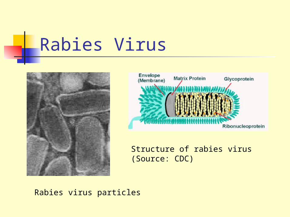
Rabies Virus
Structure of rabies virus (Source: CDC)
Rabies virus particles
Epidemiology
Rabies is a zoonosis which is prevalent in wildlife. The main animals involved differs from continent to continent.
Europe fox, bats
Middle East wolf, dog
Asia dog
Africa dog, mongoose, antelope
N America foxes, skunks, raccoons, insectivorous bats
S America dog, vampire bats
Pathogenesis The commonest mode of transmission in man is by the bite of a
rabid animal, usually a dog. Rabies is an acute infection of the CNS which is almost invariably fatal.
Following inoculation, the virus replicates in the striated or connective tissue at the site of inoculation and enters the peripheral nerves through the neuromuscular junction.
It then spreads to the CNS in the endoneurium of the Schwann cells.
Terminally, there is widespread CNS involvement but few neurons infected with the virus show structural abnormalities. The nature of the profound disorder is still not understood.
Laboratory Diagnosis Histopathology - Negri bodies are pathognomonic of rabies. However,
Negri bodies are only present in 71% of cases. Rapid virus antigen detection - in recent years, virus antigen detection
by IF had become widely used. Corneal impressions or neck skin biopsy are taken. The Direct Fluorescent Antibody test (DFA) is commonly used.
Virus cultivation - The most definitive means of diagnosis is by virus cultivation from saliva and infected tissue. Cell cultures may be used or more commonly, the specimen is inoculated intracerebrally into infant mice. Because of the difficulties involved, this is rarely offered by diagnostic laboratories.
Serology - circulating antibodies appear slowly in the course of infection but they are usually present by the time of onset of clinical symptoms.
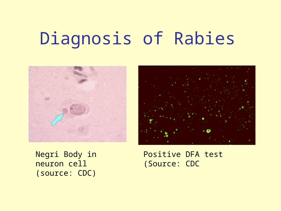
Negri Body in neuron cell (source: CDC)
Positive DFA test (Source: CDC
Diagnosis of Rabies
Management and Prevention
Pre-exposure prophylaxis - Inactivated rabies vaccine may be administered to persons at increased risk of being exposed to rabies e.g. vets, animal handlers, laboratory workers etc.
Post-exposure prophylaxis - In cases of animal bites, dogs and cats in a rabies endemic area should be held for 10 days for observation. If signs develop, they should be killed and their tissue.
Wild animals are not observed but if captured, the animal should be killed and examined. The essential components of postexposure prophylaxis are the local treatment of wounds and active and passive immunization.
Once rabies is established, there is nothing much that could be done except intensive supportive care. To date, only 2 persons with proven rabies have survived.
Postexposure Prophylaxis Wound treatment - surgical debridement should be carried out.
Experimentally, the incidence of rabies in animals can be reduced by local treatment alone.
Passive immunization - human rabies immunoglobulin around the area of the wound; to be supplemented with an i.m. dose to confer short term protection.
Active immunization - the human diploid cell vaccine is the best preparation available. The vaccine is usually administered into the deltoid region, and 5 doses are usually given.
There is convincing evidence that combined treatment with rabies immunoglobulin and active immunization is much more effective than active immunization alone. Equine rabies immunoglobulin (ERIG) is available in many countries and is considerably cheaper than HRIG.
Rabies VaccinesThe vaccines which are available for humans are present are inactivated whole virus vaccines.
Nervous Tissue Preparation e.g. Semple Vaccine - associated with the rare complication of demyelinating allergic encephalitis.
Duck Embryo Vaccine - this vaccine strain is grown in embryonated duck eggs This vaccine has a lower risk of allergic encephalitis but is considerably less immunogenic.
Human Diploid Cell Vaccine (HDCV) - this is currently the best vaccine available with an efficacy rate of nearly 100% and rarely any severe reactions. However it is very expensive.
Other Cell culture Vaccines - because of the expense of HDCV, other cell culture vaccines are being developed for developing countries. However recent data suggests that a much reduced dose of HDCV given intradermally may be just be effective.
Control of Rabies Urban - canine rabies accounts for more than 99% of all human
rabies. Control measures against canine rabies include; stray dog control.
Vaccination of dogs
quarantine of imported animals
Wildlife - this is much more difficult to control than canine rabies. However, there are on-going trials in Europe where bait containing rabies vaccine is given to foxes. Success had been reported in Switzerland.
Arenaviruses Enveloped ssRNA viruses virions 80-150nm in diameter genome consists of 2 pieces of
ambisense ssRNA. 7-8 nm spikes protrude from the
envelope. host cell ribosomes are usually seen
inside the outer membrane but play no part in replication.
Members of arenaviruses include Lassa fever, Junin and Macupo viruses.
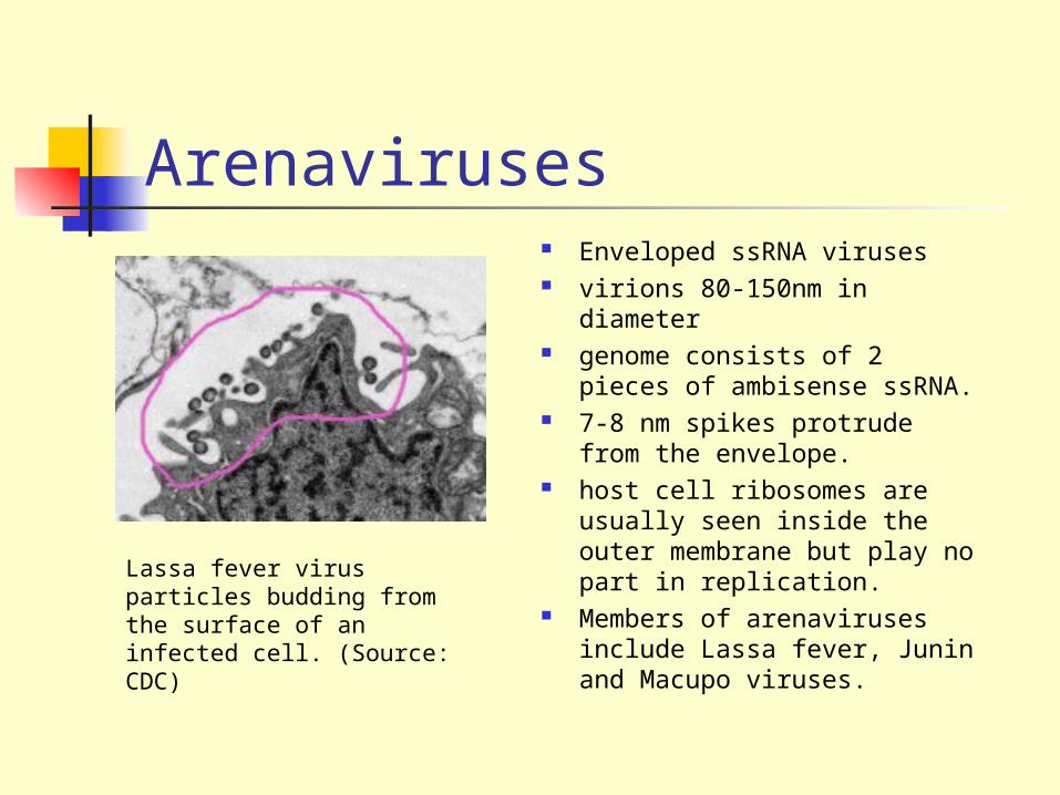
Lassa fever virus particles budding from the surface of an infected cell. (Source: CDC)
Lassa Fever
Found predominantly in West Africa, in particular Nigeria, Sierra Leone and Liberia.
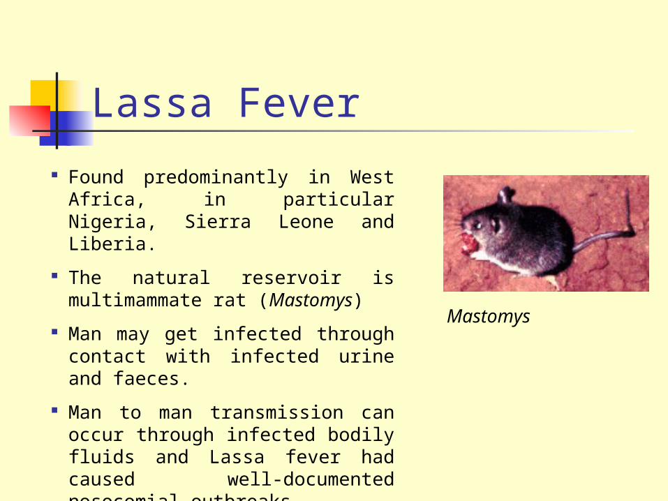
The natural reservoir is multimammate rat (Mastomys)
Man may get infected through contact with infected urine and faeces.
Man to man transmission can occur through infected bodily fluids and Lassa fever had caused well-documented nosocomial outbreaks.
Mastomys
Clinical Manifestations Incubation period of 3-5 days. Insidious onset of non-specific symptoms such as fever, malaise,
myalgia and a sore throat. Typical patchy or ulcerative pharyngeal lesions may be seen. Severe cases may develop the following:
Myocarditis Pneumonia Encephalopathy Haemorrhagic manifestations Shock
The reported mortality rate for hospitalized cases of Lassa fever is 25%. It carries a higher mortality in pregnant women.
Laboratory Diagnosis Lassa fever virus is a Group 4 Pathogen. Laboratory diagnosis should only
be carried out in specialized centers.
Detection of Virus Antigen - the presence of viral antigen in sera can be detected by EIA. The presence of viral antigen precedes that of IgM.
Serology - IgM is detected by EIA. Using a combination of antigen and IgM antibody tests, it was shown that virtually all Lassa virus infections can be diagnosed early.
Virus Isolation - virus may be cultured from blood, urine and throat washings. Rarely carried out because of safety concerns.
RT-PCR - being used experimentally.
Management and Prevention
Good supportive care is essential. Ribavirin - had been shown to be effective against Lassa fever with a 2
to 3 fold decrease in mortality in high risk Lassa fever patients. Must be given early in the illness.
Hyperimmune serum - the effects of hyperimmune serum is still uncertain although dramatic results have been reported in anecdotal case reports.
Postexposure Prophylaxis - There is no established safe prophylaxis. Various combinations of hyperimmune immunoglobulin and/or oral ribavirin may be used.
There is no vaccine available, prevention of the disease depends on rodent control.
Junin and Macupo Viruses Junin and Macupo viruses are the causative agents of Argentine
and Bolivian Haemorrhagic fever respectively. Calomys musculinis and C callosus are the rodent vectors. The clinical presentations are similar to that of Lassa fever.
Neurological signs are much more prominent than in Lassa fever.
Unlike Lassa virus, no secondary human to human spread had been recorded.
Hyperimmune serum and ribavirin had been shown to be effective in treatment.
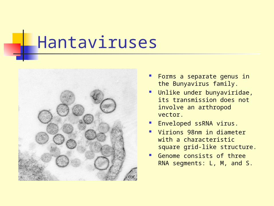
Hantaviruses
Forms a separate genus in the Bunyavirus family.
Unlike under bunyaviridae, its transmission does not involve an arthropod vector.
Enveloped ssRNA virus. Virions 98nm in diameter with a
characteristic square grid-like structure.
Genome consists of three RNA segments: L, M, and S.
History Haemorrhagic Fever with Renal Syndrome (HFRS: later
renamed hantavirus disease) first came to the attention of the West during the Korean war when over 3000 UN troops were afflicted.
It transpired that the disease was not new and had been described by the Chinese 1000 years earlier.
In 1974, the causative was isolated from the Korean Stripped field mice and was called Hantaan virus.
In 1995, a new disease entity called hantavirus pulmonary syndrome was described in the “four corners” region of the U.S.
Some Subtypes of hantaviruses associated with human disease
Hantaan, Porrogia and related viruses - This group is found in China, Eastern USSR, and some parts of S. Europe. It is responsible for the severe classical type of hantavirus disease. It is carried by stripped field mice. (Apodemus agrarius)
Seoul type - associated with moderate hantavirus disease. It is carried by rats and have a worldwide distribution. It has been identified in China, Japan, Western USSR, USA and S.America.
Puumala type - mainly found in Scandinavian countries, France, UK and the Western USSR. It is carried by bank voles (Clethrionomys glareolus) and causes mild hantavirus disease (nephropathia epidemica).
Sin Nombre - found in many parts of the US, Canada and Mexico. Carried by the Deer Mouse (Peromyscus maniculatus) and causes hantavirus pulmonary syndrome.
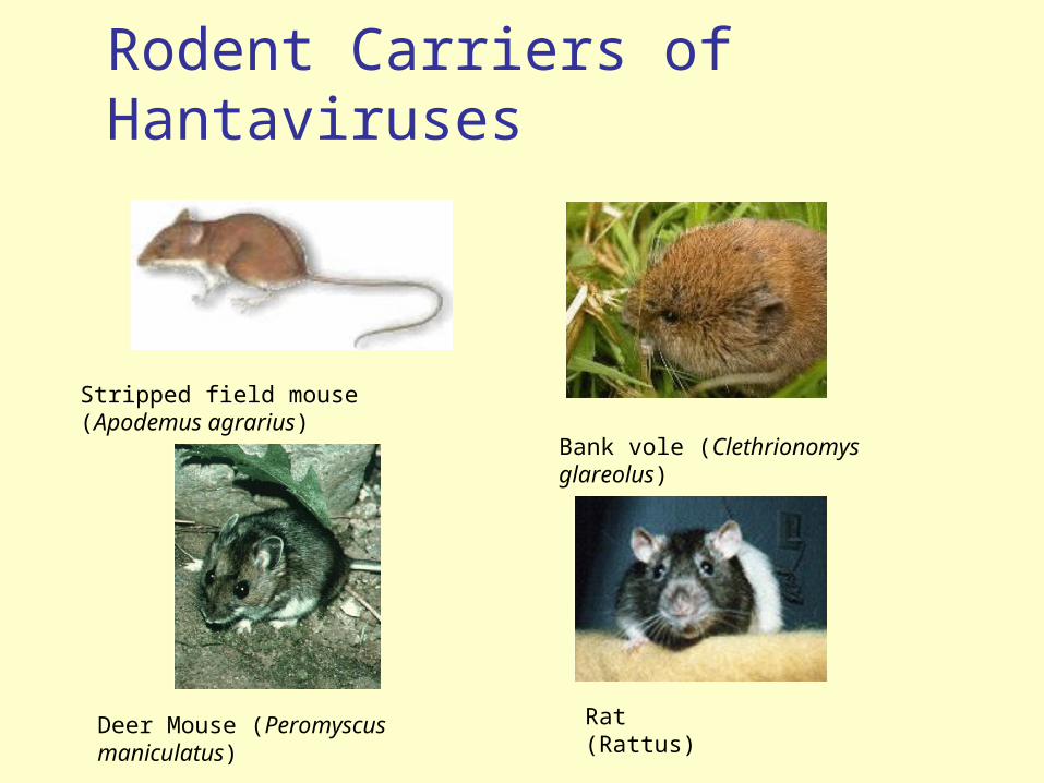
Rodent Carriers of Hantaviruses
Stripped field mouse (Apodemus agrarius)
Bank vole (Clethrionomys glareolus)
Deer Mouse (Peromyscus maniculatus) Rat (Rattus)
Clinical Features of Hantavirus Disease
The multisystem pathology of HVD is characterized by damage to capillaries and small vessel walls, resulting in vasodilation and congestion with hemorrhages.
Classically, hantavirus disease consists of 5 distinct phases. These phases may be blurred in moderate or mild cases.
Febrile phase - abrupt onset of a severe flu-like illness with a erythematous rash after an incubation period of 2-3 days.
Hypotensive phase - begins at day 5 of illness Oliguric phase - begins at day 9 of illness. The patient may develop acute
renal failure and shock. Haemorrhages are usually confined to petechiae. The majority of deaths occur during the hypotensive and oliguric phases
Diuretic phase - this occurs between days 12-14 . Convalescent phase - this may require up to 4 months.
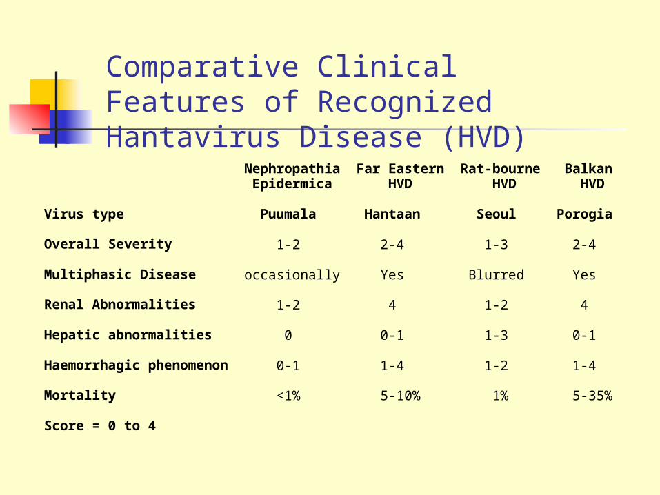
Nephropathia Far Eastern Rat-bourne Balkan Epidermica HVD HVD HVD
Virus type Puumala Hantaan Seoul Porogia
Overall Severity 1-2 2-4 1-3 2-4
Multiphasic Disease occasionally Yes Blurred Yes
Renal Abnormalities 1-2 4 1-2 4
Hepatic abnormalities 0 0-1 1-3 0-1
Haemorrhagic phenomenon 0-1 1-4 1-2 1-4
Mortality <1% 5-10% 1% 5-35%
Score = 0 to 4
Comparative Clinical Features of Recognized Hantavirus Disease (HVD)
Hantavirus Pulmonary Syndrome (HPS)
More than 250 cases of HPS have been reported throughout North and South America with a mortality rate of 50%
In common with classical HVD, HPS has a similar febrile phase.
However, the damage to the capillaries occur predominantly in the lungs rather than the kidney.
Shock and cardiac complications may lead to death.
The majority of HPS cases are caused by the Sin Nombre virus. The other cases are associated with a variety of other hantaviruses e.g. New York and Black Creek Canal viruses.
Diagnosis Serological diagnosis - a variety of tests including IF, HAI, SRH, ELISAs
have been developed for the diagnosis of HVD and HPS. Direct detection of antigen - this appears to be more sensitive than
serology tests in the early diagnosis of the disease. The virus antigen can be demonstrated in the blood or urine.
RT-PCR - found to of great use in diagnosing hantavirus pulmonary syndrome.
Virus isolation - isolation of the virus from urine is successful early in hantavirus disease. Isolation of the virus from the blood is less consistent. Sin Nombre virus has never been isolated from patients with HPS.
Immunohistochemistry - useful in diagnosing HPS.
Treatment and Prevention
Treatment of HVD and HPS depends mainly on supportive measures.
Ribavirin - reported to be useful if given early in the course of hantavirus disease. Its efficacy is uncertain in hantavirus pulmonary syndrome.
Vaccination - an inactivated vaccine is being tried out in China. Other candidate vaccines are being prepared.
Rodent Control - control measures should be aimed at reducing contact between humans and rodents.