Embed Size (px)
Citation preview

[Sample of Oral Presentations] :: Full paper for oral presentation: 4 to 7 pages
Sulfur nutrition involves in the stress tolerance in forage rapes
Bok-Rye Lee, Sang-Hyun Park, Qian Zhang, Rashed Zaman, Tae-Hwan KimDepartment of Animal Science, Institute of Agricultural Science and Technology, College of Agriculture & Life
Science, Chonnam National University, Buk-Gwangju, P.O Box 205, Gwangju 500-600, South KoreaCorrespondence: Tae-Hwan Kim, [email protected]
Key words: antioxidative system, Fe deficiency, photosynthethic organelle, salt stress, sulfur nutrition
Abstract: The objective of this study was to investigate the effect of sulfur nutrition on antioxidative system and photosynthetic mechanism under salt stress or Fe deficiency under with- and without sulfur nutrient condition. Salt stress and Fe deficiency induced oxidative stresses lead to accumulation of O2
•─ and H2O2. These indicants of oxidative stress were significantly alleviated by sulfur supply. The activity of antioxidative enzymes, ascorbate peroxidase (APOD) and catalase (CAT), was significantly decreased under salt-stress and Fe-deficiency stress conditions, whereas these increased in the presence of sulfur. Salt stress seriously resulted in decrease of photosynthetic pigments as chlorophyll and carotenoid concentration both in presence or absence of sulfur but these negative effects were more severe in the absence of sulfur. The proteomic analysis of multiple protein complexes in the thylakoid by BN-PAGE showed that expression of PS1, PSII and RuBisCO were significantly repressed under salt stress in absence of sulfur, whereas their expression was largely recovered by sulfur supply. The activity of RuBisCO was closely related to sulfur status in leaves. Twelve proteins were absent in the absence of sulfur with Fe deprived plants, whereas 13 proteins were up-regulated. The functional classification these identified proteins was estimated that 40% of the proteins belongs to S and Fe assimilation. The present results indicated that sulfur nutrition has significant role in ameliorating the damage in phtotosynthetic apparatus and oxidative stress by salt stress or Fe deficiency.
IntroductionSulfur (S) is one of six macronutrients needed for proper plant growth and development. It is present in the amino acids cysteine and methionine, and is thus an important component of proteins and peptides. Many enzymes require sulfur-containing co-enzymes and Fe-S cluster prosthetic groups cluster which have function in vital processes such as photosynthesis, respiration, S and N metabolism, plant hormone and coenzyme synthesis , for their activity (Balk and Pilon 2011). In addition, plants contain many other organic sulfur compounds, such as thiols, sulfolipids, glucosinolates or alliins, which play important roles in the normal plant lifecycle and in protection against stress and pathogens (Davidian and Kopriva 2010). Cysteine is first carbon-nitrogen-reduced sulfur product and serves as sulfur donor for the synthesis of methionine which is the precursor for S-adenosyl methionine (SAM), the precursor for ethylene, polyamines, and nicotinamine (Davidian and Kopriva 2010). Cysteine is also incorporated into tripeptide glutathione (GSH) which is major constituent of storage and transport form of reduced S in plants. This sulfur-containing metabolite, GSH, plays an important role in plant stress defense such as detoxification of reactive oxygen species and redox regulation (Khan et al. 2009). Sulfur is also significant for N assimilation. Numerous studies have defined regulatory interactions between sulfur assimilation and nitrogen metabolism and well-coordinated as the availability of one element regulates the other in higher plants (Davidian and Kopriva 2010; Carfagna et al. 2011).
In recent decades, the reduction of industrial S emissions of S to the atmosphere and the subsequent deposition of S on the soil has increased the incidence of sulfur limitation in the region of world. Sulfur availability is one of limiting factor in yield and quality parameters of crops. Sulfur deficiency decreased biomass, protein level, chlorophyll content, pigment system II (PSII) activity and ribulose 1,5-bisphosphate carboxylase (RuBisCO) content. Besides, it is lead to serious imbalance in concentration of cysteine and GSH, which are main antioxidants in the plant cell apart from ascorbate (Juszczuk and Ostasxewska 2011). Additionally, sulfur deficiency results in a reduction of nitrate reductase activity and increase of amino acids which is from hydrolysis of the previously synthesized protein (Lee et al. 2013). Conversely, adequate supply of sulfur increased chlorophyll content and photosynthetic enzymes activity due to increase iron use efficiency, and levels of enzymes of sulfur and nitrogen assimilation (Astolfi et al. 2006; Zuchi et al. 2012). A lager accumulation of N by sufficient S supply maintains high chlorophyll content and high activity of enzymes of Calvin cycle.
On the other hand, application of sulfur mitigated the adverse effects of heavy metals (Cadmium, arsenic and zinc) stress by enhancing plant growth, chlorophyll content, net photosynthetic rate, effective PSII quantum

yield Y(II) and antioxidant enzymes such as catalase, peroxidase and superoxide dismutase (Fontes and Cox 1995; Cao et al. 2004; Cai et al. 2011). Moreover, foliar-applied elemental sulfur to plant infected with fungi, viruses and harmful insects enhance growth yield and resistance to fungal pathogen and reduced fungal infection and virus accumulation (Wiliams et al. 2002; Kiraly et al. 2011). Despite extensive researches attempting to elucidate the interactions between external sulfur supply and stress tolerance, to our knowledge, the information about sulfur significance on environmental stress such as salt, drought, heat and nutrient deficiency have not yet been fully investigated.
In this study, to understand the significance of sulfur in improving abiotic stress tolerant, we investigated effect of sulfur nutrition on antioxidative system and photosynthetic mechanism under salt stress or iron deficiency stress under with- and without sulfur nutrient condition.
Materials and MethodsPlant material and treatmentSalt stress: The experimental plant, Kentucky bluegrass (Poa pratensis L.) were taken from the local golf course and were transferred to soilrite/vermiculite, and were divided in to 2 groups; 1 group were supplied with complete nutrient solution and another set of plant, were supplied with S-free nutrient solution during 4 weeks. After 4 weeks of S-treatment, each of S-supplied or S-deprived plants were exposed to salt stress with 100 mM NaCl or non-salt stress, respectively, for 3 weeks. Four groups of treatment thus were designated as sufficient S without salt stress (+S/non-salt, control), sufficient S with salt stress (+S/salt), deprived S without salt stress (-S/non-salt), and deprived S with salt stress (-S/salt) with three replicates. The samplings were done at 0, 7, 14 and 21 days of salt stress. Leaves and roots were harvested and immediately frozen in liquid nitrogen and stored in deep-freezer for further analysis.
Iron (Fe) deficiency stress: surface-sterilized seeds of Brassica napus L. cv. Mosa were germinated in wet filter paper in the petri dishes at 30ºC in the dark for 3 days. Four seedlings were transplanted and then thinned to two after 2 weeks. The seedlings were grown in 3 L plastic pots with hydroponic nutrient solution. Natural light was supplemented with 200 µmole m-2 S-1 at the canopy height for 16 h day -1. Eight-week-old plants were divided in four groups with 3 replications to receive different treatments: sufficient in S and Fe (+S/+Fe, control), sufficient S but Fe deprived (+S/-Fe), deprived S but sufficient Fe (-S/+Fe), and deprived S and Fe (-S/-Fe). After 5 and 10 days of treatment, plants were harvested and immediately frozen in liquid nitrogen and stored in deep-freezer for further analysis.
Determination of O2•─ and H2O2 content
The detection of O2•─ was made by hydroxylamine oxidation (Wang and Luo 1990). A mixture of 0.5 ml enzyme
extract and 1 cm3 of the prepared hydroxylation was incubated at 25 C for 1 h, then reacted with 1 ml of 17 mM -aminobenzene sulfonic acid and 7 mM α-naphthylamine solution at 25 C for 20 min. The absorbance was determined at 530 nm. O2
•─ concentration was obtained using a linear calibration curve of NaNO2. For H2O2
determination, about 200 mg DW was homogenized with 3 ml of 50 mM phosphate buffer (pH 6.8) and then centrifuged at 6,000 g for 25 min. The resulting extract was mixed with 1 ml of 0.1 % titanium chloride in 20 % (v/v) H2SO4 and centrifuged at 6 000 g for 15 min. The absorbance was immediately read at 410 nm. H2O2
concentration was calculated using the extinction coefficient 0.28 μM-1 cm-1 (Lin and Kao 2001).
Measurement of antioxidant enzyme activitiesFor extraction of enzymes, fresh samples (0.5 g) were homogenized with 1.5 ml of 100 mM K-PO4 buffer solution (pH 7.0) containing 2 mM phenylmethylsulfonyl fluoride (PMSF), and centrifuged at 14,000 g at 4 C for 20 min. Protein concentration was determined using the method of Bradford (1976). The activity of superoxide dismutase (SOD) was determined by measuring its ability to inhibit the photoreduction of nitroblue tetrazolium (NBT) (Giannopolitis and Ries 1977). One unit of enzyme activity was defined as the amount of enzyme required to inhibit 50 % of the NBT photoreduction in comparison with tubes lacking the plant extract. Catalase (CAT) activity was assayed using the method of Mishra et al. (1993). The reaction mixture of 1 ml contained 0.5 ml of 100 mM potassium phosphate buffer (pH 7.0), 0.1 ml of 110 mM H 2O2 and enzyme extract. The decrease in absorbance at 240 nm was recorded as a result of H2O2 degradation (extinction coefficient of 36 mM -1 cm-1). POD activities were measured using different substrates: ascorbate and guaiacol. POD activity with ascorbate as hydrogen donor (ascorbate-peroxidase; APOD) was determined by measuring the decrease in absorbance at 290 nm (extinction coefficient of 2.8 mM -1 cm-1) according to Chen and Asada (1989). One unit of enzyme activity was defined as the amount of enzyme that causes the formation of 1 μM ascorbate oxidized per min.

Photosynthetic pigments and photosynthetic activityThe content of chlorophyll and carotenoid was estimated by the method of Hiscox and Israelstam (1979). Fresh leaves were extracted with 10 ml of dimethyl sulfoxide and optical density was recorded at 480, 645, 520 and 663 nm. The content of total chlorophyll and carotenoid was calculated using the formulae given by Arnon (1949). RuBisCO activity was determined by the method given by Usuda (1985) with some modifications. Leaf samples were homogenized 0.1 M Bicine (pH 7.8) containing 20 mM MgCl2, 1 mM Na-EDTA, 5 mM DTT and 2% PVP and centrifuged at 10,000 g at 4 ℃ for 10 min. The activity was determined by monitoring NADH oxidation at 340nm.
BN – PAGE (Promeomic analysis of thylakoid protein complexes)BN-PAGE of integral thylakoid proteins was performed according to Kugler et al. (1997). Five gram of fresh leaf tissues were homogenized in liquid nitrogen and thylakoid membranes were extracted using extraction buffer (pH 7.8) containing 20 mM tricine-NaOH, 70 mM sucrose and 5 mM MgCl2 and were filtered through miracloth/cheesecloth before centrifugation at 4,500 g for 10 min. The thylakoid pellet was resupsneded in same buffer (pH 7.8) and centrifuged again. The resulting pellet containing thylakoid membranes was washed and extracted with each proper buffer (Swamy et al. 2006). An equal volume of resuspension buffer containing 2% (w/v) n-dodecyl-β-D maltoside (Sigma, St. Louis, MO, USA) was added under continuous mixing. The solubilization of membrane–protein complexes was allowed to occur for 3 min on ice. Insoluble material was removed by centrifugation at 18,000 g for 15 min. The supernatant was mixed with 0.1 volume of serva blue (5 % w/v Serva blue G, 100 mM BisTris–HCl, pH 7.0, 30 % w/v sucrose, and 500 mM ε-Amino-n-caproic acid) and loaded onto a 0.75-mm-thick 5 - 12.5 % w/v acrylamide gradient gel (180×160 mm). Electrophoresis was performed at 4°C by increasing the voltage from 100 V to 200 V overnight.
Two dimensional polyacrylamide gel electrophoresis (2DE)One gram of leaf tissues was homogenized in phosphate buffer (pH 7.6) containing 40 mM tris, 0.07 % bME (beta mercapto ethanol), 2 % PVP (Polyvinylpyrrolidone) and 1 % TritonX 100 at 4 _C using chilled pestle and mortar on ice. The homogenates were centrifuged at 15,000 rpm for 15 min, and proteins were precipitated with 10 % TCA/ acetone overnight at -20 _C. The resultant precipitate was centrifuged at 15,000 rpm for 15 min and washed with 80 % chilled acetone containing 2 mM EDTA and 0.07 % ßME. The proteins’ pellet was solubilized in solubilization buffer containing 9 M urea, 2 M thiourea, 4 % CHAPS, 2 % TritonX100, 50 mM DTT and 0.2 % ampholine (pH 4–7). For two-dimensional polyacrylamide gel electrophoresis (2DE), three hundred micrograms of proteins was separated by 2-DE in the first dimension by isoelectric focusing on 11 cm IPG strip (pI 4–7) and the second dimension by SDS-PAGE on Protean II unit (Bio-Rad, Hercules, USA) according to method given by Qureshi et al. (2010)
Results and DiscussionSulfur effects on antioxidative system under salt stress conditionSalt-stress resulted in O2
•─accumulation from day 7 both in the presence or absence of S-nutrition. At day 21, salt-stress in the absence of S-nutrition (-S/salt) increased O2
•─concentration by 70 % compared to control (+S/non-salt), but the rate of increase was much lower (50 %) in the presence of S-nutrition (+S/salt) (Figure 1A). Similarly, salt stress significantly increased the H2O2 at day 21 under salt-stressed condition in S-deprivation (-S/salt) whereas it decreased when S-nutrition was supplied (+S/salt) to plant (Figure 1B). O2
•─is converted to H2O2 by SOD in the chloroplast, mitochondrion, cytoplasm, apoplast and peroxisome (Bowler et al. 1992). H2O2 is scavenged by two groups of enzymes; catalases (CAT) and ascorbate peroxidases (APOD).

Figure 1. Changes in content of reactive oxygen species such as (A) O2•─ and (B) H2O2, activities of (C) catalase (CAT) and (D)
ascorbate peroxidase (APOD) of four sulfur and salt stress combined treatments; sufficient S without salt stress (+S/non-salt, control), sufficient S with salt stress (+S/salt), deprived S without salt stress (-S/non-salt), and deprived S with salt stress (-S/salt) in Kentucky bluegrass for 21 days. Bars labeled with the same letters are not significantly different (p>0.05) according to Duncan’s multiple range test. Vertical bars represent mean ± SE for n=3.
Many studies documented the changes in antioxidant enzyme activities in response to salt, suggests that the induction in these activities can be the basis for salt-stress tolerance (Hernández et al. 2000; Shalata et al. 2001). In this study, CAT activity was significantly decreased by 53 % in absence of S-nutrition (-S/salt) whereas it was not significantly decreased in presence of S-nutrition (+S/salt) at day 21 (Figure 1C). Salt-stress largely decreased the activity of APOD by 73 % in absence of S-nutrition (-S/salt), however, the rate of decrease was much lower (19 %) in presence of S-nutrition (+S/salt) at day 14. Salt-stress decreased the APOD activity at day 7 both in the presence or absence of S-nutrition compare to control at day 21 (Figure 1D). Therefore, these results suggest that S-nutrition reduces the accumulation of ROS accumulation as O2
•─and H2O2, and alleviates the oxidant stress.
Sulfur effects on antioxidative system under iron deficiency conditionAccumulation of oxidative stress was visualized in situ in the form of H2O2 and O2
•─ by histochemical methods. S-deprived leaves exhibited highly enhanced O2
•─ as brownish staining indicated by white arrows (Figure 2A). The staining was not increased in the +S/-Fe leaves compared to controls. In the both -S/+Fe and -S/-Fe leaves, dark brown spot areas for H2O2 were widespread, whereas the accumulation of these oxy radicals was largely reduced in the presence of S (+S/+Fe, +S/-Fe) (Figure 2B).
Figure 2. Localization of (A) O2•─ and (B) H2O2, (C) superoxide dismutase (SOD) activity, and (D) ascorbate content. as affected by four
S/Fe combined treatments; sufficient in S and Fe (+S/+Fe, control white bars), deprived S and Fe (-S/-Fe dark grey bars), deprived S but sufficient Fe (-S/+Fe light grey bars) and sufficient S but deprived Fe (+S/-Fe black bars) for 10 days in Brassica napus. Bars labeled with the same letters are not significantly different (p>0.05) according to Duncan’s multiple range test. Vertical bars represent mean ± SE for n=3.

Oxidative damage has been considered as one of the phytotoxic of reactive oxygen species (ROS) generation (Ali et al. 2005). Superoxide dismutase (SOD) is one of main consequence in ascorbate-glutathione cycle to detoxify the ROS. In this study, SOD activity was significantly increased by S-deprivation either with or without Fe at day 5 compared to control (Figure 2C). After 10 days treatment, S-and/or Fe-deprivation increased activity of SOD and the highest increase was observed in –S/-Fe treatment. Ascorbate and glutathione are the major antioxidants in plants. In our study, total ascorbate concentration was reduced by S- or Fe-deficiency. However, it was increased by presence of sulfur in absence of Fe deficiency (Figure 2D). These results showed that Fe deficiency increased the production of ROS and resulted in oxidative stress, and S-availability might alleviate the oxidative stress.
Sulfur effects on photosynthetic organelle system under salt stress conditionSalt-stress affected to photosynthetic pigments like chlorophyll and carotenoid (Figure 3A; 3B). From day 7, salt-stress seriously resulted in the decrease of chlorophyll and carotenoid content both in presence or absence of S. Similar result has been report in haematococcuspluvialis under salt-stress (Sarada et al. 2002). However, the extent of the decrease in chlorophyll and carotenoid content was largely reduced by S supply, showing 28% and 34% increase in presence of S-nutrition (+S/salt), respectively. The results obtained suggested the loss of photosynthetic pigment in S-nutrient compared S-deprivation, suggesting the possible influence of S under salt stress is positively alleviated by S-nutrition. The proteomic analysis of multiple protein complexes in the thylakoid by BN-PAGE showed in Figure 3C. The Kentucky bluegrass BN gel profile showed an interesting band identified as subcomplexes of PSI, PSII and RuBisCO. The intensity of PSI, PSII-core dimer super complex bands and RuBisCO reduced in S-deprived plants (-S/non-salt) and decreased further in the absence of S under salt- stressed plants (-S/salt), whereas these bands were abundant in the presence of S under salt-stressed plants (+S/salt) to the level of control. PSII core dimer and PSI (RCI-LHCI) complex bands were more affected. In synechococcus, salt-stress inactivated both PSI and PSII because the change in K/Na ratio (Allakhverdiev et al. 2000). In plants, RuBisCO is the key enzyme which is responsible for the primary step in CO2 fixation and its carboxylating capacity can be the limiting factor in photosynthesis (Woodrow and Berry 1988). Sulfur-deprivation resulted in a decrease of the RuBisCO activity in leaves from Day 0 (Figure 3D). At day 21, salt-stress in the absence of S (-S/salt) rapidly decreased RuBisCO activity by 80% compared control, but the rate of decrease was much lower (62%) in the presence of S (+S/salt). These results showed that the loss of photosynthetic pigments and the depressed RuBisCO under salt-stressed condition were positively alleviated by sulfur nutrition.
Figure 3. Changes in photosynthetic pigments such as (A) chlorophyll and (B) carotenoid, (C) multiple protein complexes in the thylakoid by BN-PAGE, and (D) RuBisCO activity of four sulfur and salt stress combined treatments; sufficient S without salt stress (+S/non-salt, control), sufficient S with salt stress (+S/salt), deprived S without salt stress (-S/non-salt), and deprived S with salt stress (-S/salt) in Kentucky bluegrass for 21 days. Bars labeled with the same letters are not significantly different (p>0.05) according to Duncan’s multiple range test. Vertical bars represent mean ± SE for n=3.
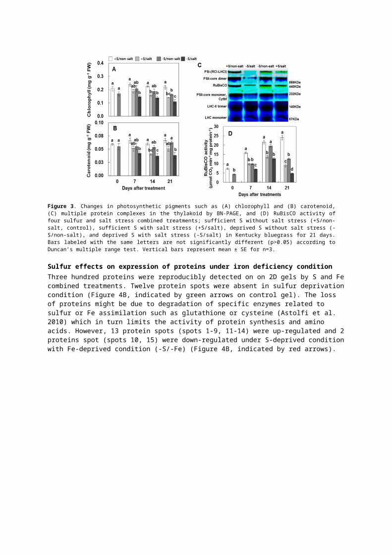
Sulfur effects on expression of proteins under iron deficiency conditionThree hundred proteins were reproducibly detected on on 2D gels by S and Fe combined treatments. Twelve

protein spots were absent in sulfur deprivation condition (Figure 4B, indicated by green arrows on control gel). The loss of proteins might be due to degradation of specific enzymes related to sulfur or Fe assimilation such as glutathione or cysteine (Astolfi et al. 2010) which in turn limits the activity of protein synthesis and amino acids. However, 13 protein spots (spots 1-9, 11-14) were up-regulated and 2 proteins spot (spots 10, 15) were down-regulated under S-deprived condition with Fe-deprived condition (-S/-Fe) (Figure 4B, indicated by red arrows).
Figure 4. 2D references images of four S/Fe combined treatments; (A) sufficient in S and Fe (+S/+Fe, control) and (B) deprived S and Fe (-S/-Fe) in roots of Brassica napus. (C) Identification of differentially expressed proteins under S/Fe combined treatment. (D) Functional classification of identified proteins from leaves analyzed by MALDI-TOF-MS as described by Beven et al. (1998).
These proteins spots were identified by MALDL-TOF-MS (Figure 4C). The seven proteins up-regulated by –S/-Fe treatment were S and Fe related proteins such as Fe-related chelates, enzymes involved in Fe uptake, induction of cysteine and phytochelatins. However, these proteins were recovered by sulfur nutrition in Fe deprived roots. The proteins identified from roots were also classified into four categories (Figure 4D) as 40% S/Fe related proteins, 30% transcriptional regulation, 10% transcription factor and 20% biosynthesis and general metabolism. ). Taken together, the results indicate that S-nutrition has a more influential role in modulating the protein expression to alleviate deleterious impacts of Fe deficiency in the roots of oilseed rape plants.
AcknowledgmentsThis work was carried out with the support of "Cooperative Research Program for Agriculture Science & Technology Development (PJ010099)" Rural Development Administration, Republic of Korea.
ReferencesAli MB, Hahn EJ, Paek KY (2005) Effect of temperature on oxidative stress defense systems, lipid peroxidation
and lipoxygenase activity in Phalaenpopsis. Plant Physiology and Biochemistry, 43: 213–223.Allakhverdiev S I, Sakamoto A, Nishiyama Y, Inaba M, Murata N (2000) Ionic and osmotic effects of NaCl-
inducedinactivation of photosystem I and II in Synechococcus sp. Plant Physiology, 123: 1047–1056.Armstrong GA, Hearst JE (1996) Carotenoids 2: genetics and molecular biology of carotenoid pigment
biosynthesis. FASEB Journal, 10: 228–237.Astolfi S, Cesco S, Zuchi S, Neumann G, Roemheld V (2006) Sulphur starvation reduces phytosiderophores
release by Fe-deficient barley plants. Soil Science & Plant Nutrition, 52: 80–85.Astolfi S, Zuchi S, Neumann G, Cesco S, Sanita di Toppi L, Pinton R (2012) Response of barley plants to Fe
deficiency and Cd contamination as affected by S starvation. Journal of Experimental Botany, 63(3): 1241–1250.
Balk J, Pilon M (2011) Ancient and essential: the assembly of iron–sulfur clusters in plants. Trends in Plant

Science, 16: 218–226Bowler C, Monagu MV, Inze D (1992) Superoxide dismutases and stress tolerance. Annual Review of Plant
Physiology and Plant Molecular Biology, 43: 83–116. Cai Y, Cao F, Cheng W, Zhang G, Wu F (2011) Modulation of exogenous glutathione in phytochelatins and
photosynthetic performance against cd stress in the two rice genotypes differing in Cd tolerance. Biological Trace Element Research, 143:1159–1173.
Cao X, Ma LQ, Tu C (2004) Antioxidative responses to arsenic in arsenichyper accumulator Chinese brake fern (Pterisvittata L.). Environmental Pollutution, 128: 317–325.
Davidian JC, Kopriva, S (2010) Regulation of sulfate uptake and assimilation – the same or not the same? Molecular Plant, 3: 314–325.
Fontes RLF, Cox FR (1995) Effects of sulfur supply on soybean plants exposed to zinc toxicity. Journal of Plant Nutrition, 18: 1893–1906.
Hernández JA, Jimenez A, Mullineaux P, Sevilla F (2000) Tolerance of pea (Pisum sativum L.) to long-term salt stress is associated with induction of antioxidant defenses. Plant Cell and Environment, 23: 853–862.
Hippler M, Klein J, Fink A, Allinger T, Hoerth P (2001) Towards functional proteomics of membrane protein complexes: analysis of thylakoid membranes from Chlamydomonas reinhardtii. Plant Journal, 28: 595–606.
Juszczuk IM, Ostaszewska M (2011) Respiratory activity, energy and redox status in sulphur-deficient bean plants. Environmental and Experimental Botany, 74: 245–254.
Khan MA, Ungar IA, Showalter AM, Dewald HD (1998) NaCl-induced accumulation of glycinebetaine in four subtropicalhalophytes from Pakistan. Physiologia Plantarum, 102: 487–492.
Kiraly L, Kunstler A, Holler K, Fattinger M, Juhasz C, Muller M, Gullner G, Zechmann B (2011) Sulfate supply influences compartment specific glutathione metabolism and confers enhanced resistance to Tobacco mosaic virus during a hypersensitive response. Plant Physiology and Biochemistry, 59: 44–54.
Lee BR, Muneer S, Kim KY, Avice JC, Ourry A, Kim TH (2013) S-deficiency responsive accumulation of amino acids is mainly due to hydrolysis of the previously synthesized proteins – not to de novo synthesis in Brassica napus. Physiologia Plantarum, 147: 369– 380.
Naumann B, Stauber EJ, Busch A, Sommer F, Hippler M (2005) N-terminal processing of Lhca3 is a key step in remodelling of the photosystem I-light-harvesting complex under iron deficiency in Chlamydomonas reinhardtii. The Journal of Biological Chemistry, 280: 20431–20441.
Sarada R, Tripathi U, Ravishankar GA (2002) Influence of stress on astaxanthin production in Haematococcuspluvialis grown under different culture conditions. Process Biochemistry, 37: 623–627.
Shalata A, Mittova V, Volokita M, Guy M, Tal M (2001) Response of the cultivated tomato and its wild salt-tolerant relativeLycopersiconpennellii to salt-dependent oxidative stress: the rootantioxidative system. Physiologia Plantarum, 112: 487–494.
Williams JS, Hall SA, Hawkesford MJ, Beale MH, Cooper RM (2002) Elemental sulfur and thiol accumulation in tomato and defense against a fungal vascular pathogen. Plant Physiology, 128: 150–159
Zuchi S, Cesco S, Astolfi S (2012) High S supply improves Fe accumulation in durum wheat plants grown under Fe limitation. Environmental and Experimental Botany, 77: 25–32.
[Sample of Poster Presentations] :: Full paper for poster presentation: 2 pages
Prediction of fermentation quality in undried corn silage by near infrared reflectance
spectroscopy
H. S. Park, S. H. Lee, J. G. Kim, K. C. Choi, E. M. Choi, W. H. Kim and Y. C. Lim
Grassland & Forages Division, National Institute of Animal Science, RDA, 330-801, Korea.
Keywords: near infrared reflectance spectroscopy, corn silage, fermentation quality
Abstract
Corn silage samples (n=112) were collected from dairy farms in Korea. Each sample was subdivided into two
treatments: i) Intact fresh (IF); ii) Liquid nitrogen grinding (LNG). From these treatments, calibration equations
were developed successfully for concentrations of all constituents except butyric acid. Prediction accuracy,
represented by standard error of prediction (SEP) and R2v(variance accounted for in validation set), was slightly
better with the LN treatment (R2 0.75-0.90) than for IF (R2 0.62-0.79) treatments. Although statistical results for
the IF treatments was the lower than those of LN treatment, intact fresh (IF) treatment may be acceptable when
processing is costly or when possible component alterations are expected.
Introduction
Near infrared reflectance spectroscopy (NIRS) has become increasingly used as a rapid, accurate method of
evaluating some chemical constituents in cereal grains and forages. If samples could be analyzed without drying
and grinding, then sample preparation time and costs may be reduced. Using NIRS directly on undried silage
can increase error due to variability in sample particle size, temperature and water content (Givens, et al., 1997).
These problems can be overcome by grinding silages in frozen state with dry ice or liquid nitrogen, but such
procedures are time-consuming and inconvenient due to cleanup required between samples and the need to thaw
the sample for subsequent use. The objective of this experiment was to assess the effect of sample preparation
methods on prediction of fermentation quality of corn silage, and to select an acceptable sample-preparation
method for wet silage.
Materials and methods
Corn silage samples (n=112) were collected from dairy farms in Korea. Each sample was subdivided into two
treatments: i) Intact fresh (IF); ii) Liquid nitrogen grinding (LNG). For LNG, samples were immersed in liquid
nitrogen (-196°C) for 30 min, then grinding. Concentrations of volatile fatty acids and lactic acid were
determined based on methods of Fussell and McCalley (1987). Samples were scanned from 400 to 2,500 nm
with an NIRS 6,500 monochromator. The samples were divided into calibration and validation sets. The spectral
data were regressed on a range of dry matter (DM), pH and short chain organic acids using modified
multivariate partial least squares (MPLS) regression with internal cross-validation after scatter correction using
SNV and Detrend.
Results
Comparisons of NIR predicted parameters for three sample preparation methods of corn silage are shown in
Table 1. Predictions of acetic acid concentrations were achieved with best accuracy in their intact fresh
condition. The best predictions of propionic acid concentrations were obtained with LNG treatment (SEP=0.08,
R2=0.65), but IF preparation methods produced poor validation R2. None of the NIRS calibration equations were
satisfactory for the estimation of butyric acid. This is possibly due to either no detectable or the lower
concentration of butyric acid in good fermentation corn silage samples. The best predictions of lactic acid
concentrations were obtained using IF preparation methods.
Table 1. Accuracy of NIRS in sample preparation methods for volatile fatty acids and lactic acidParameters Sample
preparationCalibration Validation
R2 SEC SECV 1-VR SEP R2
DM(%) IF 0.83 1.40 1.55 0.79 1.38 0.69
LNG 0.93 1.30 1.30 0.87 1.05 0.81
pH(1:5) IF 0.74 0.08 0.11 0.54 0.10 0.50
LNG 0.81 0.06 0.09 0.63 0.06 0.85
Short chain organic acids (%, DM)
Acetic acid IF 0.95 0.10 0.19 0.85 0.13 0.89
LNG 0.94 0.11 0.20 0.84 0.17 0.88
Propionic acid IF 0.63 0.09 0.11 0.44 0.12 0.27
LNG 0.83 0.06 0.09 0.67 0.08 0.65
Butyric acid IF 0.19 0.003 0.004 0.04 0.007 0.00
LNG 0.26 0.003 0.004 0.17 0.003 0.00
Lactic acid IF 0.90 0.43 0.64 0.78 0.75 0.77
LNG 0.89 0.43 0.73 0.77 0.76 0.70
Total acid IF 0.83 0.58 0.74 0.73 0.74 0.57
LNG 0.87 0.52 0.73 0.76 0.69 0.73
R2, coefficient of determination; SEC, standard error of calibration; SECV, standard error of cross-validation;
1-VR, coefficient of determination for cross-validation; SEP, standard error of prediction.
Conclusions
The results of this study have shown that NIRS analysis of undried silages can provide accurate prediction of a
wide range of fermentation products. Although predictions by NIRS analysis differ by sample preparation
method, NIRS can be used as a screening tool to predict fermentation quality in wet corn silages.
References
Fussel, R. J. and D. V. McCalley (1987). Determination of volatile fatty acids (C2-C5) and lactic acid in silage

by gas chromatography. Analyst. 112:1213-1216.
Givens, D.I., J.L. De Boever, and E.R. Deaville (1997). The principles, practices and some future applications
of near infrared spectroscopy for predicting the nutritive value of foods for animals and humans. Nutrition
GUIDELINES FOR PREPARING POSTERS
The objective is to present a simple message visually. Each poster should facilitate the reader by giving a clear
take-home message. The following should be observed:
1. Dimensions
All posters are required to conform to portrait orientation. Failure to follow this requirement will mean that the
poster will not fit on the allotted board. Poster board dimensions are 84 cm wide × 119 cm long. Alternatively, a
number of smaller sheets can be used to create a larger collage, but do not go beyond the board dimensions.
2. Layout
Divide the poster into sections e.g. Title, Message, Introduction, Materials and methods, Results and
conclusions.
3. Design
Posters should be clear and easy to read. Type size should be sufficiently large to allow people to read from 2-3
meters: a minimum of 1 cm high for the text and 2.5 cm high for the title and subtitles. Do not reduce text size
in order to fit more information onto the poster. Design the poster to convey a clear message. Avoid overload as
too much information seriously detracts from the overall impact. The simple use of color can enhance a
presentation, but avoid the temptation towards art! Consider the use of suitable photographs, but do not include
them as a background to the poster.