Embed Size (px)
Citation preview

P R E V E N T I O N OF CELL-TO-CELL S P R E A D OF H E R P E S S I M P L E X VIRUS BY L E U K O C Y T E S
BY DONALD LOUIS LODMELL,* AKIRA NIWA, KOZABURO HAYASHI, AND ABNER LOUIS NOTKINS
(From the Laboratory of Microbiology and Immunology, Virology Section, National Institute of Dental Research, National Institutes of Health,
Bethesda, Maryland 20014)
(Received for publication 20 October 1972)
There are at least three different routes by which viruses can spread from one cell to another. Firs t , viruses released from infected cells can spread to nearby or d is tan t cells b y the extracellular route (1). Second, viruses such as herpes simplex (HSV) I can spread to adjacent cells by passing through inter- cellular bridges (1-3). Third, during cell division viruses or the viral genome can be t ransmi t ted to progeny cells (4). The mechanism(s) b y which the host prevents the spread of extracellular virus through neutral izat ion by antiviral an t ibody and corrlplement has been extensively studied (5). Rela t ive ly l i t t le is known, however, about how the immunological response of the host stops those viral infections which spread direct ly from cell to cell wi thout ever being exposed to ant iviral ant ibody. The present investigation was ini t ia ted to s tudy the factors involved in preventing the spread of HSV.
Materials and Melhods
Tissue Culture and Media.--Primary rabbit kidney (PRK) cells were prepared and main- rained as previously described (6). Growth medium (GM) consisted of Eagle's minimal es- sential medium with 0.5% lactalbumin hydrolysate, 10% heat-inactivated calf serum, and antibiotics (100 U of penicillin/ml, 50 #g of neomycin/ml, and 100 U of mycostatin/ml). Media for the infectious center assay consisted of 1% Noble agar (Difco Laboratories, Inc., Detroit, Mich.) in Eagle's minimal essential medium containing 5% calf serum and anti- biotics. Dulbecco's phosphate-buffeled saline (PBS) with Ca ++ and Mg ++ was used to wash monolayers.
Virus.--HSV (type 1) was prepared and assayed in PRK cells (6). Three tissue culture plates were used for each virus dilution, and the average number of plaques per plate was determined. To prevent development of secondaD- plaques, 2% pooled human serum con- taining antibody to HSV was added to the growth medium (antibody overlay medium).
Antisera.--Antisera against HSV were prepared by immunization of New Zealand white rabbits with multiple intravenous injections of 108.0 plaque-forming units (PFU) of partially purified virus (7). The 50% neutlalizatlon tlters exceeded 10,000.
* Present address: Rocky Mountain Laboratory, Hamilton, Mont. 59840. 1Abbreviations used in this paper: GM, growth medium; HSV, herpes simplex virus;
MOI, multiplicity of infection; PBS, phosphate-buffered saline; PFU, plaque-forming units; PRK, primary labbit kidney.
706 THE JOURNAL OF EXPERIMENTAL MEDICINE • VOLUME 137, 1973
on November 29, 2018jem.rupress.org Downloaded from http://doi.org/10.1084/jem.137.3.706Published Online: 1 March, 1973 | Supp Info:
LODMELL ET AL. 707
Complement.--Pooled rabbit sera obtained flom New Zealand white rabbits served as the source of complement.
Leukocytes.--Female New Zealand white rabbits, weighing 2-3 kg, were inoculated intra- peritoneally with 35 ml of sterile 12.5% sodium caseinate (Difco Laboratories). 3 days later, the petitoneal cavity was exposed through a mldline incision and washed with 200 ml of cold PBS containing 5 U of heparin/ml and antibiotics. The cells in the exudate wele centrifuged at 800 g for 10 man, washed twice with 20 vol of PBS, and then exposed to 15 ml of 0.2% NaC1 for 30 s to lyse erythrocytes. Immediately thereafter, 15 ml of 1.6% NaC1 was added; the cells were centrifuged and, except where indicated, resuspended in antibody overlay me- dium. Approximately 75% of the cells were macrophages or monocytes, and the rest were poly- morphonuclear leukocytes or small lymphocytes. More than 95% of the cells were viable as measured by exclusion of trypan blue. The cells will be referred to hereafter as leukocytes.
Incubation of Leukocytes with Infected Cdls.--Confluent monolayers of PRK cells in tissue culture trays (Bellco Glass, Inc., Vineland, N. J.) containing 24 wells (16 mm in diameter) were washed once with PBS and, except where stated, were infected with 15-20 PFU of HSV suspended in 0.2 ml of GM. Mter incubation at 37°C for 2 h the inocula were aspirated; the monolayers were washed once and then were incubated with either 2 ml of antibody overlay medium or 2 ml of antibody overlay medium containing leukocytes at a leukocyte: PRK cell ratio of 50:1, 25:1, or 10:1. Except where indicated, leukocytes and medium were aspirated 72 h after infection; the monolayers were washed three times with PBS and were examined for plaques and/or titrated for virus. All determinations were done in quadruplicate, and the results were expressed as the average per well. Many of the experiments were repeated three or more times.
Immunofluorescence.--Anti-HSV antibody for direct immunofluorescence was prepared in rabbits by inoculation of scarified corneal surfaces with HSV. 3 wk later, the animals were bled, and the gamma globulin was conjugated with fluorescein isothiocyanate (8). To eliminate nonspecific fluorescence, the conjugated antiserum was adsorbed twice with acetone-dried powders of rabbit liver and kidney. Infected and uninfected PRK ceils grown on cover slips were fixed with acetone for 5 rain at room temperature and then were stained at 4°C over- night. Specificity of the conjugate was established by inhibition of staining with unlabeled antibody. Mter repeated washings with PBS, the cover slips were mounted in buffered glycerol (pH 9.0) and were observed with a Zeiss Universal fluorescence microscope (Carl Zeiss, Inc., New York).
Detection of Viral Antigens on the Surface of Infected Cdls.--Infected and uninfected monolayers were incubated for 1 h at 37°C with 125I-labeled anti-HSV antibody (9). The monolayers were then washed, and the amount of 1~5I that remained bound to the cells was determined.
Cell Injury Measured by Rdease of slCr.--PRK cells were labeled with Na251CrO, and infected with HSV (7). At appropriate times thereafter the monolayers were incubated for 1 h at 37°C with anti-HSV antibody plus complement. Controls were incubated with normal rabbit serum or heat-inactivated complement. The amount of 51Cr released into the medium was used to evaluate immunologically mediated cell injury (7). In other experiments the amount of 51Cr released from labeled cells after incubation with leukocytes was used to evalu- ate leukocyte-mediated cell injury.
Infectious Center Assay.--PRK cells grown in 60-ram plastic Petri dishes were infected with HSV at a multiplicity of infection (MOI) of 1.0. After a 2-h incubation, the cells were washed twice with Eagle's minimal essential medium and overlaid with 5 ml of GM contain- ing 5% human serum as the source of anti-HSV antibody. At appropriate times the mono- layers were washed, trypsinized, and transferred to centrifuge tubes containing 7 ml of GM with 5% human serum. Mter centrifugation the cells were resuspended, and the appropriate number of cells in a volume of 0.2 ml were plated on PRK monolayers. 1 ml of agar overlay medium was added and allowed to harden, and then an additional 5 ml of the same medium
708 C E L L - T O - C E L L SPREAD OF HSV: PREVENTION BY LEUKOCYTES
was added. 5 days later another 3 ml of agar overlay medium containing 0.02% neutral red was added, the monolayers were examined for plaques, and the percentage of the plated cells that were infected was calculated.
RESULTS
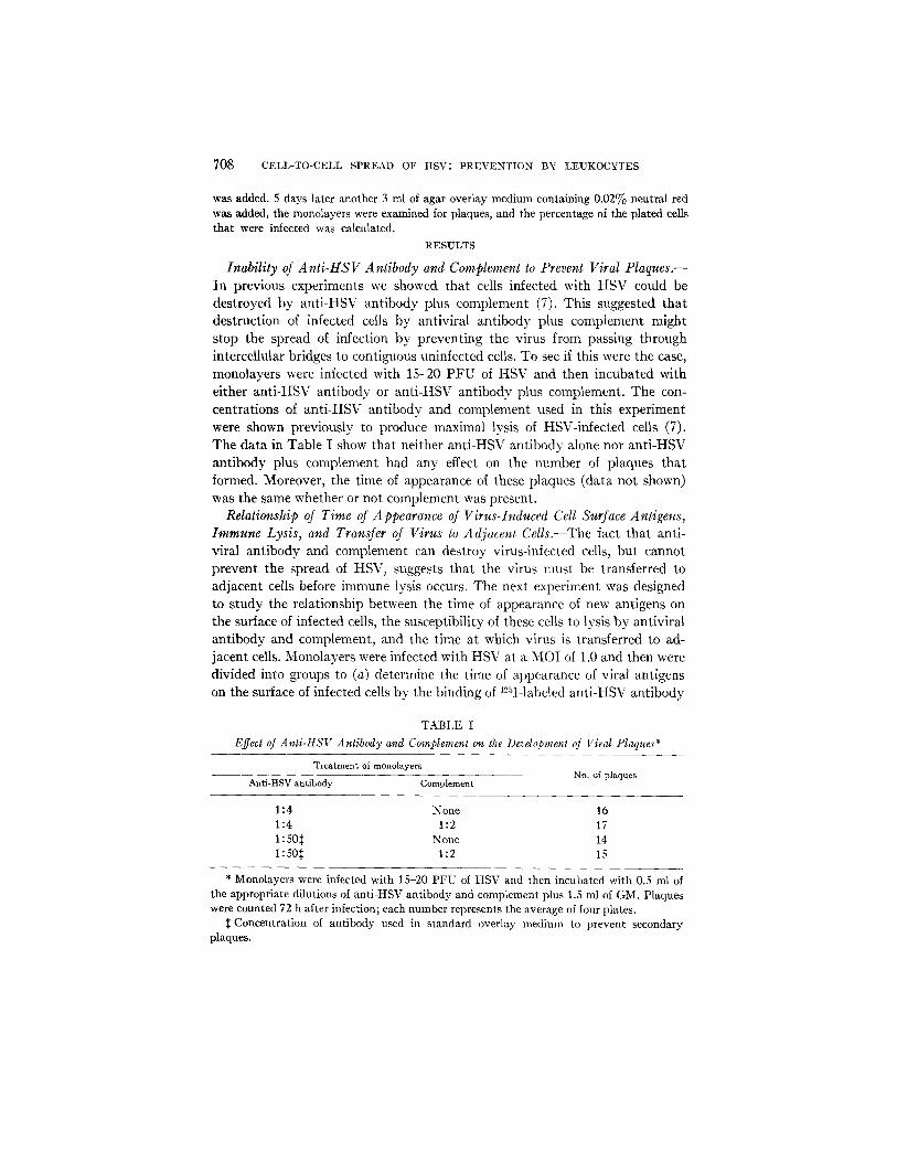
Inability of A nti-ttS V A ntibody and Complement to _Prevent Viral _Plaques.-- In previous experiments we showed that cells infected with HSV could be destroyed by anti-HSV antibody plus complement (7). This suggested that destruction of infected cells by antiviral antibody plus complement might stop the spread of infection by preventing the virus from passing through intercellular bridges to contiguous uninfected cells. To see if this were the case, monolayers were infected with 15-20 PFU of HSV and then incubated with either anti-HSV antibody or anti-HSV antibody plus complement. The con- centrations of anti-HSV antibody and complement used in this experiment were shown previously to produce maximal lysis of HSV-infected cells (7). The data in Table I show that neither anti-HSV antibody alone nor anfi-HSV antibody plus complement had any effect on the number of plaques that formed. Moreover, the time of appearance of these plaques (data not shown) was the same whether or not complement was present.
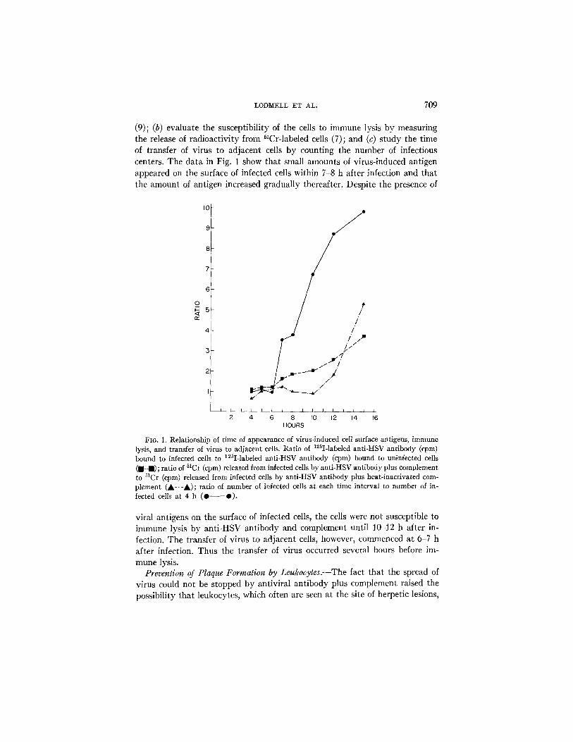
Relationship of Time of Appearance of Virus-Induced Cell Surface Antigens, Immune Lysis, and Transfer of Virus to Adjacent Cells.--The fact that anti- viral antibody and complement can destroy virus-infected cells, but cannot prevent the spread of HSV, suggests that the virus nmst be transferred to adjacent cells before immune lysis occurs. The next experiment was designed to study the relationship between the time of appearance of new antigens on the surface of infected cells, the susceptibility of these cells to lysis by antiviral antibody and complement, and the time at which virus is transferred to ad- jacent cells. Monolayers were infected with HSV at a MOI of 1.0 and then were divided into groups to (a) determine the time of appearance of viral antigens on the surface of infected cells by the binding of 1%I-labeled anti-HSV antibody
TABLE I E~ect of Anti-HSV Antibody and Complement on the Devdopment of Viral Plaques*
Treatment of monolayers
Anti-HSV antibody Complement No. of plaques
1:4 None 16 1:4 1:2 17 1:505 None 14 1:505 1:2 15
* Monolayers were infected with 15-20 PFU of HSV and then incubated with 0.5 ml of the appropriate dilutions of anti-HSV antibody and complement plus 1.5 ml of GM. Plaques were counted 72 h after infection; each number represents the average of four plates.
5 Concentration of antibody used in standard overlay medium to prevent secondary plaques.
LODM~LL ET AL. 709
(9); (b) evaluate the susceptibility of the cells to immune lysis by measuring the release of radioactivity from ~lCr-labeled cells (7); and (c) s tudy the time of transfer of virus to adjacent cells by counting the number of infectious centers. The data in Fig. 1 show that small amounts of virus-induced antigen appeared on the surface of infected cells within 7-8 h after infection and that the amount of antigen increased gradually thereafter. Despite the presence of
I0
9
8
7
6
0
n,-
4
3 r / //- / / /
'"l~ 11 ~"/~ //~ ~ ' ~ r . / /
I I I ~ _ _ 1 I t I I I
2 4 6 8 I0 12 14 16 HOURS
FIo. 1. Relationship of time of appearance of virus-lnduced ceil surface antigens, immune lysis, and transfer of virus to adjacent cells• Ratio of 12~I-labeled anfi-HSV antibody (epm) bound to infected cells to ]25I-labeled anti-HSV antibody (cpm) bound to uninfected cells ( ,-m) ; ratio of 51Ct (cpm) released from infected cells by anti-HSV antibody plus complement to 51Cr (cpm) released from infected cells by anti-HSV antibody plus heat-inactivated com- plement (A-'-A); ratio of number of infected ceils at each time interval to number of in- fected cells at 4 h (0 Q).
viral antigens on the surface of infected cells, the cells were not susceptible to immune lysis by anti-HSV antibody and complement until 10-12 h after in- fection. The transfer of virus to adjacent cells, however, commenced at 6-7 h after infection. Thus the transfer of virus occurred several hours before im- mune lysis.
Prevention of Plaque Formation by Leukocytes.--The fact that the spread of virus could not be stopped by antiviral antibody plus complement raised the possibility that leukocytes, which often are seen at the site of herpetic lesions,
710 CELL-TO-CELL SPREAD OF HSV: PREVENTION BY LEUKOCYTES
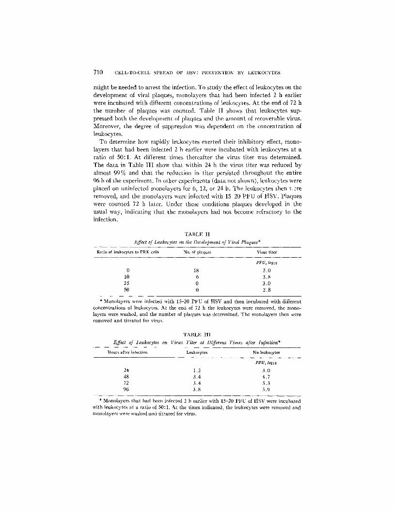
might be needed to arrest the infection. To study the effect of leukocytes on the development of viral plaques, monolayers that had been infected 2 h earlier were incubated with different concentrations of leukocytes. At the end of 72 h
the number of plaques was counted. Table I I shows that leukocytes sup- pressed both the development of plaques and the amount of recoverable virus. Moreover, the degree of suppression was dependent on the concentration of leukocytes.
To determine how rapidly leukocytes exerted their inhibitory effect, mono-
layers that had been infected 2 h earlier were incubated with leukocytes at a ratio of 50:1. At different times thereafter the virus titer was determined. The data in Table I I I show that within 24 h the virus titer was reduced by
almost 99% and that the reduction in titer persisted throughout the entire 96 h of the experiment. In other experiments (data not shown), leukocytes were placed on uninfected monolayers for 6, 12, or 24 h. The leukocytes then ¥ .'re
removed, and the monolayers were infected with 15-20 P F U of HSV. Plaques were counted 72 h later. Under these conditions plaques developed in the usual way, indicating that the monolayers had not become refractory to the infection.
TABLE II
Effect of Leukocytes on the Development of Viral Plaques*
Ratio of leukocytes to PRK cells No. of plaques Virus fiter
PFU, loglo
0 18 5.0 10 6 3.8 25 0 3.0 50 0 2.8
* Monolayers were infected with 15-20 PFU of HSV and then incubated with different concentrations of leukocytes. At the end of 72 h the leukocytes were removed, the mono- layers were washed, and the number of plaques was determined. The monolayers then were removed and titrated for virus.
TABLE III Effect of Leukocytes on Virus Titer at Different Times after Infection*
Hours after infection Leukocytes No leukocytes
PFU, loglo
24 1.2 3.0 48 3.4 4.7 72 3.4 5.3 96 3.8 5.9
* Monolayers that had been infected 2 h earlier with 15-20 PFU of HSV were incubated with leukocytes at a ratio of 50:1. At the times indicated, the leukocytes were removed and monolayers were washed and titrated for virus.

LODlV[ELL ET AL. 7 1 1
Further evidence that leukocytes in contact with infected monolayers can inhibit the development of viral plaques comes from fluorescent ant ibody studies. Monolayers were infected with HSV and at different times thereafter examined for the appearance of viral antigens. As seen in Table IV, leukocytes markedly inhibited both the number and the size of the fluorescent foci. How- ever, in those cells which showed fluorescence, the localization (intranuclear and cytoplasmic) and intensity of the fluorescence were not appreciably af- fected.
Effect of Leukocytes on Viral Replication.--The previous experiments showed that leukocytes prevented plaque formation and reduced the titer of infec- tious virus. These experiments, however, did not indicate whether this was
TABLE IV Effect of Leukocytes on Development of Viral Antigens*
Fluorescent loci*
Hours after infection With leukocytes No leukocytes
No. Size No. Size
24 3 + 14 + + 48 8 + + 25 + + + 72 7 + + 28 + + + - t -
* PRK cells on cover slips that had been infected 2 h earlier with 20-30 PFU of HSV were incubated with leukoeytes at a ratio of 50:1. At the times indicated, the leukocytes were removed and the cover slips were washed and examined by immunofluorescence for viral antigens.
~: Size of fluorescent loci: +, 1-4 cells; + + , 5-20 cells; + + + , foci with holes in center; + + + +, coalescence.
due to the inability of HSV to spread from cell to contiguous cell and/or inhibition of viral replication.
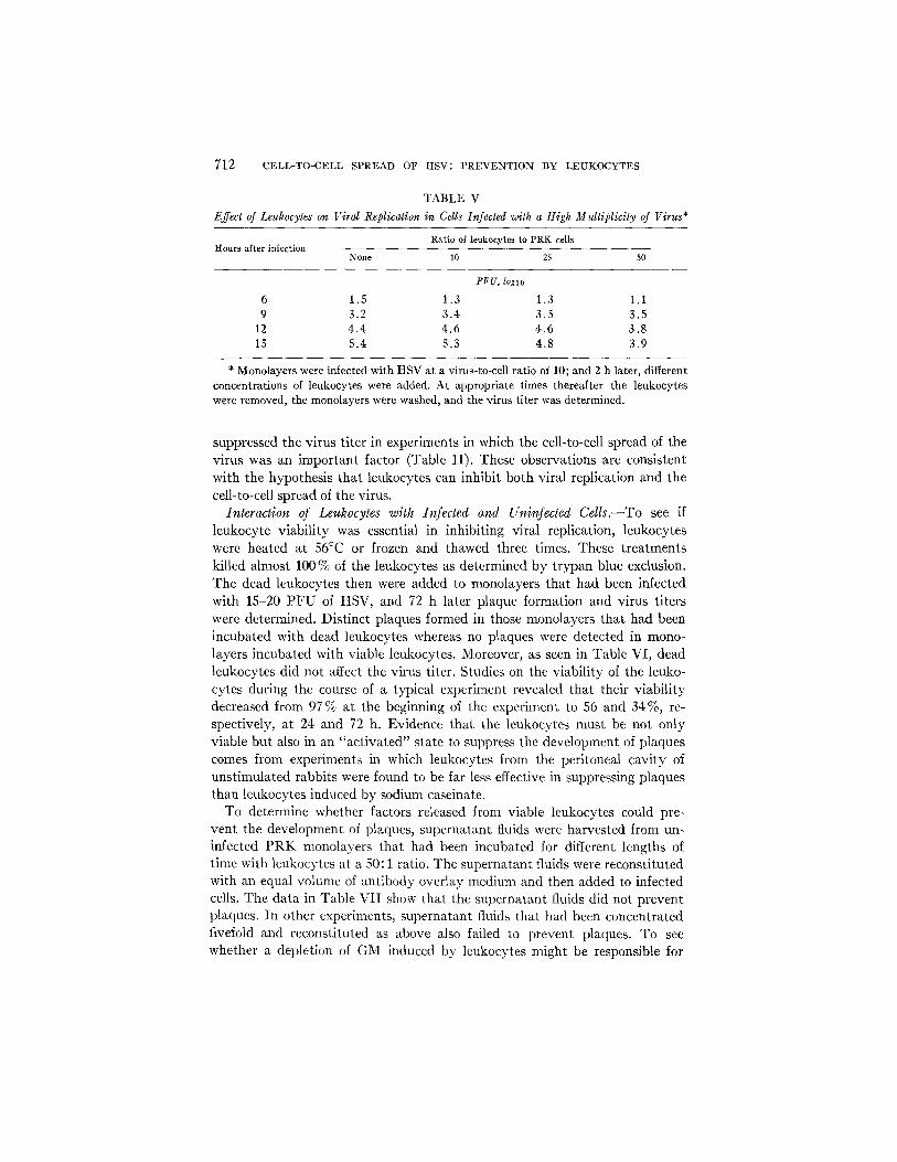
To study the effect of leukocytes on viral replication, monolayers were in- fected with concentrations of virus sufficient to infect 100 % of the cells, thereby eliminating the possibility that the depression in titer was due to the failure of the virus to spread to uninfected cells. Leukocytes at a ratio of 50:1 then were added, and at different times the P R K cells were examined by immuno- fluorescence for the appearance of viral antigens and titrated for infectious virus. In the absence of leukocytes, intranuclear and cytoplasmic fluorescence were detected in 10-20 % of the cells within 9 h and in more than 70 % of the cells within 24 h after infection. In the presence of leukocytes, substantially fewer cells showed fluorescence, approximately 10 % fluorescing at 24 h. More- over, as seen in Table V, considerably less infectious virus was recovered from monolayers incubated with high concentrations of leukocytes (50:1). Low concentrations of leukocytes (25:1 and 10:1) had little or no effect on virus titer, even though these same concentrations of leukocytes markedly
712 CELL-TO-CELL S P R E A D O F H S V : P R E V E N T I O N B Y L E U K O C Y T E S
TABLE V Effect ~ Leukocyt~ on ~ r ~ Replicati~ in Cdlslnfect~ with a H~h MMtipllcity ~ ~rus*
Hours af ter infection Rat io of lenkoeytes to P R K cells
None 10 25 50
-PFU, loglo
6 1.5 1.3 1.3 1.1 9 3.2 3.4 3.5 3.5
12 4.4 4.6 4.6 3.8 15 5.4 5.3 4.8 3.9
* Monolayers were infected with HSV at a virus-to-cell ratio of 10; and 2 h later, different concentrations of leukocytes were added. At appropriate times thereafter the leukocytes were removed, the monolayers were washed, and the virus titer was determined.
suppressed the virus titer in experiments in which the cell-to-cell spread of the virus was an important factor (Table II). These observations are consistent with the hypothesis that leukocytes can inhibit both viral replication and the cell-to-cell spread of the virus.
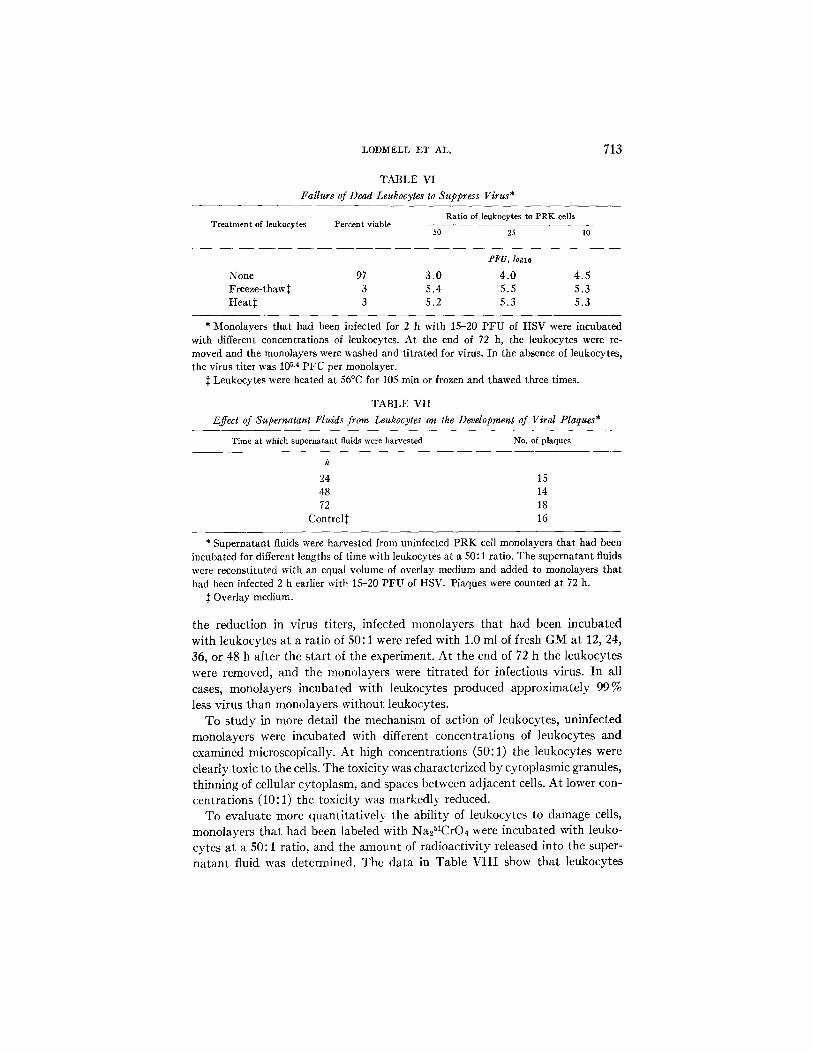
Interaction of Leukocytes with Infected and Uninfected Cells.--To see if leukocyte viability was essential in inhibiting viral replication, leukocytes were heated at 56°C or frozen and thawed three times. These treatments killed almost 100 % of the leukocytes as determined by trypan blue exclusion. The dead leukocytes then were added to monolayers that had been infected with 15-20 PFU of HSV, and 72 h later plaque formation and virus titers were determined. Distinct plaques formed in those monolayers that had been incubated with dead leukocytes whereas no plaques were detected in mono- layers incubated with viable leukocytes. Moreover, as seen in Table VI, dead leukocytes did not affect the virus titer. Studies on the viability of the leuko- cytes during the course of a typical experiment revealed that their viability decreased from 97 % at the beginning of the experiment to 56 and 34%, re- spectively, at 24 and 72 h. Evidence that the leukocytes must be not only viable but also in an "activated" state to suppress the development of plaques comes from experiments in which leukocytes from the peritoneal cavity of unstimulated rabbits were found to be far less effective in suppressing plaques than leukocytes induced by sodium caseinate.
To determine whether factors released from viable leukocytes could pre- vent the development of plaques, supernatant fluids were harvested from un- infected PRK monolayers that had been incubated for different lengths of time with leukocytes at a 50:1 ratio. The supernatant fluids were reconstituted with an equal volume of antibody overlay medium and then added to infected cells. The data in Table VII show that the supernatant fluids did not prevent plaques. In other experiments, supernatant fluids that had been concentrated fivefold and reconstituted as above also failed to prevent plaques. To see whether a depletion of GM induced by leukocytes might be responsible for
LODMELL ET AL. 713
TABLE VI Failure of Dead Leukocytes to Suppress Virus*
Treatment of leukocytes Percent viable Ratio of leukocytes to PRKcells
50 25 10
PFU, logl o
None 97 3.0 4.0 4.5 Freeze-thaw~ 3 5.4 5.5 5.3 Heat~: 3 5.2 5.3 5.3
* Monolayers that had been infected for 2 h with 15-20 PFU of HSV were incubated with different concentrations of leukocytes. At the end of 72 h, the leukocytes were re- moved and the monolayers were washed and titrated for virus. In the absence of leukoeytes, the virus titer was 105.4 PFU per monolayer.
2~ Leukocytes were heated at 56°C for 105 min or frozen and thawed three times.
TABLE VII
Effect of Supernatant Fluids from Leukocytes on the Devdopment of Viral Plaques*
Time at which supernatant fluids were harvested No. of plaques
h
24 15 48 14 72 18
Control~ 16
* Supernatant fluids were harvested from uninfected PRK cell monolayers that had been incubated for different lengths of time with leukocytes at a 50:1 ratio. The supernatant fluids were reconstituted with an equal volume of overlay medium and added to monolayers that had been infected 2 h earlier with 15-20 PFU of HSV. Plaques were counted at 72 h.
~: Overlay medium.
the reduction in virus titers, infected monolayers that had been incubated with leukocytes at a ratio of 50:1 were refed with 1.0 ml of fresh GM at 12, 24, 36, or 48 h after the start of the experiment. At the end of 72 h the leukocytes
were removed, and the monolayers were t i trated for infectious virus. In all cases, monolayers incubated with leukocytes produced approximately 99%
less virus than monolayers without leukocytes. To study in more detail the mechanism of action of leukocytes, uninfected
monolayers were incubated with different concentrations of leukocytes and examined microscopically. At high concentrations (50:1) the leukocytes were clearly toxic to the cells. The toxicity was characterized by cytoplasmic granules, thinning of cellular cytoplasm, and spaces between adjacent cells. At lower con- centrations (10: 1) the toxicity was markedly reduced.
To evaluate more quant i ta t ively the ability of leukocytes to damage cells, monolayers that had been labeled with Na251CrO4 were incubated with leuko- cytes at a 50:1 ratio, and the amount of radioactivity released into the super- na t an t fluid was determined. The data in Table VI I I show that leukocytes
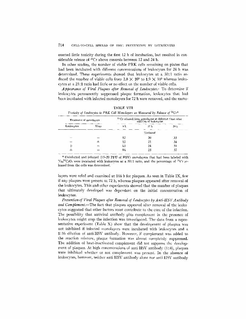
714 CELL-TO-CELL SPREAD OF HSV: PREVENTION BY LEUKOCYTES
exerted little toxicity during the first 12 h of incubation, but resulted in con- siderable release of 51Cr above controls between 12 and 24 h.
In other studies, the number of viable PRK cells remaining on plates that had been incubated with different concentrations of leukocytes for 24 h was determined. These experiments showed that leukocytes at a 50:1 ratio re- duced the number of viable cells from 2.8 X 10⁵ to 1.9 X 10⁵ whereas leuko- cytes at a 25 : 1 ratio had little or no effect on the number of viable cells.
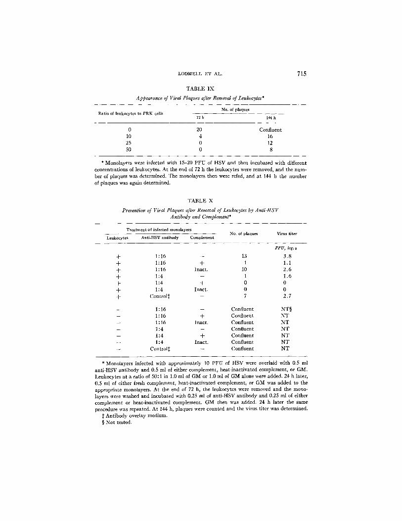
Appearance of Viral Plaques after Removal of Leukocytes.--To determine if leukocytes permanently suppressed plaque formation, leukocytes that had been incubated with infected monolayers for 72 h were removed, and the mono-
T A B L E V I I I
Toxicity of Leukocytes to P R K Call Monolayers as Measured by Release of 51Cr*
Treatment of monolayers 51Cr released from monolayers at different times after addition of leukocytes
Leukocytes Virus 6 h 12 h 24 h
%released
- - - - 12 20 3 3
- - + 13 21 3 4
+ - 13 24 54 + + 14 23 57
* Uninfected and infected (15-20 PFU of HSV) monolayers that had been labeled with Na251CrO4 were incubated with leukocytes at a 50:1 ratio, and the percentage of 5tCr re- leased from the cells was determined.
layers were refed and examined at 144 h for plaques. As seen in Table IX, few if any plaques were present at 72 h, whereas plaques appeared after removal of the leukocytes. This and other experiments showed that the number of plaques that ultimately developed was dependent on the initial concentration of leukocytes.
Prevention of Viral Plaques after Removal of Leukocytes by A nti-HS V Antibody and Complement.--The fact that plaques appeared after removal of the leuko- cytes suggested that other factors must contribute to the cure of the infection. The possibility that antiviral antibody plus complement in the presence of leukocytes might stop the infection was investigated. The data from a repre- sentative experiment (Table X) show that the development of plaques was not inhibited if infected monolayers were incubated with leukocytes and a 1:16 dilution of anti-HSV antibody. However, if complement was added to the reaction mixture, plaque formation was almost completely suppressed. The addition of heat-inactivated complement did not suppress the develop- ment of plaques. At high concentrations of anti-HSV antibody (1:4), plaques were inhibited whether or not complement was present. In the absence of leukocytes, however, neither anti-HSV antibody alone nor anti-HSV antibody
LODMELL ET AL.
T A B L E IX
Appearance of Viral Plaques after Removal of Leukocytes*
715
Ratio of leukocytes to PRK cells No. of plaques
72 h 144 h
0 20 Confluent 10 4 16 25 0 12 50 0 8
* Monolayers were infected with 15-20 P F U of HSV and then incubated with different concentiat ions of leukocytes. At the end of 72 h the leukocytes wele removed, and the num- ber of plaques was determined. The monolayers then were refed, and a t 144 h the number of plaques was again determined.
T A B L E X
Prevention of Viral Plaques after Removal of Leukocytes by Anti-HSV Antibody and Complement*
Treatment of infected monolayers
Leukocytes Anti-HSV antibody Complement No. of plaques Virus titer
PFU, logl o
-k 1:16 -- 13 3 .8 + 1:16 -[- 1 1.1 -k 1 : 16 Inact . 10 2 .6 + 1:4 -- 1 1 .6 + 1:4 + 0 0 + 1:4 Inact . 0 0 + Control~ -- 7 2 .7
1 : 16 -- Confluent NT§ 1 : 16 + Confluent N T 1 : 16 Inact . Confluent N T 1 : 4 -- Confluent N T 1 : 4 + Confluent N T 1 : 4 Inact . Confluent N T
Control:~ -- Confluent N T
* Monolayers infected with approximately 10 P F U of HSV were overlaid with 0.5 ml ant i -HSV ant ibody and 0.5 ml of either complement, heat- inactivated complement, or GM. Leukocytes a t a ratio of 50:1 in 1.0 ml of GM or 1.0 ml of GM alone were added. 24 h later, 0.5 ml of either fresh complement, heat- inactivated complement, or GM was added to the appropriate monolayers. At the end of 72 h, the leukocytes were removed and the mono- layers were washed and incubated with 0.25 ml of ant i -HSV ant ibody and 0.25 ml of either complement or heat- inact ivated complement. GM then was added. 24 h later the same procedure was repeated. At 144 h, plaques were counted and the virus titer was determined.
]~ Antibody overlay medium. § Not tested.

716 C E L L - T O - C E L L SPREAD OF HSV: P R E V E N T I O N BY LEUKOCYTES
plus complement prevented the spread of the infection or the destruction of the monolayers.
DISCUSSION
In vivo, certain viral infections persist or recur in the presence of high levels of neutralizing antibody. Presumably this happens because the virus spreads directly from one cell to another without being exposed to antiviral antibody. Although there have been few studies on how the immunological response of the host actually stops the cell-to-cell spread of viruses, it has generally been assumed that the interaction of antiviral antibody plus complement or immune lvanphocytes with virus-induced cell surface antigens destroyed the infected cells and thereby stopped the spread of the infection (7, 10-12).
To see whether antiviral antibody and complement actually could stop the infection from spreading, we used an in vitro system that resembled in many respects the in vivo state. Cells were infected with HSV, and then an antibody overlay was added. Under these conditions the virus spreads directly from one cell to another, presumably through intercellular bridges (1-3). Extracellular virus was readily neutralized by the antibody overlay, thus preventing distant cells from becoming infected. When complement was added to this system, virus-infected cells were destroyed (7); but as shown in the present study, the spread of the infection was not prevented. The failure to stop the spread of the infection suggested that the virus might be spreading from infected to uninfected cells before the infected cells were lysed by antiviral antibody and complement. Our experiments (Fig. 1) show this to be the case. The data, however, should be viewed with some caution since they are based on a popu- lation of cells rather than a specific group of contiguous cells; moreover, it is difficult to compare the precise sensitivity of the different assay procedures. Nonetheless, the data from Fig. 1, together with the fact that the virus can spread from cell to cell in the presence of antiviral antibody and complement, support the contention that the virus enters uninfected cells before the cell in which it is synthesized is immunologically destroyed. Thus the viral spread is always one step ahead of the destructive capacity of antiviral antibody and complement, and the infection persists.
Although there is still very little information as to how cellular immunity operates in viral infections, several mechanisms have been proposed. First, the interaction of immune lymphocytes with viral-induced cell surface antigens can result in cell destruction (11, 13, 14). Secondly, activation of lymphocytes or macrophages can lead to the release of biological mediators, such as inter- feron or lymphotoxin, that can inhibit viral replication and destroy cells (13, 15-20). Thirdly, macrophages might inhibit the infection by phagocytizing and digesting infectious particles (19, 21-23). The demonstration that HSV spreads from infected to uninfected cells at approximately the same time or before substantial amounts of viral antigens appear on the surface of the infected cell suggests that lymphocytes specifically immune to the virus would have
LODMELL ET AL. 717
no more opportunity to destroy the infected cell, before transfer of virus to adjacent cells, than antiviral antibody and complement. Moreover, it appears to take immune lymphocytes considerably longer to destroy target cells than antiviral antibody and complement (7, 11, 24). Evidence that interferon or other mediators released from leukocytes are not responsible for inhibiting plaque development comes from two types of experiments. First, if leukoc)/tes were placed on monolayers 6, 12, or 24 h before infection with HSV and then removed, the formation of plaques was not inhibited. Secondly, supernatant fluids from leukocyte cultures did not prevent the development of plaques. Moreover, it is known that HSV is relatively resistant to interferon (25). Phagocytosis of extracellular virus also seems to be of little consequence in our system since the virus is spreading through intercellular bridges.
The specific cell type(s) in the peritoneal exudate responsible for inhibiting the development of plaques is under study. Since at least 75 % of the cells in the exudate appear to be macrophages and since recent experiments in our laboratory with macrophages purified by adherence to glass showed that these cells can effectively suppress the development of plaques, macrophages are the prime candidate. The ability of activated macrophages to damage or destroy normal and malignant cells has been well established, and is thought to be an important component in delayed hypersensitivity reactions (26-29). Our experiments suggest that leukocytes, presumably macrophages, also may play an important role in stopping the cell-to-cell spread of viral infections. At very high concentrations (50:1 or greater), leukocytes can destroy both in- fected and uninfected cells around infectious foci, thereby eliminating potential target cells for viral replication. At lower concentrations, the cells may not be destroyed, but the leukocytes may be toxic to them. This could lead to breaking of intercellular bridges and/or depression of the cell's metabolic processes. If intercellular bridges are broken, the virus cannot be transferred to adjacent cells without being exposed to antiviral antibody. If metabolic processes are shut down, this might inhibit replication of the virus within infected cells. In one case the infection is stopped because the virus cannot reach adjacent cells, while in the other case it is halted because the virus cannot replicate. Evidence that the virus cannot reach adjacent cells without being exposed to antiviral antibody comes from microscopic observations that showed loss of continuity between adjacent cells, and from the data in Table X, which showed that leukocyte-treated cultures could be cured by antiviral antibody and complement. Evidence that high concentrations of leukocytes (50:1) can inhibit replication of the virus comes from the experiment in which a suffi- ciently high MOI was used to infect all of the cells in the culture, thereby eliminating the role of viral spread.
The demonstration that the removal of leukocytes at the end of 72 h resulted in the appearance of plaques showed that leukocytes had suppressed the in- fection, but had not destroyed all of the infected cells. Moreover, the appear- ance of plaques at 144 h suggested that intercellular bridges had been rees-
718 CELL-TO-CELL SPREAD OF HSV: PREVENTION BY LEUKOCYTES
tablished. We reasoned that if virus-infected cells were exposed to antiviral antibody and complement during the period when intercellular bridges were broken, it might be possible to stop the infection by destroying the infected cells before the virus was able to reach adjacent cells through the reestablished bridges. Our experiments showed that when low concentrations of anti-HSV (1:16) were used, the addition of complement eliminated the infection. The fact that high concentrations of anti-HSV (1:4) were able to eliminate the in- fection in the absence of complement was unexpected, but there are at least two possible explanations. First, the attachment of high concentrations of anti-HSV antibody to viral antigens on the surface of infected cells might have prevented or blocked the reestablishment of intercellular bridges. Secondly, infected cells that had been exposed to leukocytes, in contrast to infected cells that had not been exposed to leukocytes, might have been more susceptible to destruction and penetration by antiviral antibody.
On the basis of these and other observations we propose the following hy- pothesis to explain how the immune response stops viral infections that spread by a cell-to-cell route. The immunological response to HSV consists of two phases, one specific and the other nonspecific. Factors that are chemotactic for polymorphonuclear and mononuclear leukocytes are specifically generated at the site of the infection. These factors include C5a, a cleavage product generated from the fifth component of complement by the interaction of anti- HSV antibody with HSV antigens (30), and soluble mediators released from immune lymphocytes after stimulation with HSV antigens (20).~ These medi- ators attract leukocytes to the site of the infection, where they exert their toxic effect nonspecifically. This results in destruction of some of the infected cells and some of the surrounding uninfected cells to which the virus would have spread. In addition, the leukocytes can inhibit viral replication and/or break intercellular bridges and thereby prevent the virus from spreading directly to adjacent cells without being exposed to antiviral antibody. Infected cells that survive the leukocyte attack can be destroyed by antiviral antibody and complement before intercellular bridges are reestablished. Thus both humoral and cellular components acting jointly are needed to cure the infection.
Our hypothesis suggests that deficiencies in the host's immune response against viral infections must be sought not only in the amount or type of antibody produced but in the ability to generate chemotactic factors, attract and activate leukocytes, and destroy target cells. Better ways of getting leuko- cytes to the site of the infection might prove useful in treating viral infections that spread by a cell-to-cell route.
SUMMARY
Antibody to herpes simplex virus (HSV) plus complement destroyed HSV- infected cells but did not stop the spread of the infection. Studies on the re-
2 Rosenberg, G., R. Snyderman, and A. Notkins. UnpubUshed observations.
LODMELL ET AL. 719
lationship between the time of appearance of viral antigens on the cell surface, immunological destruction of the cells by antiviral antibody and comple- ment, and transfer of the virus to adjacent cells showed that the virus spread from infected to uninfected cells before the infected cells 'were susceptible to immunological destruction. Incubation of infected monolayers with leukocytes, however, stopped the spread of the virus by nonspecifically damaging both in- fected and uninfected cells and by presumably breaking intercellular bridges. When leukocytes were removed from infected monolayers, viral plaques de- veloped. If, however, antiviral antibody and complement were added to mono- layers before the leukocytes were removed, the development of plaques was prevented. These findings suggest that both antibody and leukocytes are needed to cure HSV infections.
The authors gratefully acknowledge the invaluable assistance of C. Wohlenberg, F. Shaw, G. Lewis, and M. Lombardi.
REFERENCES
1. Black, F. L., and J. L. Melnick. 1955. Micro-epidemiology of poliomyelitis and herpes-B infections. Spread of the viruses within tissue cultures. J. Immunol. 74:236.
2. Hoggan, D. M., B. Roizman, and P. R. Roane, Jr. 1961. Further studies of vari- ants of herpes simplex virus that produce syncytia or pocklike lesions in cell cultures. Am. J. ttyg. 73:114.
3. Christian, R. T., and P. P. Ludovici. 1971. Cell-to-cell transmission of herpes simplex virus in primary human amnion cells. Proc. Soc. Exp. Biol. Med. 138:1109.
4. Todaro, G. J., and R. J. Huebner. 1972. The viral oncogene hypothesis: new evi- dence. Proc. Natl. Acad. Sci. U.S.A. 69:1009.
5. Notkins, A. L. 1971. Infectious virus-antibody complexes: interaction with anti- immunoglobulins, complement, and rheumatoid factor. J. Exp. Med. 134 (3, Pt. 2):41s.
6. Hampar, B., A. L. Notkins, M. Mage, and M. A. Keehn. 1968. Heterogeneity in the properties of 7S and 19S rabbit-neutralizing antibodies to herpes simplex virus. J. Immunol. 100:586.
7. Brier, A. M., C. Wohlenberg, J. Rosenthal, M. Mage, and A. L. Notkins. 1971. Inhibition or enhancement of immunological injury of virus-infected cells. Proc. Natl. Acad. Sci. U.S.A. 66:3073.
8. A. Kawamura, Jr., editor. 1969. Fluorescent Antibody Techniques and Their Ap- plications. University of Tokyo Press, Tokyo.
9. Hayashi, K., J. Rosenthal, and A. L. Notkins. 1972. Iodine-125-1abeled antibody to viral antigens: Binding to the surface of virus-infected cells. Science (Wash. D.C.). 176:516.
10. Roane, P. R., and B. Roizman. 1964. Studies on the determinant antigens of viable cells. II. Demonstration of altered antigenic reactivity of HEp-2 cells infected with herpes simplex virus. Virology 22:1.
11. Speel, L. F., J. E. Osborn, and D. L. Walker. 1968. An immuno-cytopathogenic interaction between sensitized leukocytes and epithelial cells carrying a persis- tent noncytocidal myxovirus infection. ]. Immunol. 101:409.

720 C E L L - T O - C E L L SPREAD OF HSV: PREVENTION BY LEUKOCYTES
12. Rosenberg, G. L., P. A. Farber, and A. L. Notkins. 1972. In vitro stimulation of sensitized lymphocytes by herpes simplex virus and vaccinia virus. Proc. Natl. Acad. Sci. U.S.A. 69:756.
13. Oldstone, M. B. A., and F. J. Dixon. 1970. Tissue injury in lymphocytic chorio- meningitis viral infection: virus-induced immunologicaUy specific release of a cytotoxic factor from immune lymphoid cells. Virology. 42:805.
14. Lundstedt, C. 1969. Interaction between antigenically different ceils. Acta Pathol. Microbiol. Scand. 75:139.
15. Green, J. A., S. R. Cooperband, and S. Kibrick. 1969. Immune specific induction of interferon production in cultures of human blood leukocytes. Science (Wash. D.C.). 164:1415.
16. Glasgow, L. A. 1965. Leukocytes and interferon in the host response to viral in- fections. I. Mouse leukocytes and leukocyte-produced interferon in vaccinia virus infection in vitro. J. Exp. Med. 19.1:1001.
17. Glasgow, L. A. 1970. Transfer of interferon-producing macrophages: new ap- proach to viral chemotherapy. Science (Wash. D.C.). 170:854.
18. Tompkins, W. A. F., C. Adams, and W. E. Rawls. 1970. An in vitro measure of cellular immunity to fibroma virus. J. Immunol. 104:502.
19. Hirsch, M. S., B. Zisman, and A. C. Allison. 1970. Macrophages and age-depen- dent resistance to herpes simplex virus in mice. J. Immunol. 104:1160.
20. Wilton, J. M. A., L. Ivanyi, and T. Lehner. 1972. Cell-mediated immunity in herpes virus horninis infections. Br. Med. J. 1:723.
21. Zisman, B., M. S. Hirsch, and A. C. Allison. 1970. Selective effects of anti-macro- phage serum, silica and anti-lymphocyte serum on pathogenesis of herpes virus infection of young adult mice. J. Immunol. 104:1155.
22. Blanden: R. V. 1971. Mechanisms of recovery from a generalized viral infection: mousepox. III . Regression of infectious foci. J. Exp. Med. 133:1090.
23. Stevens, J. G., and M. L. Cook. 1971. Restriction of herpes simplex virus by macrophages. An analysis of the cell-virus interaction. J. Exp. IVied. 133:19.
24. Perlmann, P., and G. Holm. 1969. Cytotoxic effects of lymphoid cells in vitro. Advan. Irnmunol. 11:117.
25. Schachter, N., M. A. Galin, M. Weissenbacher, S. Baron, and A. Billiau. 1970. Comparison of antiviral action of interferon, interferon inducers, and IDU against herpes simplex and other viruses. Ann. Ophthalmol. 2:975.
26. Granger, G. A., and R. S. Weiser. 1966. Homograft target cells: contact destruc- tion in vtro by immune macrophages. Science (Wash. D.C.). 151:97.
27. Mackaness. G. B. 1971. Cellular immunity. Ann. Inst. Pasteur (Paris). 120:428. 28. Evans, R., and P. Alexander. 1972. Mechanism of irnmunologically specific killing
of tumor cells by macrophages. Nature (Lond.). 9.36:168. 29. Hibbs, J. B., Jr., L. H. Lambert, Jr., and J. S. Remington. 1972. Possible role of
macrophage mediated nonspecific cytotoxicity in tumor resistance. Nature ( Lond.). 9.35:48.
30. Snyderman, R., C. Wohlenberg, and A. L. Notkins. 1972. Inflammation and viral infection: chemotactic activity resulting from the interaction of antiviral anti- body and complement with cells infected with herpes simplex virus. J. Infect. Dis. 126:207.