Embed Size (px)
Citation preview
Indian Journal of Clinical Anaesthesia 2019;6(4):623–626
Content available at: iponlinejournal.com
Indian Journal of Clinical Anaesthesia
Journal homepage: www.innovativepublication.com
Case Report
Very rare and serious complication of central line insertion
Ojaswani Rai Sood1,*1Dept. of Anaesthesia, Government Medical College, Amritsar, Punjab, India
A R T I C L E I N F O
Article history:Received 29-06-209Accepted 09-10-2019Available online 20-11-2019
Keywords:Central lineComplicationPleural effusionPneumo mediastinum
A B S T R A C T
Central venous catheter (CVC) placement is indicated for various therapeutic and diagnostic purposes.However, it is important to ensure its proper placement. A CVC malposition is associated with number ofrecognized complications, some of which can be fatal.Case: In a 27 year female admitted in ICU with history of seizures and oral bleed with stable vitals, 7Fchsubclavian CVC was inserted. She had progressive dyspnea, chest pain and hypotension. Although chestX-ray showed correctly positioned CVC, CECT chest showed it was entering SVC, traversing along itsmedial wall and reaching between SVC and aorta and entering in the right atrium. Also atelectic fluidin oblique fissure, bilateral pleural effusion and small pocket of air in anterior mediastinum with minimalmediastinal high attenuation collection was seen. This was removed and patient was under observation for24 hours, after which she was shifted to ward.Conclusion: Therefore location of the catheter must be verified by radiological methods. Also real-timeultrasound reduces the number of complications associated with the technique.
© 2019 Published by Innovative Publication. This is an open access article under the CC BY-NC-NDlicense (https://creativecommons.org/licenses/by/4.0/)
1. Introduction
Central venous catheter (CVC) insertion is indicated forvarious therapeutic as well as diagnostic purposes inpatients admitted in intensive care units (ICU), variousdialysis units and in routine and emergency operationtheaters. However, it is important to be sure of its properplacement. A CVC malposition can be associated withlarge number of recognized complications, some of whichmay be fatal. These may occur either during insertionof the catheter (e.g. arterial puncture, pneumothorax,arrhythmias), and/or during maintenance of the line (e.g.infection, thrombosis). Variousmal positions of CVCcan be its placement in internal mammary vein, coilingwithin the vein itself, migration of catheter to ipsilateraljugular vein from subclavian vein or translocation to thecontralateral subclavian vein etc.1–3 Pericardial effusion andpericardial tamponade are rare but accounting for up to0.7% of central venous catheter related complications4,5
* Corresponding author.E-mail address: [email protected] (O. R. Sood).
Recommended use of post-insertion chest radiograph orusing any other radiological method for the confirmationof correct placement and detection of complications likepneumothorax should be employed.
Here we present a very rare case of mal positioned centralvenous catheter vein entering into superior part of superiorvenacava (SVC), traversing along its medial wall and itstip reaching up to the junction between SVC and aorta.However, the terminal end was in right atrium.
1.1. Case presentation
A 27 year old woman on her post-partum day 27 wasadmitted in emergency room following 5 grand mal seizureswithin a period of 6 hours. Due to the seizures she hadtongue and lip bite followed by bleeding and edema. Forthe control of fits she was shifted to ICU. On examinationshe was slightly drowsy. The pulse rate was 9 0 per minuteand blood pressure was 13 0/80 mm Hg. MRI brain waswithin normal limits. Injection Clonazepam was givenintramuscularly and phenytoin bolus followed by thrice a
https://doi.org/10.18231/j.ijca.2019.1212394-4781/© 2019 Innovative Publication, All rights reserved. 623
624 Sood / Indian Journal of Clinical Anaesthesia 2019;6(4):623–626
day dose was given. She was oxygenated using high flowoxygen mask and X-ray chest was done (Figure 1 - somehilaropacity in otherwise normal lung). As there was noperipheral venous access due to previous injuries and cutdowns, a 7 Fchpolyvinyl chloride subclavian line insertionwas planned using landmark technique in the right infraclavicular area to facilitate the administration of intravenousdrugs and fluids. A free flow of blood back down thecatheter was noted after insertion.
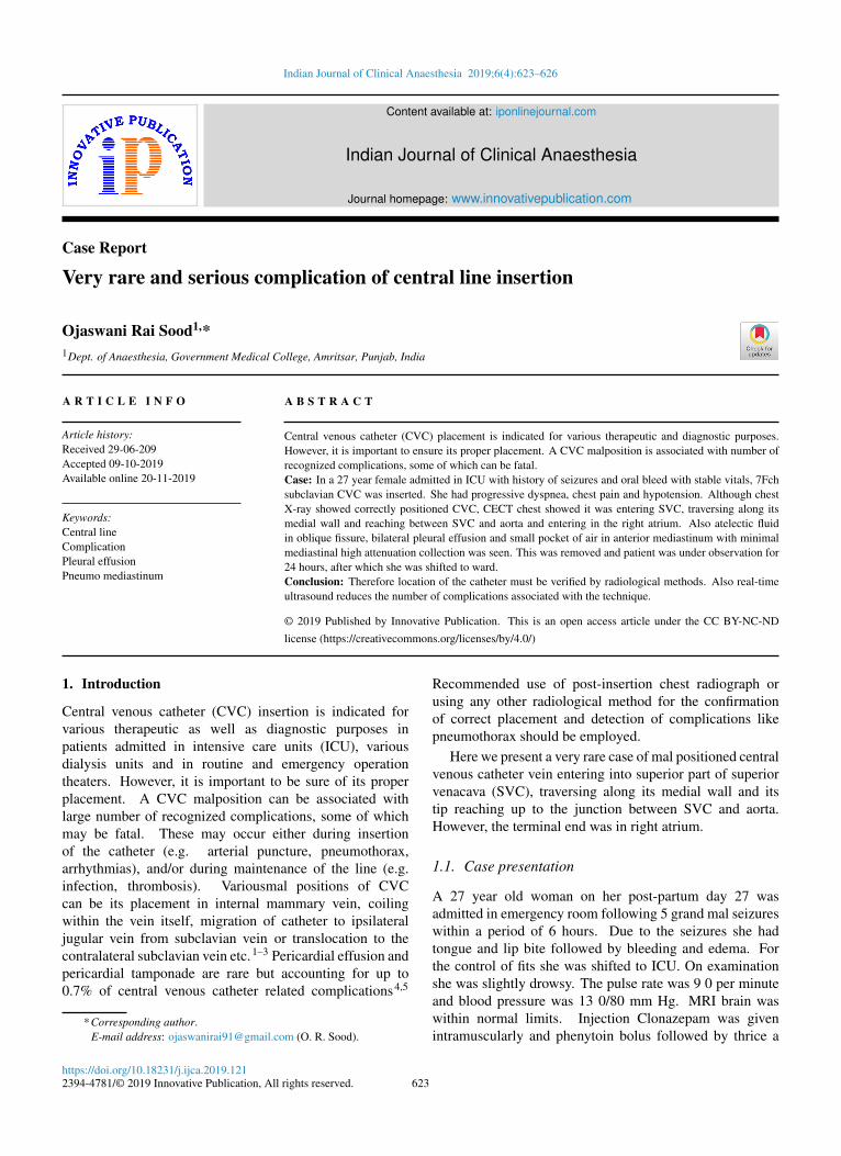
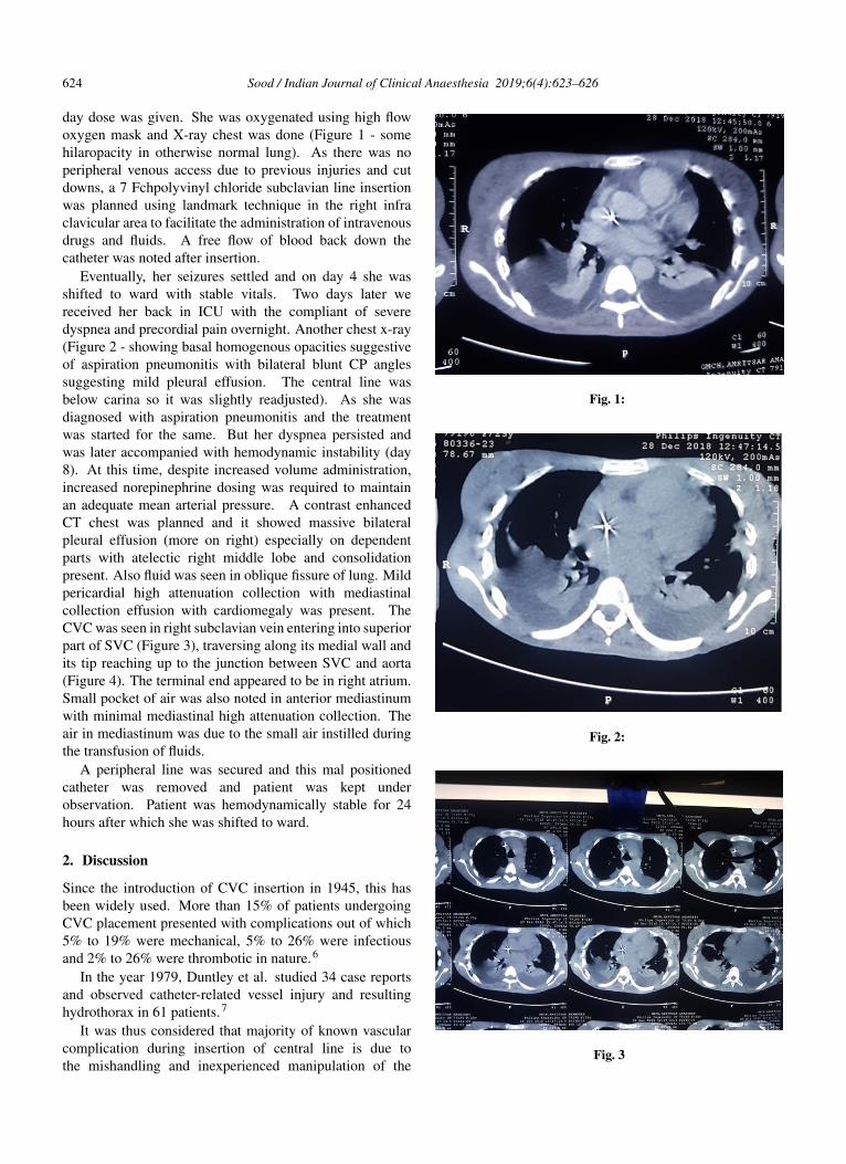
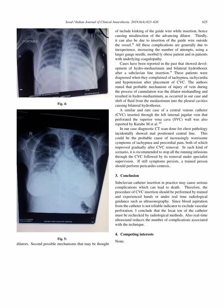
Eventually, her seizures settled and on day 4 she wasshifted to ward with stable vitals. Two days later wereceived her back in ICU with the compliant of severedyspnea and precordial pain overnight. Another chest x-ray(Figure 2 - showing basal homogenous opacities suggestiveof aspiration pneumonitis with bilateral blunt CP anglessuggesting mild pleural effusion. The central line wasbelow carina so it was slightly readjusted). As she wasdiagnosed with aspiration pneumonitis and the treatmentwas started for the same. But her dyspnea persisted andwas later accompanied with hemodynamic instability (day8). At this time, despite increased volume administration,increased norepinephrine dosing was required to maintainan adequate mean arterial pressure. A contrast enhancedCT chest was planned and it showed massive bilateralpleural effusion (more on right) especially on dependentparts with atelectic right middle lobe and consolidationpresent. Also fluid was seen in oblique fissure of lung. Mildpericardial high attenuation collection with mediastinalcollection effusion with cardiomegaly was present. TheCVC was seen in right subclavian vein entering into superiorpart of SVC (Figure 3), traversing along its medial wall andits tip reaching up to the junction between SVC and aorta(Figure 4). The terminal end appeared to be in right atrium.Small pocket of air was also noted in anterior mediastinumwith minimal mediastinal high attenuation collection. Theair in mediastinum was due to the small air instilled duringthe transfusion of fluids.
A peripheral line was secured and this mal positionedcatheter was removed and patient was kept underobservation. Patient was hemodynamically stable for 24hours after which she was shifted to ward.
2. Discussion
Since the introduction of CVC insertion in 1945, this hasbeen widely used. More than 15% of patients undergoingCVC placement presented with complications out of which5% to 19% were mechanical, 5% to 26% were infectiousand 2% to 26% were thrombotic in nature.6
In the year 1979, Duntley et al. studied 34 case reportsand observed catheter-related vessel injury and resultinghydrothorax in 61 patients.7
It was thus considered that majority of known vascularcomplication during insertion of central line is due tothe mishandling and inexperienced manipulation of the
Fig. 1:
Fig. 2:
Fig. 3
Sood / Indian Journal of Clinical Anaesthesia 2019;6(4):623–626 625
Fig. 4:
Fig. 5:dilators. Second possible mechanisms that may be thought
of include kinking of the guide wire while insertion, hencecausing misdirection of the advancing dilator. Thirdly,it can also be due to insertion of the guide wire outsidethe vessel.8 All these complications are generally due toinexperience, increasing the number of attempts, using alarger gauge needle, morbid ly obese patient and in patientswith underlying coagulopathy.
Cases have been reported in the past that showed devel-opment of hydro-mediastinum and bilateral hydrothoraxafter a subclavian line insertion.9 These patients werediagnosed when they complained of tachypnea, tachycardiaand hypotension after placement of CVC. The authorsstated that probable mechanism of injury of vein duringthe process of cannulation was the dilator mishandling andresulted in hydro-mediastinum, as occurred in our case andshift of fluid from the mediastinum into the pleural cavitiescausing bilateral hydrothorax.
A similar and rare case of a central venous catheter(CVC) inserted through the left internal jugular vein thatperforated the superior vena cava (SVC) wall was alsoreported by Kurabe M et al.10
In our case diagnostic CT scan done for chest pathologyincidentally showed mal positioned central line. Thiscould be the probable cause of increasingly worrisomesymptoms of tachypnea and precordial pain, both of whichimproved gradually after CVC removal. In such kind ofscenario, it is recommended to stop all the running infusionsthrough the CVC followed by its removal under specialistsupervision. If still symptoms persists, a trained personshould perform pericardio-centesis..
3. Conclusion
Subclavian catheter insertion in practice may cause seriouscomplications which can lead to death. Therefore, theprocedure of CVC insertion should be performed by trainedand experienced hands or under real time radiologicalguidance such as ultrasonography. Since blood aspirationfrom the catheter is not reliable indicator to exclude vascularperforation, I conclude that the locat ion of the cathetermust be rechecked by radiological methods. Also real-timeultrasound reduces the number of complications associatedwith the technique.
4. Competing interests
None.
626 Sood / Indian Journal of Clinical Anaesthesia 2019;6(4):623–626
References1. Kela M, Munde H, Raut S. Accidental placement of central venous
catheter into internal mammary vein: A rare catheter malposition. AnnCard Anaesth. 2017;20:477–485.
2. Tomar GS, Tiwari AK, Jain DG, Chawla S, Sinha R. Central venouscatheter rotation malposition: An unusual presentation. Indian JAnaesth. 2012;56(4):415–415.
3. Bansal S, Bansal S, Dogra M, Khan I. A malpositioned central venouscatheter. Pain Intensive Care. 2019;26:54–56.
4. Nowlen TT, Rosenthal GL, Johnson GL, Tom DJ, Vargo TA.Pericardial effusion and tamponade in infants with central catheters.Pediatr. 2002;110(1):137–142. PubMed.
5. Wirrell EC, Pelausa EO, Allen AC, Stinson DA, Hanna BD. Massivepericardial effusion as a cause for sudden deterioration of a very lowbirthweight infant. Am J Perinatol. 1993;10(6):419–423. PubMed.
6. Merrer J, B DJ, Golliot F, Lefrant JY, Raffy B, et al. Complications offemoral and subclavian venous catheterization in critically ill patients:a randomized controlled trial. JAMA. 2001;286:700–707.
7. Duntley P, Siever J, Korwes ML, Harpel K, Heffner JE. Vascularerosion by central venous catheters. Clinical features and outcome.
Chest. 1992;101:1633–1638.8. Hohlrieder M, Oberhammer R, Lorenz IH, Margreiter J, Khbacher G,
et al. Life-threatening mediastinal hematoma caused by extravascularinfusion through a triple-lumen central venous catheter. Anesth Analg.2004;99:31–35.
9. Naguib M, Farag H, Joshi RN. Bilateral hydrothorax andhydromediastinum after a subclavian line insertion. Can Anaesth SocJ. 1985;32:412–414.
10. Kurabe M, Watanabe T, Kohno T. Perforation of the superior venacava 5 days after insertion of a central venous catheter through the leftinternal jugular vein. J Clin Anesth. 2016;31:193–196.
Author biographyOjaswani Rai Sood Junior Resident
Cite this article: Sood OR. Very rare and serious complication ofcentral line insertion. Indian J Clin Anaesth 2019;6(4):623-626.


![Cardiogenic Shock Complicating Myocardial Infarction: An ...€¦ · Cardiogenic shock (CS) is a serious complication of acute myocardial infarction [MI] [1]. The mortality rate is](https://img.pdfslide.us/doc/110x75/5ea6f123bff5602612238709/cardiogenic-shock-complicating-myocardial-infarction-an-cardiogenic-shock-cs.jpg)