Embed Size (px)
Citation preview

Complication Avoidance and Management

65%

Complications Defined
• Access Complications• RP Bleed• Pseudo Aneurysm/hematoma
• Conduit Vessel Complications• Crossing /wire related perforations• Treatment related complications
• Perforation• Embolization
• Thrombotic complications• Renal complication• Cerebrovascular complications• Sedation related complications• Cardiac Complications

Avoidance
• Patient selection• Operator Realsm• Procedure planning • Knowing when to stop• Reducing variation• Readily available Consultation

Management
• Early Recognition• Low threshold for central large bore Venous line early• Document imagery where applicable• Maintain calm but act decisively and promptly• Have readily available tools on the shelf to fix it if you break
it• Covered stents• Coils• Reversal agents for sedatives and heparin• Pressors
• Call for help• It is preferable to fix the problem in the lab before you
transfer if at all possible, especially when ongoing.

Management
• Focus on the patient not lawyer.• Remember to keep talking to the
patient in calm reassuring tone• After stabilizing the patient, explain to
patient & family in clear honest terms what happened.
• If a Hospital transfer was needed, have a very low threshold for accompanying the pt in the ambulance to the ER.

Access Complications
• USG always• Micro Needle/Kit Always• Aim at entring the vessel at
the center of the front wall• Puncture must be where the
vessel posterior wall is supported by bone

Access Complications• Femoral access must be done with a set
protocol with zero tolerance for deviation. • USG always• Micro Needle/Kit Always• Fluoro confirmation of the micro needle
position in the CFA to further verify.• High sticks lead to 100% RP bleed • Low sticks increase the risk of hematoma or
PseudoAneurysms• Antegrade Sticks must be steep • If Needle Tip on Fluoro is too high or low,
remove, hold for 2 mins and start again.


Access Complications
• Brachial Access is still indicated in aorto iliac CTO. must be done with a set protocol with zero tolerance for deviation. • USG always• Micro Needle/Kit Always• Must stick along or very slightly below the antecubital
fossa line. • Must feel the medial and lateral humeral condyles and
draw a line between and stick there. • The brachial artery transitions medial to the bone very
quickly, and if the stick is at less than 60 degrees steep, we are likely to hit the vessel where it is no longer compressible by a bone.
• Making large arm hematomas a high likelihood.

There is no bone behind the brachial artery here to support a good hold especially with the least bit of wrist pronation thus much higher risk of hematoma and Pseudo aneurysm
Very narrow zone of wide bone behind the artery RIGHT AT THE ELBOW CREASE to allow a good brachial artery hold post op


THE BRACHIAL ARTERY WILL BOW DOWN UNDER THE MANUAL HOLD WITH NO BONE BEHIND IT IRRESPECTIVE OF HOW GOOD THE RN IS, LEADING MUCH HIGHER RISK OF HEMATOMA NO MATTER HOW HARD WE PUSH.

Access Complications• Pedal Access attrition is the
least written about complication, and very serious at that. Especially as primary/sole access.
• Our anecdotal experience puts it at 5% conservatively.
• Especially likely in diffusely diseased pedals engaged with >4 Fr sheath.

Access Complications
• Aleternate Access• Mid & Distal SFA• Profunda
• Popliteal
Must be closed
Watch for AVF,Prefer closure

Tips• Operator must own the access start to finish
• Low Threshold for early US post procedure
• Learn and perfect Thrombin injection techniques
• Consider having preemptive protective access to verify hemostasis angiographically if it cannot be done by US.
• High index of suspicion for RP bleed in Hypotension or abdominal pain post femoral access.

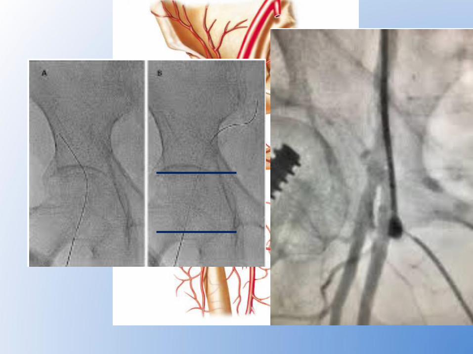
Conduit Vessel Complications • Aorto Iliac injury from
wire/sheath manipulation
• Always advance and retract interventional sheath over supportive wires
• And with the dilator• Consider advancing a soft
same size catheter to act as the long soft dilator in difficult friction cases.
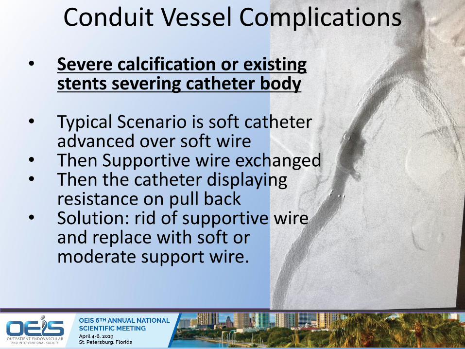
Conduit Vessel Complications
• Severe calcification or existing stents severing catheter body
• Typical Scenario is soft catheter advanced over soft wire
• Then Supportive wire exchanged• Then the catheter displaying
resistance on pull back• Solution: rid of supportive wire
and replace with soft or moderate support wire.

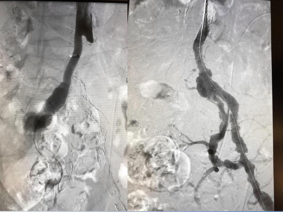
Wire Related Perforation• During crossing:
• Likely to be inconsequential perforation of a small branch or “parallel collateral”
• If in Doubt Coil embolize and continue the crossing effort
• In The calf BP cuff for 5 minutes at SYS BP usually suffices
• After crossing from wire tip left unchecked:• This could be problematic• Watch for compartment Syndrome in the calf• Watch for Renal Subcapsular or extra capsular
hematoma• We had one anecdote of intra abdominal bleeding
from .14 wire tip in a distal mesenteric branch

• Treatment Related Complications• Perforation
• Free wall perforation• Atherectomy:
• Laser• Hawk• Jetstream
• Oversized PTA.• Usually will require covered stent placement promptly.
• Embolization: • Macro:
• Hawk, Jetstream, some times DB or Laser• Micro:
• Diamond Back, Rotoblator.• Best treatment is avoidance• Use filters in moderate to high risk settings• Advance Laser at < 1 mm/sec religiously • Segmental Blades down/up technique with Jetstream is helpful• Always Treat thrombus successfully before reverting to any other modality.• The Dippel Principle “Do Not Cut Corners”

• Treatment Related Complications
Embolization: • When retrieving filters consider using a long 5 Fr
sheath not just the filter retrieving catheter.• Consider not fully constraining the filter to
reduce the risk of spilling over embolic material.
• If residual Macro emboli persist, take 4 fr long endhole catheter to the embolus and suction it out.
• If Thrombotic embolization is suspected infuse TPA and heparin for 2-4 hours and relook.
• Inject selective I.A. Nitroglycerin, many times BTK vascular spasm could look like emboli.

• Treatment Related ComplicationsEmbolization:
• Have a very low threshold for Filter use when treating the high embolic risk anatomy:• Graft Disease• ISR• Especially if
Atherectomy is used in those settings.
• Consider lesion appearance.


Thrombotic Complications
• Obtaining ACT every 20-30 mins is crucial• Especially if addressing complex
anatomy• Or having long catheter dwell times
• Thrombus can develop on the wire • Can build in the sheath• Could be part of the complex occlusion

Renal Complications• Athero Embolization• Contrast induced nephropathy • Hemoglobinuria Induced ATN (with Angiojet)
• Prevention:• Use only 4 Fr diagnostic catheters• Use soft technique• Hold ACE, ARB, Diuretics 24-48 hours ahead• Hydrate, pre, in, and post procedure• In Procedure planning define your contrast maximum load and announce
that to the team at time out.• Use more CO2 imaging• Use IVUS • Alkalinize the urine to prevent Hemoglobinuria and potential irreversible
Tubular injury• Avoid exceeding 300-500cc Angiojet• Go to definitive treatments that require less “relook angios during the
procedure”• Abort further attempts once contrast max given• Stage• Use IVUS
• Cerebrovascular Complications
• Arch catheter manipulation (with radial access or heart cath)
• Aortic valve crossing especially in AS cases.
• Excessive anticoagulation in setting of HTN

Sedation Related ComplicationsHypoxia• Use Music to reduce the need for sedation• Interactive patient engagement• Avoid Blind oxygen administration, could
be deleterious in Co2 retainers (there are many of them around) once subjected to sedation in reducing resp drive.
• Have reversal agents readily available• Correct possible airway issues before
Oxygenation. Paradoxic Agitation