Embed Size (px)
Citation preview
CASE REPORT Open Access
Vertebral artery compression of medullaoblongata causing isolated symptom ofdizziness: two case reportsMahmoud M. Mahrous1,2* and Ibrahim A. Busaad2
Abstract
Background: Symptomatic medulla oblongata compressing lesions due to dilated vertebral artery are rare in theliterature. The symptoms are extremely heterogeneous and not correlated to the severity of compression in manycases.
Case presentation: This paper describes two cases with vertebral artery compression of medulla oblongata complainingonly from dizziness without any other neurological symptoms or signs. In both cases, videonystagmography revealedpositional nystagmus. Head magnetic resonance imaging showed abnormal dilatation, elongation, and tortuosity of thevertebral artery compressing the medulla oblongata. Vestibular rehabilitation was described in both cases and had asignificant effect on symptom improvement.
Conclusion: In the study cases, the sense of vertigo and/or unsteadiness is due to vertebral artery compression of medullaoblongata and can be an isolated symptom. Positional nystagmus is the only sign in vestibular evaluation.
Keywords: Dizziness, Medulla oblongata, Vertebral artery, Dolichoectasia, Vestibular rehabilitation
BackgroundMedulla oblongata (MO) compressing lesions are rare inthe literature. Most of the reported cases are due totumor compressions, while it is less common to findvascular indentation [1]. Different vascular pathologieswere reported such as fusiform aneurysm, a persistingtrigeminal artery, a dolichoectasia of the vertebrobasilararterial system, and few reported cases of tortuous verte-bral artery (VA) compressing the brainstem.Patients’ presentations are extremely heterogeneous; it
varies between hemiparesis, leg weakness and tingling,hypertension, dysarthria, headache, visual changes, poorcognition, vertigo and nausea. Vertebral artery compres-sing the medulla can cause atypical deceiving symptoms.
Gorton et al. [2] reported a case presented with intract-able nausea and vomiting and weight loss. Symptomswere completely reversed following microvascular de-compression. Ejma et al. [3] reported a case clinicallymimicking myasthenia gravis.The largest case series of MO compression was a case
series of 9 patients published by Savitz et al. [4]. The au-thors reported various presentations of their patients.Most of the cases of VA compression were reported ofthe left VA. There is no correlation between neither thedegree nor the site of indentation and patients’ presenta-tion. Moreover, patient presentation and radiologicalfindings do not match in some cases [4], which makesMO compressing lesions difficult to be diagnosed.The management of VA compression of MO is possible
to be classified into surgical and conservative. Firstly, thereare 4 different microsurgical procedures reported in theliterature which include vessel mobilization, vessel sectionwith posterior fossa decompression, autologous materialinlay with posterior fossa decompression, and lateral
© The Author(s). 2020 Open Access This article is licensed under a Creative Commons Attribution 4.0 International License,which permits use, sharing, adaptation, distribution and reproduction in any medium or format, as long as you giveappropriate credit to the original author(s) and the source, provide a link to the Creative Commons licence, and indicate ifchanges were made. The images or other third party material in this article are included in the article's Creative Commonslicence, unless indicated otherwise in a credit line to the material. If material is not included in the article's Creative Commonslicence and your intended use is not permitted by statutory regulation or exceeds the permitted use, you will need to obtainpermission directly from the copyright holder. To view a copy of this licence, visit http://creativecommons.org/licenses/by/4.0/.
* Correspondence: [email protected] of Audio-vestibular medicine, Otorhinolaryngology Department, Facultyof Medicine, Ain Shams University, Cairo, Egypt2Unit of Audio-vestibular medicine, Otorhinolaryngology Department, Facultyof Medicine, Imam Abdulrahman Bin Faisal University, Dammam, SaudiArabia
The Egyptian Journalof Otolaryngology
Mahrous and Busaad The Egyptian Journal of Otolaryngology (2020) 36:9 https://doi.org/10.1186/s43163-020-00010-8
vessel retraction assisted with Gore-Tex. It has been re-ported that surgical decompression leads to significant im-provement [1]. In fact, other studies reported surgicalintervention did not benefit some patients or temporaryimprovement with some side effects [5]. Secondly, conser-vative management is based on addressing every patientcomplaint individually. Conservative medical managementmight include aspirin, warfarin sodium, dipyridamole, andanalgesics [4].After reviewing the literature, the table below summa-
rizes the signs, symptoms, and site of indentation ofMO, management method, and its efficacy of 4 cases.Although, all mentioned cases in Table 1 are due to VAcompression and they presented mainly by a complaintof dizziness, other uncommon symptoms are alsopresent in association with dizziness except one casethat was presented only with dizziness.
Case presentationCase 1A 36-year-old male patient complained of attacks of par-oxysmal sense of unsteadies/vertigo in the past 9months. The attacks were aggravated by turning thehead to either side or tilting his head up or down. At-tacks lasted for a few minutes. According to the patient,the attacks’ intensity were progressive and symptomsimproved by rest. It was associated with nausea withoutvomiting. No history of tinnitus or hearing loss, and noloss of consciousness or falling down. Patients did nothave history of any chronic medical diseases.Office examinations revealed normal cranial nerve
function. No spontaneous or gaze-evoked nystagmus,normal head shake test, and normal head thrust test.Videonystagmography (VNG) revealed normal saccade,smooth pursuit, optokinetic nystagmus and bilateral
normal and symmetrical caloric responses. The onlypositive finding which was found in head positional tests(head hanging positions) and Dix-Hallpike test was tor-sional nystagmus at either side which changes its direc-tion in the right and left sides. The patient did notreport a sense of vertigo while present in the provokingposition. The nystagmus was not fatigable and not sup-pressed by fixation. No nystagmus in other headpositions.Magnetic resonance imaging (MRI) was requested for
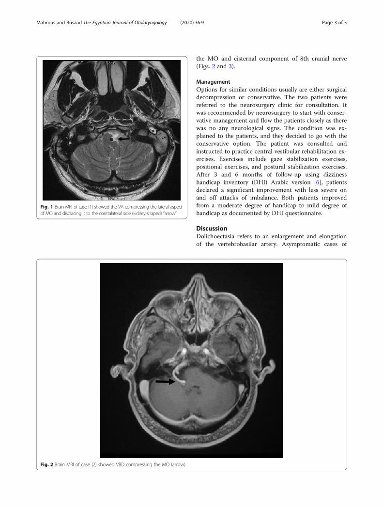
the patient to rule out central lesions. MRI demon-strated an abnormal course of the VA. The VA showedtortuous course of the lateral aspect of MO and dis-placing it to the contralateral side (kidney-shaped).There was no evidence of vascular infarction, gray-whitedifferentiation, midline shift or mass effect, or intra- orextra-axial collection. Both seventh and eighth cranialnerves are unremarkable bilaterally (Fig. 1).
Case 2Male patient, 59 years old, complained of sense of imbal-ance precipitated by standing from sitting position andafter sneezing. He is diabetic and hypertensive of 10years duration. General and cranial nerve examinationwere irrelevant. The audiological assessment revealed bi-lateral mild to moderate high-frequency sensorineuralhearing loss, with excellent speech discrimination. Pos-itional testing revealed left beating nystagmus at all headand body positions not suppressed by fixation. OtherVNG tests were within normal. Cranial nerve examin-ation was normal.MRI head with contrast showed abnormal dilatation,
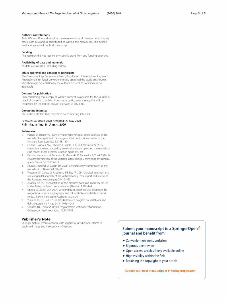
elongation, and tortuosity of V4 segment of VA suggest-ive of vertebrobasilar dolichoectasia (VBD) compressing
Table 1 Reported cases of vertebral artery compression of the medulla associated with vertigo and/or imbalance
Patientage (years) and gender
Radiographic findings Clinical presentation Treatment Outcome Reference
38/M Right VA loop compressingthe cervicomedullaryjunction/cranial nerve XI
Torticollis, vertigo,vomiting, arrhythmias
Microvasculardecompression
Cervical stiffness improved [5]
68/M Medullary compression atthe left lateral surface, base,and pyramid
Sudden 20-s imbal-ance, veering to theright
Warfarinsodium
No episodes in 1 year [4]
34/F Tortuous left vertebralcompression on the leftside at the base andtegmenta-basal junction
Multiple episodes ofunsteadiness, auralfullness, tinnitus,nausea, headache
Aspirin No further deficits at 1 year [4]
32/F Tortuous, dilated leftvertebral compression onthe left middle basilar partand pyramid
Episodes of headache,vertigo, loss ofconsciousnessTorsional nystagmusto the left, reducedleft corneal reflex
Microvasculardecompression
Episodes disappeared for 4 months, thenrecurred; magnetic resonance imagingshowed displacement of the lefttegmentum and base
[4]
Mahrous and Busaad The Egyptian Journal of Otolaryngology (2020) 36:9 Page 2 of 5
the MO and cisternal component of 8th cranial nerve(Figs. 2 and 3).
ManagementOptions for similar conditions usually are either surgicaldecompression or conservative. The two patients werereferred to the neurosurgery clinic for consultation. Itwas recommended by neurosurgery to start with conser-vative management and flow the patients closely as therewas no any neurological signs. The condition was ex-plained to the patients, and they decided to go with theconservative option. The patient was consulted andinstructed to practice central vestibular rehabilitation ex-ercises. Exercises include gaze stabilization exercises,positional exercises, and postural stabilization exercises.After 3 and 6 months of follow-up using dizzinesshandicap inventory (DHI) Arabic version [6], patientsdeclared a significant improvement with less severe onand off attacks of imbalance. Both patients improvedfrom a moderate degree of handicap to mild degree ofhandicap as documented by DHI questionnaire.
DiscussionDolichoectasia refers to an enlargement and elongationof the vertebrobasilar artery. Asymptomatic cases of
Fig. 1 Brain MRI of case (1) showed the VA compressing the lateral aspectof MO and displacing it to the contralateral side (kidney-shaped) “arrow”
Fig. 2 Brain MRI of case (2) showed VBD compressing the MO (arrow)
Mahrous and Busaad The Egyptian Journal of Otolaryngology (2020) 36:9 Page 3 of 5

dolichoectasia of vertebral and basilar arteries are notuncommon. However, there are reports suggesting thatVBD can implicate in neurologic symptoms, both bycompression of the brain stem and cranial nerves or byproducing transient ischemic attacks [7]. According tothe authors’ knowledge the isolated symptom of dizzi-ness due to VA compression of the medulla areextremely rare in the literature (Table 1). Astonishingly,there were no neurological abnormalities and cranialnerve affections especially in case (1) in spite of severecompression of the medulla. Due to its gradually pro-gressive nature, the course of brainstem compression byVBD is usually slow. Although sometimes the mass ef-fect of VBD can be very serious, the brainstem can grad-ually tolerate compression without showing obviousclinical symptoms [8]. In both cases, vertigo and nystag-mus are mostly attributed to compression of vestibularnuclei located in the dorsolateral aspect of the medulla;in addition to compression of cisternal component ofthe 8th cranial nerve in case (2). No MRI evidence ofany ischemic effects or brainstem infarction in bothcases.Although, patients had central vestibular disorders that
could not allow for central compensation and limit theamount and speed of recovery, research had shown that
patients with central vestibular disorders could benefitfrom vestibular rehabilitation therapy [9]. Accordingly,vestibular rehabilitation exercises were given to our pa-tients and they reported significant functional, physicaland emotional benefits with decreased frequency and se-verity of the dizziness attacks.
ConclusionIt would be necessary to perform a careful vestibularexamination to all vertigo cases. In our patients, the tor-tious and enlarged VA was demonstrated clearly by theMRI. The sense of vertigo and/or unsteadiness arecaused by VA compression of MO. Positional nystagmuswith central characteristics can be an isolated sign. Al-though patients have central vestibular lesions, vestibularrehabilitation exercises can be used as it showed an im-provement in our cases.
AbbreviationsDHI: Dizziness handicap inventory; MO: Medulla oblongata; MRI: Magneticresonance imaging; VA: Vertebral artery; VBD: Vertebrobasilar dolichoectasia;VNG: Videonystagmography
AcknowledgementsNot applicable
Fig. 3 Brain MRI of case (2) showed VBD compressing the MO (arrow)
Mahrous and Busaad The Egyptian Journal of Otolaryngology (2020) 36:9 Page 4 of 5
Authors’ contributionsBoth MM and IB contributed to the examination and management of studycases. Both MM and IB contributed to writing the manuscript. The authorsread and approved the final manuscript.
FundingThis research did not receive any specific grant from any funding agencies.
Availability of data and materialsAll data are available, including videos.
Ethics approval and consent to participateThe Otolaryngology Department Board, King Fahad University Hospital, ImamAbdulrahman Bin Faisal University ethically approved the study on 5/1/2018after thorough presentation by the authors. Consent to participate is notapplicable.
Consent for publicationI am confirming that a copy of written consent is available for the journal. Aproof of consent to publish from study participants is ready if it will berequested by the editors and/or reviewers at any time.
Competing interestsThe authors declare that they have no competing interests.
Received: 26 March 2020 Accepted: 20 May 2020
References1. Hänggi D, Steiger HJ (2009) Symptomatic vertebral artery conflicts to the
medulla oblongata and microsurgical treatment options: review of theliterature. Neurosurg Rev 32:143–149
2. Gorton L, Ashour AM, Lebovitz J, Cosola Di A, and Abdulrauf SI (2015)Intractable vomiting caused by vertebral artery compressing the medulla: acase report. J Craniovertebr Junction Spine 6:89-89.
3. Ejma M, Koszewicz M, Podemski R, Marianska K, Budrewicz S, Turek T (2011)Anatomical variation of the vertebral artery clinically mimicking myastheniagravis. Neurol Sci 32:715–717
4. Savitz SI, Ronthal M, Caplan LR (2006) Vertebral artery compression of themedulla. Arch Neurol 63:234–241
5. Vincentelli F, Caruso G, Rabehanta PB, Rey M (1991) Surgical treatment of arare congenital anomaly of the vertebral artery: case report and review ofthe literature. Neurosurgery 28:416–420
6. Alsanosi AA (2012) Adaptation of the dizziness handicap inventory for usein the Arab population. Neurosciences (Riyadh) 17:139–144
7. Ubogu EE, Zaidat OO (2004) Vertebrobasilar dolichoectasia diagnosed bymagnetic resonance angiography and risk of stroke and death: a cohortstudy. J Neurol Neurosurg Psychiatry 75:22–26
8. Yuan YJ, Xu K, Luo Q, Yu JL (2014) Research progress on vertebrobasilardolichoectasia. Int J Med Sci 11:1039–1048
9. Shepard NT, Telian SA (1995) Programmatic vestibular rehabilitation.Otolaryngol Head Neck Surg 112:173–182
Publisher’s NoteSpringer Nature remains neutral with regard to jurisdictional claims inpublished maps and institutional affiliations.
Mahrous and Busaad The Egyptian Journal of Otolaryngology (2020) 36:9 Page 5 of 5