Embed Size (px)
Citation preview
Version #11; 01/16/2019
Page 1 of 33
Title: Respiratory Muscle Strength Training to Improve the Vocal Function of Patients with Presbyphonia NCT number: NCT03557775 Document Approval Date: January 26th 2019
Version #11; 01/16/2019
Page 2 of 33
/post PROTOCOL TITLE:
Respiratory Muscle Strength Training to Improve the Vocal Function of Patients with Presbyphonia
PRINCIPAL INVESTIGATOR: • Heather Bonilha
Version #11; 01/16/2019
Page 3 of 33
1.0 Objectives / Specific Aims More than 10 million Americans over 65 years of age have presbyphonia, which is characterized by age-
related vocal fold atrophy that impairs their ability to speak at a normal volume for a normal amount of
time. Surgery can be used to treat presbyphonia, but non-invasive methods (voice exercises) are preferred
given the general risks of surgery and anesthesia in older individuals. Vocal Function Exercises (VFE), the
predominate type of voice exercises used to treat presbyphonia, can improve voice quality, but the
outcomes remain suboptimal, with many patients unable to successfully communicate, leading to
frustration, social isolation and feelings of helplessness.
Importantly, voice exercises target vocal fold strengthening, but underutilizes a potentially crucial second
pathway for intervention: strengthening the respiratory system. In addition to vocal fold integrity,
adequate vital capacity (airflow) and subglottal pressure are crucial for voice production. Many clinicians
do indeed devote time in their therapy sessions to ‘breathing exercises’ (diaphragmatic breathing) but
these are not evidence-based, and they are unlikely to adequately strengthen respiratory muscles to
impact the critical components of pulmonary function that effect voice. Therefore, a targeted approach
to improve the function of the respiratory system through rigorous and clinically feasible respiratory
training (training of the respiratory muscles) could augment VFE and improve voice efficiency and quality
in patients with presbyphonia.
The potential of respiratory training to improve respiratory and voice outcomes in patients with
presbyphonia is particularly relevant when one considers that the older population usually has coexisting
age-related declines in respiratory function. The clinical impact of respiratory training could be high, since
it is a low-cost/low-risk approach that can be used in clinic and at home, be delivered during voice therapy
sessions, and have a synergistic effect with voice exercises.
There are two main types of respiratory training: inspiratory training, also called inspiratory muscle
strength training (IMST), and expiratory training, also called expiratory muscle strength training (EMST) .
In this application, we propose a novel, proof-of-concept study to test whether the addition of either type
of respiratory training to the standard of care voice therapy (voice exercises) enhances respiratory and
voice outcomes. This study will permit, for the first time, the comparison between inspiratory and
expiratory training. The theoretical framework is: inspiratory training strengthens the inspiratory muscles
and thus increases both vital capacity and the ability to counteract passive recoil of the lungs, improving
Version #11; 01/16/2019
Page 4 of 33
the quantity of and control over the airflow available to drive the vocal folds. Expiratory training
strengthens the expiratory muscles to expand the expiratory reserve volume and provide more airflow for
voicing. Using pulmonary function tests and respiratory pressure measures, combined with voice
assessments, we will specifically evaluate our hypotheses and determine the independent effects of
inspiratory training and expiratory training on respiratory function and voice production. We will also
evaluate the interaction between the type of respiratory training and a patient’s baseline respiratory
function. This will provide mechanistic information to guide the future selection of the type of respiratory
training to augment voice therapy, to promote an impairment-specific intervention paradigm.
Forty-eight participants diagnosed with presbyphonia will be included in the study and will be stratified
and blocked-randomized into one of the three intervention groups (two experimental groups and one
control group), using a 3-parallel arm design: 1) inspiratory training (IMST) (experimental group),
2) expiratory training (EMST) (experimental group), or 3) voice exercises (VFE) (control group), delivered
four times during a 60-minute therapy session. Monitored home practice will consist of 10 minutes of
twice daily exercise for the duration of therapy. Participants from all three groups will carry on with their
standard of care voice therapy with a certified SLP.
Specific Aim 1: To measure the effects of respiratory training on respiratory and voice outcomes in
patients with presbyphonia. Hypothesis 1: The respiratory training groups (inspiratory and/or
expiratory) will demonstrate greater improvement in respiratory outcomes, measured through
pulmonary function tests and respiratory pressure measures, compared with the voice exercises-only
group. Hypothesis 2: The respiratory training groups (inspiratory and/or expiratory) will demonstrate
greater improvement in voice-related outcomes than the voice exercises-only group.
Secondary Aim 1: To assess the reliability of the ratings for subjective outcomes (videostroboscopy
ratings and perceptual judgements of voice quality). Hypothesis 1: Intra-judge reliability will be of
at least 80%. Hypothesis 2: Inter-judge reliability will be of at least 70%.
Specific Aim 2: To determine how baseline measures of respiratory function influence the effects of
respiratory training (respiratory and voice outcomes). Hypothesis 1: inspiratory training will have
maximal effect on patients with decreased maximum inspiratory pressure (MIP). Hypothesis 2:
Expiratory will have maximal effect on patients with decreased maximum expiratory pressure (MEP).
This is an initial feasibility and mechanistic study that has the potential to reveal that respiratory training
can significantly improve outcomes from voice therapy by acting in synergy with voice exercises. Currently,
Version #11; 01/16/2019
Page 5 of 33
much time is spent in voice therapy on subthreshold ‘breathing exercises’ that lack evidence. This project
will provide guidance for evidence-based and theory driven approaches to leverage respiratory function
to improve voice therapy. Conversely, negative findings from this study would indicate that respiratory
function is ineffective and not a viable target for therapy in this patient population.
2.0 Background Presbyphonia is characterized by vocal fold atrophy and a gap between the vocal folds. It impacts
more than 10 million Americans, inhibiting their ability to speak at a normal volume for a usual duration(1).
Disability related to presbyphonia is increasing due to: 1) the large number of older adults who work and
2) the expanding population over 65(2). Currently, non-invasive rehabilitation targeting laryngeal function
is the main treatment for presbyphonia, since surgery is associated with increased risk of negative
outcomes in older individuals and often results in suboptimal voice quality(3-5). While voice exercises
improve voice quality in patients with presbyphonia, many patients are unable to successfully
communicate in their environment even after completing treatment, i.e., they are unable to be heard
unless close to their conversational partner, cannot be heard in the setting of background noise, or are
unable to produce normal utterance lengths without taking frequent pauses to breathe. Presbyphonia is
a well-known cause of reduced quality of life and social isolation(1, 6-8).
Current therapeutic approaches underutilize a second pathway for intervention – strengthening the
respiratory system. Adequate vital capacity (airflow) and subglottal pressure are necessary to drive voice
production. While many clinicians devote substantial time in their therapy sessions to breathing exercises,
specifically diaphragmatic breathing, these exercises are not evidence-based and are unlikely to
adequately lead to neuromuscular and pulmonary function changes. Improving the respiratory support
for speech through rigorous respiratory training would, in theory, augment voice exercises to increase
vocal efficiency in patients with presbyphonia. The coexisting age-related declines in respiratory function
(in lung elasticity, ribcage mobility and inspiratory and expiratory muscle strength) further support
respiratory training’s potential to improve respiratory and voice outcomes in patients with
presbyphonia(9-12). Additionally, respiratory training is a low-cost/low-risk approach that patients can
use in clinic and at home to improve clinical measures of respiratory muscle strength and pulmonary
function(13, 14).
Version #11; 01/16/2019
Page 6 of 33
There are two types of respiratory training: inspiratory and expiratory. Inspiratory and expiratory training
have different physiological targets and result in unique and specific changes to respiratory function(13).
Inspiratory training increases vital capacity, specifically inspiratory volume, and inspiratory muscle
strength, to counteract passive recoil from positive pressures(15-17). The improved respiratory function
from inspiratory training would increase the amount of air to drive the vocal folds and allow for better
control of airflow with the respiratory system (to compensate for the lack of vocal fold closure). Expiratory
training increases vital capacity, specifically expiratory reserve volume, and expiratory muscle strength to
allow for increased contraction of the rib cage and abdominal muscles, thus providing access to more air
to drive the vocal folds(18, 19). The effects of increasing respiratory muscle strength, either inspiratory or
expiratory, have never been studied in patients with presbyphonia. Such an investigation, as we are
proposing, would improve our mechanistic understanding of the cross-system interaction for voicing
between laryngeal and respiratory function, and indicate the potential for inspiratory or expiratory
respiratory training as viable therapy modalities for patients with presbyphonia.
The objective of respiratory training is to strengthen the respiratory muscles using a hand-held pressure
threshold device in which patients breathe in (inspiratory training) or out (expiratory training) forcefully
to open a valve and allow air to flow. The overloading of the respiratory muscles activates the
neuromuscular system beyond its normal level of activity and forces the system to adapt, which results in
changes in muscle function(20). Previous studies confirm that respiratory training induces morphologic
changes in the respiratory muscles. In a study by Ramirez-Sarmiento et al.(21), post-intervention biopsies
were taken from the external intercostal muscles and the results revealed a 38% increase in the proportion
of type I fibers and a 21% increase in the size of type II fibers (21). Inspiratory and expiratory training have
also been found to improve maximum inspiratory pressure (MIP) and maximum expiratory pressure
(MEP), respectively, as well as spirometry outcomes in different populations, including elderly individuals
(15, 22). In the voice literature, inspiratory training studies have targeted increasing MIP to reduce
dyspnea in patients with upper airway obstruction diseases such as paradoxical vocal fold movement
disorder, recurrent laryngeal papilloma and bilateral abductor vocal fold paralysis (23-25). We were
unable to find published research on voice quality outcomes from inspiratory training. As for expiratory
training, three studies have assessed its effect on professional voice users with voice complaints (26-28).
Improved subglottal pressure at loud intensity and reductions in vocal symptoms were found when
combining voice therapy and expiratory training in patients with laryngeal irritation, edema, and/or
benign vocal fold lesions(28).
Version #11; 01/16/2019
Page 7 of 33
The effects of respiratory training on voice outcomes have never been studied on patients with
presbyphonia despite the declines in respiratory function that occur with aging. Both inspiratory and
expiratory pressures can be reduced in elderly patients, yielding different impairment profiles(10). Since
inspiratory muscles are crucial for increasing inspiratory lung volume and for counteracting expiratory
pressures to enhance airflow control during sustained speech, increasing their strength appears to be a
viable target to improve the loudness and duration of speech(29). However, some individuals may have
difficulty generating sufficient subglottal pressure despite initiating phonation at high lung volume
because of reduced ribcage compliance and lung recoil forces(11, 12). For those patients, early activation
of the expiratory muscles might be the most effective strategy to increase vocal efficiency, by compressing
the chest wall to a smaller volume and generating the required airway pressure for speech production
(19, 30). These patients may optimally benefit from expiratory training. Although some patients may
benefit from both inspiratory and expiratory training, there is a need to assess the individual effects of
both interventions prior to testing their combined effect in a future study to understand the mechanism
of action and optimize the use of therapy time. Thus, we propose to study how differences in patients’
baseline respiratory function impacts intervention response, specifically which patients benefit from
inspiratory training and which patients benefit from expiratory training.
The contributions of this study will be to: 1) assess the relevance of respiratory training when treating
patients with presbyphonia; 2) determine the mechanistic respiratory and voice effects of respiratory
training for patients with presbyphonia, and 3) identify profiles of patients likely to benefit from each type
of respiratory training. These contributions would be significant as they would begin to provide speech
language pathologists (SLPs) with evidence pertaining to rigorous respiratory training for presbyphonic
patients. After the study results are validated, SLPs will be able to determine, based on their clinical
assessment, which patients should undergo respiratory training and which modality would be maximally
beneficial, inspiratory or expiratory training, or both. This would improve the outcomes and efficiency of
voice therapy. If respiratory training results in meaningful voice quality changes for patients with
presbyphonia, it may also indicate a novel modality for use with other hypofunctional voice disorders (e.g.
adductor paralysis).
3.0 Intervention to be studied
Version #11; 01/16/2019
Page 8 of 33
Participants in all groups will have already undertaken their voice care (voice assessments and voice
therapy with a certified SLP) at MUSC. Depending on their group assignment, participants will also receive
either inspiratory training, expiratory training, or no respiratory intervention (standard of care group). The
experimental and standard of care interventions are described below.
Inspiratory Training [Experimental intervention]: Inspiratory training will be conducted using an
inspiratory pressure threshold trainer (Philips Respironics® Threshold IMT or POWERbreathe® Medic Plus,
for patients with a MIP of 55 cmH20 and over), which consists of a mouthpiece with a spring-loaded
valve(13, 25). The valve blocks the airflow until the threshold pressure is achieved and allows airflow as
long as the sufficient pressure is maintained. The threshold will be set at 75% of the participant’s initial
MIP, as reported in the literature with elderly participants(16). If it is too hard for the participant, the load
will be lowered until the participant is able to perform the exercises. The load will be adjusted weekly by
the study team member to maintain a threshold at 75% of the participant’s MIP. Daily practices will consist
of 5 sets of 5 breaths with the device with a break between sets, repeated twice daily (15, 18, 31).
Expiratory Training [Experimental intervention]: Expiratory training will be conducted using an expiratory
pressure threshold trainer (EMST150TM) which consists of a mouthpiece with a spring-loaded valve. The
valve blocks the airflow until the threshold pressure is produced (set at 75% of the participant’s initial
MEP, per literature with similar age groups(32-34)), and allows the airflow as long as the sufficient
pressure is maintained. Participants will be asked to exhale forcefully into the mouthpiece after a full
inspiration. If it is too hard for the participant, the load will be lowered until the participant is able to
perform the exercises. The load will be adjusted weekly by the study team member to maintain a
threshold at 75% of the participant’s MEP. Daily practices will consist of 5 sets of 5 breaths with the device
with a break between sets, repeated twice daily(32-34).
Inspiratory and expiratory training have never been conducted on patients with presbyphonia. To
determine the adequate intensity and duration of treatment, we reviewed the literature of respiratory
training interventions with patients of similar age groups. We decided on a threshold of 75% of MIP and
MEP because 1) this value was widely used across studies, resulted in significant improvements in
respiratory outcomes, and has been shown to be safe(16, 32-34); 2) maximal improvements in muscle
strength have been reported to occur with a loading at 70-90% of the maximal strength(14). The regimen
of 5 sets of 5 breaths was also widely used across studies, along with a threshold at 75% of MIP/MEP, and
resulted in improved respiratory outcomes. We chose to have the participants practice their exercises every

Version #11; 01/16/2019
Page 9 of 33
day of the week because in our opinion it is the best way to create a habit and therefore enhance
compliance to treatment.
Voice Exercises [Standard of care intervention]: Participants will be instructed to follow the four steps of
the VFE protocol, developed by Stemple (35) and commonly used by SLPs with patients with presbyphonia:
(a) sustain the vowel /i/ on the musical note F (above middle C for women; below middle C for men) for
as long as possible. (b) Glide from the lowest note to the highest note on the word "knoll". (c) Glide from
the highest note to the lowest note of the word "knoll". (d) Sustain the notes C-D-E-F-G (starting from
middle C for women and an octave below middle C for men) on the word "oll", for as long as possible.
Each note will be repeated until the participant finds the right placement (forward-focused voice), as
judged by the SLP. Humming exercises will be used to facilitate placement. Daily practices, 2x/day, will
consist of two repetitions of the VFE protocol for the IMST and EMST groups and four repetitions of the
VFE protocol for the VFE group. Recordings of the therapy sessions will be provided to facilitate home
practice.
4.0 Study Endpoints The outcome measures have been chosen to provide the most complete and accurate evaluation of
therapy results. Outcome measures are numerous in the voice field, and to allow comparison across
studies, the American Speech-Language-Hearing Association (ASHA) has recently developed a
recommended protocol for instrumental assessment of voice(36). We modeled our measurement
methods on these recommendations and added supplementary outcomes relevant to this study.
Voice measures. [Note: voice measurements will be obtained from the participants’ standard of care voice
assessments]
Videostroboscopy: Recordings of vocal fold vibration from sustained phonations of /i/ at various pitch and
loudness levels will be used 1) to assess the parameters described in the Voice-Vibratory Assessment with
Laryngeal Imaging (VALI) rating form(37), and 2) for measurements of normalized glottal gap (NGG)(38).
Acoustic measures: The Computerized Speech Lab (CSL) is used to record sustained phonation at various
pitch and intensity levels, reading of the Consensus Auditory-Perceptual Evaluation of Voice (CAPE-V)(40)
sentences, as well as a short natural conversation with the SLP. Acoustic analysis will include: 1) measures
of voice intensity: habitual and maximum sound pressure levels (SPL); 2) measures of fundamental
Version #11; 01/16/2019
Page 10 of 33
frequency (F0): mean, minimum, and maximum vocal F0, and 3) measures of voice quality: cepstral peak,
jitter, shimmer, and harmonics-to-noise ratio.
Perceptual judgments of voice quality: The CAPE-V(40) rating form will be used for the judgment of voice
quality of sustained phonation, reading and conversational speech tasks recorded via CSL. The CAPE-V
form includes ratings of overall severity, roughness, breathiness, strain, pitch, and loudness, as well as
qualitative information about resonance and additional features.
Patient self-assessments: Patients are asked to complete the 1) Voice Handicap Index (VHI): a
questionnaire that evaluates the degree of handicap experienced by the patient in relation to their voice
use(41); 2) the GFI(42): a validated disease-specific impairment instrument conceived to assess symptoms
related to glottal insufficiency; 3) the RSI(43), a validated self-administered questionnaire to assess
symptoms related to LPR; and 4) the Communicative Participation Item Bank (CPIB) general short
form(44). Patients are also asked to rate their perceived phonatory effort (PPE) on a direct magnitude
estimation scale(45) following the speaking and voicing tasks recorded with the CSL.
Aerodynamic measures: The Phonatory Aerodynamic System (PAS) is used to measure 1) mean glottal
airflow rate: estimated from sustained phonation of a vowel(36); 2) average subglottal air pressure (Ps):
measured during the production of a voiceless plosive consonant (/p/) at normal pitch and loudness, an
accepted indirect and non-invasive measurement method for Ps (46); and 3) laryngeal resistance:
calculated by dividing subglottal pressure by mean glottal airflow.
Phonation duration (MPT): The maximum phonation duration of the vowel /a/ is measured via CSL.
Respiratory measures (respiratory measures will be recorded solely as part of the study).
Spirometry: Spirometry will be used to measure 1) forced vital capacity (FVC), 2) forced expiratory volume
in one second (FEV1), and calculate the 3) FEV1/FVC ratio. The predicted values (%) of these three
measures will also be reported. The patient will be asked to take a full inspiration, blow out the air
forcefully and then exhale slowly to residual volume in the spirometer. The task is repeated at least three
times, until the two best measurements differ by no more than 0.15.
Muscle pressure measures: 1) MIP: measured at functional residual capacity, is an indicator of inspiratory
muscle strength. Patients will be instructed to exhale fully and then inhale with maximal effort with lips
sealed to the pressure meter for at least 2 seconds. 2) MEP: measured at full lung capacity, is an indicator
of expiratory muscle strength. Participants will be instructed to take a full breath and then exhale with
maximal force with lips sealed to the pressure meter for at least 2 seconds.

Version #11; 01/16/2019
Page 11 of 33
5.0 Inclusion and Exclusion Criteria/ Study Population
Patients who are diagnosed with presbyphonia and who have already sought and begun voice care at
MUSC, will undergo a brief screening interview by Dr. Halstead, after which qualifying patients will be
offered participation in the study. A study team member will meet with interested participants and will
provide a thorough description of the study prior to obtaining formal written consent. Participants who
don’t meet stratification requirements will be excluded from the study.
Inclusion Criteria
The participant:
• must receive a diagnosis of presbyphonia by a trained laryngologist. The diagnosis will be given following a visual examination of the larynx if the observations are consistent with the characteristics of a presbylarynx, as judged by the laryngologist.
• must have already undertaken their voice care at MUSC
• must be 50 years and older
• must be English-speaking
• must sign the informed consent
Exclusion Criteria
The participant
• has received voice therapy in the past year (other than current care)
• presents with a vocal fold pathology other than presbyphonia
• has a known neurologic or a progressive neuromuscular disease
• doesn’t meet the stratification requirements for muscle pressure
• has a medical condition that could be aggravated by the experimental intervention, or
any condition judged by the physician (Dr. Halstead) as being unsuitable for respiratory
training.
• has dysarthria or a language disorder
• has a hearing loss that is not adequately managed
• has a cognitive disorder that might affect treatment compliance
• is unable to give informed consent
Version #11; 01/16/2019
Page 12 of 33
Inclusion of Women
No subjects will be excluded based on gender. Based on published literature, treatment seeking patients
with presbyphonia are generally equally divided between female and male. Thus, we estimate that
approximately 50% of study participants will be female. No outreach program to recruit women is
planned, because all patients with presbyphonia who meet inclusion criteria after undergoing a voice
evaluation at the participating institution, regardless of gender, will be invited to participate. While no
subjects will be excluded based on gender, it is highly likely that the majority of participant raters will be
women as that is the current composition of speech-language pathologists.
Inclusion of Minorities
No subjects (patient or control subject groups) will be excluded based on race or ethnic origin. However,
only English-speaking participants will be included in the study.
Gender, ethnic and racial information will be collected at the study enrollment. Ethnicity (i.e.,
Hispanic/Latino, Non-Hispanic/Latino) will be recorded separately from race. All applicable racial
categories (i.e., American Indian/Alaskan Native, Asian, Native Hawaiian or other Pacific Islander,
Black/African American, White) are recorded for each subject. All ethnic and racial totals will be reported,
including specific categories for those individuals who report mixed racial descent. The Targeted/Planned
Enrollment Tables for the participating institution are included in the Targeted/Planned Enrollment
section, which indicate the anticipated ethnic and racial mixes of the subject sample. Numbers reflected
in these tables parallel the ethnic and racial mixes of the service areas of the participating institution. No
outreach program to recruit minorities is planned because all patients with presbyphonia who meet
inclusion criteria after undergoing a voice evaluation at the participating institution, regardless of race or
ethnic origin, will be invited to participate.
While no subjects will be excluded based on race/ethnicity, it is highly likely that all of the participant
raters will be White Non-Hispanic as that is the current composition of speech-language pathologists.
Inclusion of Children
Version #11; 01/16/2019
Page 13 of 33
It has been well documented that infants and children differ significantly from adults in laryngeal,
respiratory and neurological anatomy and physiology. Presbyphonia is a voice disorder caused by age-
related changes in the laryngeal, respiratory and neurological systems, including muscle atrophy, changes
in the vocal fold histology and in lung tissue and ribcage mobility. The aging process may differ between
individuals, with symptoms of presbyphonia reported to occur as early as 50 years old. No case of
presbyphonia has ever been reported in a child under 18 years old. The aim of this study is to assess the
effects of respiratory training in patients with presbyphonia, therefore, the topic is not relevant to
children. Although respiratory training could potentially be beneficial for treating other types of voice
disorders, including those affecting children, a separate, age-specific study in children, would be
warranted. Therefore, children are not a part of the current study.
6.0 Number of Subjects Forty-eight patients-participants and up to six raters-participants will be recruited for the study. 7.0 Setting Data Collection Site: All data collection and study activities will be conducted at the Medical University of
South Carolina (MUSC). The patient subject sample will include 48 treatment-seeking patients diagnosed
with presbyphonia at the Evelyn Trammell Institute for Voice and Swallowing (division of the Department
of Otolaryngology-Head & Neck Surgery). The sample will be limited to the MUSC site because historical
accruals have been shown sufficient to meet power (see participant section above). Further, the data
quality and logistical challenges faced in multi-site trials go beyond the scope and expense of this current
proposal. Participant raters will be recruited from SLPs who have an affiliation with MUSC and who have
experience in working with voice patients.
8.0 Recruitment Methods Following IRB approval, participants will be recruited from the treatment-seeking population at the Evelyn
Trammell Institute for Voice and Swallowing (division of the Department of Otolaryngology-Head & Neck
Surgery) - Medical University of South Carolina (MUSC). As part of usual care, all patients will undergo a
videostroboscopic assessment conducted by the laryngologist, Dr. Halstead and will be referred to voice
Version #11; 01/16/2019
Page 14 of 33
therapy with a certified SLP to start usual treatment. Patients will undergo a brief screening interview by
Dr. Halstead, after which qualifying patients will be offered participation in the study.
9.0 Consent Process
Interested and eligible patients will meet with a study team member shortly after being informed
of the study (on the same day if possible). The consent process will take place in Rutledge Tower.
The study member will provide a detailed description of the study and of its risks and benefits and
the patient will be handed a written document with all the information. The study member will
then discuss with the prospective participant and will ask specific questions to ensure a good
understanding of key elements related to the study. Patients will be prompted to ask any questions
they have. Finally, patients will be informed that their decision to participate or not in the study
will not impact the care that they will receive at MUSC. Following this procedure, written consent
will be obtained from interested participants. Following muscle pressure testing, participants
whose muscle pressure results don’t meet stratification requirements will be excluded from the
study.
10.0 Study Design / Methods
Overview After being enrolled in the study, treatment-seeking patients with a diagnosis of presbyphonia will
undergo baseline respiratory assessments including spirometry and respiratory muscle strength measures
as well as voice recordings (acoustic assessments). Participants will then be blocked-randomized (based
on their MIP and MEP) to one of three groups using a 3-parallel arm design: (2 experimental groups and
1 control group), using a 3-parallel arm design: 1) inspiratory training (IMST) (experimental group), 2)
expiratory training (EMST) (experimental group), or 3) standard of care (VFE) (control group), delivered
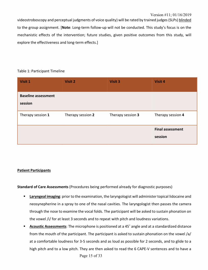
four times (see table 1 for participant timeline). The sessions will occur on the same days as the
participants’ scheduled voice therapy sessions when possible. Participants will be asked to practice their
respective exercises for 10 minutes, twice daily, seven days a week during the duration of the study. A
final assessment session, after the 4th therapy session, will mirror the baseline measures. Participants will
receive a monetary compensation of $20 for each study visit, for a maximum of $80 for the whole study.
Payments will be made via cash. Outcomes that rely on subjective judgment (visual ratings of
Version #11; 01/16/2019
Page 15 of 33
videostroboscopy and perceptual judgments of voice quality) will be rated by trained judges (SLPs) blinded
to the group assignment. [Note: Long-term follow-up will not be conducted. This study’s focus is on the
mechanistic effects of the intervention; future studies, given positive outcomes from this study, will
explore the effectiveness and long-term effects.]
Table 1: Participant Timeline
Visit 1 Visit 2 Visit 3 Visit 4
Baseline assessment
session
Therapy session 1 Therapy session 2 Therapy session 3 Therapy session 4
Final assessment
session
Patient Participants
Standard of Care Assessments (Procedures being performed already for diagnostic purposes)
§ Laryngeal imaging: prior to the examination, the laryngologist will administer topical lidocaine and
neosynepherine in a spray to one of the nasal cavities. The laryngologist then passes the camera
through the nose to examine the vocal folds. The participant will be asked to sustain phonation on
the vowel /i/ for at least 3 seconds and to repeat with pitch and loudness variations.
§ Acoustic Assessments: The microphone is positioned at a 45˚ angle and at a standardized distance
from the mouth of the participant. The participant is asked to sustain phonation on the vowel /a/
at a comfortable loudness for 3-5 seconds and as loud as possible for 2 seconds, and to glide to a
high pitch and to a low pitch. They are then asked to read the 6 CAPE-V sentences and to have a
Version #11; 01/16/2019
Page 16 of 33
short natural conversation with the speech therapist. The participant is then asked to rate their
perceived phonatory effort (PPE) on a scale.
§ Self-assessments: The participant is asked to fill out the Voice Handicap Index questionnaire, the
Reflux Symptom Index, and the Glottal Function Index.
§ Aerodynamic assessments: The participant wears a face mask over the nose and mouth, which
directs the air flow. A tube connected to a pressure transducer is passed through a hole in the
mask, and inserted between the lips. The participant is asked to produce short utterances each
comprised of minimally five /pi/ syllables at a rate approximating 1.5 - 2 syllables per second (/pi-
pi-pi-pi-pi/). Each string of at least five syllables should be produced on one breath/exhalation. The
task is repeated 3 times at comfortable pitch and loudness.
Standard of Care Intervention (Procedures being performed already for treatment purposes)
§ Vocal Function Exercises: Participants in all 3 groups will be instructed to follow the 4 steps of the
Vocal Function Exercises (VFE) protocol, developed by Stemple (2005) and commonly used by
speech language pathologists (SLP) with voice patients: (a) sustain the vowel /i/ on the musical
note F (above middle C for women; below middle C for men) for as long as possible. Repeat as
judged by the SLP. (b) Glide from the lowest note to the highest note on the word "knoll". Repeat
as judged by the SLP. (c) Glide from the highest note to the lowest note of the word "knoll". Repeat
as judged by the SLP. (d) Sustain the notes C-D-E-F-G (starting from middle C for women and an
octave below middle C for men) on the word "oll", for as long as possible. Each note will be
repeated until the participant finds the right placement (forward-focused voice), as judged by the
SLP. Humming exercises will be used to facilitate placement of the voice.
§ Counseling and patient education during the therapy sessions.
Experimental Assessments (Procedures to be performed solely as part of this research study):
Firstly, the medical chart of the participant will be reviewed to obtain the following information: height
and weight. This information will be used to perform the respiratory and muscle pressure assessments.
The medical chart will also be used to track the speech therapy appointments, since research visits will
take place on the same days. Spirometry assessments will take place at baseline and after the fourth
intervention session. Muscle pressure testing and acoustic assessments will be conducted at baseline and
before and after each intervention session.
Version #11; 01/16/2019
Page 17 of 33
§ Spirometry: Prior to the tasks, the participant will put on a nose clip and make sure that the lips
are well sealed around the tube with no air is escaping. They will be asked to breathe normally,
and then take a full inspiration and blow out with force during at least 6 seconds, in the spirometer.
Those maneuvers will be repeated at least three times, to verify test reliability. No more than eight
repetitions should be needed(47). The participant is seated to reduce risks of fall.
§ Muscle pressure (maximum inspiratory pressure-MIP- and maximum expiratory pressure-MEP):
o MIP: the participant is instructed to exhale slowly and completely, seal lips firmly around
the mouthpiece, and then breathe in forcefully. The PI will note the largest negative
pressure sustained for at least one second on the pressure gauge. The participant will rest
for about one minute and then repeat the maneuver up to 4 additional times (with the goal
of matching the highest 2 trials within 10 cmH2O)(48).
o MEP: the participant is instructed to inhale slowly and completely, seal lips firmly around
the mouthpiece, and then breathe out forcefully. The PI will note the largest positive
pressure sustained for at least one second on the pressure gauge. The participant will rest
for about one minute and then repeat the maneuver up to 4 additional times (with the goal
of matching the highest 2 trials within 10 cmH2O) (48).
§ Acoustic assessment: The microphone is positioned at a 45˚ angle and at a standardized distance
from the mouth of the participant. The participant is asked to sustain phonation on the vowels /a/
and /i/ at a comfortable loudness for 3-5 seconds and as low and as loud as possible, to glide to a
high pitch and to a low pitch, and from a soft voice to a loud voice. They are asked to sustain the
vowel /a/ for as long as possible. They are then asked to read the 6 CAPE-V sentences and to have
a short natural conversation with the speech therapist. The participant is then asked to rate their
perceived phonatory effort (PPE) on a scale.
§ Self-Assessment questionnaire: the participant will be asked to fill out the Communicative
Participation Item bank (CPIB) questionnaire, which consists of 10 questions on communication.
Participants will be stratified based on their muscle pressure measures: 1) within normal limits MIP and
MEP; 2) preserved MIP and decreased MEP; 3) preserved MEP and decreased MIP; 4) decreased MIP and
MEP. This stratification is crucial to the study design because it will ensure an equal repartition of the
Version #11; 01/16/2019
Page 18 of 33
different respiratory profiles and deficits across treatment groups. Participants that don’t meet
stratification requirements will be excluded from the study.
Participants will be blocked-randomized into three groups (allocation ratio 1:1:1): IMST, EMST and VFE
(see description of the interventions below).
Experimental Interventions (Procedures to be performed solely as part of this research study):
Participants in all groups will attend four sessions with a study team member. Intervention sessions will
last approximately 30-45 minutes each. Each session will include pre- and post-treatment muscle pressure
and acoustic measurements.
§ Inspiratory Training: Inspiratory training will be conducted using an inspiratory pressure threshold
trainer (Philips Respironics® Threshold IMT or POWERbreathe® Medic Plus, for patients with a MIP
more than 83 cmH20), which consists of a mouthpiece with a spring-loaded valve(25). The valve
blocks the airflow until the threshold pressure achieved and allows airflow as long as the sufficient
pressure is maintained. The threshold will be set at 75% of the participant’s initial MIP, as reported
in the literature with elderly participants(16). If it is too hard for the participant, the load will be
lowered until the participant is able to perform the exercises. The load will be adjusted weekly by
the study team member to maintain a threshold at 75% of the participant’s MIP. Daily practices
will consist of 5 sets of 5 breaths with the device with a break between sets, repeated twice daily
(15, 18, 31).
§ Expiratory Training: Expiratory training will be conducted using an expiratory pressure threshold
trainer (EMST150) which consists of a mouthpiece with a spring-loaded valve. The valve blocks the
airflow until the threshold pressure is produced (set at 75% of the participant’s initial MEP, per
literature with similar age groups(32-34)), and allows the airflow as long as the sufficient pressure
is maintained. Participants will be asked to exhale forcefully into the mouthpiece after a full
inspiration. If it is too hard for the participant, the load will be lowered until the participant is able
to perform the exercises. The load will be adjusted weekly by the study team member to maintain
a threshold at 75% of the participant’s MEP. Daily practices will consist of 5 sets of 5 breaths with
the device with a break between sets, twice daily(32-34).
Home Practice: Participants will be instructed to practice their respective exercises twice daily, and to log
their practice in a compliance journal (days of training, perceived effects of treatment or other comments)
Version #11; 01/16/2019
Page 19 of 33
as well as to contact a research team member if any difficulty arise. In addition to in-person therapy
sessions, weekly phone calls will clarify aspects of the training and encourage treatment adherence.
Rater Participants
Procedures that will be performed solely as part of this research study:
Raters will rate the audio recordings with the CAPE-V form and the videostroboscopy images with the VALI
form. They will rate pre- and post-intervention de-identified recordings for each participant, and an
additional sampling of 20% of the recordings (for intrarater reliability testing).
Data Collection
Data collected will be the same for Aim 1 and Aim 2. It will include information in the patient’s medical
record (obtained from standard of care assessment) and obtained through a demographic questionnaire
as well as clinical data using standardized collection instruments: spirometry measures, muscle pressure
measures and acoustic assessment.
Each video and audio recording will be de-identified when videos are converted into .mpg, .nsp or .mp3
files before distributing the recordings via Box for analysis. Participant raters will review the de-identified
digitized recordings for the purposes of data collection and score each video and audio recording using
the VALI and CAPE-V rating forms, respectively.
Spirometry, muscle pressure data, and voice data will be recorded in a data collection sheet by the study
team member. Data collection sheets will be labeled with the alphanumeric identifier of the patient-
participant. Completed data collection sheets, with ratings from the CAPE-V and VALI, will be entered in
the database by a study team member. Research records will be retained for a period of six years to allow
evaluation and repetition of the results by others.
Study personnel will enter mandatory data fields for demographics according to the NIH minimum
standards for maintaining, collecting and presenting data on gender, race and ethnicity. The demographic
and clinical data will be exported for analysis.
12.0 Data Management
Data Analysis Plan
Version #11; 01/16/2019
Page 20 of 33
Demographic characteristics will be compared across the 3 intervention groups, using a RxC contingency
table method for categorical variables (gender, smoking history, voice use, racial and ethnic category) and
a one-way ANOVA for continuous variables (age, weight, height). Baseline characteristics will be compared
across the 3 intervention groups using a Kruskal-Wallis one-way analysis of variance by ranks for ordinal
data (GFI, RSI, and VHI scores), a RxC contingency table for categorical variables (glottal gap shape, free
edge contour, vertical level, phase closure) and a one-way ANOVA for continuous variables. Aim 1: To
assess the effects of respiratory training on voice and respiratory outcomes in patients with presbyphonia.
Data will be analyzed on an intention-to-treat basis using multifactor repeated measures generalized
linear regression. Distributional assessment will be made for each outcome to confirm optimal
distributional assignment and potential need for outcome transformation (i.e. log link). In the model, the
repeated factor is time, which has two levels (pre- or post-intervention) and the independent factor is the
intervention, which has three levels (IMST, EMST, VFE). In the case of a significant between-subjects effect,
multiple comparison tests will be conducted to determine which groups differ. The post-hoc analysis will
be done using Tukey’s honestly significant difference (offering protection against type I error without
being too conservative). Spearman and Person correlations will be calculated to establish inter and intra-
rater reliability. Aim 2: To identify profiles of presbyphonic patients that would benefit the most from
respiratory training. Logistic regression will be conducted to predict which patients improved following
inspiratory training and which patients improved following expiratory training, based on their baseline
clinical characteristics. Two outcome variables will be indicative of meaningful clinical improvement:
1) MPT (to measure function), and 2) GFI (to measure patient perception of function). Meaningful clinical
improvement will be defined by an increase of at least 4 seconds in MPT or a decrease of at least 4 points
in GFI score. Participants will be dichotomized as having meaningful clinical improvement, or not, based
on these outcomes. Predictors will include MIP, MEP, maximum SPL, breaths per minute, FVC, FEV1, and
Ps. These measures were chosen because they are directly influenced by the integrity of the respiratory
system.
Sample Size Justification We considered maximum phonation time (MPT) as the primary outcome measure for sample size
calculation. This measure was chosen because 1) low MPT is a hallmark of patients with presbyphonia,
and 2) MPT is directly affected by both the laryngeal and respiratory systems. To estimate the expected
effect size between VFE and the I+IVFE and E+VFE groups, we considered MPT values from groups of an
Version #11; 01/16/2019
Page 21 of 33
elderly population with different glottal closure and spirometry profiles(49). Considering that a combined
intervention (inspiratory or expiratory training combined with voice therapy) would address both
laryngeal and respiratory deficits, and that a voice therapy-only intervention would directly address only
the laryngeal deficits, we compared MPT between the no-deficit group (complete glottal closure and
normal spirometry) and the respiratory-deficit group (complete glottal closure and altered
spirometry)(49). Based on these values and the pooled standard deviations, the expected difference
between our control and experimental groups would represent a large effect size. As for the within-
subjects effect, a study by Nam et al.(50) found a 33% increase in MPT following a respiratory training
intervention program in young adult singers. This corresponds to a very large within-subjects effect size
with values of d exceeding 1.2. For Aim 2, we will assess improvement in 2 ways: function (a MPT increase
of 4 seconds, which has been associated with a positive perceived effect following voice therapy in older
adults(51)) and patient perception of function (a Glottal Function Index (GFI) score decrease of 4 points).
An increase of 4 seconds in MPT and a decrease of 4 points on the GFI instrument both correspond to
medium-large effect sizes considering the reported standard deviations of these measures in older
subjects and in patients with glottal insufficiency (5.1 and 4.5, respectively) (42, 52). Based on the reported
studies, the expected effect size for within-subjects and between-subjects differences was set at d=0.90.
To have an 80% power to detect an effect size of d=0.90 with an alpha of 0.1 (since this is a pilot study),
15 participants are needed in each group. To account for loss to follow-up (estimated 10% dropouts rate),
up to 16 participants will be recruited for each group, 48 patients total.
Based on estimated accrual rates, we calculated an average of one presbyphonic patient per week during
the recruitment period of 16 months, for a total 64 participants. We are therefore confident of meeting
our sample size goal.
Confidentiality
Protection against the loss of confidentiality will be diminished by 1) completing study aims at one data
collection site, 2) minimizing the participants involved to only those needed to accomplish the study aims
(see sample size justification above), 3) de-identifying data at the earliest opportunity, and 4) maintaining
stored data in locked locations whose access is restricted to members of the study team and on the secure
MUSC network.
Version #11; 01/16/2019
Page 22 of 33
All data will be entered into a database, linked, and de-identified, and labeled with an alphanumeric
identifier. Further use of data will be de-identified. The link between patient identity and alphanumeric
identifier will remain confidential in a study team member’s locked office in a locked cabinet and on a
password protected server (MUSC network). Electronic databases used for this study will be password
protected and controlled by the study team. Only the study team will have access to participants’
identities. Study staff not directly involved with the patient (e.g., biostatistician) will not have access to
the linkage between identifier and subject-identifying information. Identities will be maintained in the
general study research folders as identifiers on the signed informed consent forms, HIPPA forms,
remuneration forms, and codebook. Once a participant is enrolled, he/she immediately will be identified
by an alphanumeric code unrelated to any patient’s protected health information. This code will
effectively de-identify the participant and be used for unique participant data identification in all
databases. Access to the electronic database will have a combination of physical and electronic security
protections (firewalls) to prevent unauthorized access to this data. Appropriate back-up routines also will
be performed to protect data integrity.
13.0 Provisions to Monitor the Data to Ensure the Safety of Subjects
A data and safety monitoring plan will be implemented by the PI to ensure that there are no changes in
the risk/benefit ratio during the course of the study and that confidentiality of research data is maintained.
Each member of the study team will meet with the PI and review confidentiality issues prior to having
contact with research subjects.
The monitoring of adverse events will be ongoing throughout the study. Investigators and study personnel
will meet regularly to discuss the study (study goals and modifications of those goals; participant
recruitment and retention; progress in data coding and analysis; documentation, identification of adverse
events or research participant complaints; violations of confidentiality) and address any issues or concerns
at that time. Minutes will be kept for these meetings and will be maintained in the study regulatory folder.
Any instances of adverse events will be immediately reported to MUSC’s IRB using the standard forms and
procedures that have been established by the IRB. The yearly IRB renewal for this study will include a
summary report of the Data and Safety Monitoring Plan findings from the prior year.
Version #11; 01/16/2019
Page 23 of 33
Due to the minimal risk of the study, and because no serious adverse events are expected, there will be
no specific safety endpoints. However, adverse events and unanticipated problems will be immediately
reported. The laryngologist, will review adverse events and unanticipated problems and provide an expert
assessment as to the probability of the event/problem being related to the experimental procedures. This
will be determined mainly by the temporal relationship with the procedure, the environment, and the
subject’s clinical state.
14.0 Withdrawal of Subjects
In the cases where subject should develop a health condition non-related to the study protocol, a study
team member would discuss with them that it is not appropriate at this time, given medical concerns, that
they continue with the study intervention. The laryngologist, Dr. Halstead, would be informed of the
situation and the medical team would make sure that the participant receives appropriate medical
assistance.
Subjects are allowed to withdraw at any time, they are clearly advised that this will not affect their medical
care or any aspects of their treatment.
15.0 Risks to Subjects Potential Risks Risks to subjects consist of 1) loss of confidentiality and 2) adverse events related to spirometry and
muscle pressure testing 3) adverse events related to the experimental interventions (inspiratory and
expiratory training) 4) incidental findings 5) risks associated with randomization and 6) unknown risks.
There are no known risks are associated with acoustic assessment of voice.
Loss of confidentiality: The study will involve data collection from human subjects and is therefore subject
to the risk of loss of confidentiality.
Spirometry and muscle pressure testing: Participants may feel the need to cough or may feel short of
breath and/or dizzy during or after the test. There is a risk of syncope during the forced vital capacity (FVC)
measurement.
Inspiratory training: Risks of discomfort, dizziness and light-headedness related to hyperventilation(53).
The participant may feel short of breath or notice a pulse rate increase. Inspiratory training generates a
Version #11; 01/16/2019
Page 24 of 33
negative pressure in the thorax and for this reason it could aggravate conditions such as a pneumothorax,
a middle ear pathology (such as a tympanic membrane rupture) or an acute unresolved sinusitis. Thus,
patients with any diseases that are judged by the physician to be at risk of being exacerbated by the
inspiratory training intervention will be excluded from study participation.
Expiratory training: During the expiratory training exercises, the patient has to generate sufficient
expiratory effort to open a threshold valve and allow air to flow. The action increases the intraoral and
intrathoracic pressures and has been compared to a sub-maximal Valsalva maneuver(54), which has been
associated with blood pressure, heart rate changes and abnormal cardiac findings(55). However,
expiratory training is not comparable to a typical Valsalva maneuver for two reasons: 1) the duration of
the isometric muscle contraction during expiratory training is of approximately 2 seconds, which is much
shorter than during a typical Valsalva maneuver (15-20 seconds)(54, 56); 2) during expiratory training,
complete closure of the glottis rarely occurs, and when it does it is very briefly(54). Laciuga et al.(54) tested
the effect of expiratory training on young health adults and found no acute changes in cardiovascular
response (heart rate, blood pressure, oxygen saturation) and no adverse events following a session of 25
expiratory training trials. However, expiratory training is not recommended in older patients with certain
cardiovascular disease or with untreated hypertension(18). Thus, patients with any diseases that are
judged by the physician to put the patient at risk will be excluded from participating in this study.
Expiratory training is currently used in clinics and in research with various patient populations of the same
age group as patients with presbyphonia, including patients with Parkinson’s disease, multiple sclerosis
and subacute stroke, as well as sedentary elderly participants (18, 30, 53, 57) . No serious adverse events
have been reported in the literature. Nonetheless, it is suggested that patients be cleared by a physician
before undergoing expiratory training. We will follow this recommendation.
Incidental findings: Spirometry and muscle pressure evaluations are screening tests and therefore no
diagnosis will be made based on the patients’ performance.
Risks associated with randomization: This study’s goal is to test the effectiveness of two types of
intervention (inspiratory training and expiratory training) and to compare them to usual care received by
voice patients when working with SLPs (voice exercises only). There is a chance that undergoing
respiratory training may be less effective than using the additional time to work on voice exercises.
Version #11; 01/16/2019
Page 25 of 33
Unknown risks: Respiratory training has never been tested on patients with presbyphonia. The results of
this combination are unknown. Although unlikely, there is a slight risk that unforeseen adverse events
could result from this combination.
Protections Against Risk
Loss of confidentiality: Protection against the loss of confidentiality will be diminished by 1) completing
study aims at one data collection site, 2) minimizing the participants involved to only those needed to
accomplish the study aims (see sample size justification above), 3) de-identifying data at the earliest
opportunity, and 4) maintaining stored data in locked/password protected locations whose access is
restricted to members of the study team. All data collection and study activities will be conducted at
MUSC. The patient subject sample will include up to 48 patients referred for a videostroboscopy within
the scope of their usual care at MUSC and who meet the inclusion criteria. The sample will be limited to
the MUSC site because historical accruals have been shown sufficient to meet power (see participant
section above). Six participant raters will be recruited from SLPs who have experience with voice disorders.
An alphanumeric identifier will be used instead of patient participant name and rater participant name at
the earliest possibility. In order to ensure participant’s confidentiality, database entry will be password
protected and study documents will be maintained in a locked cabinet in the PIs locked office. Patient
identity will also be protected by confidentiality policies and procedures already in place at the MUSC site.
A participant may withdraw from the study at any time without penalty or prejudice.
Spirometry and muscle pressure testing: If the patient-participant manifests signs of discomfort or
dizziness, the spirometry or muscle pressure testing maneuver will be interrupted following the American
Thoracic Society’s (ATS) recommendations(47). Stopping the maneuver will avoid a syncope, which could
follow due to the interruption of venous return to the thorax during prolonged exhalation. As a precaution,
all assessments will be done in a sitting position, in a chair with arms and without wheels to prevent falls.
Respiratory training: Respiratory training will be done in a sitting position, in a chair with arms and without
wheel to avoid falls in the case where the participant would feel dizzy or light-headed. Health screening
by Dr. Halstead will be conducted to assess suitability for the training. To reduce risks of adverse events,
patients that present with any pathology that would put them at risk when undergoing respiratory training
will be excluded. This decision will be made by Dr. Halstead based on the patient’s history and clinical
evaluation.

Version #11; 01/16/2019
Page 26 of 33
During the study, patients will be asked to report and log in their journal any adverse events they are
experiencing while conducting the respiratory training. Safety data will be monitored closely by the
research team (see Data and Safety Monitory Plan) and a decision to stop treatment will be made if a
participant presents with health concerns, as judged by the study team members and/or Dr. Halstead. In
cases of slight discomforts, the participants will be invited to take a break from the training and to breathe
normally until the discomfort passes before resuming the exercises.
Incidental findings: Spirometry and muscle pressure evaluations are screening tests and therefore no
diagnosis will be made based on the patients’ performance.
Risks associated with randomization: Prior to being enrolled in the study, the patient will be clearly advised
of the risks associated with randomization and the possibility that he or she may not be randomized to an
experimental group. Participants will also be warned that the experimental treatments may not be proven
to be effective.
Unknown risks: Study data and adverse events will be closely monitored by study personnel throughout
the study.
16.0 Potential Benefits to Subjects or Others
The experimental groups may benefit from receiving a respiratory training intervention. Respiratory
training directly targets the respiratory system and could lead to greater improvements in both respiratory
and voice outcomes than those observed with voice therapy alone.
There is a possibility that the participant will not directly benefit from being part of this research project.
For the subjects of the study, as well as for future voice patients who could benefit from this intervention
if it were to be proven effective and to be implemented in common usual care, it could lead to important
benefits:
• Improved respiratory and voice outcomes;
• Greater improvements in a person’s daily activities involving communication;
• Reduced number of voice therapy sessions needed
• Improved voice-related quality of life
Version #11; 01/16/2019
Page 27 of 33
Because communication is a social activity, these benefits are also susceptible to impact the quality
of life of the participants’ significant other, family and caregiver.
The minimal risks associated with this research project are reasonable in relation to the potential benefits
to the study participants and others. In the case where the intervention is not shown to be more
efficacious than voice therapy only, the subjects would still receive the benefits from standard of care
voice therapy.
Participant-raters will be compensated at a rate of $40 for every 10-rating package (one package includes
an endoscopy recording and an audio file). It is estimated that each rater will rate a total of 39 packages,
for a total of $160 for their participation in the study.
Both patient participants and participant raters may indirectly benefit, along with other patients, clinicians
and society from the dissemination and implementation of the results of this research. The results of this
research if in-line with our pilot data, will provide evidence that will serve as a guide for speech-language
pathologists to inform patient management decisions. In general, any study that improves the
effectiveness of voice intervention and the evidence upon which treatment recommendations are based
may improve the health status and quality of life of patients and decrease the economic burden of voice
disorders. These benefits outweigh the minimal risks that may result from loss of confidentiality and
discomforts associated with the respiratory training interventions.
Importance of the Knowledge to be Gained
The paucity of evidence supporting the therapeutic benefit of respiratory training for patients with voice
disorders is concerning considering their widespread use by speech language pathologists (SLPs) (58, 59).
Despite the well documented relationship between the respiratory and laryngeal systems, evidence-
based guidelines for respiratory training are non-existent in the field of voice therapy. This void
represents a critical barrier to optimal care for patients with voice disorders, especially for patients with
presbyphonia who have known respiratory declines due to aging.
Current breathing exercises used by SLPs, such as diaphragmatic breathing, increasing extent of thoracic
expansion, and increasing period of expiratory airflow on phonemes /s, z, a, æ, i/(29, 60) have not been
tested in clinical trials with voice patients. It is likely that most of these exercises are not adequately
intensive, in terms of loading, to induce neuromuscular and hypertrophic changes necessary for improving
Version #11; 01/16/2019
Page 28 of 33
respiratory muscle strength. Despite this lack of evidence, many clinicians devote substantial time in their
therapy sessions to these ‘breathing exercises’. In-person therapy time with a SLP is already limited and
therefore should not be spent on subthreshold ‘breathing exercises’, such as diaphragmatic breathing,
that lack evidence. If respiratory training does improve function, it would give clinicians an alternative
modality to substitute for less evidenced ‘exercises’ and change current practice by encouraging SLPs to
administer an evidence-based respiratory training. In addition, results from this study would allow to
identify profiles of patients likely to benefit from respiratory and more precisely from inspiratory or
expiratory training, further promoting efficient and impairment-specific allocation of therapy time.
Conversely, negative findings from this study would raise questions about the relevance of respiratory
training in patients with presbyphonia and help focus research as well as clinical practice on more effective
treatment approaches. If adequately rigorous respiratory training does not produce sufficient
improvement in vocal function, it would be a mandate to stop wasting treatment time on subthreshold
‘breathing exercises’.
17.0 Sharing of Results with Subjects During the course of the experimental sessions, the study team member will discuss the evolution of the
respiratory outcomes with the participant. At the conclusion of the study, a study team member will offer
to send an email to interested participants, with the findings from the study.
18.0 Drugs or Devices New inspiratory and expiratory muscle trainers will be stored in the locked office of a study team member.
The team member will bring the appropriate device (inspiratory or expiratory trainer) to the participant’s
first therapy session. The participant will be given the device for personal use (home practices), and
therefore will go back home with the device. The participant will be instructed how to use and clean the
device and will be reminded to bring it at every therapy session.
References
Version #11; 01/16/2019
Page 29 of 33
1. Golub JS, Chen PH, Otto KJ, Hapner E, Johns MM, 3rd. Prevalence of perceived dysphonia in a geriatric population. Journal of the American Geriatrics Society. 2006;54(11):1736-9. 2. Roy N, Kim J, Courey M, Cohen SM. Voice disorders in the elderly: A national database study. The Laryngoscope. 2015. 3. Johns MM, 3rd; Arviso, L. C.; Ramadan, F. Challenges and opportunities in the management of the aging voice. Otolaryngology-Head & Neck Surgery. 2011;145(1):1-6 p. 4. Gartner-Schmidt JR, C. Treatment success for age-related vocal fold atrophy. The Laryngoscope. 2011;121(3):585-9 5p. 5. Seino Y, Allen JE. Treatment of aging vocal folds: surgical approaches. Current opinion in otolaryngology & head and neck surgery. 2014;22(6):466-71. 6. Gregory ND, Chandran S, Lurie D, Sataloff RT. Voice disorders in the elderly. Journal of voice : official journal of the Voice Foundation. 2012;26(2):254-8. 7. Davids TK, A. M.; Johns, M. M., 3rd. Current dysphonia trends in patients over the age of 65: is vocal atrophy becoming more prevalent? The Laryngoscope. 2012;122(2):332-5. 8. Cohen SM, Turley R. Coprevalence and impact of dysphonia and hearing loss in the elderly. The Laryngoscope. 2009;119(9):1870-3. 9. Enright PL, Kronmal RA, Higgins MW, Schenker MB, Haponik EF. Prevalence and correlates of respiratory symptoms and disease in the elderly. Cardiovascular Health Study. Chest. 1994;106(3):827-34. 10. Enright PL, Kronmal RA, Manolio TA, Schenker MB, Hyatt RE. Respiratory muscle strength in the elderly. Correlates and reference values. Cardiovascular Health Study Research Group. American journal of respiratory and critical care medicine. 1994;149(2 Pt 1):430-8. 11. Lalley PM. The aging respiratory system--pulmonary structure, function and neural control. Respiratory physiology & neurobiology. 2013;187(3):199-210. 12. Janssens JP, Pache JC, Nicod LP. Physiological changes in respiratory function associated with ageing. The European respiratory journal. 1999;13(1):197-205. 13. Sapienza CM. Respiratory muscle strength training applications. Current opinion in otolaryngology & head and neck surgery. 2008;16(3):216-20. 14. Sapienza CM, Wheeler K. Respiratory muscle strength training: functional outcomes versus plasticity. Seminars in speech and language. 2006;27(4):236-44. 15. Souza H, Rocha T, Pessoa M, Rattes C, Brandao D, Fregonezi G, et al. Effects of inspiratory muscle training in elderly women on respiratory muscle strength, diaphragm thickness and mobility. The journals of gerontology Series A, Biological sciences and medical sciences. 2014;69(12):1545-53. 16. Huang CH, Yang GG, Wu YT, Lee CW. Comparison of inspiratory muscle strength training effects between older subjects with and without chronic obstructive pulmonary disease. Journal of the Formosan Medical Association = Taiwan yi zhi. 2011;110(8):518-26.
Version #11; 01/16/2019
Page 30 of 33
17. Matheus GB, Dragosavac D, Trevisan P, Costa CE, Lopes MM, Ribeiro GC. Inspiratory muscle training improves tidal volume and vital capacity after CABG surgery. Revista brasileira de cirurgia cardiovascular : orgao oficial da Sociedade Brasileira de Cirurgia Cardiovascular. 2012;27(3):362-9. 18. Kim J, Davenport P, Sapienza C. Effect of expiratory muscle strength training on elderly cough function. Archives of gerontology and geriatrics. 2009;48(3):361-6. 19. Kim J, Sapienza CM. Implications of expiratory muscle strength training for rehabilitation of the elderly: Tutorial. Journal of rehabilitation research and development. 2005;42(2):211-24. 20. Cognitive Behavioral Therapy for Functional Dysphonia: A Pilot Study. Annals of Otology, Rhinology & Laryngology. 2007;116(10):717-22 6p. 21. Ramirez-Sarmiento A, Orozco-Levi M, Guell R, Barreiro E, Hernandez N, Mota S, et al. Inspiratory muscle training in patients with chronic obstructive pulmonary disease: structural adaptation and physiologic outcomes. American journal of respiratory and critical care medicine. 2002;166(11):1491-7. 22. Reyes A, Cruickshank T, Nosaka K, Ziman M. Respiratory muscle training on pulmonary and swallowing function in patients with Huntington's disease: a pilot randomised controlled trial. Clinical rehabilitation. 2015;29(10):961-73. 23. Baker SE, Sapienza CM, Collins S. Inspiratory pressure threshold training in a case of congenital bilateral abductor vocal fold paralysis. International journal of pediatric otorhinolaryngology. 2003;67(4):413-6. 24. Mathers-Schmidt BA, Brilla LR. Inspiratory muscle training in exercise-induced paradoxical vocal fold motion. Journal of voice : official journal of the Voice Foundation. 2005;19(4):635-44. 25. Sapienza CM, Brown J, Martin D, Davenport P. Inspiratory pressure threshold training for glottal airway limitation in laryngeal papilloma. Journal of voice : official journal of the Voice Foundation. 1999;13(3):382-8. 26. Roy N, Weinrich B, Gray SD, Tanner K, Stemple JC, Sapienza CM. Three treatments for teachers with voice disorders: a randomized clinical trial. Journal of speech, language, and hearing research : JSLHR. 2003;46(3):670-88. 27. Tsai YC, Huang S, Che WC, Huang YC, Liou TH, Kuo YC. The Effects of Expiratory Muscle Strength Training on Voice and Associated Factors in Medical Professionals With Voice Disorders. Journal of voice : official journal of the Voice Foundation. 2016;30(6):759.e21-.e27. 28. Wingate JM, Brown WS, Shrivastav R, Davenport P, Sapienza CM. Treatment outcomes for professional voice users. Journal of Voice. 2007;21(4):433-49. 29. Mathieson L, Greene, L.C. Greene & Mathieson's The Voice & Its Disorders Sixth Edition. London: Whurr; 2006. 30. Silverman EP, Sapienza CM, Saleem A, Carmichael C, Davenport PW, Hoffman-Ruddy B, et al. Tutorial on maximum inspiratory and expiratory mouth pressures in individuals with idiopathic Parkinson disease (IPD) and the preliminary results of an expiratory muscle strength training program. NeuroRehabilitation. 2006;21(1):71-9.

Version #11; 01/16/2019
Page 31 of 33
31. Mills DE, Johnson MA, Barnett YA, Smith WH, Sharpe GR. The effects of inspiratory muscle training in older adults. Medicine and science in sports and exercise. 2015;47(4):691-7. 32. Pitts T, Bolser D, Rosenbek J, Troche M, Okun MS, Sapienza C. Impact of expiratory muscle strength training on voluntary cough and swallow function in Parkinson disease. Chest. 2009;135(5):1301-8. 33. Sapienza C, Troche M, Pitts T, Davenport P. Respiratory strength training: concept and intervention outcomes. Seminars in speech and language. 2011;32(1):21-30. 34. Troche MS, Okun MS, Rosenbek JC, Musson N, Fernandez HH, Rodriguez R, et al. Aspiration and swallowing in Parkinson disease and rehabilitation with EMST: a randomized trial. Neurology. 2010;75(21):1912-9. 35. Stemple JC. A holistic approach to voice therapy. Seminars in speech and language. 2005;26(2):131-7. 36. Shaheen Awan JB-K, Mark Courey, Dimitar Deliyski, Tanya Edie, Robert Hillman, Rita Patel, Diane Paul, Jan Svec. Recommended Protocols for Instrumental Assessment of Voice - ASHA Committee on Instrumental Voice Assessment Protocols (IVAP). 2005. 37. Poburka BJ, Patel RR, Bless DM. Voice-Vibratory Assessment With Laryngeal Imaging (VALI) Form: Reliability of Rating Stroboscopy and High-speed Videoendoscopy. Journal of voice : official journal of the Voice Foundation. 2016. 38. Bloch I, Behrman A. Quantitative analysis of videostroboscopic images in presbylarynges. The Laryngoscope. 2001;111(11 Pt 1):2022-7. 39. Fairbanks G. Voice and Articulation Drillbook 2nd Edition. New York: Harper & Row; 1960. 40. Zraick RI, Kempster GB, Connor NP, Thibeault S, Klaben BK, Bursac Z, et al. Establishing validity of the Consensus Auditory-Perceptual Evaluation of Voice (CAPE-V). American journal of speech-language pathology / American Speech-Language-Hearing Association. 2011;20(1):14-22. 41. Jacobson BH, Johnson A, Grywalski C, Silbergleit A, Jacobson G, Benninger MS, et al. The Voice Handicap Index (VHI): Development and Validation. American Journal of Speech-Language Pathology. 1997;6(3):66-9. 42. Bach KK, Belafsky PC, Wasylik K, Postma GN, Koufman JA. Validity and reliability of the glottal function index. Archives of otolaryngology--head & neck surgery. 2005;131(11):961-4. 43. Belafsky PC, Postma GN, Koufman JA. Validity and reliability of the reflux symptom index (RSI). Journal of voice : official journal of the Voice Foundation. 2002;16(2):274-7. 44. Baylor C, Yorkston K Fau - Eadie T, Eadie T Fau - Kim J, Kim J Fau - Chung H, Chung H Fau - Amtmann D, Amtmann D. The Communicative Participation Item Bank (CPIB): item bank calibration and development of a disorder-generic short form. (1558-9102 (Electronic)). 45. Stevens S. Psychophysics: introduction to its perceptual, neural, and social prospects. New York: Wiley & Sons; 1975.
Version #11; 01/16/2019
Page 32 of 33
46. Baken RJ, Orlikof, R.F. . Clinical Measurement of Speech and Voice Second Edition. San Diego: Singular Thomson Learning; 2000. 604 p. 47. Miller MR, Hankinson J, Brusasco V, Burgos F, Casaburi R, Coates A, et al. Standardisation of spirometry. The European respiratory journal. 2005;26(2):319-38. 48. Sachs MC, Enright PL, Hinckley Stukovsky KD, Jiang R, Barr RG. Performance of Maximum Inspiratory Pressure Tests and Maximum Inspiratory Pressure Reference Equations for 4 Race/Ethnic Groups. Respiratory care. 2009;54(10):1321-8. 49. Vaca M, Mora E, Cobeta I. The Aging Voice: Influence of Respiratory and Laryngeal Changes. Otolaryngology--head and neck surgery : official journal of American Academy of Otolaryngology-Head and Neck Surgery. 2015;153(3):409-13. 50. Nam DH, Lim JY, Ahn CM, Choi HS. Specially programmed respiratory muscle training for singers by using respiratory muscle training device (Ultrabreathe). Yonsei medical journal. 2004;45(5):810-7. 51. Tay EY, Phyland DJ, Oates J. The effect of vocal function exercises on the voices of aging community choral singers. Journal of voice : official journal of the Voice Foundation. 2012;26(5):672.e19-27. 52. Pessin AB, Tavares EL, Gramuglia AC, de Carvalho LR, Martins RH. Voice and ageing: clinical, endoscopic and acoustic investigation. Clinical otolaryngology : official journal of ENT-UK ; official journal of Netherlands Society for Oto-Rhino-Laryngology & Cervico-Facial Surgery. 2017;42(2):330-5. 53. Messaggi-Sartor M, Guillen-Sola A, Depolo M, Duarte E, Rodriguez DA, Barrera MC, et al. Inspiratory and expiratory muscle training in subacute stroke: A randomized clinical trial. Neurology. 2015;85(7):564-72. 54. Laciuga H, Davenport P, Sapienza C. The acute effects of a single session of expiratory muscle strength training on blood pressure, heart rate, and oxygen saturation in healthy adults. Frontiers in physiology. 2012;3:48. 55. Chaudhuri G, Hildner CD, Brady S, Hutchins B, Aliga N, Abadilla E. Cardiovascular effects of the supraglottic and super-supraglottic swallowing maneuvers in stroke patients with dysphagia. Dysphagia. 2002;17(1):19-23. 56. Luster EA, Baumgartner N, Adams WC, Convertino VA. Effects of hypovolemia and posture on responses to the Valsalva maneuver. Aviation, space, and environmental medicine. 1996;67(4):308-13. 57. Chiara T, Martin D, Sapienza C. Expiratory muscle strength training: speech production outcomes in patients with multiple sclerosis. Neurorehabilitation and neural repair. 2007;21(3):239-49. 58. Gartner-Schmidt JL, Roth DF, Zullo TG, Rosen CA. Quantifying component parts of indirect and direct voice therapy related to different voice disorders. Journal of voice : official journal of the Voice Foundation. 2013;27(2):210-6. 59. Sellars C, Carding PN, Deary IJ, MacKenzie K, Wilson JA. Characterization of effective primary voice therapy for dysphonia. The Journal of laryngology and otology. 2002;116(12):1014-8.
Version #11; 01/16/2019
Page 33 of 33
60. Boone D, McFarlane SC, Von Berg SL & Zraick R. The Voice and Voice Therapy 8th Edition. USA: Pearson; 2010.