Embed Size (px)
Citation preview

Copyright 2015 American Medical Association. All rights reserved.
Effect of Daily Antenatal Iron Supplementationon Plasmodium Infection in Kenyan WomenA Randomized Clinical TrialMartin N. Mwangi, PhD; Johanna M. Roth, MSc; Menno R. Smit, MD; Laura Trijsburg, MSc; Alice M. Mwangi, PhD; Ayşe Y. Demir, MD, PhD;Jos P. M. Wielders, PhD; Petra F. Mens, PhD; Jaco J. Verweij, PhD; Sharon E. Cox, PhD; Andrew M. Prentice, PhD, FMedSci; Inge D. Brouwer, PhD;Huub F. J. Savelkoul, PhD; Pauline E. A. Andang’o, PhD; Hans Verhoef, PhD
IMPORTANCE Anemia affects most pregnant African women and is predominantly due to irondeficiency, but antenatal iron supplementation has uncertain health benefits and can increasethe malaria burden.
OBJECTIVE To measure the effect of antenatal iron supplementation on maternalPlasmodium infection risk, maternal iron status, and neonatal outcomes.
DESIGN, SETTING, AND PARTICIPANTS Randomized placebo-controlled trial conductedOctober 2011 through April 2013 in a malaria endemic area among 470 rural Kenyan womenaged 15 to 45 years with singleton pregnancies, gestational age of 13 to 23 weeks, andhemoglobin concentration of 9 g/dL or greater. All women received 5.7 mg iron/day throughflour fortification during intervention, and usual intermittent preventive treatment againstmalaria was given.
INTERVENTIONS Supervised daily supplementation with 60 mg of elemental iron (as ferrousfumarate, n = 237 women) or placebo (n = 233) from randomization until 1 month postpartum.
MAIN OUTCOMES AND MEASURES Primary outcome was maternal Plasmodium infection atbirth. Predefined secondary outcomes were birth weight and gestational age at delivery,intrauterine growth, and maternal and infant iron status at 1 month after birth.
RESULTS Among the 470 participating women, 40 women (22 iron, 18 placebo) were lost tofollow-up or excluded at birth; 12 mothers were lost to follow-up postpartum (5 iron, 7placebo). At baseline, 190 of 318 women (59.7%) were iron-deficient. In intention-to-treatanalysis, comparison of women who received iron vs placebo, respectively, yielded thefollowing results at birth: Plasmodium infection risk: 50.9% vs 52.1% (crude difference, −1.2%,95% CI, −11.8% to 9.5%; P = .83); birth weight: 3202 g vs 3053 g (crude difference, 150 g,95% CI, 56 to 244; P = .002); birth-weight-for-gestational-age z score: 0.52 vs 0.31 (crudedifference, 0.21, 95% CI, −0.11 to 0.52; P = .20); and at 1 month after birth: maternalhemoglobin concentration: 12.89 g/dL vs 11.99 g/dL (crude difference, 0.90 g/dL, 95% CI,0.61 to 1.19; P < .001); geometric mean maternal plasma ferritin concentration: 32.1 μg/L vs14.4 μg/L (crude difference, 123.4%, 95% CI, 85.5% to 169.1%; P < .001); geometric meanneonatal plasma ferritin concentration: 163.0 μg/L vs 138.7 μg/L (crude difference, 17.5%,95% CI, 2.4% to 34.8%; P = .02). Serious adverse events were reported for 9 and 12 womenwho received iron and placebo, respectively. There was no evidence that intervention effectson Plasmodium infection risk were modified by intermittent preventive treatment use.
CONCLUSIONS AND RELEVANCE Among rural Kenyan women with singleton pregnancies,administration of daily iron supplementation, compared with administration of placebo,resulted in no significant differences in overall maternal Plasmodium infection risk. Ironsupplementation led to increased birth weight.
TRIAL REGISTRATION clinicaltrials.gov Identifier: NCT01308112
JAMA. 2015;314(10):1009-1020. doi:10.1001/jama.2015.9496Corrected on October 1, 2015.
Editorial page 1003
Related article page 1065
Supplemental content atjama.com
Author Affiliations: Authoraffiliations are listed at the end of thisarticle.
Corresponding Author: Martin N.Mwangi, PhD, Cell Biology andImmunology Group, WageningenUniversity, PO Box 338, 6700 AHWageningen, the Netherlands([email protected]).
Research
Original Investigation
(Reprinted) 1009
Copyright 2015 American Medical Association. All rights reserved.
Downloaded From: https://jamanetwork.com/ on 12/29/2021

Copyright 2015 American Medical Association. All rights reserved.
A nemia in pregnancy is a moderate or severe healthproblem in more than 80% of countries worldwide,but particularly in Africa, where it affects 57% of
pregnant women.1 Iron deficiency is the most commoncause and may be even more widespread, but global orregional estimates are lacking.
Although universal antenatal iron supplementation hasbeen recommended since 1959,2 delivery and adherencehave been notably poor in low- and middle-incomecountries.3,4 It is well established that antenatal iron supple-mentation leads to increased maternal hemoglobin concen-trations and a reduced risk of anemia at term,5 but the func-tional significance of this hematologic response is uncertain.6
Iron deficiency in pregnancy has been associated with severeanemia and maternal death, but causal evidence from ran-domized trials is inconclusive.5 A meta-analysis of random-ized trials of iron supplementation during pregnancy foundno evidence of benefits for maternal and neonatal healthoutcomes.5
Added to these uncertainties, a trial among children7
reinforced earlier concerns that iron supplementation canincrease rates of infectious diseases, including malaria.8
Antenatal supplementation nonetheless continues to berecommended,9 despite reports that iron deficiency is asso-ciated with protection against Plasmodium infection in pla-cental blood.10 In a previous randomized trial, antenataliron supplementation did not increase susceptibility toPlasmodium infection,11 but its design had limitations inallocation concealment, blinding, and data completeness.5
In highly endemic areas, P falciparum infections in preg-nancy are usually asymptomatic, but they increase the riskof adverse birth outcomes (reduced birth weight, intrauter-ine growth retardation, preterm delivery, increased neona-tal mortality) and adverse maternal outcomes (severe ane-mia and death).12
We aimed to measure the effect of daily iron supplemen-tation during pregnancy on maternal Plasmodium infectionrisk. We conducted preplanned analysis of effect modifica-tion by baseline iron status, gravidity, age, and HIV infection.We also assessed effects of iron supplementation on gesta-tional age at delivery, newborn size, and maternal and neona-tal iron status at 1 month postpartum.
MethodsThe study was a double-blind, randomized trial comparingdaily supplementation with iron vs placebo, with 2 parallelgroups of pregnant women receiving daily supplementationwith and without iron. (Additional details are provided in thetrial protocol in Supplement 1, the statistical analysis plan inSupplement 2, and the eMethods in Supplement 3.) The studyreceived ethical clearance in Kenya (Kenyatta NationalHospital/University of Nairobi) and England (London Schoolof Hygiene and Tropical Medicine). All women in the trial pro-vided written informed consent.
The study was conducted (October 2011–April 2013) inNyanza Province, Kenya, where malaria is highly endemic
and transmission is perennial. As per national and interna-tional guidelines,13-15 pregnant women should receive dailyiron supplementation and intermittent preventive treat-ment (IPT) for malaria with sulfadoxine-pyrimethamine. In2008 to 2009, 31% of women in Nyanza Province reportednot having taken any iron supplements during their lastpregnancy, while 54% took them for less than 60 days.16
SampleLocal women aged 15 to 45 years who were married or cohabi-tating were included in a community surveillance program andinvited for pregnancy screening when having missed theirmenstrual period for 10 weeks. Those who were not in stablerelationships were invited for pregnancy testing every 12 weeks.At screening, we collected stool, and we administered a urinepregnancy test, a medical examination including obstetric ul-trasonography, and preventive antihelminth chemotherapywith praziquantel and albendazole (Figure 1).
At 14 to 21 days after the initial visit but before random-ization, we collected venous blood and measured hemoglo-bin concentration (HemoCue301) and zinc protoporphyrin(ZPP; Aviv206d). Erythrocytes and plasma were stored in liq-uid nitrogen and dry ice until analysis.
Recruitment continued until the target number of womenwas reached. We calculated that we would need 225 pregnantwomen per intervention group (450 women total) to give 92%probability of excluding no effect, assuming a Plasmodium in-fection risk of 50% in the placebo group, a relative risk in-crease due to iron of 35%, 5% drop-out of the iron group, andno “drop-in” of women crossing over from the placebo groupto the iron group. To enroll this number of women, we antici-pated an 18-month recruitment period.
Women were included when they were aged 15 to 45years, written informed consent had been obtained, theywere likely to be available for study until 1 month postpar-tum, and they were planning to deliver in the predesignatedhealth facility. Women were excluded if they had obviousintellectual disabilities or a metabolic disorder (eg, diabe-tes); they had a medical history of sickle cell anemia or epi-lepsy or an obstetric history suggestive of eclampsia or pre-eclampsia; they were carrying multiples; gestational age atthe second visit was less than 13 weeks or more than 23weeks; no venous blood was collected; or hemoglobin con-centration was less than 9 g/dL (which has previously beenassociated with adverse events such as prematurity, lowbirth weight, and fetal death).17,18
Randomization and BlindingSupplements were given as capsules that were identical in ap-pearance except for shell color. The code linking each color tosupplement type was kept in sealed envelopes. One of us (H.V.)not involved in the field work used tables with random num-bers to produce sequentially numbered, sealed, opaque en-velopes containing the code, using simple randomization witha 1:1 allocation ratio. Eligible women were allocated in orderof enrollment to the color indicated in the next available en-velope. Participants and field staff were blinded to interven-tion until data analysis.
Research Original Investigation Iron Supplementation and Plasmodium Infection in Kenyan Women
1010 JAMA September 8, 2015 Volume 314, Number 10 (Reprinted) jama.com
Copyright 2015 American Medical Association. All rights reserved.
Downloaded From: https://jamanetwork.com/ on 12/29/2021

Copyright 2015 American Medical Association. All rights reserved.
InterventionsSupplements contained iron (60 mg of elemental iron as fer-rous fumarate, which has similar iron bioavailability as fer-
rous sulfate) or placebo; they contained no other micronutri-ents. Research assistants administered supplements and dailyobserved that women swallowed their supplements.
Figure 1. Participant Flow in Trial of Iron Supplementation During Pregnancy
1130 Pregnant, took preventivepraziquantel/albendazole,and started run-in
1804 Screened
660 Excluded
210 Head of household declined consent
427 Did not show for randomization visit
23 Hemoglobin <9 g/dL at randomization visit
674 Excluded
286 Unwilling to adhere to intervention
227 Not pregnant
137 Gestational age >23 wk
16 Medical reasons
8 Multiples pregnancy
211 Excluded (did not attend screening)
470 Randomized
2015 Invited for pregnancy screening
2957 to 3695 Rolling cohort of residentwomen aged 15-45 y andidentified in census a
Lost to follow-up 1 mo after birth5 Mothers
1 Moved away
4 Unknown reasons
13 Infants
2 Late fetal death c
5 Neonatal death
1 Moved away
3 Refused blood collection
2 Refused follow-up
Lost to follow-up 1 mo after birth7 Mothers
2 Death
2 Refused follow-up
1 Moved away
2 No follow-up after neonatal death
9 Infants
1 Late fetal death c
4 Neonatal death
1 Refused follow-up
1 Death after 28 d of life
2 Refused blood collection
1 Twin pregnancy
21 Lost to follow-up
11 Left study area
2 Refused follow-up
8 Plasmodium infection notdetermined at birth due tofetal death (n = 1) or homedelivery (n = 7)
0 Consumed ≤90% of scheduledsupplements
1 Twin pregnancy
15 Lost to follow-up
9 Left study area
2 Refused follow-up
4 Plasmodium infection notdetermined at birth due tofetal death (n = 2) or homedelivery (n = 2)
2 Consumed ≤90% of scheduledsupplements
210 Mothers completed trialthrough 1 mo postpartum
202 Infants completed trialthrough 1 mo postpartum
208 Mothers completed trialthrough 1 mo postpartum
206 Infants completed trialthrough 1 mo postpartum
215 Completed trial through delivery 215 Completed trial through delivery
237 Included in ITT analysis
215 Included in per-protocol analysis b
233 Included in ITT analysis
215 Included in per-protocol analysis b
237 Randomized to receive iron 233 Randomized to receive placebo
a During the study, women enteredthe cohort as they immigrated intothe study area or attained theminimum eligible age (15 years) andleft the cohort as they emigrated orattained the maximum eligible age(45 years).
b Sample sizes <215 are due to missingdata, which varied by outcome. Inthe intention-to-treat (ITT) analysis,missing values were replaced bymultiple imputation.
c Maternal samples were collected atdelivery so that primary outcomecould be established.
Iron Supplementation and Plasmodium Infection in Kenyan Women Original Investigation Research
jama.com (Reprinted) JAMA September 8, 2015 Volume 314, Number 10 1011
Copyright 2015 American Medical Association. All rights reserved.
Downloaded From: https://jamanetwork.com/ on 12/29/2021

Copyright 2015 American Medical Association. All rights reserved.
From screening until end of intervention, local mill op-erators added fortificant iron (target dose: 20 mg/kg flour) tograin routinely brought for milling by homestead members ofparticipating women. Based on weighed intake studies, we es-timate that fortification supplied on average 5.7 mg of elemen-tal iron as NaFeEDTA (ferric sodium ethylenediaminetetraac-etate) daily in pregnant women.
Follow-upWomen were referred to regular health services for routine an-tenatal and medical care during intervention, including IPT formalaria and (as required) antiretroviral therapy, but they wereinstructed not to take micronutrient supplements other thanthose supplied by our field staff. They were asked to ensurethat all diagnoses, treatments, and drugs administered wererecorded in antenatal health booklets and to alert the field teamas soon as they went into labor. An obstetric nurse assisted indelivery; complicated cases were brought to a tertiary facil-ity. Birth weight was measured (within 10 g) immediately af-ter delivery or, for those born at home, on presentation in theresearch clinic.
We collected maternal venous blood, maternal placentalblood,19 cord blood, and placental biopsies within 1 hour post-partum. For home deliveries, samples were collected within2 hours postpartum. All women received therapy with arte-mether-lumefantrine, praziquantel, and albendazole imme-diately postpartum.
Supplementation with iron or placebo continued for 1month postpartum. We then collected maternal venous bloodand neonatal capillary blood. We also extracted informationfrom antenatal health booklets about IPT use, possession ofmosquito nets, and micronutrient supplements supplied dur-ing antenatal visits.
Laboratory MeasurementsWe used dipstick tests (Access Bio) to detect histidine-richprotein-2 (HRP2) and lactate dehydrogenase (pLDH) specificto either P falciparum or to nonfalciparum human Plasmo-dium species. Whereas HRP2-based tests detect current orrecent P falciparum infection, pLDH-based tests only indicatecurrent infection.20 Placental biopsies were examinedhistologically21 to detect Plasmodium infection. We used real-time polymerase chain reaction (PCR) to detect P falciparum–specific DNA in erythrocytes and DNA in stool of intestinalhelminths associated with blood loss (Schistosoma spp,Trichuris trichiura, hookworm).
We assessed plasma iron markers (concentrations of fer-ritin, soluble transferrin receptor, transferrin) and plasma in-flammation markers (concentrations of C-reactive protein[CRP] and α1-acid glycoprotein) on a Beckman Coulter UniCelDxC 880i analyzer and HIV infection by point-of-care anti-body tests.
OutcomesThe primary outcome was defined as past or present mater-nal Plasmodium infection assessed at parturition, regardlessof species, as indicated by 1 or more positive results for the pres-ence of pLDH or HRP2 in plasma or by placental histopathol-
ogy or P falciparum DNA in maternal erythrocytes from ve-nous or placental blood by PCR test. Secondary (exploratory)outcomes were patent Plasmodium infection (similarly de-fined as primary outcome but restricted to ≥1 positive resultfrom dipstick tests or placental histopathology); presence ofP falciparum DNA (as primary outcome, but restricted to ≥1positive result for PCR tests); current or recent Plasmodium in-fection (as primary outcome, but restricted to ≥1 positive re-sult for dipstick tests or PCR tests); birth weight; gestationalage at delivery; intrauterine growth as indicated by birth-weight-for-gestational-age z score; and maternal and neona-tal iron status at 1 month postpartum.
Statistical AnalysisWe used SPSS version 21 (IBM) and a predefined plan (see thestatistical analysis plan in Supplement 2). We estimated ef-fects when possible; P values, where reported, are 2-sided.Birth-weight-for-gestational-age z scores were derived withKenyan children as a reference22 and a coefficient of varia-tion of 12.8% (estimated from the present study).
Anemia was defined as a hemoglobin concentration lessthan 11 g/dL. A participant had an iron deficiency if ferritin con-centration was less than 15 μg/L and was “iron-replete” ifferritin concentration was 15 μg/L or greater without inflam-mation. Iron status was considered uncertain if ferritin concen-tration was 15 μg/L or greater with inflammation. Inflamma-tion was defined as concentrations of CRP of greater than10 mg/L or α1-acid glycoprotein greater than 1 g/L.
The preplanned primary analysis was per protocol. Itincluded all individuals with singleton pregnancies whocomplied with intervention (ie, consumed >90% of sched-uled supplements) and for whom outcomes were available.We also conducted intention-to-treat analysis with multipleimputations to replace missing values. Results reported areby intention-to-treat analysis unless indicated otherwise. Inthe analysis of birth weight, infants born at referral facilitiesand those born at home whose weight was measured morethan 24 hours postpartum were included in intention-to-treat analysis but excluded from per-protocol analysis.
For binary outcomes, effects are reported as the absolutedifference in proportions except for the risk of low birth weight,which we also report as a relative effect to facilitate extrapo-lation of results to different settings. Continuous outcomeswere log-transformed as needed to normalize distributions; ef-fects and corresponding confidence interval limits were ex-ponentiated and expressed as proportional differences in geo-metric means. We used multiple linear regression and multiplelogistic regression to compare effect estimates with and with-out adjustment for baseline factors (gravidity, maternal age,HIV infection, Plasmodium infection status, hemoglobin con-centration, iron deficiency, and gestational age). For com-plete accounting, we report the adjusted effect for the pri-mary outcome, with the odds ratio re-expressed as a riskdifference; for secondary outcomes (birth weight, gesta-tional age at delivery, and maternal and infant hemoglobin con-centration at 30 days after birth), adjusted results are not re-ported because adjustment did not markedly affect themagnitude of the intervention effects.
Research Original Investigation Iron Supplementation and Plasmodium Infection in Kenyan Women
1012 JAMA September 8, 2015 Volume 314, Number 10 (Reprinted) jama.com
Copyright 2015 American Medical Association. All rights reserved.
Downloaded From: https://jamanetwork.com/ on 12/29/2021

Copyright 2015 American Medical Association. All rights reserved.
We anticipated that iron absorption and thus effects of ad-ministered iron would depend on baseline iron status and thatbaseline proxy markers of immunity against malaria (gravid-ity, age, HIV infection) might determine the ability to sup-press a possible increase in parasitemia resulting from admin-istered iron. We used stratified and direct multivariate analysesto assess effect modification by these baseline factors, withPlasmodium infection, birth weight, and maternal hemoglo-bin concentration at 1 month after birth as outcomes. We alsoassessed to what extent the use of IPT influenced the magni-tude of intervention effects.
ResultsOf 2015 women invited for screening, 470 (23%) were ran-domized (237 to the iron group and 233 to the placebo group)and were included in the intention-to-treat analysis (Figure 1).Per-protocol analysis included 430 or fewer cases, depend-ing on the volume of missing data for each outcome. For theprimary outcome, 363 women (77%) were available for analy-sis with missing data from 55 in the iron group and 52 in theplacebo group. Adherence in the iron and placebo groups was100% and 99.1%, respectively. The groups were similar regard-ing the percentage who received iron supplements suppliedby external sources (9.3% vs 9.9%) and who possessed insec-ticide-treated bed nets (15.2% vs 15.9%). Only on 1 occasion(at baseline) did we find a participant to be infected by aPlasmodium species other than P falciparum.
We found no evidence that iron supplementation causedserious adverse events (Figure 1 and eTable 2 in Supplement 3).Serious adverse events were reported for 9 and 12 women whoreceived iron and placebo, respectively. There were 2 maternaldeaths, which both occurred in the placebo group; one womanwas reported to have died due to postpartum hemorrhage andanother at 2 weeks postpartum due to pneumonia and cardiacarrest. There were 7 fetal or neonatal deaths in each group; inaddition, 1 child died in each group after 28 days of life.
Four hundred fifteen women (88.3%) delivered new-borns at the research clinic, 38 (8.1%) in referral hospitals, and17 (3.6%) at home. Placenta was missing or poorly preservedfor 42 women (8.9%) due to delivery at home or in referral hos-pitals; in addition, placental biopsies were unavailable for 85women first enrolled (18.1%) due to incorrect preservation ofsamples. Two mothers refused consent for neonatal blood col-lection. Plasma sample volumes from 2 infants in the controlgroup were insufficient for all biochemical tests.
BaselineIntervention groups were similar except for mild imbalancesin marital status (married or living together: 86.1% vs 79.0%in groups receiving iron or placebo) and gravidity (secundi-gravida: 24.1% vs 15.0% in groups receiving iron or placebo)(Table 1). Of women without inflammation, 59.7% (190/318)were iron-deficient. Infection by Schistosoma spp andT trichiura was relatively common, whereas Necator america-nus was rarely found. Ancylostoma DNA was absent in allstool samples.
Table 1. Baseline Characteristics of Study Participantsby Intervention Group
Characteristics Iron PlaceboNo. of participants 237 233
Height, mean (SD), cm 162.4 (5.7) 162.4 (6.7)
Weight, mean (SD), kg 58.2 (7.6) 57.5 (7.5)
Body mass index, mean (SD)a 22.1 (2.7) 21.8 (2.6)
Marital status, No. (%)
Married or living together 204 (86.1) 184 (79.0)
Divorced or separated 8 (3.4) 10 (4.3)
Never married 25 (10.5) 39 (16.7)
Age, median (IQR), y 24.0 (20.0-28.5) 24.0 (20.0-29.0)
Gestational age,median (IQR), wkb
17.6 (15.7-19.6) 17.4 (15.6-19.8)
Gravidity, No. (%)
Primigravida 37 (15.6) 48 (20.6)
Secundigravida 57 (24.1) 35 (15.0)
Multigravida 143 (60.3) 150 (64.4)
Plasmodium infection, No. (%)
Any plasmodium spp by anydipstick or PCRc
89 (37.6) 86 (36.9)
P falciparum by HRP2- orpLDH-based dipstick or PCR
88 (37.1) 86 (36.9)
Current or recent P falciparuminfection by either HRP2- orpLDH-based dipstick
49 (20.6) 46 (19.8)
P falciparum by PCR 81 (34.2) 82 (35.2)
HIV infection, No. (%)d 48 (20.3) 51 (22.1)
Plasma CRP concentration,median (IQR), mg/L
4.2 (2.1-10.1) 4.3 (2.1-11.0)
Plasma AGP concentration,mean (SD), g/L
0.8 (0.3) 0.8 (0.3)
Inflammation, No. (%)e
Plasma CRP concentration≥10 mg/L
61 (25.7) 65 (27.9)
Plasma AGP concentration≥1.0 g/L
43 (18.1) 42 (18.0)
Plasma CRP concentration≥10 mg/L or AGP ≥1.0 g/L
76 (32.1) 76 (32.6)
Hemoglobin concentration,mean (SD), g/dL
11.39 (1.09) 11.25 (1.19)
Anemia, hemoglobinconcentration <11.0 g/dL,No. (%)
82 (34.6) 93 (39.9)
Plasma ferritin concentration,median (IQR), μg/L
13.9 (8.2-29.2) 13.8 (8.3-28.5)
Plasma sTfR concentration,median (IQR), mg/L
1.9 (1.4-2.5) 2.0 (1.6-2.7)
Plasma transferrinconcentration,mean (SD), g/L
3.1 (0.5) 3.1 (0.6)
Iron deficiency, plasma ferritinconcentration <15 μg/L
All women, No. (%) 126 (53.2) 122 (52.4)
Those with CRP concentration<10 mg/L, No./total No. (%)
101/176 (57.4) 96/168 (57.1)
Those with AGP concentration<1.0 g/L, No./total No. (%)
115/194 (59.3) 106/191 (55.5)
Those with concentrations ofCRP <10 mg/L or AGP <1.0 g/L,No./total No. (%)
97/161 (60.2) 93/157 (59.2)
ZPP-heme ratio, median (IQR),μmol/mol
Whole blood 89.0 (67.8-119.3) 89.5 (67.3-126.3)
Erythrocyte 37.5 (19.8-63.3) 35.5 (19.8-72.3)
(continued)
Iron Supplementation and Plasmodium Infection in Kenyan Women Original Investigation Research
jama.com (Reprinted) JAMA September 8, 2015 Volume 314, Number 10 1013
Copyright 2015 American Medical Association. All rights reserved.
Downloaded From: https://jamanetwork.com/ on 12/29/2021

Copyright 2015 American Medical Association. All rights reserved.
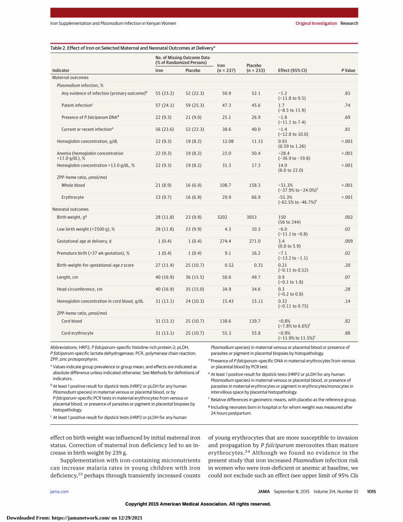
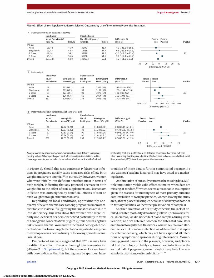
Maternal Outcomes at DeliveryForty women (22 in the iron group and 18 in the placebo group)were lost to follow-up or excluded at birth. There was no evi-dent effect of iron supplementation on Plasmodium infectionrisk (50.9% vs 52.1% for iron vs placebo groups, respectively;difference, −1.2%, 95% CI, −11.8% to 9.5%; P = .83) (Table 2).This effect was not influenced by baseline imbalances (crudeand adjusted risk difference, −1.2% vs −0.5%). Subgroup analy-sis showed no evidence that the effect of iron on Plasmodiuminfection risk depended on any of the baseline factors inves-tigated (Figure 2) or on IPT use (Figure 3).
Iron supplementation led to improved maternal iron sta-tus as indicated by hemoglobin concentrations, prevalence ofanemia, and ZPP–heme ratios (Table 2).
Neonatal Outcomes at DeliveryIron supplementation resulted in increased birth weight by150 g (95% CI, 56 to 244) (Table 2) and reduced the risk of low
birth weight by 58% (95% CI, 22% to 87%). The absolute riskreduction was 6.0% (95% CI, 0.8% to 11.1%) (Table 2). Thus,on average, 16.8 women (95% CI, 9.0 to 61.3) needed to re-ceive supplementation to prevent 1 case of low birth weight.The effect of iron on birth weight was larger in women withiron deficiency at baseline than in those who were initially iron-replete (234 g vs 39 g; difference, 195 g; 95% CI, −3 to 393;P = .05) (Figure 2). There was no evidence that the effect of ironon birth weight was influenced by IPT use (Figure 3).
Iron supplementation led to an increased gestational ageat delivery by 3.4 days and neonatal length by 0.9 cm; it re-duced the risk of premature birth by 7% (Table 2). We foundno effect of iron on neonate iron markers (hemoglobin con-centration, ZPP-heme ratios).
Maternal Outcomes at 1 Month After BirthTwelve mothers were lost to follow-up postpartum (5 in theiron group and 7 in the placebo group). Iron supplementationimproved maternal iron status. Hemoglobin concentration andgeometric mean plasma ferritin concentration increased by0.90 g/dL and 123%, respectively; geometric mean plasmatransferrin receptor concentration was reduced by 29%(Table 3). Gains in iron status were greater in women with pooriron status at baseline. Iron supplementation increased he-moglobin concentration by 1.52 g/dL and 0.44 g/dL in womenwith and without initial anemia, respectively. Correspondingvalues were 1.29 g/dL and 0.60 g/dL for women who were ini-tially iron-deficient and -replete, respectively (Figure 2).
Neonatal Outcomes at 1 Month PostpartumTwenty-two infants were lost to follow-up (13 in the iron groupand 9 in the placebo group). Iron supplementation increasedgeometric mean plasma ferritin concentration by 18%. Therewas no evidence that it affected hemoglobin and plasma trans-ferrin receptor concentrations.
Per-Protocol AnalysisResults of the per-protocol analysis were similar to those ob-tained by intention-to-treat analysis (eTables 3 and 4, eFig-ures 1 and 2 in Supplement 3). There was no evident effect ofiron on Plasmodium infection, however defined (eTables 4 and5 in Supplement 3). Iron supplementation led to a decrease inthe risk of low birth weight by 64% (95% CI, 12%-86%). Amongwomen who were initially iron deficient, iron supplementa-tion increased birth weight by 263 g (95% CI, 136-362). Therewas some evidence that IPT use modified the effect of iron onmaternal hemoglobin concentration (P = .04 for interaction),but there was no monotonic trend in effect by IPT dose.
DiscussionOverall, we found no effect of daily iron supplementation dur-ing pregnancy on risk of maternal Plasmodium infection. Ironsupplementation resulted in an increased birth weight (by150 g), gestational duration, and neonatal length; enhanced ma-ternal and infant iron stores at 1 month after birth; and a de-creased risk of low birth weight (by 58%) and prematurity. The
Table 1. Baseline Characteristics of Study Participantsby Intervention Group (continued)
Characteristics Iron PlaceboIntestinal helminth infectionsf
Schistosoma spp, No. (%)
Absent (Ct = 45) 165 (69.7) 149 (63.7)
Low (35 < Ct < 45) 10 (4.4) 10 (4.2)
Moderate (30 ≤ Ct ≤ 35) 22 (9.4) 19 (8.0)
High (Ct < 30) 39 (16.6) 56 (24.1)
Trichuris trichiura, No. (%)
Absent (Ct = 45) 185 (78.1) 198 (84.8)
Low (35 < Ct < 45) 8 (1.0) 8 (3.4)
Moderate (30 ≤ Ct ≤ 35) 12 (4.9) 13 (5.5)
High (Ct < 30) 32 (13.5) 15 (6.2)
Necator americanus, No. (%)
Absent (Ct = 45) 211 (88.9) 209 (89.8)
Low (35 < Ct < 45) 9 (3.7) 9 (3.9)
Moderate (30 ≤ Ct ≤ 35) 10 (4.2) 10 (4.2)
High (Ct < 30) 8 (3.2) 5 (2.0)
Abbreviations: AGP, α1-acid glycoprotein; CRP, C-reactive protein;Ct, cycle threshhold; HRP2, P falciparum–specific histidine-rich protein-2;IQR, interquartile range; pLDH, P falciparum-specific lactate dehydrogenase;PCR, polymerase chain reaction; sTfR, soluble transferrin receptor; ZPP, zincprotoporphyrin.a Calculated as weight in kilograms divided by height in meters squared.b All women except 1 were in the second trimester of pregnancy.c Only 1 participant (iron group) had infection by a Plasmodium species other
than P falciparum.d HIV status of 2 participants was not determined.e Only 1 participant (iron group) had current fever defined as axillary
temperature �37.5°C.f Missing values occurred because stool samples could not be collected for
some women. Cycle threshold values are inversely proportional to the amountof target DNA in the sample. Ct = 45: no detectable levels of target nucleicacid; 35 < Ct < 45: weak reactions indicative of marginal amounts of targetnucleic acid; 30 � Ct � 35: positive reactions indicative of moderate amountsof target nucleic acid, compatible with low parasite load that is unlikely to bedetectable by microscopy; Ct < 30: strong positive reactions indicative ofabundant target nucleic acid compatible with a parasite load that is probablydetectable by microscopy.
Research Original Investigation Iron Supplementation and Plasmodium Infection in Kenyan Women
1014 JAMA September 8, 2015 Volume 314, Number 10 (Reprinted) jama.com
Copyright 2015 American Medical Association. All rights reserved.
Downloaded From: https://jamanetwork.com/ on 12/29/2021
Copyright 2015 American Medical Association. All rights reserved.
effect on birth weight was influenced by initial maternal ironstatus. Correction of maternal iron deficiency led to an in-crease in birth weight by 239 g.
Supplementation with iron-containing micronutrientscan increase malaria rates in young children with irondeficiency,23 perhaps through transiently increased counts
of young erythrocytes that are more susceptible to invasionand propagation by P falciparum merozoites than matureerythrocytes.24 Although we found no evidence in thepresent study that iron increased Plasmodium infection riskin women who were iron-deficient or anemic at baseline, wecould not exclude such an effect (see upper limit of 95% CIs
Table 2. Effect of Iron on Selected Maternal and Neonatal Outcomes at Deliverya
Indicator
No. of Missing Outcome Data(% of Randomized Persons)
Iron(n = 237)
Placebo(n = 233) Effect (95% CI) P ValueIron Placebo
Maternal outcomes
Plasmodium infection, %
Any evidence of infection (primary outcome)b 55 (23.2) 52 (22.3) 50.9 52.1 −1.2(−11.8 to 9.5)
.83
Patent infectionc 57 (24.1) 59 (25.3) 47.3 45.6 1.7(−8.5 to 11.9)
.74
Presence of P falciparum DNAd 22 (9.3) 21 (9.0) 25.1 26.9 −1.8(−11.1 to 7.4)
.69
Current or recent infectione 56 (23.6) 52 (22.3) 38.6 40.0 −1.4(−12.8 to 10.0)
.81
Hemoglobin concentration, g/dL 22 (9.3) 19 (8.2) 12.08 11.15 0.93(0.59 to 1.26)
<.001
Anemia (hemoglobin concentration<11.0 g/dL), %
22 (9.3) 19 (8.2) 22.0 50.4 −28.4(−36.9 to −19.8)
<.001
Hemoglobin concentration >13.0 g/dL, % 22 (9.3) 19 (8.2) 31.3 17.3 14.0(6.0 to 22.0)
<.001
ZPP-heme ratio, μmol/mol
Whole blood 21 (8.9) 16 (6.9) 108.7 158.3 −31.3%(−37.9% to −24.0%)f
<.001
Erythrocyte 23 (9.7) 16 (6.9) 29.9 66.9 –55.3%(–62.5% to –46.7%)f
<.001
Neonatal outcomes
Birth weight, gg 28 (11.8) 23 (9.9) 3202 3053 150(56 to 244)
.002
Low birth weight (<2500 g), % 28 (11.8) 23 (9.9) 4.3 10.3 −6.0(−11.1 to −0.8)
.02
Gestational age at delivery, d 1 (0.4) 1 (0.4) 274.4 271.0 3.4(0.8 to 5.9)
.009
Premature birth (<37 wk gestation), % 1 (0.4) 1 (0.4) 9.1 16.2 −7.1(−13.2 to −1.1)
.02
Birth-weight-for-gestational-age z score 27 (11.4) 25 (10.7) 0.52 0.31 0.21(−0.11 to 0.52)
.20
Length, cm 40 (16.9) 36 (15.5) 50.6 49.7 0.9(−0.1 to 1.8)
.07
Head circumference, cm 40 (16.9) 35 (15.0) 34.9 34.6 0.3(−0.2 to 0.8)
.28
Hemoglobin concentration in cord blood, g/dL 31 (13.1) 24 (10.3) 15.43 15.11 0.32(−0.11 to 0.75)
.14
ZPP-heme ratio, μmol/mol
Cord blood 31 (13.1) 25 (10.7) 138.6 139.7 −0.8%(−7.8% to 6.6%)f
.82
Cord erythrocyte 31 (13.1) 25 (10.7) 55.3 55.8 −0.9%(−11.9% to 11.5%)f
.88
Abbreviations: HRP2, P falciparum–specific histidine-rich protein-2; pLDH,P falciparum-specific lactate dehydrogenase; PCR, polymerase chain reaction;ZPP, zinc protoporphyrin.a Values indicate group prevalence or group mean, and effects are indicated as
absolute difference unless indicated otherwise. See Methods for definitions ofindicators.
b At least 1 positive result for dipstick tests (HRP2 or pLDH for any humanPlasmodium species) in maternal venous or placental blood, or byP falciparum–specific PCR tests in maternal erythrocytes from venous orplacental blood, or presence of parasites or pigment in placental biopsies byhistopathology.
c At least 1 positive result for dipstick tests (HRP2 or pLDH for any human
Plasmodium species) in maternal venous or placental blood or presence ofparasites or pigment in placental biopsies by histopathology.
d Presence of P falciparum–specific DNA in maternal erythrocytes from venousor placental blood by PCR test.
e At least 1 positive result for dipstick tests (HRP2 or pLDH for any humanPlasmodium species) in maternal venous or placental blood, or presence ofparasites in maternal erythrocytes or pigment in erythrocytes/monocytes inintervillous space by placental histopathology.
f Relative differences in geometric means, with placebo as the reference group.g Including neonates born in hospital or for whom weight was measured after
24 hours postpartum.
Iron Supplementation and Plasmodium Infection in Kenyan Women Original Investigation Research
jama.com (Reprinted) JAMA September 8, 2015 Volume 314, Number 10 1015
Copyright 2015 American Medical Association. All rights reserved.
Downloaded From: https://jamanetwork.com/ on 12/29/2021

Copyright 2015 American Medical Association. All rights reserved.
Figure 2. Effect of Iron Supplementation on Selected Outcomes by Subgroup
–40 –10 302010–20 0
Difference, % (95% CI)
–30
P ValueFavors
IronFavorsPlacebo
Iron GroupNo. of Participants/Total No. Risk, %
Gravida status
Difference, %(95% CI)
.18
.56
.22
.23
.48a
Placebo GroupNo. of Participants/Total No. Risk, %
27/37 73 34/48 71Primigravid 2 (–26 to 31)
30/57 52 23/35 66Secundigravid –14 (–37 to 9)
Maternal age
34/48 71 31/49 64<20 y 7 (–19 to 33)
89/189 47 89/184 48≥20 y –1 (–12 to 11)
HIV infection
15/48 32 23/51 44Yes –13 (–36 to 11)
109/189 57 97/182 54No 4 (–8 to 16)
Anemia
49/82 60 47/93 50Yes 9 (–8 to 26)
75/155 48 73/140 52No –4 (–18 to 10)
67/143 47 63/150 42Multigravid 5 (–8 to 18)
Iron status
62/126 50 53/122 43Deficient 6 (–8 to 20)
33/64 51 34/64 54Replete –2 (–23 to 18)
29/47 61 33/47 70Uncertain –9 (–34 to 16)
121/237 51 121/233 52Overall –1 (–12 to 10)
Plasmodium infection assessed at deliveryA
–200 100 4000 300200
Difference, g (95% CI)
–100
P ValueFavors
PlaceboFavorsIron
Iron GroupNo. ofParticipants
Birth WeightMean (SE), g
Birth WeightMean (SE), g
Gravida status
Difference, g(95% CI)
.80
.98
.95
.72
.05a
Placebo GroupNo. ofParticipants
37 3051 (95) 48 2928 (81)Primigravid 123 (–134 to 380)
57 3223 (65) 35 3101 (75)Secundigravid 122 (–75 to 319)
Maternal age
48 3095 (77) 49 2974 (79)<20 y 121 (–100 to 341)
189 3230 (37) 184 3073 (36)≥20 y 156 (54 to 258)
HIV infection
48 3127 (74) 51 2989 (71)Yes 138 (–63 to 338)
189 3222 (38) 182 3071 (38)No 151 (43 to 259)
Anemia
82 3134 (54) 93 3009 (54)Yes 125 (–23 to 272)
155 3238 (42) 140 3081 (41)No 157 (38 to 276)
143 3233 (42) 150 3081 (41)Multigravid 152 (38 to 266)
Iron status
126 3288 (44) 122 3055 (42)Deficient 234 (116 to 351)
64 3168 (59) 64 3129 (67)Replete 39 (–134 to 211)
47 3019 (82) 47 2943 (81)Uncertain 76 (–155 to 307)
237 3202 (34) 233 3053 (33)Overall 150 (56 to 244)
Birth weightB
–0.5 1.0 2.50.5 2.01.5
Difference, g/dL (95% CI)
0
P ValueFavors
PlaceboFavorsIron
Iron GroupNo. ofParticipants
HemoglobinMean (SE), g/dL
HemoglobinMean (SE), g/dL
Gravida status
Difference, g/dL(95% CI)
.73
.24
.52
.07
.02a
Placebo GroupNo. ofParticipants
37 12.38 (0.30) 48 11.89 (0.27)Primigravid 0.49 (–0.29 to 1.27)
57 12.90 (0.17) 35 12.01 (0.25)Secundigravid 0.88 (0.30 to 1.47)
Maternal age
48 12.41 (0.23) 49 11.89 (0.24)<20 y 0.51 (–0.13 to 1.16)
189 13.01 (0.11) 184 12.01 (0.13)≥20 y 1.00 (0.67 to 1.33)
HIV infection
48 12.59 (0.22) 51 11.74 (0.24)Yes 0.85 (0.20 to 1.50)
189 12.96 (0.11) 182 12.06 (0.13)No 0.91 (0.57 to 1.24)
Anemia
82 12.52 (0.18) 93 11.00 (0.17)Yes 1.52 (1.05 to 2.00)
155 13.08 (0.12) 140 12.64 (0.12)No 0.44 (0.10 to 0.77)
143 13.01 (0.13) 150 12.01 (0.14)Multigravid 1.00 (0.63 to 1.38)
Iron status
126 12.88 (0.13) 122 11.58 (0.16)Deficient 1.29 (0.88 to 1.71)
64 13.23 (0.18) 64 12.62 (0.16)Replete 0.60 (0.14 to 1.07)
47 12.45 (0.22) 47 12.17 (0.26)Uncertain 0.29 (–0.36 to 0.93)
237 12.89 (0.10) 233 11.99 (0.11)Overall 0.90 (0.61 to 1.19)
Maternal hemoglobin concentration at 1 mo after birthC
Analyses were by intention-to-treat, with multiple imputations to replace missingvalues. Where pooling of results from multiple imputations led to nonintegercounts, we rounded those values. P values indicate the 2-sided probability that
group effects are as different as observed or more extreme when assuming thatthey are identical. Dashed lines indicate overall effect; solid lines, no effect.a Analysis restricted to those who were iron-deficient or iron-replete.
Research Original Investigation Iron Supplementation and Plasmodium Infection in Kenyan Women
1016 JAMA September 8, 2015 Volume 314, Number 10 (Reprinted) jama.com
Copyright 2015 American Medical Association. All rights reserved.
Downloaded From: https://jamanetwork.com/ on 12/29/2021
Copyright 2015 American Medical Association. All rights reserved.
in Figure 2). Should this raise concerns? P falciparum infec-tions in pregnancy notably cause increased risks of low birthweight and severe anemia.12 In our study, however, womenwho were initially iron-deficient benefited most in terms ofbirth weight, indicating that any potential decrease in birthweight due to the effect of iron supplements on Plasmodiuminfection was outweighed by beneficial effects of iron onbirth weight through other mechanisms.
Depending on local conditions, approximately one-quarter of severe anemia cases among pregnant women are at-tributable to malaria,12 suggesting that most cases are due toiron deficiency. Our data show that women who were ini-tially iron-deficient or anemic benefited particularly in termsof hemoglobin concentrations (Figure 2), thus decreasing theirrisk of severe anemia. Women with increased hemoglobin con-centrations due to iron supplementation may also be less proneto develop severe anemia during or following episodes of ma-larial illness.
Per-protocol analysis suggested that IPT use may havemodified the effect of iron on hemoglobin concentration(eFigure 2 in Supplement 3), but the absence of a clear trendwith dose indicates that this finding may be spurious. Inter-
pretation of these data is further complicated because IPTuse was not a baseline factor and may have acted as a mediat-ing factor.
One limitation of our study concerns the missing data. Mul-tiple imputation yields valid effect estimates when data aremissing at random,25 which seems a reasonable assumptiongiven the reasons for missingness of most primary outcomedata (exclusion of twin pregnancies, women leaving the studyarea, absent placental samples because of delivery at home orin tertiary facilities, or incorrect preservation of samples).
Another limitation of our study concerns the lack of de-tailed, reliable morbidity data during follow-up. To avoid ethi-cal dilemmas, we did not collect blood samples during inter-vention, and we referred women for antenatal care afterenrollment to regular health services, where they received stan-dard service. Plasmodium infection was determined in samplescollected at delivery, which may not have captured all infec-tions or symptomatic episodes during intervention. Plasmo-dium pigment persists in the placenta, however, and placen-tal histopathology probably captures most infections in thesecond half of pregnancy, even though it may have low sen-sitivity in capturing earlier infections.21,26
Figure 3. Effect of Iron Supplementation on Selected Outcomes by Use of Intermittent Preventive Treatment
–30 –10 302010–20 0
Difference, % (95% CI)
P ValueFavors
IronFavorsPlacebo
Iron GroupNo. of Participants/Total No. Risk, %
IPT use
Difference, %(95% CI)
.91
Placebo GroupNo. of Participants/Total No. Risk, %
20/48 41.0 20/43 45.4None –4.3 (–28.5 to 19.8)
23/47 48.5 19/39 47.7Single dose 0.8 (–26.8 to 28.5)
29/51 56.6 31/60 51.53 Doses 5.0 (–17.1 to 27.2)
49/91 54.2 52/90 57.52 Doses –3.3 (–19.0 to 12.4)
121/237 50.9 121/233 52.1Overall –1.2 (–11.9 to 9.5)
Plasmodium infection assessed at deliveryA
–200 100 400 5000 300200
Difference, g (95% CI)
–100
P ValueFavors
PlaceboFavorsIron
Iron GroupNo. ofParticipants
Birth WeightMean (SE), g
Birth WeightMean (SE), g
IPT use
Difference, g(95% CI)
.63
Placebo GroupNo. ofParticipants
48 3130 (91) 43 2962 (84)None 167 (–91 to 426)
47 3176 (83) 39 3101 (92)Single dose 76 (–164 to 316)
51 3264 (76) 60 3054 (64)3 Doses 210 (19 to 401)
91 3221 (51) 90 3073 (57)2 Doses 149 (0 to 297)
237 3202 (34) 233 3053 (33)Overall 150 (56 to 244)
Birth weightB
–0.5 1.0 2.00.5 1.5
Difference, g/dL (95% CI)
0
P ValueFavors
PlaceboFavorsIron
Iron GroupNo. ofParticipants
HemoglobinMean (SE), g/dL
HemoglobinMean (SE), g/dL
IPT use
Difference, g/dL(95% CI)
.21
Placebo GroupNo. ofParticipants
48 12.72 (0.24) 43 11.84 (0.30)None 0.88 (0.15 to 1.61)
47 12.67 (0.26) 39 12.24 (0.32)Single dose 0.43 (–0.37 to 12.4)
91 12.83 (0.17) 90 11.93 (0.20)2 Doses 0.90 (0.40 to 1.40)
51 13.36 (0.23) 60 12.01 (0.21)3 Doses 1.34 (0.73 to 1.96)
237 12.89 (0.10) 233 11.99 (0.11)Overall 0.90 (0.61 to 1.19)
Maternal hemoglobin concentration at 1 mo after birthC
Analyses were by intention-to-treat, with multiple imputations to replacemissing values. Where pooling of results from multiple imputations led tononinteger counts, we rounded those values. P values indicate the 2-sided
probability that group effects are as different as observed or more extremewhen assuming that they are identical. Dashed lines indicate overall effect; solidlines, no effect; IPT, intermittent preventive treatment.
Iron Supplementation and Plasmodium Infection in Kenyan Women Original Investigation Research
jama.com (Reprinted) JAMA September 8, 2015 Volume 314, Number 10 1017
Copyright 2015 American Medical Association. All rights reserved.
Downloaded From: https://jamanetwork.com/ on 12/29/2021

Copyright 2015 American Medical Association. All rights reserved.
The use of placebo in a population that included womenwith anemia and iron deficiency was justified by our concernthat risks inherent to iron supplementation outweighed pos-sible benefits; however, it also provided a unique opportu-nity to assess benefits of antenatal iron supplementation.Recent meta-analyses, published after we started our trial,found no27 or only small effects (31 g28 and 41 g29) of antena-tal iron supplementation on birth weight. In these meta-analyses, however, subgroup analysis by initial iron statuscould not be investigated, because women with anemia oriron deficiency were excluded from the studies reviewed,women (also in the placebo group) received iron as rescuetherapy during intervention, or initial anemia status and ironstatus had not been specified.27 Consistent with the findingsreported in these meta-analyses,27-29 our data showed no evi-dent effect of iron on birth weight among iron-repletewomen.
The beneficial effect of maternal iron supplementation onbirth weight found in our study may also be explained in partby a superior adherence compared with previous studies.27-29
Whereas we observed daily whether women swallowed supple-mental capsules, supervision was unclear in other studies, orcontact with participants was limited to repeated visits or tele-phone calls. Although several studies used capsules or coated
tablets,30,31 iron was mostly given as tablets with ferrous salts,which produce a strong, unpleasant taste that may have dis-couraged adherence. Attrition was high in many studies, al-though it is unclear whether this occurred selectively in theiron groups. Adherence in most studies was not reported orpoor, or supplement use was assessed by tablet counts or self-reports, which tend to overestimate adherence.32,33
The gain in birth weight was due at least in part to a lon-ger gestational duration, with perhaps some contribution byimproved fetal growth. In magnitude, this effect (150 g) is com-parable or exceeds effects of interventions to prevent malariain pregnancy, namely, IPT (79-135 g, depending on frequencyof administration) and insecticide-treated nets (55 g),34 and isparticularly important because low birth weight is associatedwith neonatal and postneonatal mortality, morbidity, inhib-ited growth, cognitive development, and potentially chronicdiseases later in life.35 This comparison should not detract fromthe benefits of these malaria control interventions, but is rathermade to indicate the importance that should be given to in-creased coverage of iron supplementation.
In accordance with our finding on birth weight, the he-moglobin response to iron was larger in women who were ini-tially iron-deficient as compared with those who were iron-replete. A differential benefit of iron by anemia status was
Table 3. Effect of Iron on Selected Maternal and Neonatal Outcomes at 1 Month After Birtha
Indicator
No. of Missing Outcome Data(% of Randomized Persons)
Iron(n = 237)
Placebo(n = 233) Effect (95% CI) P ValueIron Placebo
Maternal outcomes
Hemoglobin concentration, g/dL 22 (9.3) 19 (8.2) 12.89 11.99 0.90(0.61 to 1.19)
<.001
Anemia (hemoglobin concentration<12.0 g/dL), %
22 (9.3) 19 (8.2) 43.2 65.7 −22.4(−31.4 to −13.5)
<.001
Plasma ferritin concentration, μg/L 23 (9.7) 21 (9.0) 32.1 14.4 123.4%(85.5% to 169.1%)b
<.001
Iron deficiency (plasma ferritin concentration<15 μg/L)
All persons, % 23 (9.7) 21 (9.0) 19.8 56.0 −36.2(−44.9 to −27.5)
<.001
Those without inflammation, %c 23 (9.7) 21 (9.0) 21.1 59.5 −38.5(−49.7 to −27.3)
<.001
Plasma transferrin concentration, g/L 24 (10.1) 21 (9.0) 2.67 3.07 −0.40(−0.49 to −0.30)
<.001
Plasma transferrin receptor concentration, mg/L 23 (9.7) 21 (9.0) 1.81 2.53 −28.6%(−35.5% to −21.0%)b
<.001
ZPP-heme ratio, μmol/mol
Whole blood 22 (9.3) 19 (8.2) 100.4 152.0 −33.9%(−40.3% to −26.8%)b
<.001
Erythrocyte 24 (10.1) 19 (8.2) 31.9 74.5 −52.7%(−64.0% to −49.1%)b
<.001
Neonatal outcomes
Hemoglobin concentration, g/dL 33 (13.9) 30 (12.9) 13.45 13.32 0.13(−0.52 to 0.78)
.69
Plasma ferritin concentration, μg/L 34 (14.3) 31 (13.3) 163.0 138.7 17.5%(2.4% to 34.8%)b
.02
Plasma transferrin receptor concentration, mg/L 35 (14.8) 31 (13.3) 1.21 1.27 −4.4%(−11.3% to 2.9%)b
.23
Abbreviation: ZPP, zinc protoporphyrin.a Values indicate group prevalence or group mean, and effects are absolute
differences unless indicated otherwise. See Methods for definitions ofindicators.
b Relative differences in geometric means, with placebo as the reference group.c Concentrations of C-reactive protein <10 mg/L and α1-acid glycoprotein
<1.0 g/L.
Research Original Investigation Iron Supplementation and Plasmodium Infection in Kenyan Women
1018 JAMA September 8, 2015 Volume 314, Number 10 (Reprinted) jama.com
Copyright 2015 American Medical Association. All rights reserved.
Downloaded From: https://jamanetwork.com/ on 12/29/2021

Copyright 2015 American Medical Association. All rights reserved.
apparent for hemoglobin concentration but not birth weight(Figure 2), consistent with initial anemia being due only partlyto iron deficiency and partly to other causes.
Our finding of increased plasma ferritin concentrations at1 month postpartum adds to growing evidence,36 so far con-firmed only in a single trial,37 that antenatal iron supplemen-tation improves neonatal iron stores, thus delaying the age atwhich iron deficiency is likely to develop during infancy.
The baseline characteristics of our study population weretypical for pregnant women in many rural settings in low- andmiddle-income countries. There was no evidence that gainsin birth weight depended on gravidity, maternal age, HIV in-fection, anemia, and IPT use, suggesting benefits from iron forall subgroups thus defined, including primigravidae and thosewho did not receive IPT. Thus, our results may apply to preg-nant women in other low- and middle-income countries, al-though the effect on birth weight can vary depending on theprevalence of iron deficiency. Our results cannot be extrapo-
lated to daily antenatal supplementation with 120 mg, forwhich safety data are lacking, or to children, for whom thereis substantial evidence that iron supplementation can in-crease malaria rates.38
In low- and middle-income countries, it is generally im-practical to screen for iron status, and most countries have poli-cies for universal iron supplementation for pregnant women.Based on our results, we believe that the benefits of universalsupplementation outweigh possible risks.
ConclusionsAmong rural Kenyan women with singleton pregnancies, ad-ministration of daily iron supplementation, compared with ad-ministration of placebo, resulted in no significant differencesin overall maternal Plasmodium infection risk. Iron supple-mentation led to increased birth weight.
ARTICLE INFORMATION
Author Affiliations: Cell Biology and ImmunologyGroup, Wageningen University, Wageningen, theNetherlands (M. N. Mwangi, Roth, Smit, Trijsburg,Savelkoul, Verhoef); School of Public Health andCommunity Development, Maseno University,Maseno, Kenya (M. N. Mwangi, Andang’o); KITBiomedical Research, Royal Tropical Institute,Amsterdam, the Netherlands (Roth, Mens); AppliedNutrition Programme, University of Nairobi,Nairobi, Kenya (A. M. Mwangi); Laboratory forClinical Chemistry, Meander Medical Centre,Amersfoort, the Netherlands (Demir, Wielders);Laboratory for Medical Microbiology andImmunology, St Elisabeth Hospital, Tilburg, theNetherlands (Verweij); MRC International NutritionProgramme, London School of Hygiene andTropical Medicine, England (Cox, Prentice,Verhoef); MRC International Nutrition Programme,Medical Research Council, Keneba, the Gambia(Cox, Prentice, Verhoef); Division of HumanNutrition, Wageningen University, Wageningen, theNetherlands (Brouwer).Author Contributions: Drs M. N. Mwangi andVerhoef had full access to all of the data in the studyand take responsibility for the integrity of the dataand the accuracy of the data analysis.Study concept and design: M. Mwangi, Cox, Verhoef.Acquisition, analysis, or interpretation of data:M. Mwangi, Roth, Smit, Trijsburg, Demir, Wielders,Mens, Verweij, Prentice, Savelkoul, Andang’o, Verhoef.Drafting of the manuscript: M. Mwangi, Verhoef.Critical revision of the manuscript for importantintellectual content: All authors.Statistical analysis: M. Mwangi, Verhoef.Obtained funding: Brouwer, Verhoef.Administrative, technical, or material support:M. Mwangi, Roth, Smit, Trijsburg, A. Mwangi, Demir,Wielders, Mens, Verweij, Brouwer, Savelkoul,Andang’o, Verhoef.Study supervision: Andang’o, Verhoef.Conflict of Interest Disclosures: All authors havecompleted and submitted the ICMJE Form forDisclosure of Potential Conflicts of Interest.Dr Verhoef reported having received grants fromthe European Union, having received nonfinancialsupport from Luchtvaart Zonder Grenzen, andhaving intellectual property rights to his invention
relating to an iron supplement for use in thetreatment and/or prevention of infant low birthweight. No other disclosures were reported.Funding/Support: This work was supported by theINSTAPA project, which received funding from theEuropean Union’s Seventh Framework Programme(FP7/2007–2013) under grant agreement 211484.Fortitech and Swiss Precision Diagnostics donatedfortification premix and urine pregnancy tests,respectively.Laboratory and Allied, Nairobi, prepared supplementalcapsules (without financial compensation).
Role of the Funder/Sponsor: The funders had norole in the design and conduct of the study;collection, management, analysis, andinterpretation of the data; preparation, review, orapproval of the manuscript; and decision to submitthe manuscript for publication.
Additional Contributions: We thank localauthorities, field staff, community workers,research assistants, and students; StephenRogerson, MBBS, MRCP, DTM&H, FRACP, PhD(University of Melbourne/Royal MelbourneHospital, Australia), Paul Milligan, PhD, and TimClayton, PhD (London School of Hygiene andTropical Medicine), and Meghna Desai, MPH, PhD(US Centers for Disease Control and Prevention,Kisumu, Kenya), for data and safety monitoringboard oversight; Mark Londema, MD, and StephenRulisa, BCh MB, PhD, for assistance in staff trainingand trial implementation; Kephas Otieno, MSc(KEMRI/CDC, Kenya), for help in placentalexaminations; Paul Hulshof (WageningenUniversity, the Netherlands) for measuring ironcontent in flour; Jenny Howard (London School ofHygiene and Tropical Medicine), Chris van Kreij, andLucy Elburg (Wageningen University, theNetherlands) for administrative assistance; Karlijnvan Rijzingen and Rini Geurts (St Elisabeth Hospital,the Netherlands) for laboratory assistance; andLuchtvaart Zonder Grenzen for free logisticssupport. Data and safety monitoring boardmembers, Stephen Rulisa, and Paul Hulshof did notreceive financial compensation for theircontributions; the other individuals did.Correction: This article was corrected onlineOctober 1, 2015, to add 2 individuals to theAdditional Contributions section.
REFERENCES
1. De Benois B, McLean E, Egli I, Cogswell M, eds.Worldwide Prevalence of Anaemia 1993-2005: WHOGlobal Database on Anaemia. Geneva, Switzerland:World Health Organization; 2008.
2. Iron deficiency anaemia: report of a study group [29September to 4 October 1958]. World HealthOrganization. http://www.who.int/nutrition/publications/micronutrients/anaemia_iron_deficiency/WHO_TRS_182/en/. Accessed August 13, 2015.
3. Galloway R, McGuire J. Determinants ofcompliance with iron supplementation: supplies,side effects, or psychology? Soc Sci Med. 1994;39(3):381-390.
4. Mason J, Lotfi M, Dalmiya N, Sethuraman K,Deitchler M. The Micronutrient Report: CurrentProgress and Trends in the Control of Vitamin A,Iodine, and Iron Deficiencies. Ottawa, ON:Micronutrient Initiative; 2001.
5. Peña-Rosas JP, Viteri FE. Effects and safety ofpreventive oral iron or iron+folic acidsupplementation for women during pregnancy.Cochrane Database Syst Rev. 2009;(4):CD004736.
6. McCarthy M. Evidence for iron deficiencyscreening “inadequate,” US panel concludes. BMJ.2015;350:h1841.
7. Sazawal S, Black RE, Ramsan M, et al. Effects ofroutine prophylactic supplementation with iron andfolic acid on admission to hospital and mortality inpreschoolchildreninahighmalariatransmissionsetting:community-based,randomised,placebo-controlledtrial.Lancet. 2006;367(9505):133-143.
8. Oppenheimer SJ. Iron and its relation toimmunity and infectious disease. J Nutr. 2001;131(2S-2):616S-633S.
9. Guideline: daily iron and folic acidsupplementation in pregnant women. World HealthOrganization. http://apps.who.int/iris/bitstream/10665/77770/1/9789241501996_eng.pdf.Accessed August 13, 2015.
10. Sangaré L, van Eijk AM, Ter Kuile FO, Walson J,Stergachis A. The association between malaria andiron status or supplementation in pregnancy:
Iron Supplementation and Plasmodium Infection in Kenyan Women Original Investigation Research
jama.com (Reprinted) JAMA September 8, 2015 Volume 314, Number 10 1019
Copyright 2015 American Medical Association. All rights reserved.
Downloaded From: https://jamanetwork.com/ on 12/29/2021

Copyright 2015 American Medical Association. All rights reserved.
a systematic review and meta-analysis. PLoS One.2014;9(2):e87743.
11. Menendez C, Todd J, Alonso PL, et al. Theeffects of iron supplementation during pregnancy,given by traditional birth attendants, on theprevalence of anaemia and malaria. Trans R Soc TropMed Hyg. 1994;88(5):590-593.
12. Desai M, ter Kuile FO, Nosten F, et al.Epidemiology and burden of malaria in pregnancy.Lancet Infect Dis. 2007;7(2):93-104.
13. WHO policy brief for the implementation ofintermittent preventive treatment of malaria inpregnancy using sulfadoxine-pyrimethamine(IPTp-SP): April 2013 (revised January 2014). WorldHealth Organization. http://www.who.int/malaria/publications/atoz/iptp-sp-updated-policy-brief-24jan2014.pdf?ua=1. Accessed August 13, 2015.
14. Kenya National Technical Guidelines forMicronutrient Deficiency Control. Nairobi, Kenya:Ministry of Public Health and Sanitation; 2008.
15. National Guidelines for the Diagnosis,Treatment, and Prevention of Malaria in Kenya. 3rded. Nairobi, Kenya: Ministry of Public Health andSanitation/Ministry of Medical Services; 2010.
16. Kenya Demographic and Health Survey2008-09. Calverton, MD: Kenya National Bureau ofStatistics/ICF Macro; 2010.
17. Beaton GH. Iron needs during pregnancy: do weneed to rethink our targets? Am J Clin Nutr. 2000;72(1)(suppl):265S-271S.
18. Dietary Reference Intakes for Vitamin A, VitaminK, Arsenic, Boron, Chromium, Copper, Iodine, Iron,Manganese, Molybdenum, Nickel, Silicon,Vanadium, and Zinc. Washington, DC: US Instituteof Medicine; 2001.
19. Othoro C, Moore JM, Wannemuehler K, et al.Evaluation of various methods of maternalplacental blood collection for immunology studies.Clin Vaccine Immunol. 2006;13(5):568-574.
20. Moody A. Rapid diagnostic tests for malariaparasites. Clin Microbiol Rev. 2002;15(1):66-78.
21. Bulmer JN, Rasheed FN, Francis N, Morrison L,Greenwood BM. Placental malaria: I: Pathologicalclassification. Histopathology. 1993;22(3):211-218.
22. Mikolajczyk RT, Zhang J, Betran AP, et al.A global reference for fetal-weight and birthweightpercentiles. Lancet. 2011;377(9780):1855-1861.
23. Veenemans J, Milligan P, Prentice AM, et al.Effect of supplementation with zinc and othermicronutrients on malaria in Tanzanian children:a randomised trial. PLoS Med. 2011;8(11):e1001125.
24. Clark MA, Goheen MM, Fulford A, et al. Hostiron status and iron supplementation mediatesusceptibility to erythrocytic stage Plasmodiumfalciparum. Nat Commun. 2014;5:4446.
25. Puma MJ, Olsen RB, Bell SH, Price C. What toDo When Data Are Missing in Group RandomizedControlled Trials (NCEE 2009-0049). Washington,DC: US Dept of Education; 2009.
26. Cottrell G, Deloron P, Fievet N, Sow S, Gaye O,Le Hesran J-Y. Prediction of Plasmodium falciparumplacental infection according to the time ofinfection during pregnancy. Acta Trop. 2006;98(3):255-260.
27. Peña-Rosas JP, De-Regil LM, Dowswell T, ViteriFE. Daily oral iron supplementation duringpregnancy. Cochrane Database Syst Rev. 2012;12:CD004736.
28. Vucic V, Berti C, Vollhardt C, et al. Effect of ironintervention on growth during gestation, infancy,childhood, and adolescence: a systematic reviewwith meta-analysis. Nutr Rev. 2013;71(6):386-401.
29. Haider BA, Olofin I, Wang M, Spiegelman D,Ezzati M, Fawzi WW; Nutrition Impact Model StudyGroup (anaemia). Anaemia, prenatal iron use, andrisk of adverse pregnancy outcomes: systematicreview and meta-analysis. BMJ. 2013;346:f3443.
30. Zeng L, Dibley MJ, Cheng Y, et al. Impact ofmicronutrient supplementation during pregnancyon birth weight, duration of gestation, and perinatalmortality in rural western China: double blindcluster randomised controlled trial. BMJ. 2008;337:a2001.
31. Cogswell ME, Parvanta I, Ickes L, Yip R,Brittenham GM. Iron supplementation duringpregnancy, anemia, and birth weight: a randomizedcontrolled trial. Am J Clin Nutr. 2003;78(4):773-781.
32. Cramer JA, Mattson RH, Prevey ML, ScheyerRD, Ouellette VL. How often is medication taken asprescribed? a novel assessment technique. JAMA.1989;261(22):3273-3277.
33. Olivieri NF, Matsui D, Hermann C, Koren G.Compliance assessed by the Medication EventMonitoring System. Arch Dis Child. 1991;66(12):1399-1402.
34. Kayentao K, Garner P, van Eijk AM, et al.Intermittent preventive therapy for malaria duringpregnancy using 2 vs 3 or more doses ofsulfadoxine-pyrimethamine and risk of low birthweight in Africa: systematic review andmeta-analysis. JAMA. 2013;309(6):594-604.
35. Low Birthweight: Country, Regional and GlobalEstimates. Geneva, Switzerland: UN Children’sFund/World Health Organization; 2004.
36. Scholl TO. Maternal iron status: relation to fetalgrowth, length of gestation, and iron endowmentof the neonate. Nutr Rev. 2011;69(suppl 1):S23-S29.
37. Preziosi P, Prual A, Galan P, Daouda H,Boureima H, Hercberg S. Effect of ironsupplementation on the iron status of pregnantwomen: consequences for newborns. Am J Clin Nutr.1997;66(5):1178-1182.
38. Prentice AM, Cox SE. Iron and malariainteractions: research needs from basic science toglobal policy. Adv Nutr. 2012;3(4):583-591.
Research Original Investigation Iron Supplementation and Plasmodium Infection in Kenyan Women
1020 JAMA September 8, 2015 Volume 314, Number 10 (Reprinted) jama.com
Copyright 2015 American Medical Association. All rights reserved.
Downloaded From: https://jamanetwork.com/ on 12/29/2021