Embed Size (px)
Citation preview

VERBAL LEARNING IN MILD COGNITIVE
IMPAIRMENT AND ALZHEIMER’S DISEASE:
BEHAVIOURAL AND NEURAL APPROACHES
Petra Grönholm-Nyman
Supervised by Professor Matti Laine, PhD Department of Psychology Åbo Akademi University Finland Reviewed by Docent Laura Hokkanen, PhD Department of Psychology University of Helsinki Finland Professor Lars Nyberg, PhD Department of Integrative Medical Biology Umeå University Sweden Opponent Professor Lars Nyberg, PhD Department of Integrative Medical Biology Umeå University Sweden

To Nalle and Freja
4
CONTENTS
ACKNOWLEDGEMENS 5
LIST OF ORIGINAL PUBLICATIONS 8
SWEDISH SUMMARY – SVENSK SAMMANFATTNING 9
ABSTRACT 12
1. INTRODUCTION 14
1.1. WORD LEARNING 16
1.1.1. Memory mechanisms underlying word learning 16 1.1.2. Neural correlates of word learning 18
1.2. MILD COGNITIVE IMPAIRMENT (MCI) AND ALZHEIMER’S DISEASE (AD) 19 1.2.1. Cognitive changes in MCI and AD 20 1.2.2. Neural changes in MCI and AD 21 1.2.3. Cognitive screening for MCI and AD 23
2. AIMS OF THE PRESENT THESIS 24
3. PARTICIPANTS AND METHODS 25
3.1. PARTICIPANTS 25
3.1.1. Study I 26 3.1.2. Studies II-IV 26 3.2. METHODS 28 3.2.1. CERAD (study I) 28 3.2.2. Experimental word learning paradigm (studies II-IV) 29 3.2.2.1. Stimuli 29 3.2.2.2. Training 30 3.2.2.3. Follow-up (study II) 31 3.2.3. PET imaging (studies III-IV) 31 3.2.3.1. Experimental conditions in the PET session 32 3.2.3.2. PET data acquisition and processing 32 3.2.4. Statistical analyses 33
4. RESULTS 36
4.1. Performance on the CERAD in MCI and AD 36 4.2. Verbal learning in MCI and AD 39 4.3. Neural correlates (PET) of verbal learning in healthy elderly 43 4.4. Neural correlates (PET) of verbal learning in MCI 44
5. DISCUSSION 47
5.1. Screening for MCI and AD with the Finnish CERAD 47
5.2. Verbal learning in MCI and AD 50 5.3. Neural correlates of verbal learning in healthy elderly and MCI 56 5.4. Methodological considerations and future directions 64
6. CONCLUSIONS 67
REFERENCES 68
ORIGINAL PUBLICATIONS 83
5
ACKNOWLEDGEMENTS
This work was carried out at the Department of Psychology at Åbo Akademi University, the
Turku PET Centre, and at the Centre for Cognitive Neuroscience, University of Turku.
I wish to express my deepest gratitude to my supervisor Professor Matti Laine for his
invaluable guidance and support throughout this thesis. Over the years he has always
found the time to give skillful supervision. His vast knowledge in the field of science is
immensely appreciated.
I am indebted to Professor Juha Rinne for diagnosing the patients and for his friendly help
in carrying out the PET studies, as well as for his encouraging and helpful comments on
my work.
I want to express my sincere thanks to Mira Karrasch, PhD, for collaboration and for
unfailing encouragement and support over the years, as well as for being such a good
friend. Her constructive comments on this thesis are much appreciated.
Warmest thanks go to Victor Vorobyev, PhD, for his enormous help with the PET analyses
and for his helpful comments on the PET studies.
The official reviewers, Docent Laura Hokkanen and Professor Lars Nyberg, are warmly
thanked for their comments on this thesis. Professor Lars Nyberg is also thanked for
accepting to serve as opponent in the doctoral defence.
Warm thanks to all my colleagues at the Department of Psychology at Åbo Akademi
University for making the work atmosphere so pleasurable and positive. Special thanks to
the Girls for their endless support and for many marvellous moments at the department
and elsewhere. I thank Susanne Maikola for always being so helpful in solving practical
problems. I also wish to thank Benny Salo for standing in for me during my leave of
absence, which made it possible for me to finish this thesis.

6
Carina Söderholm, MA, is thanked for collaboration with the neuropsychological
assessments. Kim Bast, MA, and Valeska Stadie, MA, are thanked for their much
appreciated inputs. Co-author Eija Sinervä, MA. is also thanked for collaboration. I am
indebted to neurologist Riitta Niskanen for her help with the recruitement of patients. Anna
Soveri, MA, is thanked for proofreading parts of this thesis.
I also wish to express my appreciation to Professor Juhani Knuuti, Director of the Turku
PET Centre, for allowing me to use the PET facilities, and the personnel at the Turku PET
Centre for their invaluable help. Many thanks to Mika Teräs, PhD, for technical assistance
and to Tarja Järvenpää, MD, for her input.
Professor Heikki Hämäläinen, the head of the Centre for Cognitive Neuroscience at the
University of Turku, is gratefully acknowledged for collaboration.
I also wish to express my gratitude to Professor Christina Krause, who introduced me to
the field of neuroscience. She has taught me a great deal about making research and was
the first person who could explain statistics in such a way that it made sense to me.
I am very grateful to all the participants and their relatives whose contributions have made
this thesis possible.
On a more personal level, I want to thank all my friends outside science for their support
during this process. Special thanks go to my faithful friend Katja for all the good moments
that we have shared over the past few years. Your home has always been a soft place to
fall.
I want to thank my parents Maija and Bo for their help and support and for believing in me,
and for giving me encouragement for an academic education. I am also grateful for their
help with babysitting. My sister Pia and her son Rasmus are warmly thanked for their love
and support.
7
I owe my warmest gratitude to my husband Nalle for his unfailing love and faith in me, and
to our daughter Freja for bringing so much joy and happiness in our lives and for showing
me what is most precious.
The thesis was financially supported by the Finnish Graduate School of Psychology, and
grants from the Research Institute of the Åbo Akademi University Foundation, Åbo
Akademi University, the Paulo Foundation, the Foundation for Swedish Culture in Finland,
the Oskar Öflund Foundation, Samfundet Folkhälsan i svenska Finland r.f. and the
Alzheimer Foundation of Finland, all of which I gratefully acknowledge.
Åbo, March 2009
Petra Grönholm-Nyman
8
LIST OF ORIGINAL PUBLICATIONS
This thesis is based on the following original publications:
I Karrasch, M., Sinervä, E., Grönholm, P., Rinne, J., & Laine, M. (2005). CERAD test
performances in amnestic mild cognitive impairment and Alzheimer’s disease. Acta
Neurologica Scandinavica, 111, 172-179.
II Grönholm-Nyman, P., Rinne, J., & Laine, M. (manuscript submitted for publication).
Learning and forgetting new names and objects in MCI and AD.
III Grönholm, P., Rinne, J., Vorobyev, V. & Laine, M. (2005). Naming of newly learned
objects: a PET activation study. Cognitive Brain Research, 25, 359-371.
IV Grönholm, P., Rinne, J., Vorobyev, V., & Laine, M. (2007). Neural correlates of
naming newly learned objects in MCI. Neuropsychologia, 45, 2355-2368.
The original articles have been reproduced by permission of the copyright holders.
9
SWEDISH SUMMARY - SVENSK SAMMANFATTNING
Med åldern blir kognitiva symptom, såsom minnessvårigheter, allt vanligare. I en åldrad
population blir den kognitiva prestationsnivån likaså en allt viktigare fråga med tanke på
livskvaliteten. Det har även blivit allt viktigare att kunna skilja åt normalt åldrande från
degenerativa hjärnsjukdomar som förorsakar demens, eftersom det nuförtiden finns
effektiva mediciner tillgängliga för den vanligaste orsaken till demens, d.v.s. Alzheimers
sjukdom. Dessa mediciner kan inte bota sjukdomen i sig, men de kan lindra symptomen
för en viss period, vilket gör det viktigare att upptäcka sjukdomen i ett så tidigt skede så
möjligt. Med tanke på den tidiga upptäckten av sjukdomen har diagnosen mild kognitiv
svikt (Mild Cognitive Impairment, MCI) väckt allt mer intresse. Personer som lider av MCI
har tydliga minnessvårigheter även om nedsättningen av minnesfunktionerna ännu inte
väsentligt påverkar det dagliga livet. Ett flertal forskningsresultat tyder på att MCI-patienter
har en förhöjd risk att insjukna i Alzheimers sjukdom. Med andra ord kan MCI ses som ett
slags mellanstadium mellan normalt åldrande och lindrig Alzheimers sjukdom. För att få en
djupare insikt i de neurokognitiva förändringar som demenssjukdomar och normalt
åldrande för med sig, bör man kunna integrera psykologiska, psykofysiologiska och
neurala aspekter. Detta är viktigt ur diagnostisk synvinkel och även med tanke på att på
lång sikt kunna förbättra livskvaliteten för såväl demenspatienter som deras anhöriga.
Syftet med föreliggande avhandling var att med hjälp av både behaviorella
undersökningsmetoder och funktionell hjärnavbildning (positronemissionstomografi, PET,
syre-15-metoden) undersöka de underliggande neurokognitiva mekanismerna i episodiska
och semantiska minnessystem vid verbal inlärning hos friska äldre personer, patienter
med MCI och patienter med Alzheimers sjukdom. Dessutom undersöktes användbarheten
av det kognitiva testbatteriet CERAD som kliniskt verktyg vid diagnostisering av MCI och
tidig Alzheimers sjukdom.
I studie I undersöktes skillnader i testprestationer i CERAD mellan MCI-patienter och friska
kontrollpersoner med fokus på uppgiften inlärning av ordlista. Därutöver utforskades
sensitiviteten och specificiteten av CERAD som screeninginstrument vid MCI och
Alzheimers sjukdom. Resultaten tydde på att MCI-patienter hade svårigheter med
minnesinkodningen vid inlärning av ordlista, vilket dessutom var den enda uppgiften i
vilken det upptäcktes skillnader mellan MCI-patienter och kontrollpersoner. I motsats till
10
inkodningen, upptäcktes inga svårigheter med att bevara den inlärda ordlistan i minnet hos
MCI patienter, d.v.s. det fanns inga signifikanta skillnader i fördröjd minnesåterkallning
mellan MCI-patienter och kontrollpersoner. Angående CERAD som screeninginstrument,
tydde resultaten på att testbatteriet inte är tillräckligt sensitivt för att upptäcka MCI. Med
andra ord finns det en risk för att personer med preklinisk Alzheimers sjukdom inte blir
upptäckta ifall CERAD används som det enda screeninginstrumentet. Likaså tydde
resultaten på att det kan finnas en risk för att en del kognitivt friska äldre personer kan få
ett falskt positivt resultat. Dessa riskfaktorer borde beaktas med tanke på att CERAD blivit
ett populärt screeninginstrument i Finland vid demensutredningar.
I studie II undersöktes verbal inlärning och glömska hos patienter med MCI och
Alzheimers sjukdom samt hos friska äldre kontrollpersoner. Detta undersöktes med ett
experimentellt ordinlärningsparadigm där försökspersonerna fick lära sig benämningar på
föremål från äldre tider (d.v.s. sådana föremål som existerar på riktigt, men som
försökspersonerna inte kände till) på så vis att hälften av föremålen och deras
benämningar inövades med semantiskt stöd (semantiskt stöd = vad föremålet används till)
och hälften inövades utan semantiskt stöd. Orsaken till att semantiskt stöd användes vid
träningen, var tanken om att det kunde stöda ordinlärningen speciellt hos MCI-patienterna,
vilkas semantiska minne är mera intakt än deras episodiska minne. Träningsfasen tog en
vecka och utöver detta utfördes en uppföljningsundersökning en vecka, fyra veckor och
åtta veckor efter träningsperioden. Vid uppföljningen undersöktes, förutom nivån på
minnesåterkallningen av benämningarna på föremålen, även återkallningen av det
semantiska stödet, igenkännandet av föremålen samt effekten av fonologiskt stöd vid
återkallningen av benämningarna på föremålen. Resultaten från studie II indikerade att
MCI-patienterna led av försämrad inkodningsförmåga jämfört med friska äldre
kontrollpersoner, vilket framkom i MCI-patienternas försämrade förmåga att lära sig
benämningarna på de nya föremålen. Patienterna med Alzheimers sjukdom lärde sig
benämningarna på de nya föremålen ännu sämre än MCI-patienterna. Däremot fanns det
under uppföljningen inga skillnader mellan de tre grupperna gällande glömskan av de
nyligen inlärda benämningarna på föremålen. Med andra ord tydde resultaten på att en
inlärningssvårighet var kännetecknande för både MCI och Alzheimers sjukdom, medan de
inlärda benämningarna på föremålen bevarades i minnet på samma vis såväl i
patientgrupperna som i kontrollgruppen. Därtill drog MCI-patienterna nytta av det
11
semantiska stödet vid återkallningen av ord vid sista uppföljningstillfället, vilket indikerade
att patienternas bättre bevarade semantiska minnesfunktioner i viss mån kompenserade
deras mera gravt försämrade episodiska minnesfunktioner. Resultaten för
minnesåterkallningen av det semantiska stödet, liksom även igenkänningsuppgiften,
uppvisade inga skillnader mellan MCI-patienterna och kontrollpersonerna, vilket antydde
att dessa minnesområden var väl bevarade i MCI-gruppen.
I studie III och IV användes funktionell hjärnavbildning, PET, för att undersöka
hjärnaktiveringsmönster vid benämning av nyligen inlärda, sällsynta föremål jämfört med
bl.a. benämning av vanliga föremål hos friska äldre personer (studie III) och MCI-patienter
(studie IV). Resultaten från studie III visade att benämning av nyligen inlärda, sällsynta
föremål aktiverar ett nätverk av hjärnområden i vänstra hemisfären (frontotemporala
områden och lillhjärnan) som är mera omfattande än det som aktiveras vid benämning av
vanliga föremål. Detta i sin tur tyder på att benämning av nyligen inlärda, sällsynta föremål
kräver en mera intensiv fonologisk och semantisk processering än benämning av vanliga
föremål. I studie IV syntes en signifikant ökning av aktiveringen i främre delen av gyrus
cinguli (anterior cingulate) hos MCI-patienter jämfört med kontrollpersoner vid
benämningen av nyligen inlärda föremål som hade tränats utan semantiskt stöd.
Resultaten indikerade att en högre grad av exekutiv uppmärksamhet krävdes hos MCI-
patienterna än hos kontrollpersonerna.
12
ABSTRACT
The main focus of the present thesis was at verbal episodic memory processes that are
particularly vulnerable to preclinical and clinical Alzheimer’s disease (AD). Here these
processes were studied by a word learning paradigm, cutting across the domains of
memory and language learning studies. Moreover, the differentiation between normal
aging, mild cognitive impairment (MCI) and AD was studied by the cognitive screening test
CERAD.
In study I, the aim was to examine how patients with amnestic MCI differ from healthy
controls in the different CERAD subtests. Also, the sensitivity and specificity of the CERAD
screening test to MCI and AD was examined, as previous studies on the sensitivity and
specificity of the CERAD have not included MCI patients. The results indicated that MCI is
characterized by an encoding deficit, as shown by the overall worse performance on the
CERAD Wordlist learning test compared with controls. As a screening test, CERAD was
not very sensitive to MCI.
In study II, verbal learning and forgetting in amnestic MCI, AD and healthy elderly controls
was investigated with an experimental word learning paradigm, where names of 40
unfamiliar objects (mainly archaic tools) were trained with or without semantic support. The
object names were trained during a 4-day long period and a follow-up was conducted one
week, 4 weeks and 8 weeks after the training period. Manipulation of semantic support
was included in the paradigm because it was hypothesized that semantic support might
have some beneficial effects in the present learning task especially for the MCI group, as
semantic memory is quite well preserved in MCI in contrast to episodic memory. We found
that word learning was significantly impaired in MCI and AD patients, whereas forgetting
patterns were similar across groups. Semantic support showed a beneficial effect on
object name retrieval in the MCI group 8 weeks after training, indicating that the MCI
patients’ preserved semantic memory abilities compensated for their impaired episodic
memory. The MCI group performed equally well as the controls in the tasks tapping
incidental learning and recognition memory, whereas the AD group showed impairment.
Both the MCI and the AD group benefited less from phonological cueing than the controls.
Our findings indicate that acquisition is compromised in both MCI and AD, whereas long-
13
term retention is not affected to the same extent. Incidental learning and recognition
memory seem to be well preserved in MCI.
In studies III and IV, the neural correlates of naming newly learned objects were
examined in healthy elderly subjects and in amnestic MCI patients by means of positron
emission tomography (PET) right after the training period. The naming of newly learned
objects by healthy elderly subjects recruited a left-lateralized network, including fronto-
temporal regions and the cerebellum, which was more extensive than the one related to
the naming of familiar objects (study III). Semantic support showed no effects on the PET
results for the healthy subjects. The observed activation increases may reflect lexical-
semantic and lexical-phonological retrieval, as well as more general associative memory
mechanisms. In study IV, compared to the controls, the MCI patients showed increased
anterior cingulate activation when naming newly learned objects that had been learned
without semantic support. This suggests a recruitment of additional executive and
attentional resources in the MCI group.
14
1. INTRODUCTION
The maintenance of cognitive capabilities is important for the quality of life as people get
older. Cognitive problems become increasingly common with advancing age and their
appearance often has a detrimental effect on subjective well-being and ability to lead an
independent life. It has also become increasingly important to be able to differentiate
normal aging from neurodegenerative disorders that cause dementia, as there is now
effective medication that can slow down the progression of the most common cause of
dementia, namely Alzheimer’s disease (AD) (e.g. Ballard, 2000). Of special interest is the
condition that has been coined as mild cognitive impairment (MCI), as it entails an
increased risk of developing AD over the next few years (Petersen, 2004). To achieve a
deeper understanding of the evolvement of dementia, both the cognitive and neural
aspects of this process need to be studied and related to each other. This is important not
only for the diagnostics but also for the treatment where the aim is to improve the quality of
the patients’ and their relatives’ lives and to support the patients’ cognition.
Memory impairment is the key cognitive deficit in preclinical and early Alzheimer’s disease
(e.g. Collie & Maruff, 2000). However, memory is a very broad and complex concept,
including many different functions that can be selectively disrupted. Memory and learning
has traditionally been investigated by asking the subjects to encode and retrieve familiar
items such as words or objects. The topic of the present thesis, learning of new
information, has received much less attention. A striking example of the immense learning
capacity of the human brain is our ability to acquire, maintain and update a massive
storage of words (usually tens of thousands of actively used words), which is functional
throughout the life-span.
Among researchers there is still some disagreement about which subcomponents
constitute the function we generally refer to as memory, and different models of memory
overlap to a great extent. However, virtually all contemporary models distinguish between
immediate and long-term memory. Short-term memory (STM) refers to temporary retention
of a limited amount of information that may then be incorporated into a more stable,
potentially more permanent memory store, i.e. into long-term memory (LTM) (Baddeley,
2000b; Jonides et al., 2008). Currently, the most influential theory about the structure and
15
function of the STM is the concept of working memory (WM) (Baddeley, 2000b). Working
memory refers to a multicomponent system with limited capacity that provides temporary
storage of information for the facilitation of complex cognitive activities, such as learning.
One of the components is coined as the phonological loop that is thought to hold
information that can be rehearsed verbally. The visuospatial sketchpad is suggested to
hold visuospatial information. The central executive component is suggested to control the
overall system. The episodic buffer (Baddeley, 2000a) that was added to the WM model
afterwards is thought to integrate information from several sources.
Long-term memory can be divided into declarative and procedural memory, i.e., memory
for facts and episodes vs. memory for skills and other cognitive operations, respectively
(Squire, 1987). Declarative memory can further be divided into episodic and semantic
memory, with episodic memory referring to memory for personally experienced and
temporally specific events or episodes, whereas semantic memory refers to a store of
knowledge including facts, concepts and word meanings (Tulving, 2002). Both lesion and
neuroimaging studies have shown that the medial temporal lobes are crucial to episodic
memory functioning (Gabrieli, 2001), whereas semantic memory seems to entail a broader
network of cortical regions, including temporal and frontal areas (Martin, 2001).
Memory includes encoding, maintenance and retrieval, and is thus a highly active process
that requires executive functions. These functions refer to goal-directed, flexible use of
cognitive abilities, e.g., sustaining, dividing and shifting attention according to task
demands, inhibiting inappropriate responses, and solving problems. They seem to
represent a cluster of closely related but partly separate cognitive processes that to a great
extent rely on prefrontal brain regions (Miyake et al., 2000).
Learning can be either intentional or incidental. Whereas intentional learning denotes an
active intent to learn something, incidental learning refers to passive learning that happens
as a by-product of other information processing. For example, unknown words may be
learned incidentally during normal reading (for review, see Swanborn & de Glopper, 1999).
The main focus of the present thesis is at verbal episodic memory processes that are
particularly vulnerable to preclinical and clinical dementia. The tasks used for this purpose
16
involve word learning, thus cutting across traditional memory studies and the domain of
language learning research. Moreover, the differentiation between normal aging, MCI and
AD is studied by a current cognitive screening test.
1.1. WORD LEARNING
Several alternative models for word learning have been put forward, and their
methodological and empirical background varies considerably. The cognitive mechanisms
underlying word learning have also been much debated (Baddeley, Gathercole, &
Papagno, 1998; Gupta & MacWhinney, 1997; Markson & Bloom, 1997; Martin & Gupta,
2004: Waxman & Booth, 2000). At question is to which degree language learning is either
domain specific or non-specific and which memory and learning mechanisms are involved
in vocabulary acquisition. While some researchers argue strongly that working memory
(Baddeley et al., 1998) or short-term memory (Martin & Gupta, 2004) is essential in word
learning, others stress the importance of declarative memory processes (Ullman, 2004)
and incidental learning (Saffran, Newport, Aslin, Tunick, & Barrueco, 1997).
1.1.1. Memory mechanisms underlying word learning
According to Baddeley (2000a, 2000b), a crucial functional element in word learning is the
verbal WM (phonological loop) that consists of two components, the phonological store
and the subvocal articulatory rehearsal process. The acoustic or phonological memory
trace is held in the phonological store, and is assumed to decay within a few seconds
unless it is maintained by the rehearsal process. In word learning, the verbal WM enables
the temporary storage of unfamiliar sound patterns of words until long-term
representations are established. It should be mentioned, though, that although verbal WM
is important for the learning of phonological forms of words, it does not account for the
buildup of visual and semantic representations. In fact, Duyck, Szmalez, Kemps, and
Vandierendonck (2003) have suggested that the learning of word associations can rely on
other resources, such as the visuospatial sketchpad. An integrative model on word
learning by Gupta and MacWhinney (1997) assumes that verbal STM and word learning
involve common underlying cognitive mechanisms. Firstly, they are related because they
depend on the same core phonological processing mechanisms. Secondly, they are
17
related because of their use of rehearsal and chunking. This would also explain the
correlation found between performances in these two cognitive domains. In other words,
the model views verbal STM and word learning as involving common underlying
mechanisms, without any implication that they are causally related. Furthermore, Ullman
(2004) has proposed an influential model, called the declarative/procedural model, of how
memory circuits contribute to language, including word learning. More specifically, it claims
that word learning relies heavily on declarative memory subserved by temporal lobe
structures, whereas mental grammar underlies procedural memory that is subserved by a
network of brain structures, including frontal/basal ganglia circuits, with a probable role for
parts of the parietal cortex, superior temporal cortex and the cerebellum. The
lexical/declarative and the grammatical/procedural memory are thought to interact in
several ways and similar types of knowledge may be acquired by both systems. An
impairment of the declarative system is expected to lead to altered processing by the
procedural system, and vice versa. Finally, Saffran et al. (1997) have stressed the
incidental memory mechanisms in word learning, as both children and adult language
learners are remarkably skillful at automatically absorbing detailed linguistic information
from language input. More specifically, Saffran et al. (1997) showed that children and
adults can extract words from a speech stream by exploiting the sequential probabilities of
syllable sequences. This statistical learning mechanism may support not only word
segmentation but also the acquisition of other aspects of language.
To summarize, the abovementioned models have aimed at identifying the explicit and
implicit memory mechanisms involved in word learning. It has even been suggested that
one of these mechanisms, verbal WM, has evolved primarily in order to serve vocabulary
development (Baddeley et al., 1998). This is supported by the significant correlations
between nonword repetition performance (a verbal WM measure) and vocabulary
development across a wide range of ages. It should also be noted that the models shortly
presented above show considerable overlap. For example, the model by Gupta and
McWhinney (1997) can be seen as an extension of the WM model by Baddeley (2000a,
2000b). Certain aspects of WM are also brought up in relation to the
declarative/procedural model by Ullman (2004). At a more general level, word learning can
be seen as an associative learning task. For example, to learn a novel concrete noun, one
18
needs to associate an object, a name (phonological representation) and a concept with
each other.
1.1.2. Neural correlates of word learning
With regard to the neural basis of the proposed cognitive mechanisms underlying word
learning (see above), the verbal WM has been associated with frontal and parietal brain
activation. More specifically, it has been suggested that the phonological store is
associated with left parietal activation, while the rehearsal process is associated with left
posterior-inferior frontal activation (Broca’s area). Also cerebellar activations are often
found in verbal working memory tasks (for review, see Cabeza & Nyberg, 2000). The
declarative/procedural model by Ullman (2004) strongly emphasizes the role of the medial
temporal lobe structures in word learning. However, also other areas, such as the
ventrolateral prefrontal cortex, have been suggested to be involved in the encoding of new
memories and the selection or retrieval of declarative knowledge (for review, see Buckner
& Wheeler, 2001), i.e., the same brain areas that are also consistently found to be
activated in WM tasks (for review, see Smith & Jonides, 1999). Ventral prefrontal cortex
activation has also been related to associative learning that can be linked to word learning,
where one should learn and associate three components (word, concept, and external
referent) with each other. In general terms, the ventral prefrontal cortex is seen as a part of
a system where associations are made between visual cues and the choices that they
represent (for review, see Passingham, Toni, & Rushworth, 2000).
Thus far, the neural mechanisms of word learning (more specifically the retrieval of newly
acquired words) have received scant attention in the functional neuroimaging literature.
Neuroimaging experiments on word learning conducted on healthy subjects have
suggested predominantly left hemisphere mechanisms (Breitenstein et al., 2005; James &
Gauthier, 2004; Raboyeau et al., 2004). With regard to acquisition of lexical-semantic
knowledge (object meaning), the most prominent brain correlates that have been put
forward are the left inferior frontal cortex (James & Gauthier, 2004) and the left temporal
lobe, more specifically posterior superior temporal sulcus and middle temporal cortex
(Raboyeau et al., 2004), and left medial temporal structures (Breitenstein et al., 2005).
Acquisition of lexical-phonological knowledge (object name) has been related to inferior
19
parietal lobe (Breitenstein et al., 2005, Cornelissen et al., 2004) and left temporal cortex
function (Hulten, Vihla, Laine, & Salmelin, 2009). In addition, anterior cingulate activation
has been found in association with lexical learning, interpreted to reflect attentional
processes needed to access recently learned words (Raboyeau et al., 2004). The
abovementioned results partly overlap with those found in functional neuroimaging studies
on naming familiar objects, but differences are also found (Bookheimer, Zeffiro, Blaxton,
Gaillard, & Theodore, 1995; Martin, Wiggs, Ungerleider, & Haxby, 1996; Murtha,
Chertkow, Beaugard, & Evans, 1999; Zelkowicz, Herbster, Nebes, Mintun, & Becker,
1998). For instance, inferior frontal activation is found in retrieval of both familiar and
recently learned new words, but the temporal activation in retrieving familiar names has
been found in posterior inferior areas (fusiform gyrus), and have thus differed from that
found in studies on word learning, which report activation increases in left medial temporal
structures, as well as superior and posterior middle temporal cortex. Also, the inferior
parietal lobe activation observed in some studies on naming of newly learned objects has
not been characteristic for naming of familiar objects.
1.2. MILD COGNITIVE IMPAIRMENT (MCI) AND ALZHEIMER’S DISEASE (AD)
Mild cognitive impairment (MCI) has become an important research topic, as patients with
this condition have been shown to be at risk of developing Alzheimer’s disease (AD) or
other neurodegenerative diseases (Collie & Maruff, 2000; Petersen, 2004; Petersen et al.,
2001). There is heterogeneity concerning the MCI criteria, but generally MCI refers to
persons who do not fulfil the criteria for AD or dementia, but who show some form of
cognitive decline (for review, see Palmer, Fratiglioni, & Winbland, 2003). Of particular
interest is amnestic MCI that refers to subjects with isolated episodic memory impairment
(Collie & Maruff, 2000; Petersen et al., 1999; Petersen et al., 2001; Petersen, 2004).
Amnestic MCI is the form that most often leads to AD, the turnover rate being
approximately 12% per year (Petersen & Morris, 2003).
20
1.2.1. Cognitive changes in MCI and AD
Although particularly amnestic MCI has attracted considerable research interest, the
nature of the memory impairment in this condition is still fairly little studied. Recent studies
have shown that the episodic memory impairment in MCI is characterized by a decreased
learning efficacy (Moulin, James, Freeman, & Jones, 2004; Ribeiro, Guerreiro, &
Mendonca, 2007) and impaired delayed recall (Fernandez-Ballesteros, Zamarrón, &
Tàrraga, 2005; Moulin et al., 2004; Petersen & Morris, 2003). Verbal memory tasks appear
to cause somewhat more problems than nonverbal ones (for review, see Collie & Maruff,
2000). The episodic memory impairment in amnestic MCI may not be totally isolated, as it
has recently been argued that other domains of cognition may be affected as well (see e.g.
Bäckman, Jones, Berger, Laukka, & Small, 2004). Subtle preclinical deficits in amnestic
MCI, i.e. impairments that cannot be found in standard neuropsychological tests, have
been found in cognitive domains such as executive functioning (Collie, Maruff, & Currie,
2002; Davie et al., 2004).
When the disease progression leads to the diagnosis of AD, the neuropsychological
deficits are widespread and marked (for reviews, see Collie & Maruff, 2000; Spaan,
Raaijmakers, & Jonker, 2003). Alzheimer’s disease is characterized by impairments of
episodic and semantic memory, attention, executive function and visuospatial ability, with
episodic memory problems appearing first (cf. amnestic MCI) and visuospatial impairments
appearing at a later stage (Belleville, Peretz, & Malefant, 1996; Binetti et al., 1998; Colette,
Van der Linden, Bechet, & Salmon, 1999; De Jager, Hogervorst, Combrinck, & Budge,
2003; Dudas, Clague, Thompson, Graham, & Hodges, 2005; Greene, Baddeley, &
Hodges, 1996; Hodges, Salmon, & Butters, 1992; Laatu, Portin, Revonsuo, Tuisku, &
Rinne, 1997; Nebes & Brady, 1991; Perry, Watson, & Hodges, 2000).
Acquisition of new words has not previously been studied in MCI. In AD, lexical acquisition
has been investigated by studying new verb learning through incidental learning
(Grossman et al., 2007; Grossman, Mickanin, Onishi, Robinson, & D’Esposito, 1997).
These studies showed that the AD patients were impaired at acquiring the new word’s
meaning compared with controls, reflecting the AD patients’ semantic memory difficulties.
However, the AD patients did acquire grammatical knowledge associated with a new word
21
inci, indicating that AD patients can learn about a new verb and maintain the newly
acquired knowledge over a week following incidental learning.
1.2.2. Neural changes in MCI and AD
Regarding the biological basis of the cognitive deficits in MCI, structural magnetic
resonance imaging (MRI) studies have indicated gray matter reduction in medial temporal
areas (De Toledo-Morell et al., 2004, Dickerson et al., 2001; Karas et al., 2004; Pennanen
et al., 2005). Functional imaging imaging has revealed alterations in regional glucose
metabolism and blood flow in temporoparietal areas (for review, see Wolf et al., 2003).
There are also PET studies that have aimed at identifying characteristic patterns of
glucose metabolism in MCI patients that have converted to AD or whose cognitive abilities
have deteriorated significantly over time. The changes predicting cognitive decline
included reduced glucose metabolism in temporoparietal and posterior cingulate cortex, as
well as frontal cortical areas (Chetelat et al., 2005; Drzezga et al., 2003; Mosconi et al.,
2004). In other words, although changes in medial temporal areas have been heavily
emphasized in MCI because of their well-known link to episodic memory, other brain areas
may be affected at the preclinical phase of AD as well (see also Bäckman et al., 2004). In
AD, the cognitive decline is related to progressive temporoparietal brain atrophy,
especially in the entorhinal cortex and hippocampus, as shown by structural imaging
studies (for review, see Nestor, Scheltens, & Hodges, 2004) and neuropathological
findings (Braak & Braak, 1991). In addition, a recent structural neuroimaging study by
Thomann et al. (2008) found significantly larger cerebellar atrophy in AD patients
compared with controls. Furthermore, the temporoparietal distribution (extending further to
different frontal areas) of these neural changes has been verified by resting-state
metabolic studies of brain function in AD (for review, see Salmon, Lekeu, Bastin, Garraux,
& Collette, 2008; Silverman, 2004). In sum, the structural and functional brain changes are
more widespread in AD compared with MCI (De Santi et al., 2001) which is in line with the
more severe and global cognitive deficits in AD compared with MCI.
There are fewer cognitive neuroimaging studies on MCI than on AD. The few relevant
studies in MCI have mostly focused on task-related activation patterns in medial temporal
structures and the posterior cingulate, and they have found decreased activation in MCI
22
patients compared with healthy controls during encoding (Johnson et al., 2006; Machulda
et al., 2003) and recognition tasks (Johnson et al., 2006; Ries et al., 2006). However,
Saykin et al. (2004) reported reduced activation in frontoparietal areas during a working
memory task in MCI patients relative to controls. Recently, Dannhauser et al. (2008) found
that verbal encoding related to decreased activation in the left ventrolateral prefrontal
cortex in MCI compared with controls. In contrast, Yetkin, Rosenberg, Weiner, Purdy, and
Cullum (2006) found increased activation in the MCI group compared with the control
group during a working memory task in frontal and temporal areas, as well as the anterior
cingulate. Dickerson et al. (2004) studied medial temporal lobe function by fMRI in MCI
patients and found that the right parahippocampal gyrus was recruited to a larger extent
during memory encoding in MCI patients showing greater clinical impairment.
Furthermore, Bokde et al. (2006) studied functional connectivity of the right middle fusiform
gyrus in MCI during a face-matching task. They found that MCI affected functional
connectivity from the fusiform gyrus to visual areas and medial frontal areas, including
anterior cingulate. Finally, a recent PET study by Moulin et al. (2007) on word-pair learning
suggested different activation patterns in MCI patients vs. elderly controls. In the MCI
group, incremental learning failed to elicit changes in frontal activations but instead
showed increased occipital activation. During retrieval, the MCI patients only showed a left
frontal activation increase and no right frontal or left temporal activation increases as the
controls did.
Although cognitive neuroimaging studies in MCI are scarce, the corresponding literature in
AD is more extensive (for review, see Almqvist, 2000; Wermke, Sorg, Wohlschläger, &
Drzezga, 2008). The findings of these studies have been quite variable: they have showed
loss of activated regions, emergence of newly activated regions (compensatory activation),
reduced activation, or no change at all. There are also cognitive activation studies with
fMRI conducted with people at genetic risk for AD (=carriers of the APOE ,4 allele) that
have found differences in the activation patterns between the risk group and the healthy
controls (Bookheimer et al., 2000; Smith et al., 2002).
In sum, the existing cognitive neuroimaging studies on both MCI and AD have revealed
differences in brain activation patterns when compared to controls, but the observed
23
changes have been quite variable. A number of factors may explain the heterogeneity of
the results, including differences in tasks, experimental designs and patient samples.
1.2.3. Cognitive screening for MCI and AD
Cognitive screening tests are used to detect cognitive impairment, and to assess the need
for more detailed neuropsychological assessment and medical examination, which could
then lead to a diagnosis. Efforts have been made to develop short, easily and quickly
administered cognitive tests that would be sensitive and specific for AD. Until the mid-90’s
the Mini-Mental State Examination (MMSE) was the most used screening test for
dementia. When it comes to specificity, the MMSE has been shown to be excellent, but its
sensitivity to clinically diagnosed probable AD has been found to be relatively low (~.65)
(Gallassi, Morreale, Di Sarro, & Lorusso, 2002; Tangalos et al., 1996; Wind et al., 1997).
Its sensitivity to preclinical AD has been shown to be even lower (Tang-Wai et al., 2003;
Tierney, Szalai, Dunn, Geslani, & McDowell, 2000). The CERAD test battery (The
Consortium to Establish a Registry for Alzheimer´s Disease) (Welsh et al., 1994)
encompasses measures in the cognitive domains where impairments associated with AD
first occur, and it has been found to have high re-test reliability, inter-rater agreement and
longitudinal validity (Welsh-Bohmer & Mohs, 1997). Delayed recall and savings scores (i.e.
delayed recall adjusted for acquisition) on the CERAD Wordlist learning test are well
preserved in normal aging but impaired early on in dementia, which is important for the
detection of early impairment in cognitive function (Welsh et al., 1994). The CERAD test
battery has been recommended as a screening instrument for memory problems in
persons aged >55 years in Finland (Hänninen et al., 1999). If individuals perform below the
cut-off score in the memory tests or in several of the non-memory tests, subsequent
neurological and neuropsychological assessments are recommended. If the performances
on one or two non-memory tests fall below the cut-off scores, a follow-up testing (6-12
months) is recommended, as well as an evaluation of possible mood-related reasons for
the cognitive decline.
24
2. AIMS OF THE PRESENT THESIS
The primary aim of the present thesis was to explore verbal learning in amnestic MCI, AD
and healthy elderly. Study I was directly related to clinical practice, whereas studies II-IV
were experimental with a specific word learning paradigm, including both behavioural as
well as neural measures.
In study I, the aim was to examine how patients with amnestic MCI differ from healthy
controls in the different CERAD subtests. Of particular interest were the performances on
the Wordlist learning test that was expected to be a sensitive measure here. Also the
sensitivity and specificity of the CERAD screening test to MCI and AD was studied. One
should note that previous studies on the sensitivity and specificity of the CERAD have not
included MCI patients.
In study II, verbal learning and forgetting in amnestic MCI, AD and healthy elderly controls
were investigated with an experimental word learning paradigm, where the names of
unfamiliar objects were trained with or without semantic support. Verbal memory in MCI
and AD has traditionally been investigated by tasks where patients are asked to encode
and retrieve familiar items, e.g., words and objects. To date, the learning of new names of
objects has not been studied in MCI and AD. Manipulation of semantic support was
expected to have some beneficial effects in the present learning task especially for the
MCI group, as semantic memory is quite well preserved in MCI in contrast to episodic
memory.
In studies III and IV, the neural correlates of naming newly learned objects were
examined by means of positron emission tomography (PET). The former study addressed
only healthy elderly subjects and the latter examined whether amnestic MCI would induce
changes in the brain activation patterns related to naming of newly learned objects,
compared with healthy elderly controls. In both studies, it was also explored whether
provision of semantic information would show an effect on naming of newly learned
objects at the neural level. Studies on novel word learning in healthy subjects are scant in
the functional neuroimaging literature, and the neurocognitive mechanisms of word
learning in MCI patients have not been examined before.
25
3. PARTICIPANTS AND METHODS
3.1. PARTICIPANTS
Subject characteristics are shown in Table 1. Neuropsychological assessments (studies I-
IV), CERAD tests (study I) and the experimental word learning paradigm (studies II-IV)
were conducted at the Department of Psychology at the Åbo Akademi University, Finland.
The PET scans (study III-IV) were carried out at the Turku PET Centre, Finland. All the
healthy elderly subjects were recruited from various community sources. They all
volunteered in the study and were native speakers of Finnish. None of the control subjects
reported subjective cognitive impairments, linguistic dysfunctions, neurological illnesses or
psychiatric problems. Neither did the controls show any cognitive deficits in the
neuropsychological assessments as compared with age-appropriate norms.
The MCI and AD patients were referred to the studies by a neurologist. Neurological
findings for the MCI patients did not meet the NINCDS-ADRDA (McKhann et al., 1984)
criteria for probable AD, and there were no other neurological or psychiatric disorders
explaining the subjective memory complaint in these patients. Based on neurological
examination and a neuropsychological assessment they all met the criteria for amnestic
MCI (Petersen et al., 2001). These criteria are as follows: (1) memory complaint preferably
corroborated by an informant, (2) impaired memory function for age and education, (3)
preserved general cognitive function, (4) intact activities of daily living, and (5) not
demented. The patients with probable AD met the NINCDS-ADRDA criteria (McKhann et
al., 1984). The specific criteria are as follows: (1) dementia established by clinical
examination and documented by MMSE or some similar examination and confirmed by
neuropsychological tests, (2) deficits in two or more areas of cognition, (3) progressive
worsening of memory and other cognitive functions, (4) no disturbance of consciousness,
(5) onset between ages 40-90 (most often after the age 65), and (6) absence of systemic
disorders or other brain diseases that in and of themselves could account for the
progressive deficits in memory and cognition. A signed informed consent in keeping with
the Declaration of Helsinki was received from all subjects before participation. All study
protocols were approved by the local ethical committee.
26
3.1.1. Study I
Fifteen healthy elderly, 15 amnestic MCI patients and 15 patients with probable AD
participated in study I. Five of the patients with probable AD also participated in study II. All
subjects underwent a neuropsychological assessment, including the Finnish versions of
the following tests: Wechsler Memory Scale-Revised (Wechsler, 1996), four subtests from
the Wechsler Adult Intelligence Scale-Revised (Wechsler, 1992) (Digit Span, Similarities,
Block Design, Digit Symbol), the Benton Visual Retention Test-C, Trail Making A+B and
Boston Naming Test (Laine, Koivuselkä-Sallinen, Hänninen, & Niemi, 1997). There were
no significant differences between the three groups in age or years of education (means
and standard deviations shown in Table 1).
3.1.2. Studies II-IV
Altogether 34 subjects participated in studies II-IV (Table 1). All were native speakers of
Finnish. Twelve of the participants in study II were healthy elderly, 13 were amnestic MCI
patients and 9 were mild AD patients. Ten of the healthy elderly subjects in study II
participated in the PET experiment (studies III and IV), and 10 of the MCI patients served
as subjects in study IV. All 34 subjects were neuropsychologically assessed by the Finnish
versions of the following tests: the CERAD (Welsh et al., 1994; Pulliainen, Hokkanen,
Salo, & Hänninen, 1999), the Wechsler Adult Intelligence Scale-Revised (Wechsler, 1992)
(Digit Span, Block Design, Digit Symbol, Similarities), the Wechsler Memory Scale-
Revised (Wechsler, 1996), the Benton Visual Retention Test-C, the Trail Making A+B, the
Stroop test, the Boston Naming Test (Laine et al., 1997) and the Word Fluency test
(semantic and phonological). In addition to the standard neuropsychological assessment,
three tests were administered to all subjects, namely a non-verbal semantic test
(Category-specific odd-one-out test; Laine, Schmied, & Trefzer, 1998), a word span/non-
word span test, and a synonym judgement task (with both the auditory and written
synonym judgements performed orally) derived from the experimental Finnish translation
of the Psycholinguistic Assessments of Language Processing in Aphasia (PALPA; Kay,
Lesser, & Coltheart, 1992). The subject groups were matched by age and years of
education (controls/MCI/AD in study II and controls/MCI in study IV) (means and standard
deviations are shown in Table 1). All subjects participating in the PET experiments (studies
27
III-IV) were right-handed. In studies III and IV, a structural T1-weighted MRI image of the
brain was taken from each subject.
Hereafter, the amnestic MCI patients in the present thesis will be referred to as MCI
patients.
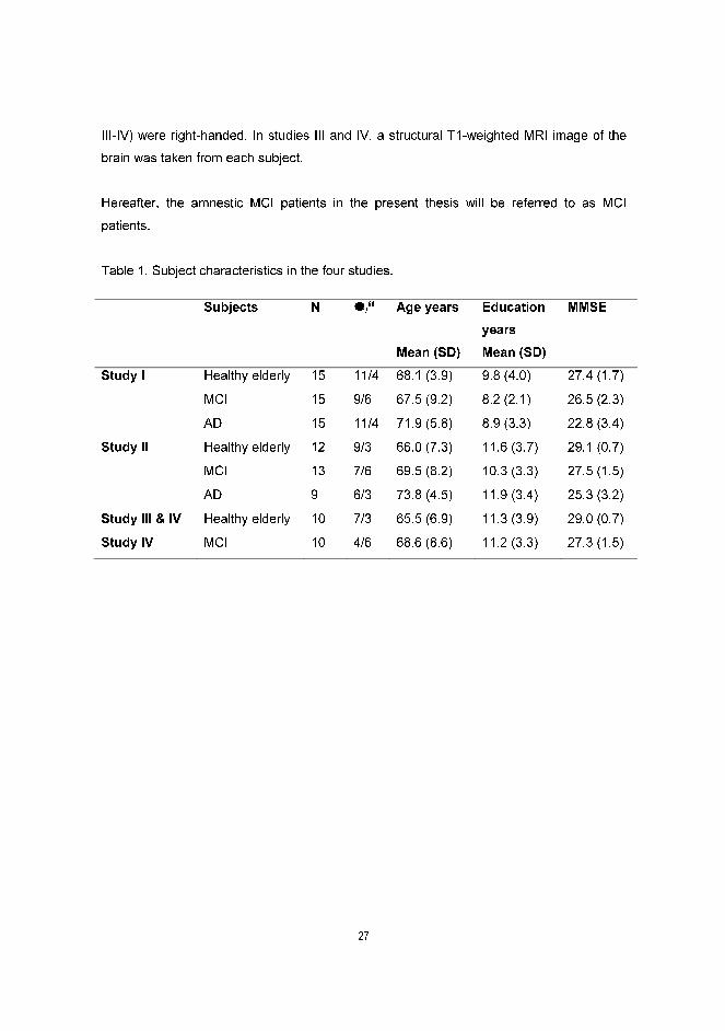
Table 1. Subject characteristics in the four studies.
Subjects N �/� Age years
Mean (SD)
Education
years
Mean (SD)
MMSE
Study I Healthy elderly 15 11/4 68.1 (3.9) 9.8 (4.0) 27.4 (1.7)
MCI 15 9/6 67.5 (9.2) 8.2 (2.1) 26.5 (2.3)
AD 15 11/4 71.9 (5.8) 8.9 (3.3) 22.8 (3.4)
Study II Healthy elderly 12 9/3 66.0 (7.3) 11.6 (3.7) 29.1 (0.7)
MCI 13 7/6 69.5 (8.2) 10.3 (3.3) 27.5 (1.5)
AD 9 6/3 73.8 (4.5) 11.9 (3.4) 25.3 (3.2)
Study III & IV Healthy elderly 10 7/3 65.5 (6.9) 11.3 (3.9) 29.0 (0.7)
Study IV MCI 10 4/6 68.6 (8.6) 11.2 (3.3) 27.3 (1.5)

28
3.2. METHODS
3.2.1. CERAD (study I)
In study I the Finnish CERAD (Pulliainen et al., 1999) was administered to all subjects.
The test battery consists of 9 subtests that are described in Table 2. Note that the CERAD
also includes another screening instrument, namely the Mini-Mental State Examination
(MMSE).
Table 2. CERAD subtests.
Subtest Description Max.
score
Cut-off
score
1. Verbal fluency Generating animal names (60 seconds) - <15
2. Naming Visual confrontation naming of 15 pictures 15 <11
3. MMSE Thirty questions and tasks (orientation in time and
space, memory, cognitive control)
30 <25
4. Wordlist learning List of 10 words shown to the patient 3 times, free
recall after each presentation
30 -
5. Constructional
praxis
Copy of 4 line drawings 11 -
6 a. Wordlist learning
(del)
Delayed recall of 10 words 10 -
6 b. Wordlist learning
(savings)
Delayed recall adjusted for acquisition (Delayed
recall/Immediate recall x 100)
100% <80%
7. Wordlist
recognition
Recognition (yes/no) of the 10 stimulus words
amongst 10 distractors
100% <80%
8 a. Constructional
praxis (del)
Delayed recall of 4 line drawings 11 -
8 b. Constructional
praxis (savings)
Delayed recall adjusted for acquisition (Delayed
recall/Immediate recall x 100)
100% <60%
9. Clock drawing Draw a clock showing the time ten past eleven 6 <5
29
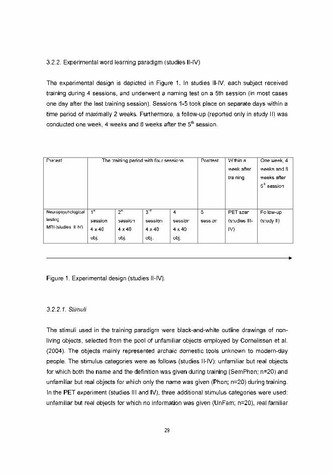
3.2.2. Experimental word learning paradigm (studies II-IV)
The experimental design is depicted in Figure 1. In studies II-IV, each subject received
training during 4 sessions, and underwent a naming test on a 5th session (in most cases
one day after the last training session). Sessions 1-5 took place on separate days within a
time period of maximally 2 weeks. Furthermore, a follow-up (reported only in study II) was
conducted one week, 4 weeks and 8 weeks after the 5th session.
Figure 1. Experimental design (studies II-IV).
3.2.2.1. Stimuli
The stimuli used in the training paradigm were black-and-white outline drawings of non-
living objects, selected from the pool of unfamiliar objects employed by Cornelissen et al.
(2004). The objects mainly represented archaic domestic tools unknown to modern-day
people. The stimulus categories were as follows (studies II-IV): unfamiliar but real objects
for which both the name and the definition was given during training (SemPhon; n=20) and
unfamiliar but real objects for which only the name was given (Phon; n=20) during training.
In the PET experiment (studies III and IV), three additional stimulus categories were used:
unfamiliar but real objects for which no information was given (UnFam; n=20), real familiar
Pretest The training period with four sessions Posttest Within a
week after
training
One week, 4
weeks and 8
weeks after
5th
session
Neuropsychological
testing
MRI (studies III-IV)
1st
session
4 x 40
obj.
2nd
session
4 x 40
obj.
3rd
session
4 x 40
obj.
4th
session
4 x 40
obj.
5th
session
PET scan
(studies III-
IV)
Follow-up
(study II)
30
non-living objects (Fam; n=20), and visual noise patterns (VNP; n=20). The names of the
SemPhon and Phon objects were matched on several linguistic features (word length,
number of syllables, number of vowels, number of consonants). Visual complexity between
the four groups of objects (SemPhon, Phon, UnFam, Fam) as well as associative potential
(only for SemPhon, Phon, UnFam objects) were also checked for.
3.2.2.2. Training
To ensure that the SemPhon and Phon objects were originally unfamiliar to the subjects,
they were presented for naming at the first session prior to any training. Maximally 2 out of
the 40 object names were allowed to be familiar to a subject. During each training session,
all 40 objects were shown four times in a pseudorandomized order. The objects were
shown as a PowerPoint slide show, one picture at a time for a period of 10 seconds. The
subjects were asked to read aloud the name printed below the object. If the object’s
definition was also given, they were to read that aloud as well. However, the subjects were
instructed to learn only the object names provided. The experimenter was present during
the whole training procedure.
Training sessions 2-4 were preceded by a naming test where the objects were presented
on the computer screen, thus yielding 3 measurements during the training and one after
the training (the 5th session). In the naming test, the subjects were instructed to name the
object as soon as possible. They were given 10 seconds to name each object, and the
correct answer was not provided. Furthermore, each training session (sessions 1-4) was
followed by a pointing-and-naming test, where all the objects were presented on a paper
sheet and the examiner pointed at the objects one at a time in a pseudorandomized order
and asked the subjects to name each one. If the subjects were not able to name the object
in 10 seconds, the correct answer was given to them, and thus even the pointing-and-
naming test included some training.
31
3.2.2.3. Follow-up (study II)
The naming test described above was accompanied by a cueing procedure during the
follow-up (sessions 5-8). If the subjects could not name the object in 10 seconds, a
phonological cue (the first syllable of the object name) was given, and the subjects were
thereafter given 10 additional seconds to name the object. In sessions 5-8, a recognition
test and a semantic test were also performed. The recognition test preceded the naming
test. It consisted of the 40 trained objects and 40 similar but untrained objects that were
shown to the subjects for 5 seconds on the computer screen in a pseudorandomized
order. The subjects were to decide whether or not the object had been among the 40
trained ones. A semantic test was performed after the naming test. The subjects were
shown the 40 trained objects in a pseudorandomized order and asked to decide whether
or not the object was presented with a definition during training. If yes, the subjects were
asked to report the definition as fully as possible. The definitions given at this task were
scored by two raters. The inter-rater reliability was high.
3.2.3. PET imaging (studies III-IV)
Positron emission tomography (PET) as a functional neuroimaging method relies on the
positive correlation between the level of neural function and regional cerebral blood flow
(rCBF) increases that support the activity. By contrasting rCBF patterns during the task of
interest with those in a reference condition, one can reveal brain regions that are
participating in the task of interest. While the non-invasive functional magnetic resonance
imaging (fMRI) method is nowadays more common in cognitive neuroimaging, the PET
method suited well for the present purposes as overt verbal responses were collected from
the subjects during scanning. The silence of the PET scanner as well as its somewhat
lower sensitivity to small head movements that can be elicited by articulation were positive
features for the present studies (for review, see Buckner & Logan, 2001; Rugg, 2000).
PET imaging was conducted within a week after training. Each subject underwent 12 PET
scans with 15O-water, including a rest condition (eyes open, blank screen) and 5
experimental conditions (SemPhon, Phon, UnFam, Fam, VNP). All the conditions were
presented twice, yielding two separate blocks with 6 conditions in a pseudorandomized
32
order, so that no condition was immediately repeated. The presentation order of the two
blocks was counterbalanced across participants.
3.2.3.1. Experimental conditions in the PET session
(1) SemPhon condition. The subjects were shown 20 trained SemPhon objects. The
subjects were instructed to name each object aloud. If they could not retrieve the name of
the object, they were instructed to stay silent and concentrate on the next object. In this
and the four following conditions each object was shown twice within a scan, with no
objects being immediately repeated. (2) Phon condition. The subjects were shown 20
trained Phon objects and were instructed to name the objects aloud. (3) UnFam condition.
The subjects were shown 20 untrained UnFam objects. The subjects had seen them only
once before the PET-scanning, on the 5th session as a part of a recognition test. The
subjects’ task was to say “picture” every time an UnFam object appeared on the screen,
(4) Fam condition. The subjects were shown 20 familiar objects and the instruction was to
name each of them. The subjects had seen and named them once before the PET
scanning (on the 5th session), (5) VNP condition. The subjects were shown 20 black and
white visual noise patterns and the task was to say “picture” every time a new random
pattern appeared.
3.2.3.2. PET data acquisition and processing
In order to register relative changes in the regional cerebral blood flow (rCBF) between the
experimental conditions, 12 emission PET scans were obtained for each subject using a
GE Advance PET scanner (General Electric Medical Systems, Milwaukee, WI, USA),
providing 35 transverse slices covering the entire brain and spaced 4.25 mm apart (centre
to centre). The task block that lasted approximately 3 minutes and was initiated 15 s prior
to the intravenous bolus (10 ml in 10-15 s) administration of 300 MBq 15O-water. Emission
data were acquired in 3D mode for 90 s starting when the true coincidence rate exceeded
15 kcps. Scans were separated by approximately 10 minutes. The images were
reconstructed using a filtered back-projection algorithm into a series of 35 slices including
128 x 128 voxels each, yielding an in-plane pixel dimension of 2.34 x 2.34 mm.
33
The PET image preprocessing and statistical analysis was performed using the Statistical
Parametric Mapping software (Friston 2004; Friston et al., 1995). The SPM99 was used in
study III and the SPM2 in study IV, implemented in Matlab version 6.1 (Mathworks Inc.,
USA). In order to compensate for inter-scan head motion, a two-step image realignment
procedure was performed. Each reconstructed PET image was realigned to the first image
in the series and a mean of the realignment images was created. Then all images were
realigned to the mean one. After this the realigned images were spatially normalized into a
coordinate space defined by the Montreal Neurological Institute (MNI) PET brain template
that approximates the standard stereotactic space of the Talairach and Tournoux brain
atlas (Talairach & Tournoux, 1988). An isotropic Gaussian filter of 16 mm full width at half
maximum was applied to smooth each normalized image to compensate for residual inter-
individual differences in brain shape and to suppress high frequency noise in the images.
An inter-scan difference in global signal was removed by proportional scaling of gray
matter voxel values to their mean value.
3.2.4. Statistical analyses
In study I, MANOVA was used to study overall differences between the three groups on
the combined CERAD measure. Subsequent one-way ANOVAs were conducted to further
study the differences between the three groups in the following CERAD subtests: Verbal
Fluency, Naming, MMSE, Wordlist learning (sum of 3 trials), Wordlist delayed recall,
Wordlist savings, Wordlist recognition, Constructional praxis, Constructional praxis
(delayed recall), Constructional praxis (savings) and Clock drawing. The Tukey post hoc
test was used to analyze pairwise group differences. Because several statistical
comparisons were performed, a Bonferroni-corrected alpha level was used both in the
ANOVAs and in the post hoc tests. A repeated measures ANOVA was used to analyse the
learning of the wordlist (trial 1, 2 and 3) in the three groups. Sensitivity [=correct
positives/(correct positives+false negatives)] and specificity [correct negatives/(correct
negatives+false positives)] were calculated for the following subtests using the current cut-
off scores based on Finnish normative data (Pulliainen et al., 1999): Verbal fluency,
Naming, MMSE, Wordlist savings, Wordlist recognition, Constructional praxis savings and
Clock drawing. The sensitivity and specificity of the Wordlist recognition test were also
explored in the cut-off range 81-95, and the optimal cut-off scores were reported. In the
34
Finnish CERAD no cut-off scores have been put forth for the Wordlist learning and delayed
recall tests, and thus the sensitivity and specificity of these subtests were explored using
different cut-off scores (16-20 for the Wordlist learning test and 5-8 for the Wordlist
delayed recall test). The optimal cut-off scores for these subtests were reported.
In study II, a three-way mixed model ANOVA was conducted to study the naming
performance of the newly learned objects in the three subject groups, separately for the
training period (sessions 2-5) and for the follow-up (sessions 5-8). Statistically significant
interactions were analyzed further by subsequent two-way mixed model ANOVAs (training
period) and by subsequent paired-samples t-tests (follow-up). Cued recall (phonological
cueing), recall of the semantic definitions (both quantitative and qualitative performance),
and recognition memory in the three groups were analyzed by two-way mixed model
ANOVAs. As the sample sizes were somewhat different and the assumption of
homogeneity of variance as shown by Levene’s test was not always met, the Games-
Howell post hoc test was used in all the analyses when examining the pairwise group
differences. For within-subject factors with more than two levels, corrected probabilities
(the Huynh-Feldt procedure) were reported.
In study III, the results from the verbal training were analyzed by a repeated measures
ANOVA. The PET results were analyzed with a fixed-effect model to estimate the effects
of conditions. The conditions were compared to each other as linear contrasts using t-
statistics and the following threshold criteria: height threshold T=4.67 corresponding to
p<.05, corrected for multiple non-independent comparisons (Worsley et al., 1996) along
with the cluster extent threshold of 50 contiguous voxels. Anatomical location of the
activated foci were found by directly transferring the MNI coordinates of the rCBF peaks
into the atlas of Talairach and Tournoux (1988).
In study IV, the behavioural results were analyzed by a three-way mixed model ANOVA.
The statistical analysis of the PET results was done in the following steps. At the first step,
each individual PET data were fit to a single-subject model and subject-specific inter-
condition contrasts were calculated. At the next step, in order to make inferences at the
population level, a second level analysis treating subjects as random effects was done by
creating a separate model for each inter-condition contrast and entering one contrast file

35
per subject into the random effects (RFX) model. Due to the relatively small sample size
(10 subjects per group), a non-parametric permutation-based method [Statistical non-
parametric mapping (SnPM); Nichols & Holmes, 2002)] was chosen. More specifically, the
SnPM3b software run under SPM2 with 1024 permutations and 16 mm variance
smoothing was used for both within- and between-group analyses.
Previous research indicates that the left inferior frontal cortex and the left temporal cortex
are particularly important in verbal episodic recall (Cabeza et al., 2003; James & Gauthier,
2004; Lundström, Ingvar, & Petersson, 2005; Nyberg et al., 2003; Raboyeau et al., 2004).
Additionally, the anterior cingulate is thought to participate in more general modulation of
executive and attentional control processes in particularly demanding tasks (for review,
see Bush, Luu, & Posner, 2000). Therefore, the activation patterns between the MCI
patients and the controls were compared when they named newly learned objects vs. saw
unfamiliar objects (and said “picture”) by performing volume of interest (VOI) analyses for
three selected brain regions. Binary masks for the VOIs were generated with the MARINA
software, version 0.6.1 (Walter et al., 2003). The VOI masks covered the following brain
structures: the anterior cingulate VOI that included bilateral anterior cingulate cortex, the
left frontal (BA 45, 47) VOI that included the triangular (BA 45) and orbital (BA 47) parts of
the left inferior frontal gyrus, and the left temporal VOI (BA 21, 22, 38, 20, 37) that included
the superior, middle, and inferior gyri of the left temporal lobe. The results of the RFX
SnPM analyses were assessed with pseudo-t-statistics and thresholded at a voxel-level
family-wise error (FWE) chance probability p<.05 corrected either for the whole brain
volume (in the within-group comparisons) or for a volume of interest (in the VOI-based
between-group comparisons). Anatomical location of the activated foci were found by
directly transferring the MNI coordinates of the rCBF peaks into the coordinate space of
the Talairach and Tournoux atlas (1988).
Additionally, correlations between naming success in the SemPhon and Phon conditions
and brain activation were explored separately for the control and the MCI group at the
second level in a series of SnPM analyses. For each of the contrasts SemPhon vs. UnFam
and Phon vs. UnFam, the corresponding contrast files (one per subject) were entered into
a non-parametric permutation test along with the regression vector representing the
naming score. In addition to RFX SnPM analyses for the whole-brain volume, such
36
analyses were repeated separately for each VOI. The threshold of significance was set as
p<.05, FWE-corrected for the analysed volume.
4. RESULTS
4.1. PERFORMANCE ON THE CERAD IN MCI AND AD (study I)
Significant overall group differences on the combined CERAD variable including 14
subtest measures were observed. At the subtest level, a significant main effect of group
was found on the following CERAD variables: Verbal fluency, Naming, MMSE, Wordlist
learning (sum of 3 trials), Wordlist learning (delayed recall), Wordlist savings, Wordlist
recognition, Constructional praxis (delayed recall) and Constructional praxis (savings).
Post hoc tests showed significant differences between the control group and the AD group
in all the abovementioned tests, with the AD patients performing significantly worse. In
MMSE, Wordlist learning (delayed recall), Wordlist learning (savings) and Wordlist learning
(recognition) the MCI group outperformed the AD group. No statistically significant pair-
wise differences were found between the controls and the MCI patients on these
measures.
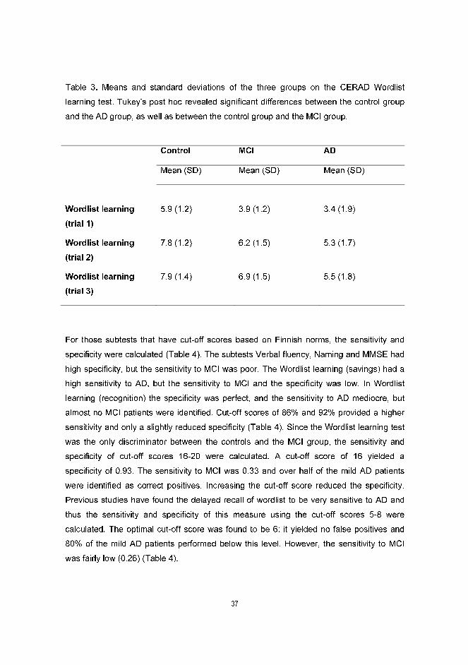
The analysis of the Wordlist learning trials showed that the number of remembered items
increased across subject groups during the three trials, albeit the amount of items learned
differed between the three groups. Post hoc analyses revealed significant differences
between the controls and the AD group, as well as between the controls and the MCI
group, but not between the MCI and AD group, indicating that the Wordlist learning of the
MCI patients was almost as poor as that of the AD group (Table 3). The interaction term
was non-significant, indicating that the relative degree of learning over trials was similar in
all three groups.
37
Table 3. Means and standard deviations of the three groups on the CERAD Wordlist
learning test. Tukey’s post hoc revealed significant differences between the control group
and the AD group, as well as between the control group and the MCI group.
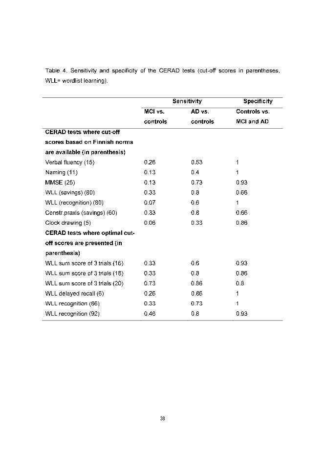
For those subtests that have cut-off scores based on Finnish norms, the sensitivity and
specificity were calculated (Table 4). The subtests Verbal fluency, Naming and MMSE had
high specificity, but the sensitivity to MCI was poor. The Wordlist learning (savings) had a
high sensitivity to AD, but the sensitivity to MCI and the specificity was low. In Wordlist
learning (recognition) the specificity was perfect, and the sensitivity to AD mediocre, but
almost no MCI patients were identified. Cut-off scores of 86% and 92% provided a higher
sensitivity and only a slightly reduced specificity (Table 4). Since the Wordlist learning test
was the only discriminator between the controls and the MCI group, the sensitivity and
specificity of cut-off scores 16-20 were calculated. A cut-off score of 16 yielded a
specificity of 0.93. The sensitivity to MCI was 0.33 and over half of the mild AD patients
were identified as correct positives. Increasing the cut-off score reduced the specificity.
Previous studies have found the delayed recall of wordlist to be very sensitive to AD and
thus the sensitivity and specificity of this measure using the cut-off scores 5-8 were
calculated. The optimal cut-off score was found to be 6: it yielded no false positives and
80% of the mild AD patients performed below this level. However, the sensitivity to MCI
was fairly low (0.26) (Table 4).
Control MCI AD
Mean (SD) Mean (SD) Mean (SD)
Wordlist learning
(trial 1)
5.9 (1.2)
3.9 (1.2)
3.4 (1.9)
Wordlist learning
(trial 2)
7.8 (1.2) 6.2 (1.5) 5.3 (1.7)
Wordlist learning
(trial 3)
7.9 (1.4) 6.9 (1.5) 5.5 (1.8)
38
Table 4. Sensitivity and specificity of the CERAD tests (cut-off scores in parentheses,
WLL= wordlist learning).
Sensitivity Specificity
MCI vs.
controls
AD vs.
controls
Controls vs.
MCI and AD
CERAD tests where cut-off
scores based on Finnish norms
are available (in parenthesis)
Verbal fluency (15) 0.26 0.53 1
Naming (11) 0.13 0.4 1
MMSE (25) 0.13 0.73 0.93
WLL (savings) (80) 0.33 0.8 0.66
WLL (recognition) (80) 0.07 0.6 1
Constr.praxis (savings) (60) 0.33 0.8 0.66
Clock drawing (5) 0.06 0.33 0.86
CERAD tests where optimal cut-
off scores are presented (in
parenthesis)
WLL sum score of 3 trials (16) 0.33 0.6 0.93
WLL sum score of 3 trials (18) 0.33 0.8 0.86
WLL sum score of 3 trials (20) 0.73 0.86 0.8
WLL delayed recall (6) 0.26 0.86 1
WLL recognition (86) 0.33 0.73 1
WLL recognition (92) 0.46 0.8 0.93
39
4.2. VERBAL LEARNING IN MCI AND AD (study II)
For the training period, significant overall group differences were found (see Table 5), with
controls expectedly showing the best performance and AD patients showing the lowest
overall performance. Post hoc analyses showed significant differences between all three
groups. The overall naming performance of the newly learned objects increased
significantly during training. Also, a significant interaction between test session and group
was found, i.e. the learning curves of the 3 groups differed from each other. Subsequent
analyses showed that all learning curves differed significantly from each other (controls vs.
MCI group, MCI vs. AD group and control vs. AD group), with controls learning the fastest
and the AD patients the slowest while the MCI patients were situated in between.
Regarding semantic support, the only significant finding was the stimulus type and group
interaction, and subsequent analyses showed that the stimulus type and group interaction
failed to reach statistical significance between the control vs. AD group or between the
MCI vs. AD group, but it was statistically significant between the control vs. MCI group.
This was due to the fact that the control subjects learned more names of Phon than
SemPhon objects, whereas the MCI patients learned an approximately equal amount of
SemPhon and Phon objects.
For the follow-up (see Table 5), significant overall group differences were found again, with
the controls showing the best performance and the AD patients showing the worst
performance. Post hoc analyses showed significant differences between all three groups.
Also, the overall forgetting of the newly learned object names increased significantly during
follow-up. In contrast to the training period, the test session and group interaction was non-
significant, reflecting similar forgetting curves in the three groups. Concerning semantic
support, the only significant finding was the interaction of test session and stimulus type.
Subsequent analyses showed that during session 8 (i.e., 8 weeks after training) more
SemPhon object names than Phon object names were recalled. Even though the three-
way interaction of test session, stimulus type and group failed to reach statistical
significance, further analyses were conducted, based on the hypothesis concerning the
beneficial effects of semantic support in MCI patients. This effect would be due to the
relatively well preserved semantic memory in MCI that could compensate for a deficit in
episodic memory. Consequently, if semantic support would show a benefit at recall, its
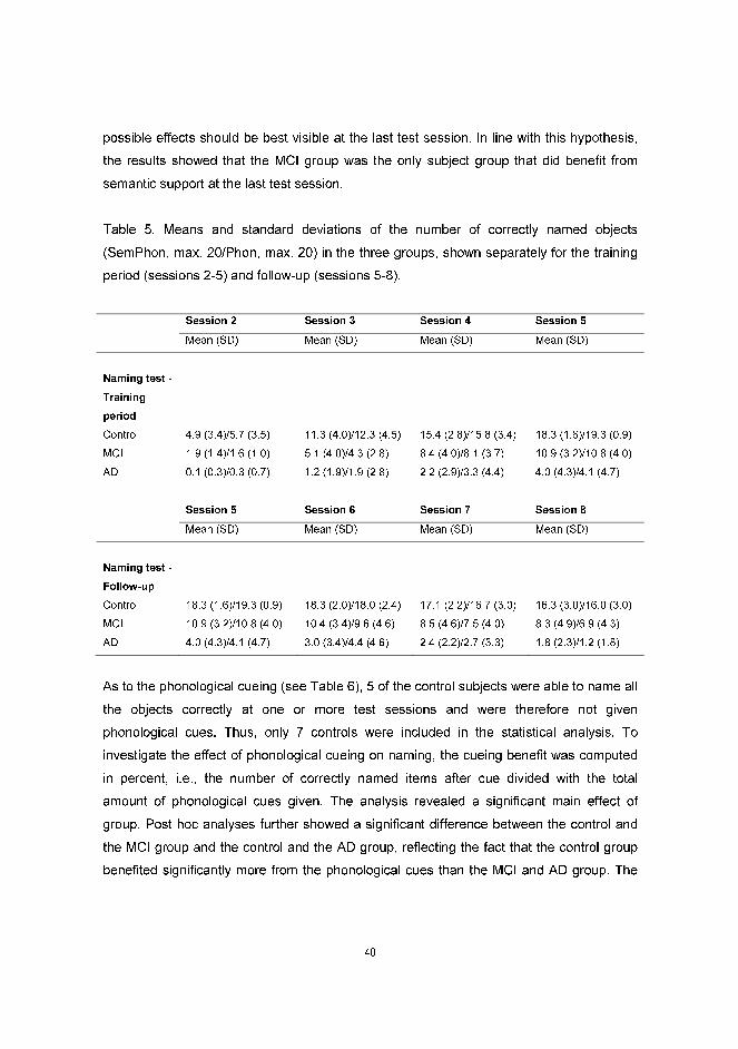
40
possible effects should be best visible at the last test session. In line with this hypothesis,
the results showed that the MCI group was the only subject group that did benefit from
semantic support at the last test session.
Table 5. Means and standard deviations of the number of correctly named objects
(SemPhon, max. 20/Phon, max. 20) in the three groups, shown separately for the training
period (sessions 2-5) and follow-up (sessions 5-8).
Session 2 Session 3 Session 4 Session 5
Mean (SD) Mean (SD) Mean (SD) Mean (SD)
Naming test -
Training
period
Control 4.9 (3.4)/5.7 (3.5) 11.3 (4.0)/12.3 (4.5) 15.4 (2.8)/15.8 (3.4) 18.3 (1.6)/19.3 (0.9)
MCI 1.9 (1.4)/1.6 (1.0) 5.1 (4.0)/4.3 (2.8) 8.4 (4.0)/8.1 (3.7) 10.9 (3.2)/10.8 (4.0)
AD 0.1 (0.3)/0.3 (0.7) 1.2 (1.9)/1.9 (2.8) 2.2 (2.9)/3.3 (4.4) 4.0 (4.3)/4.1 (4.7)
Session 5
Session 6
Session 7
Session 8
Mean (SD) Mean (SD) Mean (SD) Mean (SD)
Naming test -
Follow-up
Control 18.3 (1.6)/19.3 (0.9) 18.3 (2.0)/18.0 (2.4) 17.1 (2.2)/16.7 (3.0) 16.3 (3.0)/16.0 (3.0)
MCI 10.9 (3.2)/10.8 (4.0) 10.4 (3.4)/9.6 (4.6) 8.5 (4.6)/7.5 (4.0) 8.3 (4.9)/6.9 (4.3)
AD 4.0 (4.3)/4.1 (4.7) 3.0 (3.4)/4.4 (4.6) 2.4 (2.2)/2.7 (3.3) 1.8 (2.3)/1.2 (1.8)
As to the phonological cueing (see Table 6), 5 of the control subjects were able to name all
the objects correctly at one or more test sessions and were therefore not given
phonological cues. Thus, only 7 controls were included in the statistical analysis. To
investigate the effect of phonological cueing on naming, the cueing benefit was computed
in percent, i.e., the number of correctly named items after cue divided with the total
amount of phonological cues given. The analysis revealed a significant main effect of
group. Post hoc analyses further showed a significant difference between the control and
the MCI group and the control and the AD group, reflecting the fact that the control group
benefited significantly more from the phonological cues than the MCI and AD group. The
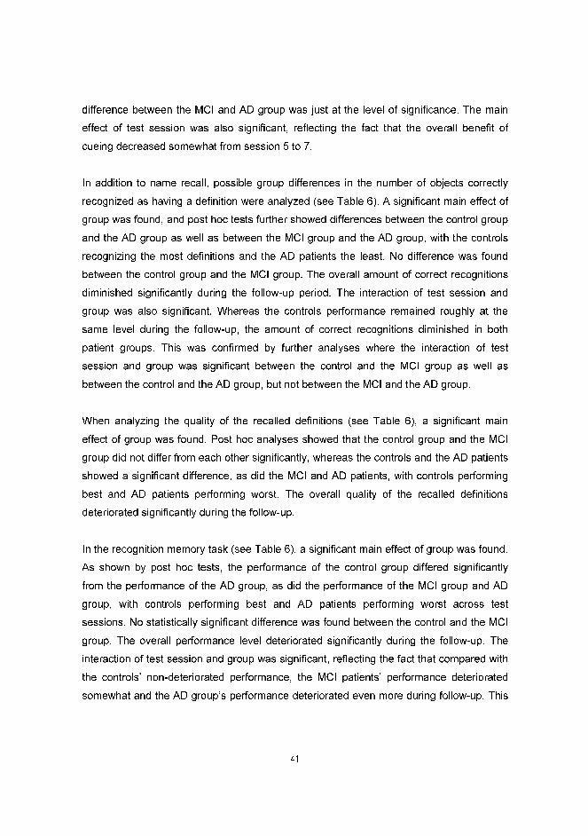
41
difference between the MCI and AD group was just at the level of significance. The main
effect of test session was also significant, reflecting the fact that the overall benefit of
cueing decreased somewhat from session 5 to 7.
In addition to name recall, possible group differences in the number of objects correctly
recognized as having a definition were analyzed (see Table 6). A significant main effect of
group was found, and post hoc tests further showed differences between the control group
and the AD group as well as between the MCI group and the AD group, with the controls
recognizing the most definitions and the AD patients the least. No difference was found
between the control group and the MCI group. The overall amount of correct recognitions
diminished significantly during the follow-up period. The interaction of test session and
group was also significant. Whereas the controls performance remained roughly at the
same level during the follow-up, the amount of correct recognitions diminished in both
patient groups. This was confirmed by further analyses where the interaction of test
session and group was significant between the control and the MCI group as well as
between the control and the AD group, but not between the MCI and the AD group.
When analyzing the quality of the recalled definitions (see Table 6), a significant main
effect of group was found. Post hoc analyses showed that the control group and the MCI
group did not differ from each other significantly, whereas the controls and the AD patients
showed a significant difference, as did the MCI and AD patients, with controls performing
best and AD patients performing worst. The overall quality of the recalled definitions
deteriorated significantly during the follow-up.
In the recognition memory task (see Table 6), a significant main effect of group was found.
As shown by post hoc tests, the performance of the control group differed significantly
from the performance of the AD group, as did the performance of the MCI group and AD
group, with controls performing best and AD patients performing worst across test
sessions. No statistically significant difference was found between the control and the MCI
group. The overall performance level deteriorated significantly during the follow-up. The
interaction of test session and group was significant, reflecting the fact that compared with
the controls’ non-deteriorated performance, the MCI patients’ performance deteriorated
somewhat and the AD group’s performance deteriorated even more during follow-up. This
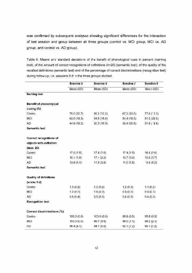
42
was confirmed by subsequent analyses showing significant differences for the interaction
of test session and group between all three groups (control vs. MCI group, MCI vs. AD
group, and control vs. AD group).
Table 6. Means and standard deviations of the benefit of phonological cues in percent (naming
test), of the amount of correct recognitions of definitions (n=20) (semantic test), of the quality of the
recalled definitions (semantic test) and of the percentage of correct discriminations (recognition test)
during follow-up, i.e. sessions 5-8 in the three groups studied.
Session 5 Session 6 Session 7 Session 8
Mean (SD) Mean (SD) Mean (SD) Mean (SD)
Naming test
Benefit of phonological
cueing (%)
Control 75.0 (33.7) 89.2 (12.2) 67.2 (22.5) 77.8 (13.3)
MCI 63.0 (18.3) 54.8 (18.4) 54.4 (16.5) 51.5 (20.0)
AD 44.9 (18.2) 35.3 (16.3) 35.4 (20.6) 31.6 (19.6)
Semantic test
Correct recognitions of
objects with definition
(max. 20)
Control 17.0 (2.5) 17.4 (2.0) 17.4 (2.5) 16.8 (2.6)
MCI 18.1 (1.9) 17.1 (2.2) 15.7 (3.4) 15.9 (3.7)
AD 13.9 (4.4) 11.9 (3.8) 11.0 (3.8) 10.9 (6.2)
Semantic test
Quality of definitions
(scale: 0-2)
Control 1.3 (0.2) 1.2 (0.2) 1.2 (0.3) 1.1 (0.2)
MCI 1.2 (0.2) 1.0 (0.2) 0.9 (0.2) 0.9 (0.2)
AD 0.6 (0.4) 0.5 (0.3) 0.4 (0.3) 0.4 (0.3)
Recognition test
Correct discriminations (%)
Control 100.0 (0.0) 100.0 (0.0) 99.8 (0.5) 99.8 (0.5)
MCI 100.0 (0.0) 99.7 (0.5) 99.0 (1.3) 98.2 (2.3)
AD 96.4 (4.1) 94.7 (6.0) 92.1 (7.2) 90.7 (7.2)
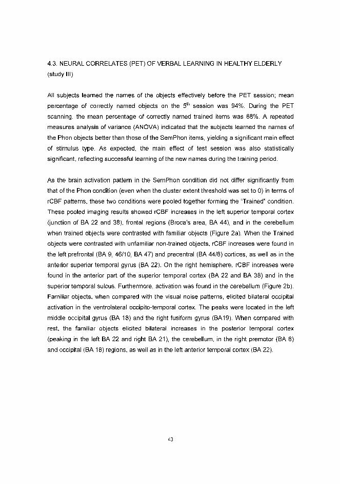
43
4.3. NEURAL CORRELATES (PET) OF VERBAL LEARNING IN HEALTHY ELDERLY
(study III)
All subjects learned the names of the objects effectively before the PET session; mean
percentage of correctly named objects on the 5th session was 94%. During the PET
scanning, the mean percentage of correctly named trained items was 88%. A repeated
measures analysis of variance (ANOVA) indicated that the subjects learned the names of
the Phon objects better than those of the SemPhon items, yielding a significant main effect
of stimulus type. As expected, the main effect of test session was also statistically
significant, reflecting successful learning of the new names during the training period.
As the brain activation pattern in the SemPhon condition did not differ significantly from
that of the Phon condition (even when the cluster extent threshold was set to 0) in terms of
rCBF patterns, these two conditions were pooled together forming the “Trained” condition.
These pooled imaging results showed rCBF increases in the left superior temporal cortex
(junction of BA 22 and 38), frontal regions (Broca’s area, BA 44), and in the cerebellum
when trained objects were contrasted with familiar objects (Figure 2a). When the Trained
objects were contrasted with unfamiliar non-trained objects, rCBF increases were found in
the left prefrontal (BA 9, 46/10, BA 47) and precentral (BA 44/6) cortices, as well as in the
anterior superior temporal gyrus (BA 22). On the right hemisphere, rCBF increases were
found in the anterior part of the superior temporal cortex (BA 22 and BA 38) and in the
superior temporal sulcus. Furthermore, activation was found in the cerebellum (Figure 2b).
Familiar objects, when compared with the visual noise patterns, elicited bilateral occipital
activation in the ventrolateral occipito-temporal cortex. The peaks were located in the left
middle occipital gyrus (BA 18) and the right fusiform gyrus (BA19). When compared with
rest, the familiar objects elicited bilateral increases in the posterior temporal cortex
(peaking in the left BA 22 and right BA 21), the cerebellum, in the right premotor (BA 6)
and occipital (BA 18) regions, as well as in the left anterior temporal cortex (BA 22).

44
Figure 2. Areas of relative rCBF increase (p<.05 corrected, k>50 voxels) in the a) trained
objects vs. familiar objects contrast and the b) trained objects vs. unfamiliar objects
contrast projected onto the reconstructed lateral and medial surfaces of the anatomical
MRI/SPM99 brain template.
4.4. NEURAL CORRELATES (PET) OF VERBAL LEARNING IN MCI (study IV)
The MCI patients learned 56% of the object names before the PET session. During the
PET scanning, the mean percentage of correctly named trained items was 53%. The MCI
patients’ overall performance was significantly lower than that of the controls, even though
both groups showed a learning effect during the training period. As in study II, the
significant interaction of test session and group showed that the learning curves of the two
groups differed significantly from each other. The interaction of stimulus type and group
reached significance, too. This was due to the fact that the control subjects learned more
names of Phon than SemPhon objects, whereas the MCI patients learned an
approximately equal amount of SemPhon and Phon object names.
The between-group PET results (VOI analysis) revealed that when newly learned objects
that had been learned without semantic support (Phon) were contrasted with unfamiliar
non-trained objects, a significantly higher increase in rCBF for the MCI patients than for
the controls was found in the anterior cingulate VOI, with maxima in BA 32 (Figure 3a).
When newly learned objects that had been learned with semantic support (SemPhon)
were contrasted with unfamiliar non-trained objects, no significant differences were found
a b
45
in the anterior cingulate. Albeit only approaching statistical significance in the left frontal
VOI (left BA 45, 47), an activation peak in the SemPhon vs. UnFam contrast was found
when the MCI patients were compared with the controls (Figure 3b). The Phon vs. UnFam
contrast did not yield any significant effects in the frontal VOI. In the left temporal VOI (BA
21, 22, 38, 20, 37) (Figure 3c), no group differences were found for either contrast. The
control subjects did not demonstrate any activation increases in any of the VOIs in either
contrast when compared with the MCI group. Furthermore, the whole-brain correlation
analyses showed a significant positive correlation between naming performance and
cerebellar activity for the Phon condition in the MCI group in the cerebellum. The VOI-
related results in the left frontal area (peak in BA 47/11) showed a negative correlation for
the Phon condition in the MCI group. No significant correlations were found in the control
group.
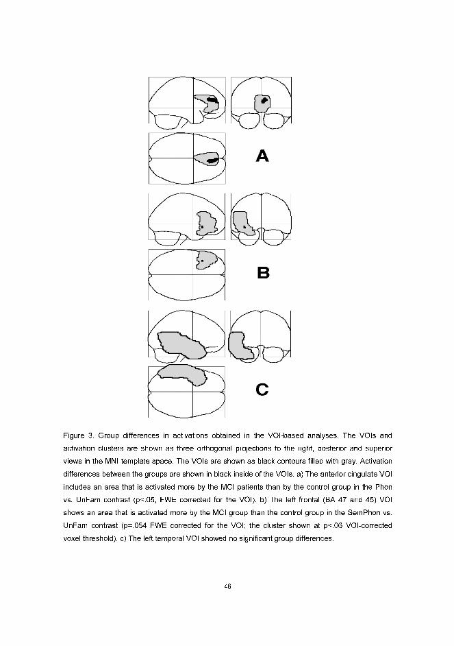
46
Figure 3. Group differences in activations obtained in the VOI-based analyses. The VOIs and
activation clusters are shown as three orthogonal projections to the right, posterior and superior
views in the MNI template space. The VOIs are shown as black contours filled with gray. Activation
differences between the groups are shown in black inside of the VOIs. a) The anterior cingulate VOI
includes an area that is activated more by the MCI patients than by the control group in the Phon
vs. UnFam contrast (p<.05, FWE corrected for the VOI). b) The left frontal (BA 47 and 45) VOI
shows an area that is activated more by the MCI group than the control group in the SemPhon vs.
UnFam contrast (p=.054 FWE corrected for the VOI; the cluster shown at p<.06 VOI-corrected
voxel threshold). c) The left temporal VOI showed no significant group differences.
47
5. DISCUSSION
There has been an increasing research interest in memory and aging. As deficits in
learning and memory are often the first symptoms of the most common form of dementia,
AD, it would be very important to be able to differentiate these symptoms from more
benign age-related changes in memory. The first study addressed the sensitivity and
specificity of a cognitive screening battery CERAD in the differential diagnostics of MCI
and AD. A special emphasis was put on a wordlist learning subtest expected to be
sensitive to memory disorders in these two patient groups. The other three studies focused
at the ability to learn new words by healthy elderly, MCI patients, and AD patients. In this
regard, it was somewhat surprising to find out that relatively few studies have focused on
the learning capacity of patients suffering from preclinical and early AD. Nevertheless, it is
common for geriatric patients to complain not only about difficulties in remembering
names, but also about learning new words or a new language. The present thesis presents
new results on the ability of healthy elderly, MCI patients and AD patients to learn new
words. Word learning was investigated by an experimental word learning paradigm and by
employing both behavioural and neural measures.
5.1. SCREENING FOR MCI AND AD WITH THE FINNISH CERAD (study I)
In study I, the only CERAD subtest that showed significant differences between healthy
controls and MCI patients was the Wordlist learning test, with the MCI patients showing a
worse overall learning performance than the controls. The AD patients’ performance was
significantly worse than that of the controls on almost all CERAD subtests. The results
indicate that encoding of material to episodic memory is compromised in MCI, which is in
line with findings from previous studies on MCI and preclinical AD (Collie & Maruff, 2000;
Grober & Kawas, 1997; Morris et al., 1991; Petersen, Smith, Ivnik, Kokmen, & Tangalos,
1994; Petersen et al., 1999; Wang & Zhou, 2002). As regards the CERAD, also Moulin et
al. (2004) reported an overall difference between MCI patients and controls in the CERAD
Wordlist learning test. However, Moulin et al. (2004) also found that the learning curves of
MCI patients and controls differed from each other, with MCI patients showing a less steep
curve. No group differences in the learning curves were found in study I, and one reason
for this discrepancy could be that the present patient sample was smaller than that of
48
Moulin et al. (2004). An alternative explanation is that the MCI patients in the Moulin et al.
(2004) study were cognitively slightly more impaired the patients in study I: the average
MMSE score in their MCI group was 25.7 compared with 26.5 in study I. In any case, the
present results give further support for the view that MCI is characterized by an encoding
impairment, which can impede learning of new information, as seen in early AD patients
(for review, see Germano & Kinsella, 2005). Furthermore, no cut-off score has been
assigned to the Wordlist learning test in the Finnish CERAD. This hampers the clinical use
of the test, as previous studies have shown that verbal episodic learning problems are
amongst the earliest signs of preclinical AD (e.g., Collie & Maruff, 2000; Morris et al., 1991;
Linn et al., 1995; Petersen et al., 1994). Accordingly, the sensitivity and specificity of the
sum score of the three trials of the Wordlist learning test was explored using a range of
cut-off scores. A cut-off score of 16 yielded a sensitivity of 0.33 to MCI and 0.6 to AD with
a specificity of 0.93. The sensitivity of the test was raised when the cut-off score was
raised, but the number of false positives also increased. Interestingly, a recent Finnish
normative study on CERAD (Pulliainen et al., 2007) including 321 cognitively healthy
persons between the ages 63-79 found similar results to ours. Although the study by
Pulliainen et al. (2007) did not address the sensitivity and specificity of CERAD to MCI and
AD, their results showed that 90% of the healthy elderly learned more than 15 words in the
Wordlist learning test, i.e., a score of 15 or less was seen as significantly deviant.
It is somewhat surprising that the controls and the MCI patients did not show significant
differences in the delayed recall of the Wordlist learning test either in absolute terms or in
the delayed recall adjusted for acquisition (Savings score). Previous findings have shown
that MCI patients are impaired especially on delayed recall in verbal episodic memory
tasks (Petersen et al., 1999). One reason for this discrepancy might be that the delays in
the CERAD memory tests are too short (usually approximately 5 minutes) to fully tap the
episodic memory consolidation problems in MCI. For example, the delayed recall on the
WMS-R Logical memory test, where the delay is usually 30 minutes, has been found to be
a good predictor of patients who will later convert to AD (Elias et al., 2000; Marquis et al.,
2002). The findings from study I may also indicate that retention is not impaired to the
same extent as acquisition in MCI, in line with e.g. Grober and Kawas (1997). Also for the
recall performance in the Wordlist learning test, a cut-off score is lacking in the Finnish
CERAD, although this test has been shown to be the best discriminator between normal
49
aging and early AD (Welch-Bohmer & Mohs, 1997). Calculations on the present data
showed that a cut-off score of 6 seemed to be very specific and sensitive to clinically
diagnosed AD, but only 26% of the MCI patients were identified using this cut-off.
Furthermore, the Savings scores in both Wordlist learning and Constructional praxis were
found to be relatively unspecific, i.e., several healthy controls were identified as false
positives using this measure, which was quite unexpected. This suggests that the current
cut-off scores (80 and 60, respectively) in the Finnish CERAD Savings scores might be too
high. Results from the recent CERAD study by Pulliainen et al. (2007) with healthy elderly
subjects also indicated that the current cut-off scores may be rather high, but the authors
argued that a false positive result may not be as detrimental as a negative one.
Previous studies have shown results close to ceiling for healthy elderly people in the
Wordlist recognition test (Karrasch & Laine, 2003), suggesting that the current cut-off
score (80%) in the Finnish CERAD might be fairly low. A cut-off score of 86% for Wordlist
recognition in the present study yielded a moderate sensitivity for MCI and relatively good
sensitivity for AD, with a perfect specificity. This finding suggests that the cut-off in this
subtest might be adjusted to increase sensitivity without losing specificity. Likewise,
normative results from healthy elderly subjects have indicated that the cut-off score for
Wordlist recognition may be too high (Pulliainen et al., 2007). There were no significant
differences in the MMSE scores between the MCI patients and the controls. This finding is
in line with previous studies showing a low sensitivity of the measure in preclinical AD
(Grober, Hall, Lipton, & Teresi, 2007; Tang-Wai et al., 2003; Tierney et al., 2000), and also
clearly shows how important it is to develop new screening tools for the detection of milder
and earlier phases of AD. When comparing normal elderly controls and clinically
diagnosed probable AD patients, the results in study I clearly show that almost all CERAD
tests are sensitive enough to reveal significant differences. This reflects the fact that in
order to fulfil the diagnostic criteria of probable AD, the cognitive decline has to affect other
cognitive domains than just memory.
In sum, the results from study I suggest that MCI patients suffer from an encoding
impairment when trying to learn a wordlist. Interestingly, neither the delayed recall
performance nor the Savings score of the Wordlist learning test showed significant
differences between controls and MCI patients, indicating intact memory retention for the
50
MCI group in this subtest. In fact, no CERAD subtests, except Wordlist learning, showed
significant differences between controls and MCI patients. Furthermore, part of the
individuals with preclinical AD might not be detected, when using the CERAD test battery
as the sole screening instrument. There is also a risk that some cognitively healthy elderly
people might receive a false positive result. These risk factors should be considered,
especially as CERAD is becoming an increasingly popular instrument in Finland for
detecting people at risk for dementia or already showing signs of dementia.
5.2. VERBAL LEARNING IN MCI AND AD (STUDY II)
In study II, the aim was to explore how subjects with mild cognitive impairment (MCI),
subjects with early Alzheimer’s disease (AD) and age-matched controls learned and
maintained the names of previously unfamiliar rare objects that were trained with or
without semantic support (object definitions). Word learning in MCI has not previously
been studied. This is somewhat surprising, as word learning is such an important part of
our everyday life - we develop and modify our mental lexicon throughout life. The study of
word learning abilities of MCI patients and AD patients could also shed light on cognitive
plasticity in MCI and AD. The present paradigm also enabled a comparison between
semantic memory and episodic memory functions. It was hypothesized that semantic
support might have some beneficial effects in the present learning task especially in the
MCI group, as semantic memory functions are relatively well preserved in MCI compared
to episodic memory. In the follow-up, the effects of phonological cueing on the naming of
newly learned objects and retrieval of the object definitions, as well as recognition of the
objects were also studied.
The MCI patients learned the names of the unfamiliar objects more poorly than the
controls, and the AD patients’ performance was inferior to that of the MCI group. The
results are in line with recent studies that have showed a decreased learning efficacy in
MCI (Moulin et al., 2004; Ribeiro et al., 2007). There is also increasing evidence that early
AD patients suffer from an encoding impairment that impedes the acquisition of new
information (for review, see Germano & Kinsella, 2005). One should bear in mind, though,
that previous studies have used paradigms that are very different from the one used in the
present study, measuring learning over much shorter time periods. Moreover, episodic
51
verbal learning tests are usually based on previously familiar materials. In fact, the results
from the word learning paradigm used in the present study probably reflect at least partly
also semantic memory functions and not merely episodic memory processes (see also the
discussion in 5.3. concerning the PET findings). It should also be noted that the MCI
patients could nevertheless retrieve 54% of the object names at the end of the training,
and even the AD group could retrieve 20% of the object names. In other words, repeated
exposure and training did in fact result in learning in both patient groups, showing that their
verbal learning abilities were impaired but by no means lost.
One explanation for the difference between the learning performances of the controls and
both patient groups might be the difference in their ability to use memory strategies. When
asked after training, the controls reported plenty of self-generated strategies. For example,
they often associated the name of the object either to a familiar name that phonologically
resembled it or to some well-known object in order to use that as a memory cue. This is in
line with previous studies that have shown that healthy individuals usually call upon prior
knowledge when encoding and retrieving new information (for review, see Brown & Craik,
2000). However, the MCI patients and AD patients reported hardly any self-generated
strategies. It has been shown that AD patients are impaired in manipulating, integrating
and organizing relevant features of new information in order to facilitate acquisition (for
review, see Germano & Kinsella, 2005), and considering MCI as a prodromal stage of AD,
this type of impairment might be reflected in the MCI patients’ results as well.
With regard to forgetting during the follow-up, all three groups showed similar forgetting
curves despite the fact that their overall performance differed. As mentioned above, there
is some controversy as to which aspects of verbal episodic memory are predominantly
affected in MCI and AD: acquisition (e.g. Greene et al., 1996; Grober & Kawas, 1997),
consolidation (e.g. Hart, Kwentus, Harkins, & Taylor, 1988), or both (Moulin et al., 2004).
The present findings indicate that, also when learning is measured over a longer time
period, acquisition seems to be the key deficit in MCI and AD. According to the standard
model of memory consolidation (for review, see Moscovitch et al., 2005), the hippocampal
formation is necessary for the acquisition and retrieval of recent memories, but is no longer
needed when long-term consolidation is complete and permanent memory storage has
taken place in the neocortical networks. Subsequently, as hippocampal regions are first
52
affected in MCI and AD, the present findings would be in line with the idea that impaired
acquisition of the names of the unfamiliar objects in the MCI and AD group was at least
partly due to hippocampal damage. However, those names that had been successfully
acquired despite hippocampal dysfunction were consolidated and stored in less affected
brain areas and therefore accessible during follow-up. When interpreting the present
results, one should also note that the follow-up consisted of several test trials that may
boost learning (for review, see Brown & Craik, 2000). In other words, the test trials per se
might have influenced the follow-up performance to some extent. Nevertheless, the
possible beneficial effect from the test trials should have been similar in all three groups.
It should also be noted that the AD patients in the present study were quite well-performing
when looking at their mean MMSE score (25.3). For example, Greene et al. (1996)
considered AD patients with a MMSE score of 24 or above to have a “minimal” deficit.
Semantic support did not enhance verbal learning during training in MCI and AD. In
contrast, the control group learned more names of the Phon objects than the SemPhon
objects during training. Similar results on healthy subjects were found in a study by
Whiting, Chenery, Chalk, Darnell, and Copland (2007), who studied the effects of
dexamphetamine on new word learning with a paradigm similar to the present study.
However, in contrast to the present study, the learning of the semantic support was
encouraged in their study, which might have led to learning of the descriptions at the
expense of the names. In studies by Cornelissen et al. (2004) and Hulten et al. (2009) who
also used a similar paradigm, the object names with and without semantic support were
learned equally fast. In sum, although there is some variation in whether semantic support
decreases the learning performance of new names or not in healthy individuals, none of
the abovementioned studies, including the present one, have neither found a beneficial
effect of semantic support on learning. This is somewhat surprising when considering
studies that show that “deep” processing enhances learning (e.g. Craik & Tulving, 1975).
On the one hand, given the difficulty of the task, it is plausible that the subjects used any
means they had in order to learn the names, such as self-generated associations,
regardless of semantic support. It has even been suggested, based on behavioural
evidence, that sometimes indirectly available information may be learned more effectively
than observable object features (Bloom, 2000). In the present task, the lack of additional
53
verbal information (the definition) might even have enabled a better exploration of the
Phon objects and their names in healthy subjects.
Furthermore, it is somewhat surprising that semantic support did not facilitate word
learning during training to a greater extent in the MCI group, as semantic memory is better
preserved than episodic memory in MCI. However, it is possible that the semantic support
would have shown effects with a larger subject sample and with a longer training period. It
is also possible that the ability to use any kind of support or memory strategies is limited in
MCI. The results may also have been different had the patients explicitly been asked to
use the semantic support when encoding the names. It could also be that the definitions
that were employed in the present paradigm were not salient enough to support word
learning. Another issue that needs to be taken into account is the possibility that the MCI
patients had already slight subclinical problems in semantic memory functions. As for the
AD patients, the task was very difficult and an easier task with fewer object names to learn
might have given different results for this group.
However, during follow-up, the MCI group did benefit from semantic support at the last test
session, i.e., 8 weeks after training. Hence, even though the MCI patients did not show an
effect of semantic support during training, they showed an effect at the stage where
forgetting of the names had started to take place. This result might indicate that with the
aid of their relatively well-preserved semantic memory, the MCI patients had created richer
associations for the SemPhon object names than for the Phon object names in the long-
term memory store. The fact that the controls did not benefit from semantic support at the
end of the follow-up is not unexpected, as they reported of self-generated meanings and
form-related associations for both SemPhon and Phon object names. In other words,
regardless of stimulus type, the controls seemed to spontaneously incorporate the
SemPhon and Phon object names in pre-existing semantic and lexical-phonological
networks. The fact that the AD patients did not significantly benefit from semantic support
at the end of the follow-up is probably due to the fact that their semantic memory functions
were already compromised and therefore they could not benefit from this type of support
anymore. Finally, it is possible that both MCI and AD patients would have shown larger
benefits from semantic support both during training and retrieval, if the semantic definitions
would have been given as cues during the naming tests. It has namely been found that AD
54
patients are able to benefit from semantic retrieval cues when encoding has been
combined with support at retrieval (Almkvist, Fratiglioni, Agüero-Torres, Viitanen, &
Bäckman, 1999).
Phonological cueing benefited the controls’ naming performance to a significantly greater
extent than it did for the MCI and the AD group. The present results are not in line with
previous findings, showing that phonemic cueing can benefit AD patients and controls to
the same extent (Hodges, Salmon & Butters, 1993). However, Hodges et al. (1993)
studied phonemic cueing in relation to naming of famous faces, and their study is thus not
directly comparable with the present one. The results from study II suggest that both the
MCI and AD group might be impaired in accessing the phonological representations of the
object names when compared with healthy controls, although overall, both patient groups
in the present study did benefit from the phonological cues to a certain extent.
To study maintenance in semantic memory, recall of the semantic support that half of the
objects had been trained with was also checked for. Note that this can be considered as
an incidental learning task, as the subjects had been told to remember only the name.
Both with respect to the number of objects correctly recognized as having a definition as
well as the quality of the retrieved definitions, no differences were found between the MCI
patients and the controls. These findings speak for well-preserved incidental learning
ability in MCI. Studies on incidental learning in MCI are scant. A recent fMRI study by
Mandzia, Pat McAndrews, Grady, Graham & Black (in press) showed that MCI patients
performed significantly worse than normal controls on an incidental memory task.
However, they used a recognition task while our task required free recall, which makes a
comparison difficult. The MCI patients’ ability to learn the definitions to the same degree as
the controls did is probably related to the nature of the task. The definitions already had
some kind of representation in semantic memory and the learning process was probably
less dependent on episodic memory functions that are first affected in MCI. In this context,
it is of interest to consider a study by Holdstock, Mayes, Isaac, Gong, & Roberts (2002)
where two patients with damage in the hippocampus vs anterolateral temporal cortex were
examined. Their findings suggested that whereas rapid acquisition of episodic and
semantic information is critically dependent on the hippocampus, the hippocampal
processing is less important for the gradual acquisition of semantic information through
55
repeated exposure. In other words, in their view, consolidation of semantic memories can
occur without hippocampal processing. The AD patients recognized significantly fewer
definitions than the other subject groups. The definitions given by the AD group were also
less precise than those of the MCI group and the control group. This was not unexpected,
since previous studies have shown that tests on incidental learning are sensitive to AD
(Demakis, Sawyer, Fritz, & Sweet, 2001; Lindeboom, Schmand, Tulner, Walstra, & Jonker,
2002). During the follow-up, the retention of the number of recognized definitions in both
patient groups was inferior to that of the controls.
Recognition memory was probed by a test that consisted of the 40 trained objects and 40
similar but untrained objects that were shown to the subjects on the computer screen and
the subjects were to decide whether or not the object had been among the 40 trained
ones. The performance of the MCI group did not differ significantly from that of the
controls, showing cognitive plasticity in MCI in this task. Recent studies on recognition
memory based on face and object recognition (Dudas et al., 2005; Wolk, Signoff, &
DeKosky, 2008) have shown impaired item recognition in MCI. The present findings are,
again, difficult to compare with previous studies, as recognition was measured after
extensive training. In other words, it is possible that the MCI patients’ recollection would
have been impaired compared with controls, if the recognition test had been performed in
the beginning of the training period. Also, in comparison to traditional recognition memory
tasks that tap episodic memory functions, the presently used might have reflected rather
semantic memory functions, as the objects and their features probably at least partly were
incorporated to pre-existing semantic networks. It is also interesting to note that findings
from animal studies have shown that the medial temporal lobes are less important in more
difficult object discrimination tasks that are learned gradually than in simple quickly learned
object discrimination tasks. In addition, similar object discrimination tasks in human
amnesic patients have suggested that the first few trials of each testing day are especially
sensitive to memory deficits (for review, see Zola & Squire, 2000). It is thus possible that
the recognition memory task in the present paradigm, where the objects are repeated
several times, was less dependent on the medial temporal lobes, i.e. the brain areas first
affected in MCI. At a more general level, recognizing objects is easier than retrieving the
names for them, and requires less self-initiated processes. One may also speculate, to
what extent the memory traces for the physical features of the objects are different from
56
those of the object names (cf. dual coding hypothesis that proposes modality-specific
semantic stores; for review, see Brown & Craik, 2000). The AD patients in study II
performed more poorly than both the controls and the MCI patients in the recognition task.
This is not unexpected, as the AD patients probably already suffered from more
widespread brain pathology. However, the AD patients still managed to discriminate on the
average 93% of the objects correctly during follow-up, which can be regarded as a rather
good performance. Finally, although the MCI patients’ overall recognition performance did
not differ from that of the controls’, it did deteriorate somewhat during the follow-up,
whereas the controls’ did not, suggesting problems in long-term retention in MCI in this
type of task. The AD group showed even more deterioration over time.
To summarize, the results from study II show that new word learning is possible in MCI
and even to some extent in AD, although (as expected) learning is not as efficient as for
healthy controls. Forgetting of the newly learned names was similar across groups,
pointing to a word learning deficit instead of a retention deficit in MCI and early AD. In
addition, the MCI patients were able to benefit from semantic support when naming the
newly learned words at the last test session, suggesting that they can indeed benefit from
their better preserved semantic memory functions to compensate for their more impaired
episodic memory abilities. Likewise, the results concerning the retrieval of the definitions
and recognition memory, suggest that these memory functions are well preserved in MCI.
These well-preserved domains of memory may thus be potentially useful when planning
behavioural treatment attempts.
5.3. NEURAL CORRELATES OF VERBAL LEARNING IN HEALTHY ELDERLY AND MCI
(STUDIES III-IV)
In study III, the aim was to investigate the naming-related brain activity when healthy
elderly subjects retrieved the names of successfully learned unfamiliar objects, which they
had trained with or without semantic support. As in study II, the healthy elderly subjects in
study III could recall somewhat better the names of the Phon than the SemPhon objects.
Even though semantic support did not have a facilitating effect on learning at the
behavioural level in these healthy elderly subjects, it was of interest to explore whether it
would have an impact on brain activation patterns.
57
The naming-related activation patterns for the SemPhon and Phon objects did not differ
significantly from each other. In a previous study, James and Gauthier (2004) found
activation in the inferior frontal cortex in relation to matching objects that had been learned
with a name and semantic features, as compared with objects that had been trained with a
name only. The discrepancy between their results and the present findings, are probably
explained by differences in the research paradigms. Firstly, all novel objects in their study
were highly similar and did not resemble any well-known objects. Secondly, their subjects
performed a matching task (they indicated by a button press if two objects were the same
or different), whereas the subjects in study III performed a language production task, i.e.
retrieved the names of the objects. Training was also conducted in a different manner, and
the results of the two subject groups employed by James and Gauthier (2004) indicate that
even slight differences in the training protocol and stimuli can yield partly different brain
activation patterns. The lack of difference in brain activation patterns between the
SemPhon and Phon objects in the present study might be due to the fact that the healthy
subjects often used self-generated semantic and phonological associations in both the
SemPhon and the Phon condition. Given the difficulty of the task, it seems plausible that
the subjects used any means they had to learn the objects and their names, including self-
generated associations. Finally, the results from study III replicate those of Cornelissen et
al. (2004) and Hulten et al. (2009) who employed a similar paradigm and failed to find
differences in the learning of SemPhon and Phon item names either at the behavioural or
at the neural level as measured by magnetoencephalography (MEG).
The SemPhon and Phon objects were thus pooled together to form a category coined as
Trained objects. The naming of the Trained objects activated Broca’s area, left anterior
temporal areas, and the cerebellum, when compared with the naming of familiar objects.
When the naming of the newly learned objects was contrasted with unfamiliar control
objects, a more extensive bilateral activation pattern was observed than in the Trained vs.
familiar objects contrast. This was expected, since the contrast is less specific in the way
that the unfamiliar objects do not match with any lexical-semantic representations. As the
subjects merely produced a generic response (“picture”) to the unfamiliar objects, the
naming of Trained objects required more phonological processing and more complex
articulations. Therefore, it seems natural to find a more extensive left fronto-temporal
58
activation, with emphasis on prefrontal areas as well as additional cerebellar activation,
than in the Trained vs. familiar objects contrast. This probably reflects involvement of both
semantic and phonological processes that were not seen to such an extent in the Trained
vs. familiar objects contrast, in which both conditions required retrieval of unique lexical
entities. The fact that right temporal activation was also found might reflect recruitment of
more general resources for memory retrieval.
Broca’s area and nearby regions have been found to be activated in a number of
neuroimaging studies on object naming (for review, see Martin, 2001), suggested to reflect
both semantic and phonological processes (Poldrack et al., 1999; Woodward et al., 2006).
The activation of Broca’s area that was found when naming Trained objects can reflect
one or both of the abovementioned processes. The subjects reported of self-generated
meaning and form-related associations, which suggests that they had incorporated the
Trained objects and their names in pre-existing semantic and lexical-phonological
networks by the time of the PET scanning. Furthermore, one could assume that the
semantic retrieval associated with naming of the familiar objects is different from that of the
Trained objects, as the familiar objects have been learned early on in childhood. The same
argument can be made for retrieval of phonological output representations. It is thus
plausible that the retrieval of the phonological representations of the newly learned Trained
items required enhanced phonological retrieval.
The temporal lobe activation found in the Trained vs. familiar contrast was anterior.
Temporal activation, although occasionally found in anterior temporal regions (Murtha et
al., 1999; Etard et al., 1999), tends to be posterior-inferior (fusiform gyrus) when naming
familiar objects (Bookheimer et al., 1995; Martin et al., 1996; Murtha et al., 1999;
Zelkowicz et al., 1998). However, the present task was different in that brain activation
patterns for naming of newly learned objects was studied. Which processes might then
underlie the anterior temporal activation that was found in the present study? Based on
case studies, Holdstock et al. (2002) have shown that the anterolateral temporal cortex
underlies the slow acquisition of semantic information through repeated exposure,
whereas the initial encoding of information depends on the hippocampus. Furthermore,
based on single case reports and PET studies, Markowitsch (1995) has argued for a
memory retrieval system that encompasses both the anterolateral temporal and the
59
ventrolateral frontal regions. In short, the frontal part would be related to effortful initiation
of retrieval while the temporal component would serve as an important interface to more
posterior areas responsible for long-term memory storage. Markowitsch (1995) argues that
this system is left-lateralized for retrieval of semantic knowledge. The left anterior temporal
activation that was observed during naming of Trained objects could thus reflect enhanced
functioning of such a retrieval system. The Trained items would thus have become at least
partly integrated with the existing lexical-semantic networks in the brain, rather than being
represented as episodic memory traces. In other words, the retrieval of newly learned
names of objects would be slowly approaching the naming of familiar objects. This fits with
the findings of McCandliss, Posner, and Givon (1997), who found a shift in processing
artificial language stimuli in the direction of English stimuli after 20 hours of training, as
measured by ERPs. Nevertheless, it is plausible that the naming of newly learned objects
requires much more retrieval effort than naming familiar objects. Enhanced retrieval effort
can incorporate semantic and phonological processes discussed above, as well as
associative memory processes related to the ventral prefrontal cortex (for review, see
Passingham et al., 2000), and to the cerebellum (for review, see Baillieux, DeSmet,
Paquier, De Deyn, & Mariën, 2008; Desmond & Fiez, 1998), i.e. areas that showed
increased activation during naming of the Trained objects in the present study.
The cerebellar activation that was found in study III might also reflect the search of lexical
items via procedural memory processes, as proposed in the declarative/procedural (DP)
model by Ullman (2004). Also, Broca’s area is another critical component of Ullman’s
procedural memory system, where it is seen as an important area when selecting
declarative knowledge, i.e. the role of this region is to recall and maintain information that
is actually stored in temporal and temporo-parietal regions. The present results fit nicely
with this thought, as well. However, in contrast to Ullman’s model, activation in the parietal
cortex, thought to play an important role in procedural memory, especially in phonological
processing, was not found in the present study. It is possible that the present paradigm
might have enhanced semantic processing to a larger extent than phonological processing
[cf. also discussion below concerning the discrepancy between the present results and
those of Cornelissen et al. (2004)]. Based on neuroimaging and lesion studies, the
cerebellum has also been related to verbal working memory functions (for review, see
60
Baillieux et al., 2008). In other words, the cerebellar activation that was found may also
reflect enhanced phonological processing for the newly learned objects.
In cognitive neuroimaging studies on episodic memory, a commonly observed pattern of
left frontal activity during encoding and right frontal activity during retrieval is captured by
the hemispheric encoding/retrieval asymmetry (HERA) model (for review, see Habib,
Nyberg, & Tulving, 2003). There may be several reasons for the fact that the HERA
pattern was not observed in the present study. First, the participants performed a naming
task and the naming of the newly learned objects may have involved semantic memory to
a significant degree. Second, encoding and retrieval was studied over a much longer time
period in the present study. Third, the experimental stimuli used in the present study were
novel.
The few previous studies on neural correlates of word learning have, in line with the results
in the present study, also found predominantly left-lateralized activation in spite of having
used different paradigms (Breitenstein et al., 2005, Cornelissen et al., 2004; Hulten et al.,
2009; James & Gauthier, 2004; McCandliss et al., 1997; Raboyeau, 2004). The MEG
studies by Cornelissen et al. (2004) and Hulten et al. (2009) had a similar paradigm to the
present study, but differed in that they employed a delayed naming paradigm that might
have enhanced the role of the phonological storage component in the naming task. This, in
turn, might account for the fact that the naming-related activation in the Cornelissen et al.
(2004) study was found in the inferior parietal cortex, suggested to reflect phonological
storage (Awh et al., 1996). The study by Hulten et al. (2009) further differed from the
present one and the Cornelissen et al. (2004) study in that they asked the subjects to learn
both the names and the definitions of the objects, thus encouraging a broader learning
experience. This might have enhanced semantic processing, as they found a strong left
temporal effect in relation to naming newly learned objects. Breitenstein et al. (2006) found
modulations of activation in the left hippocampus in relation to vocabulary acquisition.
Given their research paradigm, the results seem to reflect novel, implicit language
learning, whereas the present results tap slower, more explicit acquisition processes in
word learning. This interpretation would also be in line with the findings of Holdstock et al.
(2002).
61
When familiar objects were contrasted with both visual noise patterns and a rest condition,
they elicited activation increases in bilateral occipital areas and the fusiform gyrus. The
findings are in line with previous neuroimaging studies on naming familiar objects
(Bookheimer et al., 1995; Martin et al., 1996; Murtha et al., 1999; Zelkowicz et al., 1998).
These results give further support to the claim that the retrieval of newly learned names
recruits specific brain areas.
At a more general level, the findings from study III showed that the naming of newly
learned objects recruits a distributed neural network. This network encompasses Broca’s
area that is typically activated in language-related tasks. Both the frontal and temporal
activation that was observed in the present study was left-lateralized, suggesting a
domain-specific verbal mechanism, although not necessarily specific to word
learning/retrieval only. On the other hand, the present study found activations that are
usually not observed in relation to naming familiar objects and that may reflect more
general memory mechanisms. First, the temporal activation we found was anterior, as
opposed to posterior temporal activation that is typical during naming of familiar objects.
Second, activation increases were found in the cerebellum, an area that has traditionally
not been regarded as important for linguistic processing, although recent studies have
shown that it may have a larger contribution in language processing than previously
thought (for review, see Baillieux et al., 2008). In sum, the results from study III indicated
that the naming of newly learned unfamiliar objects entails neural processes that are partly
different from the naming of familiar objects. This, in turn, prompted us to extend the
experiment to MCI patients (study IV).
In study IV, the naming of newly learned objects in subjects with MCI compared with
healthy elderly controls by means of PET was studied. Although an effect of semantic
support was not found in healthy elderly either at the behavioural or the neural level in
study III, it was hypothesized that semantic support might have an effect in the MCI group,
as semantic memory functions are relatively well preserved in MCI in contrast to episodic
memory. As in study II, the behavioural results of study IV study showed a decreased
learning efficacy in the MCI group. Semantic support did not facilitate the learning
performance in either subject group (for a discussion on this issue, see section 5.2).
62
The MCI patients showed increased activation in the anterior cingulate compared with the
controls when naming newly learned objects that had been learned without semantic
support (Phon) vs. seeing unfamiliar control objects (and saying “picture”) (UnFam).
Increased activity in the anterior cingulate has been related to executive and attentional
control processes in tasks requiring substantial cognitive effort (for review, see Bush et al.,
2000), and it has also been argued that the anterior cingulate interacts with e.g. prefrontal
cortical regions (Bush et al., 2000; Markela-Lerenc et al., 2004). The fact that the MCI
patients showed greater activation than the controls is reasonable, as the task was
cognitively more demanding for the MCI patients than for the controls, as shown by their
decreased naming performance. Task difficulty has also been found to be positively
correlated with activity in the anterior cingulate (Barch et al., 1997). Anterior cingulate
activations have been found in studies targeting similarities in regionally specific
activations across different kinds of cognitive tasks (Cabeza et al., 2003; Nyberg et al.,
2003), including working memory, episodic memory and semantic memory tasks.
Accordingly, it is plausible that the present findings reflect executive and attentional
demands, required to a greater extent for the patients, rather than task-specific processes.
It is also of interest to note that Yetkin et al. (2006) found increased anterior cingulate
activation for MCI patients compared with controls in a working memory task. A possible
explanation for the larger difference between the MCI patients and the controls in the Phon
vs. UnFam than in the SemPhon vs. UnFam contrast could be related to the fact that the
controls learned the names of Phon objects more efficiently than those of the SemPhon
objects. Thus one could argue that the retrieval of the Phon object names was easier and
less effort-demanding (i.e. posing less executive and attention demands) than the retrieval
of the SemPhon object names for the controls than for the MCI patients.
In addition to the anterior cingulate activation, a nonsignificant trend of increased activation
in the left prefrontal cortex was found in the MCI group compared with the controls in the
SemPhon vs. UnFam contrast. The prefrontal cortex has been found to be activated in a
number of tasks (for review, see e.g. Cabeza & Nyberg, 2000), and it is possible that the
activation trend found in the present study may also reflect enhanced executive,
attentional and/or other control processes in the MCI group. Along the same lines, as the
left mid-ventrolateral prefrontal cortex has been found to be activated in episodic, semantic
and working memory tasks (Nyberg et al., 2003), it is possible that the activation reflects
63
working-memory processes that contribute to long-term memory tasks by updating and
maintaining information. Furthermore, the left prefrontal cortex has been shown to be
involved in semantic processing (Fiez, 1997; Fletcher & Henson, 2001; Gabrieli, Poldrack,
& Desmond, 1998; Hagoort, 2005) and in such executive processes as response
selection, memory search, effortful initiation of retrieval, and maintenance of information
on-line (Gabrieli et al., 1998; Hagoort, 2005; Markowitsch, 1995). There is also evidence
for more task-specific left prefrontal activation, as the results of Cabeza et al. (2003) have
suggested. They found that activity in the left prefrontal cortex and frontopolar regions was
greater for episodic retrieval than for visual attention, thus reflecting greater
verbal/semantic demands in the episodic retrieval task. Therefore it is possible that the
activation that was found in the present study may reflect task-specific semantic processes
that are recruited to a greater extent in the MCI group as a compensatory mechanism [cf.
also Moulin et al. (2007) who proposed that the different activation patterns in MCI patients
vs. controls in an episodic memory task would reflect the patients’ higher reliance on
compensatory semantic processing]. This could also explain why the Phon vs. UnFam
contrast did not show a significant group difference, as one would expect more semantic
processing for the SemPhon objects, especially regarding the patients who apparently did
not use self-made strategies for the Phon objects.
The correlational analyses between brain activation and task performance showed
significant results for the MCI group only: the better the performance in naming Phon
objects was, the more activation in the cerebellum was found. This might reflect
associative memory processes (for a review, see Desmond & Fiez, 1998), or, verbal
working memory processes (for review, see Baillieux et al., 2008). Another possible
reason that needs to be taken into account is that the increased cerebellar activity for
better naming performance might reflect enhanced search of lexical items, as proposed by
the declarative/procedural model by Ullman (2004). Furthermore, the better the naming of
Phon objects was, the less activity was seen in the left frontal area. This correlation might
reflect intense but unsuccessful attempts, requiring additional executive efforts, to retrieve
the name of a recently learned object.
64
5.4. METHODOLOGICAL CONSIDERATIONS AND FUTURE DIRECTIONS
The number of subjects in all four studies was small, which limits the generalizability of the
results. Larger samples should be studied in order to validate the findings, as all significant
group differences might not be detected due to limitations in statistical power. The small
group sizes are of special concern in study I, as it was a clinical study that was concerned
with the application of cut-off scores of CERAD to MCI and AD. The results from study I
can thus be regarded only as preliminary results. In study III, the number of participants
was not considered sufficient for a random-effect statistical analysis of the PET data, and
therefore a fixed-effect model was used to estimate effects of conditions. In study IV, the
PET data was analyzed by a non-parametric permutation-based method that is suitable for
small sample sizes. The group sizes were also somewhat different in study II, which was
controlled for by using the Games-Howell post hoc test. In addition, the distribution of
gender was uneven in all studies, and should be better balanced in future studies. Another
important factor that should be taken into consideration, especially in relation to study I, is
the role of educational background. As educational background has been shown to affect
CERAD performances (Karrasch & Laine, 2003), its role in the CERAD test scores of MCI
and AD patients would be worth studying (the subjects in study I were matched on the
group level for age and level of education). Furthermore, although all the MCI patients in
the present thesis met the criteria for amnestic MCI, the group is probably heterogeneous.
While some subjects may progress to AD, others may remain stable or develop other
dementing disorders.
As with all new paradigms, the present word learning task has its strengths and its
weaknesses. Creating a paradigm that would not be too easy for healthy subjects and not
too difficult for the patients, especially the AD patients is a challenging task. In hindsight
the learning of the object names proved to be very difficult for the AD patients and put
them under great strain, and was thus not a very motivating and comfortable task to
perform. On the other hand, if the task had been modified for each group individually, it
would not have been possible to directly compare the groups with each other. This also led
to the situation that in the PET study (study IV), the naming performance of the MCI group
was worse than that of the control group. One could thus claim that the PET results of the
65
MCI patients might be less reliable. However, a very strict criterion was used, and all
answers that were even slightly wrong or had a longer latency were excluded. It is possible
that the MCI group would have performed somewhat better if they had been given more
time. Furthermore, it was evident that the MCI patients were trying hard to retrieve the
names of the newly learned objects during PET scanning, instead of behaving randomly.
PET as a method has its limitations. It has poor temporal resolution and allows only for
blocked designs, as opposed to event-related designs that are possible to conduct with
fMRI. Blocked designs used in PET activation studies have their limitations in that they
make it difficult to assess which effects are stimulus-related. Blocked designs do not allow
post hoc analyses of e.g. correct and false answers that could be analyzed separately. In
addition, the number of scans per subject is limited when using PET, which restricts the
number of blocks that can be employed. This also leads to the fact that it is usually
necessary to pool data across subjects to gain sufficient statistical power to detect effects.
This, in turn, requires that each subject’s images are spatially normalized into a standard
space and smoothed to compensate for inter-individual differences. This has two
consequences. Firstly, the spatial resolution of data across subjects is lower than the
resolution of the PET method. Secondly, there is no possibility to study individual
differences in brain activation patterns. It would also have been ideal to be able to collect
PET data before, during and after training, but this was not possible due to several
reasons. Despite its limitations, PET as a method does have advantages compared with
e.g. fMRI, one of them being that PET detects activity in all brain regions with roughly
equal sensitivity. With regard to this, it is interesting to note that fMRI is less sensitive to
e.g. activation changes in the anterior temporal lobes (for review, see Buckner & Logan,
2001), an area that was found to be important when naming newly learned objects. Also,
PET is less sensitive to head movements, which is an important aspect when the
experimental design requires overt speech, as the present paradigm did.
In the present paradigm, definitions of the objects were used in order to study whether the
MCI group could benefit from their relatively well preserved semantic memory in the word
learning task. Some of the definitions used in the present paradigm were quite detailed
and should be made more salient if used in the future. Also, the semantic support could
have shown a larger effect on naming performance, had the definitions been given as cues
66
to the subjects at the naming tests. It would also be interesting to study whether
encouraging the patients to come up with self-generated definitions and associations to
the objects would affect the learning of the object names. This might have helped the
patients to store the names in long-term memory even more effectively than the semantic
support provided in the present paradigm. Furthermore, it would also be interesting to see
if some form of enhanced phonological processing in relation to learning object names
would benefit MCI and AD patients, as phonological processing is thought to be relatively
intact in both MCI and AD. Another interesting idea comes from studies where subjects are
verbally instructed to perform actions (e.g. “roll the ball”) according to verbal instructions.
The typical result in such studies is that memory for enacted action phrases is superior
compared to events encoded without enactment (for review, see Nilsson, 2000). This type
of multimodal processing could be interesting to apply to new word learning as well. It
would also be of interest (although probably difficult to implement) to use real objects
instead of pictures. Finally, it is also important to note that the present results might have
been somewhat different, if living objects had been used instead of nonliving objects, as
lesion and neuroimaging studies have indicated object category specific deficits and
category specific brain activations, respectively (for review, see Martin, 2001).
Concerning the recognition memory task in study II, the recognition of the newly learned
objects was investigated, but it would also have been interesting to study the recognition
performance of the objects with their corresponding names between groups. How that
could be implemented without influencing later naming performance is a more difficult
question.
Finally, the present results showed that naming of newly learned objects recruited specific
brain areas. In the light of previous studies, the results of each activated brain area have
been interpreted separately, as the data does not provide information about the possible
interactions of the different brain areas. Future studies are needed to shed light on the
question how these different aspects of areas of brain activation are functionally tied
together, and also how the different aspects of memory, word learning and executive
functions work in concert.
67
6. CONCLUSIONS
Studies I and II indicated that MCI is characterized by an encoding deficit, as shown by the
overall worse performance on the CERAD Wordlist learning test compared with controls,
as well as a decreased learning efficacy in learning new object names. As a screening
test, CERAD was not very sensitive to MCI. Based on the present data, the CERAD
subtests Wordlist learning, Wordlist delayed recall and Wordlist recognition could,
however, show a higher sensitivity to MCI if cut-off scores are modified.
The experimental word learning paradigm showed, as expected, that the learning ability of
the mild AD patients was more compromised than that of the MCI patients. Nevertheless,
forgetting of the object names showed a similar pattern in all subject groups, indicating that
when learning is measured over a longer time period, acquisition seems to be the key
deficit both in MCI and early AD. Semantic support did not have an effect on learning
during training. It nevertheless showed a beneficial effect on long-term retention of the
object names in the MCI group, but not in the AD group, suggesting that MCI patients can
compensate for their episodic memory impairment by better preserved semantic memory
functions. Incidental learning seemed to be well preserved in the MCI patients, as their
performance did not differ from that of the controls when retrieving the incidentally learned
object definitions. Also their object recognition memory did not differ significantly from that
of the controls. These well-preserved domains of memory are potentially useful when
planning behavioural treatment attempts.
At the neural level, naming of newly learned objects by healthy elderly subjects recruited a
left-lateralized network, including fronto-temporal regions and the cerebellum, which was
more extensive than the one related to the naming of familiar objects. Semantic support
showed no effects on the PET results for the healthy subjects. The observed activation
increases may reflect lexical-semantic and lexical-phonological retrieval, as well as more
general associative memory mechanisms. Compared to the controls, the MCI patients
showed increased anterior cingulate activation when naming newly learned objects that
had been learned without semantic support. This suggests a recruitment of additional
executive and attentional resources in the MCI group.
68
REFERENCES
Almkvist, O. (2000). Functional brain imaging as a looking-glass into the degraded brain: reviewing
evidence from Alzheimer disease in relation to normal aging. Acta Psychologica, 105, 255-277.
Almkvist, O., Fratiglioni, L., Agüero-Torres, H., Viitanen, M., & Bäckman, L. (1999). Cognitive
support at episodic encoding and retrieval: similiar patterns of utilization in community-based
samples of Alzheimer’s disease and vascular dementia patients. Journal of Clinical and
Experimental Neuropsychology, 21, 816-830.
Awh, E., Jonides, J., Smith, E. E., Schumacher, E. H., Koeppe, R.A, & Katz, S. (1996). Dissociation
of storage and rehearsal in verbal working memory: evidence from positron emission tomography.
Psychological Science, 7, 25-31.
Bäckman, L., Jones, S., Berger, A.-K., Laukka, E.J., & Small, B.J. (2004). Multiple cognitive deficits
during transition to Alzheimer’s disease. Journal of Internal Medicine, 256, 195-204.
Baddeley, A. (2000a). The episodic buffer: a new component of working memory? Trends in
Cognitive Sciences, 4, 417-423.
Baddeley, A. (2000b). Short-term and working memory. In E. Tulving, & F.I.M. Craik (Eds.), The
Oxford Handbook of Memory (pp. 77-92). New York: Oxford University Press.
Baddeley, A., Gathercole, S., Papagno, C. (1998). The phonological loop as a language learning
device. Psychological Review, 105, 158-173.
Baillieux, H., De Smet, H. J., Paquier, P. F., De Deyn, P. P., & Marïen, P. (2008). Cerebellar
neurocognition: insights into to bottom of the brain. Clinical Neurology and Neurosurgery, 110, 763-
773.
Ballard, C. (2000). Criteria for the diagnosis of dementia. In J. O’Brien, D. Ames, & A. Burns (Eds.),
Dementia, Second Edition (pp. 29-40). London: Arnold.
Barch, D.M., Braver, T.S., Nystrom, L.E., Forman, S.D., Noll, D.C., & Cohen, J.D. (1997).
Dissociating working memory from task difficulty in human prefrontal cortex. Neuropsychologia, 35,
1373-1380.
69
Belleville, S., Peretz, I., & Malenfant, D. (1996). Examination of the working memory components in
normal aging and in the dementia of the Alzheimer type. Neuropsychologia, 34, 195-207.
Binetti, G., Cappa, S. F., Magni, E., Padovani, A., Bianchetti, A. & Trabucchi, M. (1998). Visual and
spatial perception in the early phase of Alzheimer’s disease. Neuropsychology, 12, 29-33.
Bloom, P. (2000). How Children Learn the Meanings of Words. Cambridge: The MIT Press, MA,
Cambridge, 2000.
Bokde, A. L. W., Lopez-Bayo, P., Meindl, T., Pechler, S., Born, C., Faltraco, F., et al. (2006).
Functional connectivity of the fusiform gyrus during a face-matching task in subjects with mild
cognitive impairment. Brain, 129, 113-1124.
Bookheimer, S. Y, Strojwas, M. H., Cohen, M. S., Saunders, A. M., Periack-Vance, M. A.,
Mazziotta, J. C, et al. (2000). Patterns of brain activation in people at risk for Alzheimer’s disease.
The New England Journal of Medicine, 343, 450-456.
Bookheimer, S.Y., Zeffiro, T.A., Blaxton, T., Gaillard, W., & Theodore, W. (1995). Regional cerebral
blood flow during object naming and word reading. Human Brain Mapping, 3, 93-106.
Braak, H., & Braak, E. (1991). Neuropathological stageing of Alzheimer-related changes. Acta
Neuropathologica, 82, 239-259.
Breitenstein, C, Jansen, A., Deppe, M., Foerster, A.-F., Sommer, J, Wolbers, T., et al. (2005).
Hippocampus activity differentiates good from poor learners of a novel lexicon. NeuroImage, 25,
958-968.
Brown, S. C., & Craik, F. I. M. (2000). Encoding and retrieval of information. In E. Tulving, & F. I. M.
Craik (Eds.), The Oxford Handbook of Memory (pp. 93-107). New York: Oxford University Press.
Buckner, R. L., & Logan, J. M. (2001). Functional neuroimaging methods: PET and fMRI. In R.
Cabeza, & A. Kingstone (Eds.), Handbook of Functional Neuroimaging of Cognition (pp. 27-48).
Cambridge: The MIT Press.
Buckner, R.L., & Wheeler, M.E. (2001). The cognitive neuroscience of rememebering. Nature
Reviews Neuroscience, 2, 624-634.
70
Bush, G., Luu, P., & Posner, M.P. (2000). Cognitive and emotional influences in anterior cingulate
cortex. Trends in Cognitive Sciences, 4, 215-222.
Cabeza, R., Dolcos, F., Prince, S.E., Rice, H.J., Weissman, D.H., & Nyberg, L. (2003). Attention-
related activity during episodic memory retrieval: a cross-function fMRI study. Neuropsychologia,
41, 390-399.
Cabeza, R., & Nyberg, L. (2000). Imaging cognition II: an empirical review of 275 PET and fMRI
studies. Journal of Cognitive Neuroscience, 12, 1-47.
Chetelat, G., Eustache, F., Viader, F., De La Sayette, V., Pelerin, A., Mezenge, F., et al. (2005).
FDG-PET measurement is more accurate than neuropsychological assessments to predict global
cognitive deterioration in patients with mild cognitive impairment. Neurocase, 11, 14-25.
Collette, F., Van der Linden, S., Bechet, S., & Salmon, E. (1999). Phonological loop and central
executive functioning in Alzheimer’s disease. Neuropsychologia. 37, 905-918.
Collie, A., & Maruff, P. (2000). The neuropsychology of preclinical Alzheimer’s disease and mild
cognitive impairment. Neuroscience and Biobehavioral Reviews, 24, 365-374.
Collie, A., Maruff, P., & Currie, J. (2002). Behavioral characterization of mild cognitive impairment.
Journal of Clinical and Experimental Neuropsychology, 24, 720-733.
Cornelissen, K., Laine, M., Renvall, K., Saarinen, T., Martin, N., & Salmelin, R. (2004). Learning
new names for new objects: cortical effects as measured by magnetoencephalograpy. Brain and
Language, 89, 617-622.
Craik, F.I.M., & Tulving, E. (1975). Depth of processing and the retention of words in episodic
memory. Journal of Experimental Psychology: General, 104, 268-294.
Dannhauser, T. M., Shergill, S. S., Stevens, T., Lee, L., Seal, M., Walker, W. H. et al. (2008). An
fMRI study of verbal episodic memory encoding in amnestic mild cognitive impairment. Cortex, 44,
869-880.
71
Davie, J.E., Azuma, T., Goldinger, S.D., Connor, D.J., Sabbagh, M.N., & Silverberg, N.B. (2004).
Sensitivity to expectancy violations in healthy aging and mild cognitive impairment.
Neuropsychology, 18, 269-275.
De Jager, C. A., Hogervorst, E., Combrinck, M., & Budge, M. M. (2003). Sensitivity and specificity of
neuropsychological tests for mild cognitive impairment, vascular cognitive impairment and
Alzheimer’s disease. Psychological Medicine, 33, 1039-1050.
Demakis, G.J., Sawyer, T.P., Fritz, D., & Sweet, J.J. (2001). Incidental recall on WAIS-R digit
symbol discriminates Alzheimer’s and Parkinson’s diseases. Journal of Clinical Psychology, 57,
387-394.
De Santi, S., de Leon, M. J., Rusinek, H., Convit, A., Tarshish, C. Y., Roche, A., et al. (2001).
Hippocampal formation glucose metabolism and volume losses in MCI and AD. Neurobiology of
Aging, 22, 529-539.
Desmond, J.E., & Fiez, A.F. (1998). Neuroimaging studies of the cerebellum: language, learning
and memory. Trends in Cognitive Sciences, 2, 355-362.
De Toledo-Morrell, L., Stoub, T. R., Bulgakova, M., Wilson, R. S., Bennet, D. A., Leurgans, S., et al.
(2004). MRI-derived entorhinal volume is a good predictor of conversion from MCI to AD.
Neurobiology of Aging, 25, 1197-1203.
Dickerson, B.C., Goncharova, I., Sullivan, M.P., Forchetti, C., Wilson, R.S., Bennett, D.A., et al.
(2001). MRI-derived entorhinal and hippocampal atrophy in incipient and very mild Alzheimer’s
disease. Neurobiology of Aging, 22, 747-754.
Dickerson, B. C., Salat, D. H., Bates, J. F., Atiya, M., Killiany, R. J., Greve, D. N., et al. (2004).
Medial temporal lobe function and structure in mild cognitive impairment. Annals of Neurology, 56,
27-35.
Drzezga, A., Lautenschlager, N., Siebner, H., Riemenschneider, M., Willoch, F., Minoshima, S., et
al. (2003). Cerebral metabolic changes accompanying conversion of mild cognitive impairment into
Alzheimer's disease: a PET follow-up study. European Journal of Nuclear Medicine and Molecular
Imaging, 30, 1104-13.
72
Dudas, R.B., Clague, F., Thompson, S.A., Graham, K.S., & Hodges, J.R. (2005). Episodic and
semantic memory in mild cognitive impairment. Neuropsychologia, 43, 1266-1276.
Duyck, W., Szmalec, A., Kemps, E., & Vandierendonck, A. (2003). Verbal working memory is
involved in associative word learning unless visual codes are available. Journal of Memory and
Language, 48, 527-541.
Elias, M. F., Beiser, A., Wolf, P. A., Au, R., White, R. F., & D’Agostino, R. B. (2000). The preclinical
phase of Alzheimer’s disease: a 22-year prospective study of the Framingham cohort. Archives of
Neurology, 57, 808-813.
Etard, O., Mellet, E., Papathanassiou, D., Benali, K., Houdé, O., Mazoyer, B., Tzourio-Mazoyer, N.
(1999). Picture naming without Broca’s and Wernicke’s area. Neuroreport, 11, 617-622.
Fernández-Ballesteros, R., Zamarrón, M.D., & Tàrraga, L. (2005). Learning potential: a new method
for assessing cognitive impairment. International Psychogeriatrics, 17, 119-128.
Fiez, J.A. (1997). Phonology, semantics, and the role of the left inferior prefrontal cortex. Human
Brain Mapping, 5, 79-83.
Fletcher, P.C., & Henson, R.N.A. (2001). Frontal lobes and human memory, insights from functional
neuroimaging. Brain, 124, 849-881.
Friston, K.J. (2004). Experimental design and statistical parametric mapping. In R.S.J. Frackowiak,
K.J. Friston, C.D. Frith, R.J. Dolan, C.J. Price, S. Zeki, J. Ashburner & W. Penny (Eds.), Human
brain function, 2nd
edition (pp. 599-632). Boston: Elsevier Academic Press.
Friston, K.J., Holmes, A.P., Worsley, K.J., Poline, J.B., Frith, C.D., & Frackowiak, R.S.J. (1995).
Statistical parametric maps in functional imaging: a general linear approach. Human Brain Mapping,
2, 189-210.
Gabrieli, J. D. E. (2001). Functional neuroimaging of episodic memory. In R. Cabeza, & A.
Kingstone (Eds.), Handbook of Functional Neuroimaging of Cognition (pp. 253-291). Cambridge:
The MIT Press.
Gabrieli, J.D.E., Poldrack, R.A., & Desmond, J.E. (1998). The role of left prefrontal cortex in
language and memory. Proceedings of the National Academy of Sciences USA, 95, 906-913.
73
Gallassi, R., Morreale, A., Di Sarro, R., & Lorusso, S. (2002). Value of clinical data and
neuropsychological measures in probable Alzheimer's disease. Archives of Gerontology and
Geriatrics, 34, 123-134.
Germano, C., & Kinsella, G.J. (2005). Working memory and learning in early Alzheimer’s disease.
Neuropsychology Review, 15, 1-10.
Greene, J. W., Baddeley, A. D., & Hodges, J. R. (1996). Analysis of episodic memory deficit in early
Alzheimer’s disease: evidence from the doors and people test. Neuropsychologia, 34, 537-551.
Grober, E., Hall, C., Lipton, R. B., & Teresi, J. A. (2007). Primary care screen for early dementia.
Journal of the American Geriatrics Society, 56, 206-213.
Grober, E., & Kawas, C. (1997). Learning and retention in preclinical and early Alzheimer’s disease.
Psychology and Aging, 12, 183-188.
Grossman, M., Mickanin, J., Onishi, K., Robinson, K.M., & D’Esposito, M. (1997). Lexical acquisition
in probable Alzheimer’s disease. Brain and Language, 60, 443-463.
Grossman, M., Murray, R., Koenig, P., Ash, S., Cross, K., Moore, P., & Troiani, V. (2007). Verb
acquisition and representation in Alzheimer’s disease. Neuropsychologia, 45, 2508-2518.
Gupta, P., & MacWhinney, B. (1997). Vocabulary acquisition and verbal short-term memory:
computational and neural bases. Brain and Language, 59, 267-333.
Habib, R., Nyberg, L., & Tulving, E. (2003). Hemispheric assymmetries of memory: the HERA
model revisited. Trends in cognitive sciences, 7, 241-245.
Hagoort, P. (2005). On Broca, brain, and binding: a new framework. Trends in Cognitive Sciences,
9, 416-423.
Hänninen, T., Pulliainen, V., Salo, J., Hokkanen, L., Erkinjuntti, T., Koivisto, K., et al. (1999).
Kognitiiviset testit muistihäiriöiden ja alkavan dementian varhaisdiagnostiikassa: CERAD
tehtäväsarja. Suomen Lääkärilehti, 54, 1967-1975.
74
Hart, R.P., Kwentus, J.A., Harkins, S.W., & Taylor, J.R. (1988). Rate of forgetting in mild
Alzheimer’s type dementia. Brain and Cognition, 7, 31-38.
Hodges, J.R., Salmon, D.P., & Butters, N. (1993). Recognition and naming of famous faces in
Alzheimer’s disease: a cognitive analysis. Neuropsychologia, 31, 775-788.
Hodges, J. R., Salmon, D. P., & Butters, N. (1992). Semantic memory impairment in Alzheimer’s
disease: failure of access or degraded knowledge? Neuropsychologia, 30, 301-304.
Holdstock, J.S., Mayes, A.R., Isaac, C.L., Gong, Q., & Roberts, N. (2002). Differential involvement
of the hippocampus and temporal lobe cortices in rapid and slow learning of new information.
Neuropsychologia, 40, 748-768.
Hulten, A., Vihla, M., Laine, M., & Salmelin, R. (2009). Accessing newly learned names and
meanings in the native language. Human Brain Mapping, 30, 976-989.
James, T.W., & Gauthier, I. (2004). Brain areas engaged during visual judgements by involuntary
access to novel semantic information. Vision Research, 44, 429-439.
Johnson, S. C., Schmitz, T. W., Moritz, C. H., Meyerand, M. E., Rowley, H. A., Alexander, A. L., et
al. (2006). Activation of brain regions vulnerable to Alzheimer’s disease: the effect of mild cognitive
impairment. Neurobiology of Aging, 27, 1604-1612.
Jonides, J., Lewis, R. L., Nee, D. E., Lustig, C. A., Berman, M. G., Sledge Moore, K. (2008). The
mind and brain of short-term memory. Annual Review of Psychology, 59, 193-224.
Karrasch, M., & Laine, M. (2003). Age, education and test performance on the Finnish CERAD.
Acta Neurologica Scandinavica, 108, 97-101.
Karas, G. B., Scheltens, P., Rombouts, S. A. R. B., Visser, P. J., van Schijndel, R. A., Fox, N. C., et
al. (2004). Global and local gray matter loss in mild cognitive impairment and Alzheimer’s disease.
NeuroImage, 23, 708-716.
Kay, J., Lesser, R., & Coltheart, M. (1992). Psycholinguistic assessments of language processing in
aphasia (PALPA). Hove: Lawrence Erlbaum Associates Ltd.
75
Laatu, S., Portin, R., Revonsuo, A., Tuisku, S., & Rinne, J. (1997). Knowledge of concept meanings
in Alzheimer’s disease. Cortex, 33, 27-45.
Laine, M., Koivuselkä-Sallinen, P., Hänninen, R., & Niemi, J. (1997). Bostonin nimentätesti.
Suomenkielinen versio. [Boston naming test. Finnish version]. Helsinki: Psykologien kustannus.
Laine, M., Schmied, W., & Trefzer, K. (1998). Category-specific odd-one-out test [for research
purposes only]. Department of Psychology, University of Turku.
Lindeboom, J., Schmand, B., Tulner, L., Walstra, G., & Jonker, C. (2002). Visual association test to
detect early dementia of the Alzheimer type. Journal of Neurology, Neurosurgery, and Psychiatry,
73, 126-133.
Linn, R. T., Wolf, P. A., Bachman, D. L., Knoefel, J. E., Cobb, J. I., Belanger, A. J., et al. (1995).
The “preclinical phase” of probable Alzheimer’s disease: a 13-year prospective study of the
Framingham cohort. Archives of Neurology, 52, 485-490.
Lundström, B.N., Ingvar, M., & Petersson, K.M. (2005). The role of the precuneus and left inferior
frontal cortex during source memory episodic retrieval. NeuroImage, 27, 824-834.
Machulda, M. M., Ward, H. A., Borowski, B., Gunter, J. L., Cha, R. H., O’Brien, P. C., et al. (2003).
Comparison of memory fMRI response among normal, MCI, and Alzheimer’s patients. Neurology,
61, 500-505.
Markela-Lerenc, J., Ille, N., Kaiser, S., Fiedler, P., Mundt, C., & Weisbrod, M. (2004). Prefrontal-
cingulate activation during executive control: which comes first? Cognitive Brain Research, 18, 278-
287.
Markowitsch, H.J. (1995). Which brain regions are critically involved in the retrieval of old episodic
memory? Brain Research Reviews, 21, 117-127.
Markson, L., & Bloom, P. (1997). Evidence against a dedicated system for word learning in children.
Nature, 385, 813-815.
Marquis, S., Moore, M., Howieson, D. B., Sexton, G., Payami, H., Kaye, J., et al. (2002).
Independent predictors of cognitive decline in healthy elderly persons. Archives of Neurology, 59,
601-606.
76
Martin, A. (2001). Functional neuroimaging of semantic memory. In R. Cabeza, & A. Kingstone
(Eds.), Handbook of Functional Neuroimaging of Cognition (pp. 153-186). Cambridge: The MIT
Press.
Martin, N., & Gupta, P. (2004). Exploring the relationship between word processing and verbal
short-term memory: evidence from associations and dissociations. Cognitive Neuropsychology, 21,
213-228.
Martin, A., Wiggs, C.L., Ungerleider, L.G., & Haxby, J.V. (1996). Neural correlates of category-
specific knowledge. Nature, 379, 649-652.
McCandliss, B.D., Posner, M.I., & Givon, T. (1997). Brain plasticity in learning visual words.
Cognitive Psychology, 33, 88-110.
McKhann, G., Drachman, D., Folstein, M., Katzman, R., Price, D., & Stadlan, E.-M. (1984). Clinical
diagnosis of Alzheimer’s disease: report of the NINCDS-ADRDA work group under the auspices of
department of health and human services task force on Alzheimer’s disease. Neurology, 34, 939-
944.
Miyake, A., Friedman, N. P., Emerson, M. J., Witzki, A. H., Howerter, A., & Wager, T. D. (2000).
The unity and diversity of executive functions and their contributions to complex “frontal lobe” tasks:
a latent variable analysis. Cognitive Psychology, 41, 49-100.
Morris, J. C., McKeel, D. W., Storandt, M., Rubin, E. H., Price, D., Grant, E. A., et al. (1991). Very
mild Alzheimer’s disease: informant –based clinical, psychometric, and pathologic distinction from
normal aging. Neurology, 41, 469-478.
Mosconi, L., Perani, D., Sorbi, S., Herholz, K., Nacmias, B., Holthoff, V., et al. (2004). MCI
conversion to dementia and the APOE genotype: a prediction study with FDG-PET. Neurology, 63,
2332-40.
Moscovitch, M., Rosenbaum, R.S., Gilboa, A., Addis, D.R., Wesmacott, R., Grady, C., et al. (2005).
Functional neuroanatomy of remote episodic, semantic and spatial memory: a unified account
based on multiple trace theory. Journal of Anatomy, 207, 35-66.
77
Moulin, C. J. A., James, N., Freeman, J. E., & Jones, R. W. (2004). Deficient acquisition and
consolidation: intertrial free recall performance in Alzheimer’s disease and mild cognitive
impairment. Journal of Clinical and Experimental Neuropsychology, 26, 1-10.
Moulin, C. J. A., Laine, M., Rinne, J. O., Kaasinen, V., Sipilä, H., Hiltunen, J. et al. (2007). Brain
function during multi-trial learning in mild cognitive impairment: a PET activation study. Brain
Research, 1136, 132-141.
Murtha, S., Chertkow, H., Beaugard, M., & Evans, A. (1999). The neural substrate of picture
naming. Journal of Cognitive Neuroscience, 11, 399-423.
Nebes, R.D., & Brady, C.B. (1991). The effect of contextual constraint on semantic judgements by
Alzheimer patients, Cortex, 27, 237-246.
Nestor, P.J., Scheltens, P., & Hodges, J.R. (2004). Advances in the early detection of Alzheimer’s
disease. Nature Reviews Neuroscience, 5, S34-S41.
Nichols, T.E., & Holmes, A.P. (2002). Nonparametric permutation tests for functional neuroimaging:
a primer with examples. Human Brain Mapping, 15, 1-25.
Nilsson, L.-G. (2000). Remembering actions and words. In E. Tulving, & F. I. M. Craik (Eds.), The
Oxford Handbook of Memory (pp. 137-148). New York: Oxford University Press.
Nyberg, L., Marklund, P., Persson, J., Cabeza, R., Forkstam, C., Petersson, K.M., et al. (2003).
Common prefrontal activations during working memory, episodic memory, and semantic memory.
Neuropsychologia, 41, 371-377.
Palmer, K., Fratiglioni, L, & Winblad, B. (2003). What is mild cognitive impairment? Variations in
definitions and evolution of nondemented persons with cognitive impairment. Acta Neurologica
Scandinavica, 107 (Suppl.179), 14-20.
Passingham, R. E., Toni, I., & Rushworth, M. F. S. (2000). Specialisation within the prefrontal
cortex: the ventral prefrontal cortex and associative learning. Experimental Brain Research, 133,
103-113.
78
Pennanen, C., Testa, C., Laakso, M. P., Hallikainen, M., Helkala, E.-L., Hänninen, T., et al. (2005).
A voxel based morphometry study on mild cognitive impairment. Journal of Neurology,
Neurosurgery and Psychiatry, 76, 11-14.
Perry, R.J., Watson, P., & Hodges, J.R. (2000). The nature and staging of attention dysfunction in
early (minimal and mild) Alzheimer’s disease: relationship to episodic and semantic memory
impairment. Neuropsychologia, 38, 252-271.
Petersen, R. C. (2004). Mild Cognitive Impairment as a diagnostic entity. Journal of Internal
Medicine, 256, 183-194.
Petersen, R. C., Doody, R., Kurz, A., Mohns, R. C., Morris, J. C., Rabins, P. V., et al. (2001).
Current concepts in Mild Cognitive Impairment. Archives of Neurology, 58, 1985-1992.
Petersen, R. C., & Morris, J. C. (2003). Clinical features. In R.C. Petersen (Ed.), Mild cognitive
impairment (pp.15-40). New York: Oxford University Press.
Petersen, R. C., Smith, G. E., Ivnik, R. T., Kokmen, T., & Tangalos, E. G. (1994). Memory function
in very early Alzheimer’s disease. Neurology, 44, 867-872.
Petersen, R. C., Smith, G. E., Waring, S.C., Ivnik, R.J., Tangalos, E.G., & Kokmen, E. (1999). Mild
cognitive impairment, clinical characterization and outcome. Archives of Neurology, 56, 303-308.
Poldrack, R. A., Wagner, A. D., Prull, M. W., Desmond, J. E., Glover, G. H., Gabrieli, J. D. E.
(1999). Functional specialization for semantic and phonological processing in the left inferior
prefrontal cortex. NeuroImage, 10, 15-35.
Pulliainen, V., Hänninen, T., Hokkanen, L., Tervo, S., Vanhanen, M., Pirttilä, T., et al. (2007).
Muistihäiriöiden seulonta – suomalaiset normit CERAD tehtäväsarjalle. Suomen Lääkärilehti, 62,
1235-1241.
Pulliainen, V., Hokkanen, L., Salo, J., & Hänninen, T. (1999). CERAD-kognitiivinen tehtäväsarja.
Käsikirja. [Manual of the Finnish version of CERAD]. Kuopio: Suomen Alzheimertutkimusseura ry.
Raboyeau, G., Marie, N., Balduyck, S., Gros, H., Démonet, J.-F., & Cardebat, D. (2004). Lexical
learning of the English language: a PET study in healthy French subjects. NeuroImage, 22, 1808-
1818.
79
Ribeiro, F., Guerreiro, M., & De Mendonca, A. (2007). Verbal learning and memory deficits in mild
cognitive impairment. Journal of Clinical and Experimental Neuropsychology, 29, 187-197.
Ries, M. L., Schmitz, T. W., Kawahara, T. N., Torgerson, B. M., Trivedi, M. A., & Johnson, S. C.
(2006). Task-dependent posterior cingulate activation in mild cognitive impairment. NeuroImage,
29, 485-492.
Rugg, M. D.(2000). Functional neuroimaging in cognitive neuroscience. In C. M. Brown, & P.
Hagoort (Eds.), The Neurocognition of Language (pp. 15-36). New York: Oxford University Press.
Salmon, E., Lekeu, F., Bastin, C., Garraux, G., & Collette, F. (2008). Functional imaging of cognition
in Alzheimer’s disease using positron emission tomography. Neuropsychologia, 46, 1613-1623.
Saffran, J. R., Newport, E. L., Aslin, R. N., Tunick, R. A., & Barrueco, S. (1997). Incidental language
learning: listening (and learning) out of the corner of your ear. Psychological Science, 8, 101-105.
Saykin, A. J., Wishart, H. A., Rabin, L. A., Flashman, L. A., McHugh, T. L., Mamourian, A. C., et al.
(2004). Cholinergic enhancement of frontal lobe activity in mild cognitive impairment. Brain, 127,
1574-1583.
Silverman, H.S. (2004). Brain 18
F-FDG PET in the diagnosis of neurogenerative dementias:
comparison with perfusion SPECT and with clinical evaluations lacking nuclear imaging. The
Journal of Nuclear Medicine, 45, 594-607.
Smith, C. D., Andersen, A. H., Kryscio, R. J., Schmitt, F. A., Kindy, M. S., Blonder, L. X., et al.
(2002). Women at risk for AD show increased parietal activation during a fluency task. Neurology,
58, 1197-1202.
Smith, E.E., & Jonides, J. (1999). Storage and executive processes in the frontal lobes. Science,
283, 1657-1661.
Spaan, P. E. J., Raaijmakers, J. G. W., & Jonker, C. (2003). Alzheimer’s disease versus normal
ageing: a review of the efficiency of clinical and experimental memory measures. Journal of Clinical
and Experimental Neuropsychology, 25, 216-233.
Squire, L .R. (1987). Memory and Brain (pp. 151-174). New York: Oxford University Press.
80
Swanborn, M.S.L., & de Glopper, K. (1999). Incidental word learning while reading: a meta-analysis.
Review of Educational Research, 69, 261-285.
Talairach, J, & Tournoux, P. (1988). Co-planar stereotaxic atlas of the human brain (pp.84-110).
New York: Thieme Medical Publishers.
Tang-Wai, D., Knopman, D., Geda, Y., Edland, S., Smith, G. E., Ivnik, R. J., et al. (2003).
Comparison of the Short Test of Mental Status and the Mini-Mental State Examination in Mild
Cognitive Impairment. Archives of Neurology, 60, 1777-1781.
Tangalos, E. G., Smith, G. E., Ivnik, R. J., Petersen, R. C., Kokmen, E. Kurland, L. T., et al. (1996).
The Mini-Mental State Examination in general medical practice: Clinical utility and acceptance.
Mayo Clinic Proceedings, 71, 829-837.
Thomann, P. A., Schläfer, C., Seidl, U., Dos Santos, V., Essig, M., & Schröder, J. (2008). The
cerebellum in mild cognitive impairment and Alzheimer’s disease – a structural MRI study. Journal
of Psychiatric Research, 42, 1198-1202.
Tierney, M. C., Szalai, J. P., Dunn, E., Geslani, D., & McDowell, I. (2000). Prediction of probable
Alzheimer Disease in patients with symptoms suggestive of memory impairment: Value of the Mini-
Mental State Examination. Archives of Family Medicine, 9, 527-532.
Tulving, E. (2002). Episodic memory and common sense: how far apart? In A. Baddeley, M.
Conway, & J. Aggleton (Eds.), Episodic Memory: New Directions in Research (pp. 269-287). New
York: Oxford University Press.
Ullman, M. T. (2004). Contributions of memory circuits to language: the declarative/procedural
model. Cognition, 92, 231-270.
Walter, B., Blecker, C., Kirsch, P., Sammer, G., Schienle, A., Stark, R., et al. (2003). MARINA: An
easy to use tool for the creation of masks for region of interest analyses [abstract]. NeuroImage, 19,
Suppl.1, e1899.
Wang, Q.-S., & Zhou, J.-N. (2002). Retrieval and encoding of episodic memory in normal aging and
patients with mild cognitive impairment. Brain Research, 924, 113-115.
81
Waxman, S. R, & Booth, A. E. (2000). Principles that are invoked in the acquisition of words, but not
facts. Cognition, 77, B33-B43.
Wechsler, D. (1992). WAIS-Revised [Finnish manual]. Helsinki: Psykologien kustannus Oy.
Wechsler, D. (1996). WMS-Revised [Finnish manual]. Helsinki: Psykologien kustannus Oy.
Welsh, K. A., Butters, N., Mohs, R. C., Beekly, D., Edland, S., Fillenbaum, G., et al. (1994). The
Consortium to Establish a Registry for Alzheimer´s Disease (CERAD). Part V. A normative study of
the neuropsychological battery. Neurology, 44, 609-614.
Welsh-Bohmer, K. A., & Mohs, R. C. (1997). Neuropsychological assessment of Alzheimer´s
disease. Neurology, 49 (Suppl. 3), S11-S13.
Wermke, M., Sorg, C., Wohlschläger, A. M., Drzezga, A. (2008). A new integrative model of
cerebral activation, deactivation and default mode function in Alzheimer’s disease. European
Journal of Nuclear Medicine and Molecular Imaging, doi. 10.1007/s00259-007-0698-5.
Whiting, E., Chenery, H., Chalk, J., Darnell, R., & Copland, D. (2007). Dexamphetamine enhances
explicit new word learning for novel objects. International Journal of Neuropsychopharmacology,
doi: 10.1017/S1461145706007516.
Wind, A.W., Schellevis, F.G., Van Staveren, G., Scholten, R.J.P.M., Jonker, C., & Van Eijk, J.T.M.
(1997). Limitations of the Mini-Mental State Examination in diagnosing dementia in the general
practice. International Journal of Geriatric Psychiatry, 12, 101-108.
Wolf, H., Jelic, V., Gertz, H. -J., Nordberg, A., Julin, P., & Wahlund, L. -O. (2003). A critical
discussion of the role of neuroimaging in mild cognitive impairment. Acta Neurologica Scandinavica,
107, 52-76.
Wolk, D.A., Signoff, E.D., & DeKosky, S.T. (2008). Recollection and familiarity in amnestic mild
cognitive impairment. Neuropsychologia, 46, 1965-1978.
Woodward, T. S., Cairo, T. A., Ruff, C. C., Takane, Y., Hunter, M. A., & Ngan, E. T. C. (2006).
Functional connectivity reveals load dependent neural systems underlying encoding and
maintenance in verbal working memory. Neuroscience, 139, 317-325.
82
Worsley, K. J., Marrett, S., Neelin, P., Vandal, A. C., Friston, K. J., & Evans, A. C. (1996). A unified
statistical approach for determining significant signals in images of cerebral activation. Human Brain
Mapping, 4, 58-73.
Yetkin, F.Z., Rosenberg, R.N., Weiner, M.F., Purdy, P.D., & Cullum, C.M. (2006). FMRI of working
memory in patients with mild cognitive impairment and probable Alzheimer’s disease. European
Radiology, 16, 193-206.
Zelkowicz, B. J., Herbster, A. N., Nebes, R. D., Mintun, M. A., & Becker, J. T. (1998). An
examination of regional cerebral blood flow during object naming tasks. Journal of the International
Neuropsychological Society, 4, 160-166.
Zola, S. M., & Squire, L. R. (2000). The medial temporal lobe and the hippocampus. In E. Tulving, &
F. I. M. Craik (Eds.), The Oxford Handbook of Memory (pp. 485-500). New York: Oxford University
Press.

83
ORIGINAL PUBLICATIONS
Petra Grönholm
-Nym
an VERB
AL LEA
RN
ING
IN M
ILD C
OG
NITIVE IM
PAIR
MEN
T AN
D A
LZHEIM
ER’S D
ISEASE 2009
ISBN 978-952-12-2255-9
UNIPRINTÅbo 2009


![Galantamine for Alzheimer's disease and mild cognitive impairment … · [Intervention Review] Galantamine for Alzheimer’s disease and mild cognitive impairment Clement Loy1, Lon](https://img.pdfslide.us/doc/110x75/5f171c96f6354220cc16d6f0/galantamine-for-alzheimers-disease-and-mild-cognitive-impairment-intervention.jpg)