Embed Size (px)
Citation preview

Contents lists available at ScienceDirect
NeuroImage: Clinical
journal homepage: www.elsevier.com/locate/ynicl
Ventricular shape and relative position abnormalities in preterm neonates
N. Paquettea,1, J. Shib,1, Y. Wangb, Y. Laoa, R. Ceschinc, M.D. Nelsona, A. Panigrahyc,1,N. Leporea,⁎,1
a Department of Radiology, University of Southern California and Children's Hospital of Los Angeles, CA, USAb School of Computing, Informatics, and Decision Systems Engineering, Arizona State University, Tempe, AZ, USAc Department of Radiology, Children's Hospital of Pittsburgh UPMC, Pittsburgh, PA, USA
A R T I C L E I N F O
Keywords:PrematurityMultivariate tensor-based morphometry(mTBM)Relative poseLateral ventriclesPutamenThalamus
A B S T R A C T
Recent neuroimaging findings have highlighted the impact of premature birth on subcortical development andmorphological changes in the deep grey nuclei and ventricular system. To help characterize subcortical mi-crostructural changes in preterm neonates, we recently implemented a multivariate tensor-based method(mTBM). This method allows to precisely measure local surface deformation of brain structures in infants. Here,we investigated ventricular abnormalities and their spatial relationships with surrounding subcortical structuresin preterm neonates. We performed regional group comparisons on the surface morphometry and relative po-sition of the lateral ventricles between 19 full-term and 17 preterm born neonates at term-equivalent age.Furthermore, a relative pose analysis was used to detect individual differences in translation, rotation, and scaleof a given brain structure with respect to an average. Our mTBM results revealed broad areas of alterations onthe frontal horn and body of the left ventricle, and narrower areas of differences on the temporal horn of theright ventricle. A significant shift in the rotation of the left ventricle was also found in preterm neonates.Furthermore, we located significant correlations between morphology and pose parameters of the lateral ven-tricles and that of the putamen and thalamus. These results show that regional abnormalities on the surface andpose of the ventricles are also associated with alterations on the putamen and thalamus. The complementarity ofthe information provided by the surface and pose analysis may help to identify abnormal white and grey mattergrowth, hinting toward a pattern of neural and cellular dysmaturation.
1. Introduction
Studies using conventional MRI techniques such as volumetric ormorphometric analyses have highlighted structural irregularities incortical and subcortical grey matter in preterm infants. For instance,volumetric decreases were measured within the orbitofrontal and cin-gulate cortices, the anterior temporal lobe, the thalamus, and the hip-pocampus of infants and children born prematurely (Isaacs et al., 2000;Kesler et al., 2004; Maalouf et al., 1999; Nosarti et al., 2002, 2008).These structural abnormalities have been shown to persist well intoadolescence and adulthood (Allin et al., 2004, 2011; Counsell et al.,2008; Nosarti et al., 2002, 2008, 2014; Skranes et al., 2007; Woo Namet al., 2015). More recently, brain imaging studies have highlighted theimpact of premature birth on subcortical development and morpholo-gical changes in the deep grey nuclei. Volumetric reduction in thethalamus and globus pallidus, as well as altered hippocampal shape,were measured in prematurely born children (Boardman et al., 2010;
Thompson et al., 2013). In Ball et al. (2012), the degree of prematuritywas associated with reduced volume of the thalamus, which predictedoverall cortical volume decreases (Ball et al., 2012). Ventricular en-largement has also been frequently documented. In preterm-bornadults, significant volumetric increase was a strong predictor for overallvolumetric decrease in subcortical grey matter and periventricularwhite matter (Allin et al., 2004). Similarly, larger ventricular volume onMRI at neonatal equivalent age was correlated with several structuralabnormalities in the frontal lobes, cerebellum, brainstem, basal gangliaand thalami (Maunu et al., 2009). Ventricular enlargement might thuscontribute to abnormal patterns of grey and white matter development,and heighten the risk of adverse outcomes in preterm infants. However,conventional volume-based MRI analysis still lack in precision andspecificity when characterizing subcortical changes in neonates. In-novative ways to evaluate morphological subcortical data in pretermneonates are needed to provide more specific phenotypic neuromarkersof their cerebral development. To our knowledge, few studies have
http://dx.doi.org/10.1016/j.nicl.2017.05.025Received 5 April 2017; Received in revised form 24 May 2017; Accepted 26 May 2017
⁎ Corresponding author at: Department of Radiology, University of Southern California & Children's Hospital Los Angeles, 4650 Sunset Blvd, Los Angeles, CA 90027, USA.
1 Equal contribution.E-mail address: [email protected] (N. Lepore).
NeuroImage: Clinical 15 (2017) 483–493
Available online 28 May 20172213-1582/ Published by Elsevier Inc. This is an open access article under the CC BY-NC-ND license (http://creativecommons.org/licenses/BY-NC-ND/4.0/).
MARK
aimed at precisely quantifying and localizing regions of ventricularenlargement and their relation to surrounding cerebral structures ab-normalities.
We recently implemented a multivariate tensor-based method(mTBM) to measure local surface deformations of brain structures inpreterm children (Shi et al., 2013b; Shi et al., 2012; Wang et al., 2010;Wang et al., 2011b). This method showed greater detection power ofstructural brain alterations in preterm neonates without major visiblebrain injury compared to the more traditional univariate version oftensor-based morphometry (Shi et al., 2013b; Wang et al., 2010; Wanget al., 2011c). Complementary to the morphometry analysis, we im-plemented a relative pose algorithm to measure in translation, rotation,and scale changes of a given brain structure, after removing the effect ofthe size and alignment of the patient's head within the scanner (Bossaand Olmos, 2006; Bossa et al., 2011a, 2011b; Lao et al., 2014a, 2014b).In this regard, the relative pose analysis offer important informationabout the anatomical changes in terms of the size, location and or-ientation of a structure within the brain (Bossa et al., 2011a, 2011b).This type of change is most likely to reflect the impact of diffuse al-terations or abnormal growth within both the white and the greymatters (Back, 2015; Back and Miller, 2014).
In previous works, morphological changes were located usingmTBM within the anterior and inferior portion of the putamen, and theventral portion of the thalamic nuclei (Lao et al., 2014b; Shi et al.,2013b). Furthermore, broader left than right difference, suggesting aleft vulnerability to preterm birth. Significant shifts in the relative po-sition of the left thalamus and the two putamen were also observed (Laoet al., 2014b; Shi et al., 2013b). Here, to further investigate a leftvulnerability of subcortical development in preterm neonates, we ex-tend our methods to the analysis of the left and right ventricles. Tocharacterize the subcortical growth pattern in preterm infants, weperform a correlation analysis of the ventricular surfaces and poseparameters with that of the putamen and thalamus described in Laoet al. (2014b) & Shi et al. (2013b).
2. Materials and methods
2.1. Neonatal sample
Our sample included 17 preterm neonates prospectively recruitedwith clinically acquired neonatal MRI, and 19 healthy full-term bornneonates. Demographic information and birth data of the sample aredisplayed in Table 1. Demographic data were compared between groupusing SPSS version 24 (SPSS Inc., Chicago, IL, USA). The group differ-ence in the birth weight was assessed using a parametric t-test for in-dependent samples. Non-parametric Mann-Whitney U tests were per-formed on categorical (gender) or non normally distributed variables(Apgar scores at 1 and 5 min, gestational age, and postnatal age attesting). Structural MR images were qualitatively classified by a boardcertified neonatal neuroradiologist. To be included in the pretermgroup, infants had to meet the following criteria: 1) fewer than 37
gestational weeks at birth, and 2) visually normal scans on conventionalMRI. Participants were excluded based on abnormal neurological exam,or if they exhibited brain lesions including 1) focal or diffuse whitematter injury, 2) intra-ventricular hemorrhage I-IV, 3) ven-triculomegaly, and 4) significant increase of the subarachnoid spaceand/or sulcal enlargement.
Mean and (SD in parentheses) unless stated otherwise.Postnatal age at testing included corrected age for preterm infants
(tested at term-equivalent age).N/A = missing data.This study was approved by the Children Hospital Los Angeles
Committee on Clinical Investigations and the University of PittsburghInternal Review Board. Written consent for use of each child's MRI dataand for participation in additional neurodevelopmental and neuroi-maging studies were obtained from the parents on behalf of the patientsby a research coordinator. The ethics committee approved this consentprocess. As this study involved a retrospective review of all clinicallyacquired neonatal data for the period between 2005 and 2011, whichincluded neonates who were not enrolled into prospective studies, ap-proval was also obtained from the CHLA Committee on ClinicalInvestigations and the University of Pittsburgh Internal Review Boardfor the retrospective use of all neonatal MRI data clinically acquired atCHLA between 2005 and 2011.
2.2. MRI acquisition and pre-processing
MR images were acquired with a dedicated neonatal head coil on a1.5 T GE scanner, with a coronal 3D SPGR sequence (TE= 6ms,TR= 25 ms, FOV= 18 cm, Matrix = 256×160), axial and sagittal T1-weighted FLAIR sequences (TE= 7.4 ms, TR= 2100 ms, TI = 750,FOV= 20 cm, Matrix = 256×160) and an axial T2-weighted FSE se-quence (TE= 85 ms, TR= 5000 ms, FOV= 20 cm, Matrix = 320×160or 256×128). The scans were optimized for the best grey and white mattercontrast, both at the cortical and at the subcortical levels.
T1-weighted MRI scans were registered to a common template spacethrough linear registration, aligned to the MNI space. Each image wasbias and motion corrected, and pre-segmented through a neonatal pi-peline. The subcortical structures were manually edited by an experi-enced pediatric neuro-radiologist using the Insight Toolkit's SNAPprogram (Yushkevich et al., 2006). The intra-rater overlap for mea-suring the lateral ventricles was 0.88 in four participants (two pretermand two term-born participants) at two subsequent times spanningseveral months. Ventricular surfaces were reconstructed from the seg-mented images using a topology-preserving level set method (Hanet al., 2003) and represented as triangular meshes using the marchingcube algorithm (Lorensen and Cline, 1987). To reduce the noise in theimage acquisition and the partial volume effects, we then applied asmoothing process to all subcortical surfaces, consisting of mesh sim-plification with “progressive meshes” (Hoppe, 1996) and refinementwith Loop subdivision surface (Loop, 1987). The smoothed surfaces areaccurate approximations of the original surfaces with higher resolutionand less noise (Shi et al., 2013a).
2.3. Topology optimization and surface parameterization
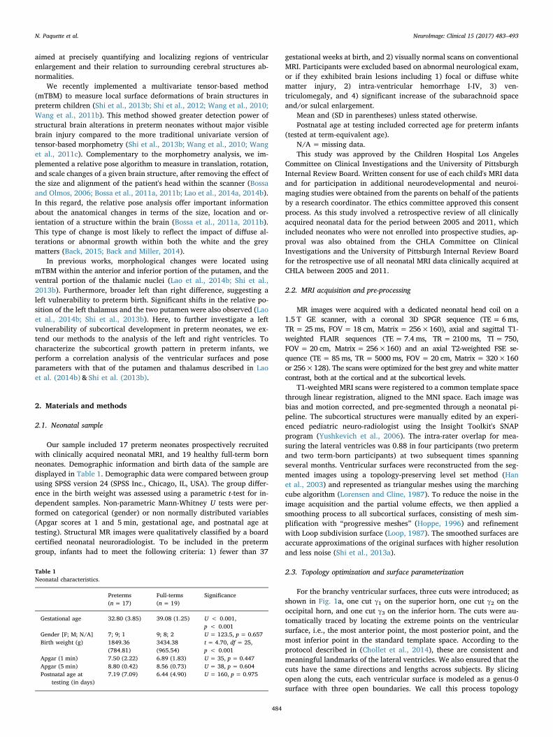
For the branchy ventricular surfaces, three cuts were introduced; asshown in Fig. 1a, one cut γ1 on the superior horn, one cut γ2 on theoccipital horn, and one cut γ3 on the inferior horn. The cuts were au-tomatically traced by locating the extreme points on the ventricularsurface, i.e., the most anterior point, the most posterior point, and themost inferior point in the standard template space. According to theprotocol described in (Chollet et al., 2014), these are consistent andmeaningful landmarks of the lateral ventricles. We also ensured that thecuts have the same directions and lengths across subjects. By slicingopen along the cuts, each ventricular surface is modeled as a genus-0surface with three open boundaries. We call this process topology
Table 1Neonatal characteristics.
Preterms(n = 17)
Full-terms(n = 19)
Significance
Gestational age 32.80 (3.85) 39.08 (1.25) U < 0.001,p < 0.001
Gender [F; M; N/A] 7; 9; 1 9; 8; 2 U = 123.5, p = 0.657Birth weight (g) 1849.36
(784.81)3434.38(965.54)
t = 4.70, df = 25,p < 0.001
Apgar (1 min) 7.50 (2.22) 6.89 (1.83) U = 35, p = 0.447Apgar (5 min) 8.80 (0.42) 8.56 (0.73) U = 38, p = 0.604Postnatal age at
testing (in days)7.19 (7.09) 6.44 (4.90) U = 160, p = 0.975
N. Paquette et al. NeuroImage: Clinical 15 (2017) 483–493
484
optimization (Shi et al., 2015; Shi et al., 2013a; Wang et al., 2010;Wang et al., 2011c).
For the surface registration, we computed a conformal para-meterization using the Hyperbolic Ricci Flow method (Shi et al., 2015).The ventricular surfaces resulting from topology optimization admithyperbolic geometry and their conformal parameterizations can be ef-ficiently computed with this method (Hamilton, 1988; Shi et al., 2015;Zeng et al., 2010). It is a general and powerful tool to compute surfaceconformal parameterization based on the correspondence between thesurface Riemannian metric and the Gaussian curvature i.e., when thesurface metric changes, the Gaussian curvature will change accord-ingly. The hyperbolic Ricci flow allows to compute conformal para-meterizations for general topological surfaces (i.e., high-genus surfacesor genus-0 surfaces with more the two open boundaries) with minimalangle distortions and no singular points. With the conformal para-meterization, a 3D ventricular surface can be embedded onto the 2DPoincaré disk. For embedding, each ventricular surface is sliced openalong the three cuts and two paths τ1 and τ2, which connect γ1 and γ2, γ1and γ3, respectively, as shown in Fig. 1b. The Poincaré disk embeddingof a ventricular surface is illustrated in Fig. 1c. For details about hy-perbolic Ricci flow and Poincaré disk embedding, please refer to (Shiet al., 2015).
2.4. Ventricular surface registration
As shown in Fig. 1c, the three cuts become geodesics (hyperboliclines) in the Poincaré disk. However, the two paths, where each splitsinto two identical segments τi and τi
−1 , i=1,2, are not geodesics andnot consistent across subjects. For ventricular surface registration, thelocations of the paths need to be determined. In the Poincaré disk, weuse the Fuchsian group generators (Shi et al., 2015) to tile a finiteportion of the universal covering space, i.e. the whole Poincaré disk, ofeach ventricular surface, as shown in Fig. 1d. In the universal coveringspace, we recompute the locations of the paths as unique and consistentgeodesic curves. By slicing the universal covering space along the newpaths, we obtain the canonical fundamental domain of a ventricularsurface, as shown in Fig. 1e. These new geodesics, when lifted to ori-ginal 3D ventricular surfaces, are also consistent (Fig. 1g) and can beused as boundary conditions of the surface registration. Finally, weconvert the canonical fundamental domain to the Klein model, asshown in Fig. 1f. The Klein model is used as the canonical parameterdomain to register each ventricular surface to a common template.Please refer to (Shi et al., 2015) for details of the ventricular surfaceregistration algorithm.
Fig. 1. Computation steps in the proposed ventricular morphometry registration.
N. Paquette et al. NeuroImage: Clinical 15 (2017) 483–493
485
2.5. Multivariate tensor-based morphometry
We used multivariate tensor-based morphometry (mTBM) (Leporeet al., 2008; Wang et al., 2008) to measure ventricular surface de-formations that occur in the registration process. First, the Jacobianmatrix (J) is computed at each vertex from the transformation betweenthe population-based template and the subject's shape. The logarithm ofthe deformation tensor matrix log √JJT is used to measure local dif-ferences on a brain structure morphology with respect to the populationshape (Arsigny et al., 2006; Lepore et al., 2008; Wang et al., 2010).Multivariate statistics on the deformation tensor in the Euclidean spacehave been shown to improve group effect sizes, thus increasing statis-tical power (Shi et al., 2013b; Wang et al., 2010; Wang et al., 2011c).Furthermore, we applied the heat kernel smoothing algorithms (Chunget al., 2005) to increase the signal-to-noise-ratio (SNR) and improve thesensitivity of the statistical analysis. As in (Shi et al., 2013b), we cov-aried the smoothed mTBM measure to correct for post-conception ageeffects, by applying a general linear model.
Group differences on mTBM were computed vertex-wise using themultivariate Hotelling's T2 test, which is a multivariate generalizationof the t-test (Hotelling, 1931). Permutation tests (10, 000) were used tocorrect for multiple comparisons (Nichols and Holmes, 2002). We usedthe group comparison p-map to visualize the shape morphometry pat-terns between the two groups. These p-values are computed by com-paring the data to a permutation distribution in order to eliminate theassumption of normal distribution. The whole structure-based permu-tation corrected p-value was used to measure the overall significance ofthe p-map at the 0.05 alpha level. Furthermore, to see the direction ofchanges between groups, we mapped the detJ matrix at each vertex k ina ratio map according to the following formula:
=∑
∑R
det J
det JNN
k jN
ik
jN
jk
11
22
2
1 (1)
where J1i k and J2j kare the Jacobian matrices for ith subject in onegroup and jth in the other group, and N1 and N2 are the number ofsubjects in each groups (Shi et al., 2013a; Wang et al., 2011b). Thismatrix indicates the regional difference in surface area in the individualsubject with respect to the common template where the two groups ofsubjects are registered. Rk values smaller than 1 indicate atrophicchanges at a given vertex in preterm group, while values greater than 1indicates enlargement at a particular vertex.
2.6. Relative pose analysis
To compute the relative position of each individual's lateral ven-tricles, we used a Procrustes alignment of each subject's structure inrelation with a population-based template (Bossa and Olmos, 2006;Bossa et al., 2011a; Dryden and Mardia, 1998). The population-basedtemplate was calculated iteratively and then align to the subject's shape(Bossa et al., 2011a; Lao et al., 2014a, 2014b). For each structure, wecomputed a transformation matrix corresponding to the rotation,translation and scaling. The transformation matrix can be written as:
= ⎡⎣
⎤⎦
T SRXT
d1 (2)
where S is the scalar scaling factor, R is a 3 × 3 rotation matrix, and d isthe translation vector (x, y, z)T (Bossa et al., 2011a). A schematic re-presentation of the Procrustes transform applied to the left and rightlateral ventricles is shown in Fig. 2. 13 sets of parameters were com-puted for each structure. Univariate parameters (9) comprised: logS,||logR||, ||logd||, respectively representing the total scale, total rota-tion and total translation of each structure; θx,θy,θz, representing the 3rotation parameters; and x, y, and z, representing the 3 translationparameters. Multivariate parameters (4) included: (θx, θy, θz); (x, y, z);(logS, ||logR||, ||logd||); and a combination of 7 parameters (1 scale, 3
rotations, 3 translations). For statistical computation, all transforma-tions were centered on the center mass of the template, and projectedonto the tangent plane at the origin of the manifold of transformations(Arsigny et al., 2006; Bossa and Olmos, 2006). As with the mTBManalyses, group analyses on the relative pose parameters were per-formed on post-conception age-covaried data using linear regression.
Between-group statistical comparisons were performed structure-wise on all 9 sets of univariate parameters (logS, ||logR||, ||logd||, θx,θy, θz, x, y, z) using univariate t-test. For the multivariate parameters((θx, θy, θz), (x, y, z), (logS, ||logR||, ||logd||), combination of 7parameters), the multivariate Hotelling's T2 test was used. For each test,10,000 permutations were performed to avoid the normal distributionassumption (Nichols and Holmes, 2002).
2.7. Surface and pose correlations
The structural relationship between the bilateral ventricles, theputamen and the thalamus described and discussed in Lao et al. (2014a,2014b) and Shi et al. (2013b) were further investigated using Pearson'scorrelation analysis for both surface morphometry deformation andrelative pose measures. For the surface deformation parameters, Pear-son's correlations were performed between the determinant (surfacearea) of each vertex of the ventricles (LVent, RVent) and the total vo-lume of the putamen (LPut, RPut) and the thalamus (LThal, RThal). Wealso ran two permutation tests on the surface-based correlations: avertex-based one that allowed us to avoid normal distribution as-sumptions, and one over the total segmented images to correct formultiple comparisons (Lepore et al., 2008; Nichols and Holmes, 2002;Wang et al., 2011c). Similarly, to test for the relationship in terms ofrelative position between the two ventricles, and the two putamen andthalami, we computed Pearson's correlations on the pose parameterslogS, ||logR||, and ||logd|| for each pair (LVent vs. LThal; LVent vs.LPuta; RVent vs. RThal; RVent vs. RPuta). For each correlation set,10,000 permutations were computed to avoid the normal distributionassumption.
3. Results
3.1. Sociodemographic characteristics
Groups differences on demographic and birth data are displayed inTable 1. A t-test calculated on the birth weight and nonparametricMann-Whitney U tests calculated on the gestational age revealed sig-nificant differences between preterm and full-term neonates (GA andBirth weight: p < 0.001), as expected. However, Mann–Whitney Utests revealed that the two groups were equivalent in terms of gender(p = 0.657), Apgar scores at 1 min (p= 0.447) and at 5 min(p = 0.604), and postnatal age at scan time (p= 0.975).
3.2. Surface morphometry results
Fig. 3a shows the results of the mTBM analysis on the ventricularsurface morphometry of premature and full-term neonates. The p-valuemap shows significant group differences in the shape of the lateralventricles with p = 0.0005, corrected for multiple comparisons. Fig. 3bshow the direction of changes with values smaller than 1 indicatingatrophic changes and values greater than 1 indicating enlargement inthe preterm group. Local comparison analysis revealed broad areas ofdifference (at pt. = 0.05 level) where preterm neonates show significantventricular enlargement. These regions are located primarily within thefrontal horn and the body of the left ventricle, and more restricted, butsignificant, areas of differences on the temporal horn and body of theright ventricle.
N. Paquette et al. NeuroImage: Clinical 15 (2017) 483–493
486

3.3. Relative position results
Results from the relative pose analysis (rotation, translation andscale) for all parameters are displayed in Table 2, and illustrated inFig. 4. Significant differences between preterm and full-term neonateswere found on the rotation parameter of the left ventricle, for bothunivariate (p = 0. 0165) and multivariate measures (p= 0.0499). Forthe right ventricle, only marginal differences were found on both therotation (p = 0.0542) and translation (p = 0.0634) measures.
3.4. Correlations results
To further investigate the relationship between ventricular shapeand pose, and the surrounding structures, we tested the surface mor-phometry and relative pose correlations between the ventricle and re-sults previously found on the thalamus (Lao et al., 2014b) and putamen
(Shi et al., 2013b). All p-values are computed using 10,000 permuta-tions to correct for multiple comparisons, at 0.05 alpha level of sig-nificance.
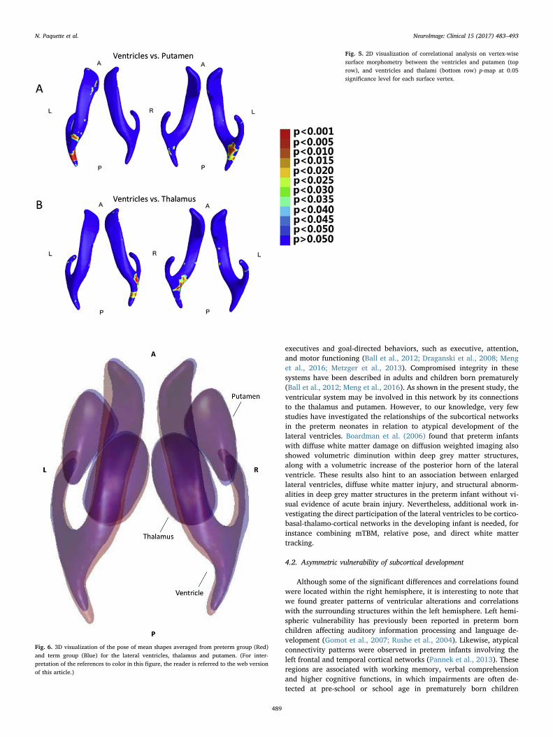
3.4.1. Surface morphometry correlationsFig. 5 shows the p-map of the correlational analysis on the surface
morphometry of the ventricles and the surface of the putamen andthalamus (see Lao et al., 2014b and Shi et al., 2013b respectively, foradditional details on the putamen and thalamus between-group ana-lysis). The correlations on the shape of the bilateral ventricles and thatof the bilateral thalamus and putamen were all nonsignificant (ventriclevs thalamus: p = 0.3320; ventricle vs putamen: p = 0.3866). Likewise,correlation analysis between these structures when tested within theleft or the right hemispheres failed to reach significance. Corrected p-value after permutation tests were: (a) LVent vs. LThal: p= 0.8043; (b)LVent vs. LPuta: p= 0.3866: (c) RVent vs. RThal: p= 0.1357: (d)
Fig. 2. Relative pose analysis pipeline. Individual shapes (A) are first Procrustes aligned to an average template shape (B), which results in a set of 7 parameters for each of subjects (C).Group analysis results (D) are then obtained through univariate and multivariate tests.
Fig. 3. The top row (A) show Statistical p-map representation ofgroup differences detected between the preterm and term-borngroups, using the smoothed and covaried mTBM (0.05 significancelevel for each surface vertex). The non-blue colors illustrate areaswith statistically significant differences between the two groups.The bottom row (B) shows the direction of changes betweengroups. Values smaller than 1 (in blue) indicates atrophic changesat a given vertex in preterm group, while values greater than 1 (inred) indicate enlargement in the preterm group at the givenvertex. (For interpretation of the references to color in this figurelegend, the reader is referred to the web version of this article.)
N. Paquette et al. NeuroImage: Clinical 15 (2017) 483–493
487

RVent vs. RPuta: p = 0.7296.
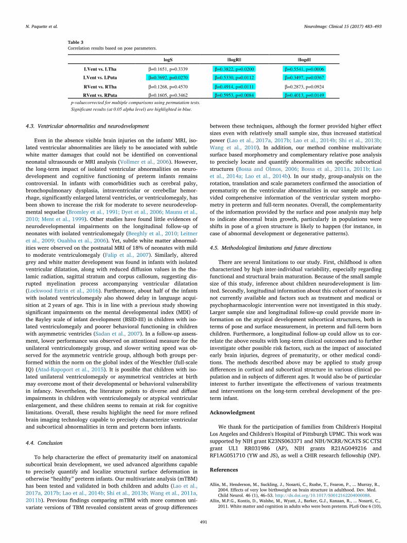
3.4.2. Pose correlationsFig. 6 shows the 3D representation of the relative position for the
lateral ventricles, putamen and thalamus for the preterm (in red) andfull-term (in blue) neonates, along with areas of shape overlappingbetween the two groups (in purple). Fig. 7 shows the correlation plotsfor the logS, ||logR||, and ||logd|| pose parameters between eachstructure (LVent vs. LTha, LVent vs. LPuta, RVent vs. RTha, and RVentvs. RPuta). The correlation coefficient and corrected p-values are dis-played in Table 3 for each of the pose parameter tested (total scale, totalrotation, total translation). Overall, significant correlations were foundbetween each structure in terms of rotation (||logR||). Significant cor-relations were also found on the translation parameter (||logd||) be-tween the left ventricle, and the left putamen and left thalamus, alongwith a significant correlation between the right ventricle and rightputamen. Significant correlations on the scale parameter (logS) werefound only between the left ventricle and left putamen.
4. Discussion
Here, we examined the morphological features of the lateral ven-tricles in a population of preterm and full-term born neonates. We
applied a novel pipeline combining surface deformation and poseanalysis to precisely characterize and localize ventricular abnormalitiesin these infants. Significant areas of differences were detected on thefrontal horn and the body of the left ventricle, along with narrowerdifferences on the temporal horn of the right ventricle. These results areconsistent with prior works on the thalamus and putamen, showingchanges in shape and pose located primarily within the left hemisphere(Lao et al., 2014b; Shi et al., 2013b). In addition, we found a significantshift in terms of rotation of the left ventricle in preterm neonates, with atrend for the right ventricle. This is also in line with previous findingsshowing significant pose alterations in the left thalamus and the rightputamen, and trends in the left putamen (Lao et al., 2014b). Com-plementary to these results, we examined the relationship betweenchanges in surface morphometry and pose parameters of the lateralventricles, and those of the putamen and thalamus described pre-viously. We found significant correlations between each of thesestructures in terms of rotation. We also measured significant correla-tions on the translation parameter between the left ventricle, and theleft putamen and left thalamus, as well as between the right ventricleand right putamen. Significant correlation on the scale parameter wasalso found between the left ventricle and left putamen. Overall, theseresults further corroborate our previous works suggesting a left sub-cortical vulnerability to preterm birth. These results also show the in-terdependence between the ventricles and its surrounding structures aswell as an atypical organization of the subcortical network in oursample of relatively healthy preterm neonates.
4.1. Impact of prematurity on subcortical organization
Here, pose abnormalities found in the lateral ventricles correlatedsignificantly with those found in thalamus (Shi et al., 2013b) and pu-tamen (Lao et al., 2014b). The ventricular pose differences in the pre-sent study and its relation to the pose differences in the thalamus andputamen, especially in terms of rotation and translation of the struc-tures, suggest altered growth of the white matter tracts surroundingthese structures. It is possible that altered growth, or dysmaturation, ofthe white matter is also found in synergy to impaired growth of the greymatter (Back, 2015; Back and Miller, 2014). The cerebral white andgrey matter dysmaturation in preterm infants has been proposed toaccount for myelination and neural disturbance related to pretermbirth, impacting the pre-oligodendrocytes (myelin promoting cells) andimmature neurons development (Back, 2015; Back and Miller, 2014).This is further supported by our results showing group differences andstructure inter-correlation in scale, or general volume, and shape sur-face, suggesting impaired neural development of the deep grey matterstructures. These results need to be further validated in future workusing diffusion based tractography analyses.
The effect of pose and surface changes of subcortical structureswithin the thalamo-cortical network in preterm infants have beenpreviously proposed and discussed (Lao et al., 2014b; Shi et al., 2013b).The cortico-basal-thalamo-cortical networks is involved in a number of
Table 2Results of the relative pose analysis.
Pose Parameters LVent RVent Pose parameters (Cont) LVent RVent
logS 0.3979 0.8591 ||logR|| 0.5312 0.1421
θx 0.0606 0.0542 ||logd|| 0.5073 0.4515
θy 0.1395 0.5210 (θx,θy,θz) 0.0499 0.2123
θz 0.0165 0.4299 (x, y, z) 0.7387 0.1650
x 0.4644 0.0634 (logS, ||logR||, ||logd||) 0.784 0.5260
y 0.9694 0.3414 All7parameters 0.2405 0.1389
z 0.5519 0.9378
p-valuecorrected formultiple comparisons using permutation tests.
Significant results (at 0.05 alpha level) are highlighted in blue; marginal differences arehighlighted in light grey.
Fig. 4. 2D visualization of mean shapes averaged from preterm group (Red) and termgroup (Blue) for the left (LVent) and right (RVent) ventricle. Areas where the mean shapesof two groups overlaid appear in purple. (For interpretation of the references to color inthis figure legend, the reader is referred to the web version of this article.)
N. Paquette et al. NeuroImage: Clinical 15 (2017) 483–493
488
executives and goal-directed behaviors, such as executive, attention,and motor functioning (Ball et al., 2012; Draganski et al., 2008; Menget al., 2016; Metzger et al., 2013). Compromised integrity in thesesystems have been described in adults and children born prematurely(Ball et al., 2012; Meng et al., 2016). As shown in the present study, theventricular system may be involved in this network by its connectionsto the thalamus and putamen. However, to our knowledge, very fewstudies have investigated the relationships of the subcortical networksin the preterm neonates in relation to atypical development of thelateral ventricles. Boardman et al. (2006) found that preterm infantswith diffuse white matter damage on diffusion weighted imaging alsoshowed volumetric diminution within deep grey matter structures,along with a volumetric increase of the posterior horn of the lateralventricle. These results also hint to an association between enlargedlateral ventricles, diffuse white matter injury, and structural abnorm-alities in deep grey matter structures in the preterm infant without vi-sual evidence of acute brain injury. Nevertheless, additional work in-vestigating the direct participation of the lateral ventricles to be cortico-basal-thalamo-cortical networks in the developing infant is needed, forinstance combining mTBM, relative pose, and direct white mattertracking.
4.2. Asymmetric vulnerability of subcortical development
Although some of the significant differences and correlations foundwere located within the right hemisphere, it is interesting to note thatwe found greater patterns of ventricular alterations and correlationswith the surrounding structures within the left hemisphere. Left hemi-spheric vulnerability has previously been reported in preterm bornchildren affecting auditory information processing and language de-velopment (Gomot et al., 2007; Rushe et al., 2004). Likewise, atypicalconnectivity patterns were observed in preterm infants involving theleft frontal and temporal cortical networks (Pannek et al., 2013). Theseregions are associated with working memory, verbal comprehensionand higher cognitive functions, in which impairments are often de-tected at pre-school or school age in prematurely born children
Fig. 5. 2D visualization of correlational analysis on vertex-wisesurface morphometry between the ventricles and putamen (toprow), and ventricles and thalami (bottom row) p-map at 0.05significance level for each surface vertex.
Fig. 6. 3D visualization of the pose of mean shapes averaged from preterm group (Red)and term group (Blue) for the lateral ventricles, thalamus and putamen. (For inter-pretation of the references to color in this figure, the reader is referred to the web versionof this article.)
N. Paquette et al. NeuroImage: Clinical 15 (2017) 483–493
489

(Anderson et al., 2011; Arpino et al., 2010; Sansavini et al., 2010).Recent evidences also suggest that left hemisphere myelination is morerapid than the right in many brain regions during early the perinatalperiod (Deoni et al., 2011). Region within the left hemisphere showingearlier onset of myelin development also appears to have slower sub-sequent myelination process than regions showing later myelinationonset in the right hemisphere (Deoni et al., 2011). It is possible that
preterm infants are more at risk of oligodendrocyte and myelinationdisturbances within the left hemisphere following perinatal injuries.Yet, the hemispheric vulnerability of subcortical network remainspoorly investigated. Longitudinal and birth cohort studies that includescomprehensive neuroimaging, white matter tracking, neuropsycholo-gical, and behavioral assessments are needed to better understand anddocument the etiology and outcomes of this vulnerability.
Fig. 7. Correlation plots between the ventricles and thalamus (A and C), and the ventricles and putamen (B and D) in the left (A and B) and right (C and D) hemispheres, tested using poseparameters: logS (left), ||logR|| (middle), ||logd|| (right).
N. Paquette et al. NeuroImage: Clinical 15 (2017) 483–493
490
4.3. Ventricular abnormalities and neurodevelopment
Even in the absence visible brain injuries on the infants' MRI, iso-lated ventricular abnormalities are likely to be associated with subtlewhite matter damages that could not be identified on conventionalneonatal ultrasounds or MRI analysis (Vollmer et al., 2006). However,the long-term impact of isolated ventricular abnormalities on neuro-development and cognitive functioning of preterm infants remainscontroversial. In infants with comorbidities such as cerebral palsy,bronchopulmonary dysplasia, intraventricular or cerebellar hemor-rhage, significantly enlarged lateral ventricles, or ventriculomegaly, hasbeen shown to increase the risk for moderate to severe neurodevelop-mental sequelae (Bromley et al., 1991; Dyet et al., 2006; Maunu et al.,2010; Ment et al., 1999). Other studies have found little evidences ofneurodevelopmental impairments on the longitudinal follow-up ofneonates with isolated ventriculomegaly (Beeghly et al., 2010; Leitneret al., 2009; Ouahba et al., 2006). Yet, subtle white matter abnormal-ities were observed on the postnatal MRI of 18% of neonates with mildto moderate ventriculomegaly (Falip et al., 2007). Similarly, alteredgrey and white matter development was found in infants with isolatedventricular dilatation, along with reduced diffusion values in the tha-lamic radiation, sagittal stratum and corpus callosum, suggesting dis-rupted myelination process accompanying ventricular dilatation(Lockwood Estrin et al., 2016). Furthermore, about half of the infantswith isolated ventriculomegaly also showed delay in language acqui-sition at 2 years of age. This is in line with a previous study showingsignificant impairments on the mental developmental index (MDI) ofthe Bayley scale of infant development (BSID-III) in children with iso-lated ventriculomegaly and poorer behavioral functioning in childrenwith asymmetric ventricles (Sadan et al., 2007). In a follow-up assess-ment, lower performance was observed on attentional measure for theunilateral ventriculomegaly group, and slower writing speed was ob-served for the asymmetric ventricle group, although both groups per-formed within the norm on the global index of the Weschler (full-scaleIQ) (Atad-Rapoport et al., 2015). It is possible that children with iso-lated unilateral ventriculomegaly or asymmetrical ventricles at birthmay overcome most of their developmental or behavioral vulnerabilityin infancy. Nevertheless, the literature points to diverse and diffuseimpairments in children with ventriculomegaly or atypical ventricularenlargement, and these children seems to remain at risk for cognitivelimitations. Overall, these results highlight the need for more refinedbrain imaging technology capable to precisely characterize ventricularand subcortical abnormalities in term and preterm born infants.
4.4. Conclusion
To help characterize the effect of prematurity itself on anatomicalsubcortical brain development, we used advanced algorithms capableto precisely quantify and localize structural surface deformation inotherwise “healthy” preterm infants. Our multivariate analysis (mTBM)has been tested and validated in both children and adults (Lao et al.,2017a, 2017b; Lao et al., 2014b; Shi et al., 2013b; Wang et al., 2011a,2011b). Previous findings comparing mTBM with more common uni-variate versions of TBM revealed consistent areas of group differences
between these techniques, although the former provided higher effectsizes even with relatively small sample size, thus increased statisticalpower (Lao et al., 2017a, 2017b; Lao et al., 2014b; Shi et al., 2013b;Wang et al., 2010). In addition, our method combine multivariatesurface based morphometry and complementary relative pose analysisto precisely locate and quantify abnormalities on specific subcorticalstructures (Bossa and Olmos, 2006; Bossa et al., 2011a, 2011b; Laoet al., 2014a; Lao et al., 2014b). In our study, group-analysis on therotation, translation and scale parameters confirmed the association ofprematurity on the ventricular abnormalities in our sample and pro-vided comprehensive information of the ventricular system morpho-metry in preterm and full-term neonates. Overall, the complementarityof the information provided by the surface and pose analysis may helpto indicate abnormal brain growth, particularly in populations wereshifts in pose of a given structure is likely to happen (for instance, incase of abnormal development or degenerative patterns).
4.5. Methodological limitations and future directions
There are several limitations to our study. First, childhood is oftencharacterized by high inter-individual variability, especially regardingfunctional and structural brain maturation. Because of the small samplesize of this study, inference about children neurodevelopment is lim-ited. Secondly, longitudinal information about this cohort of neonates isnot currently available and factors such as treatment and medical orpsychopharmacologic intervention were not investigated in this study.Larger sample size and longitudinal follow-up could provide more in-formation on the atypical development subcortical structures, both interms of pose and surface measurement, in preterm and full-term bornchildren. Furthermore, a longitudinal follow-up could allow us to cor-relate the above results with long-term clinical outcomes and to furtherinvestigate other possible risk factors, such as the impact of associatedearly brain injuries, degrees of prematurity, or other medical condi-tions. The methods described above may be applied to study groupdifferences in cortical and subcortical structure in various clinical po-pulation and in subjects of different ages. It would also be of particularinterest to further investigate the effectiveness of various treatmentsand interventions on the long-term cerebral development of the pre-term infant.
Acknowledgment
We thank for the participation of families from Children's HospitalLos Angeles and Children's Hospital of Pittsburgh UPMC. This work wassupported by NIH grant K23NS063371 and NIH/NCRR/NCATS SC CTSIgrant UL1 RR031986 (AP), NIH grants R21AG049216 andRF1AG051710 (YW and JS), as well a CHIR research fellowship (NP).
References
Allin, M., Henderson, M., Suckling, J., Nosarti, C., Rushe, T., Fearon, P., ... Murray, R.,2004. Effects of very low birthweight on brain structure in adulthood. Dev. Med.Child Neurol. 46 (1), 46–53. http://dx.doi.org/10.1017/S0012162204000088.
Allin, M.P.G., Kontis, D., Walshe, M., Wyatt, J., Barker, G.J., Kanaan, R., ... Nosarti, C.,2011. White matter and cognition in adults who were born preterm. PLoS One 6 (10),
Table 3Correlation results based on pose parameters.
logS ||logR|| ||logd||
LVent vs. LTha β=0.1651, p=0.3339 β=0.3822, p=0.0200 β=0.5541, p=0.0006
LVent vs. LPuta β=0.3692, p=0.0270 β=0.5330, p=0.0112 β=0.3497, p=0.0367
RVent vs. RTha β=0.1268, p=0.4570 β=0.4914, p=0.0111 β=0.2873, p=0.0924
RVent vs. RPuta β=0.1605, p=0.3462 β=0.5953, p=0.0084 β=0.4013, p=0.0149
p-valuecorrected for multiple comparisons using permutation tests.
Significant results (at 0.05 alpha level) are highlighted in blue.
N. Paquette et al. NeuroImage: Clinical 15 (2017) 483–493
491

e24525. http://dx.doi.org/10.1371/journal.pone.0024525.Anderson, P.J., De Luca, C.R., Hutchinson, E., Spencer-Smith, M.M., Roberts, G., Doyle,
L.W., 2011. Attention problems in a representative sample of extremely preterm/extremely low birth weight children. Dev. Neuropsychol. 36 (1), 57–73. http://dx.doi.org/10.1080/87565641.2011.540538.
Arpino, C., Compagnone, E., Montanaro, M.L., Cacciatore, D., Luca, A. De, Cerulli, A., ...Curatolo, P., 2010. Preterm birth and neurodevelopmental outcome: a review. ChildsNerv. Syst. http://dx.doi.org/10.1007/s00381-010-1125-y.
Arsigny, V., Fillard, P., Pennec, X., Ayache, N., 2006. Log-Euclidean metrics for fast andsimple calculus on diffusion tensors. Magn. Reson. Med. 56 (2), 411–421. http://dx.doi.org/10.1002/mrm.20965.
Atad-Rapoport, M., Schweiger, A., Lev, D., Sadan-Strul, S., Malinger, G., Lerman-Sagie, T.,2015. Neuropsychological follow-up at school age of children with asymmetricventricles or unilateral ventriculomegaly identified in utero. BJOG 122, 932–938. AnInternational Journal of Obstetrics and Gynaecology. http://dx.doi.org/10.1111/1471-0528.13312.
Back, S.A., 2015. Brain injury in the preterm infant: new horizons for pathogenesis andprevention. Pediatr. Neurol. 53 (3), 185–192. http://dx.doi.org/10.1016/j.pediatrneurol.2015.04.006. (Brain).
Back, S.A., Miller, S.P., 2014. Brain injury in premature neonates: A primary cerebraldysmaturation disorder? Ann. Neurol. http://dx.doi.org/10.1002/ana.24132.
Ball, G., Boardman, J.P., Rueckert, D., Aljabar, P., Arichi, T., Merchant, N., ... Counsell,S.J., 2012. The effect of preterm birth on thalamic and cortical development. Cereb.Cortex 22 (5), 1016–1024. http://dx.doi.org/10.1093/cercor/bhr176.
Beeghly, M., Ware, J., Soul, J., Du Plessis, A., Khwaja, O., Senapati, G.M., ... Levine, D.,2010. Neurodevelopmental outcome of fetuses referred for ventriculomegaly.Ultrasound Obstet. Gynecol. 35 (4), 405–416. http://dx.doi.org/10.1002/uog.7554.
Boardman, J.P., Counsell, S.J., Rueckert, D., Kapellou, O., Bhatia, K.K., Aljabar, P., ...Edwardsa, A.D., 2006. Abnormal deep grey matter development following pretermbirth detected using deformation-based morphometry. NeuroImage 32 (1), 70–78.http://dx.doi.org/10.1016/j.neuroimage.2006.03.029.
Boardman, J.P., Craven, C., Valappil, S., Counsell, S.J., Dyet, L.E., Rueckert, D., ...Edwards, a D., 2010. A common neonatal image phenotype predicts adverse neuro-developmental outcome in children born preterm. NeuroImage 52 (2), 409–414.http://dx.doi.org/10.1016/j.neuroimage.2010.04.261.
Bossa, M.N., Olmos, S., 2006. Statistical model of similarity transformations: building amulti-object pose model of brain structures. In: Proceedings of the IEEE ComputerSociety Conference on Computer Vision and Pattern Recognition. Vol. 2006http://dx.doi.org/10.1109/CVPRW.2006.198.
Bossa, M., Zacur, E., Olmos, S., 2011a. Statistical analysis of relative pose information ofsubcortical nuclei: Application on ADNI data. NeuroImage 55 (3), 999–1008. http://dx.doi.org/10.1016/j.neuroimage.2010.12.078.
Bossa, M., Zacur, E., Olmos, S., The Alzheimer's Disease Neuroimaging Initiative, 2011b.Statistical analysis of relative pose information of subcortical nuclei: Application onADNI data. NeuroImage 55 (3), 999–1008. http://dx.doi.org/10.1016/j.neuroimage.2010.12.078.Statistical.
Bromley, B., Frigoletto, F.D., Benacerraf, B.R., 1991. Mild fetal lateral cerebral ven-triculomegaly: clinical course and outcome. Am. J. Obstet. Gynecol. 164 (3),863–867. http://dx.doi.org/10.1016/0002-9378(91)90530-5.
Chollet, M.B., Aldridge, K., Pangborn, N., Weinberg, S.M., DeLeon, V.B., 2014.Landmarking the brain for geometric morphometric analysis: an error study. PLoSOne 9 (1). http://dx.doi.org/10.1371/journal.pone.0086005.
Chung, M.K., Robbins, S.M., Dalton, K.M., Davidson, R.J., Alexander, A.L., Evans, A.C.,2005. Cortical thickness analysis in autism with heat kernel smoothing. NeuroImage25 (4), 1256–1265. http://dx.doi.org/10.1016/j.neuroimage.2004.12.052.
Counsell, S.J., Edwards, A.D., Chew, A.T.M., Anjari, M., Dyet, L.E., Srinivasan, L., ...Cowan, F.M., 2008. Specific relations between neurodevelopmental abilities andwhite matter microstructure in children born preterm. Brain 131, 3201–3208. AJournal of Neurology. http://dx.doi.org/10.1093/brain/awn268.
Deoni, S.C.L., Mercure, E., Blasi, A., Gasston, D., Thomson, A., Johnson, M., ... Murphy,D.G.M., 2011. Mapping infant brain myelination with magnetic resonance imaging. J.Neurosci. 31 (2), 784–791. http://dx.doi.org/10.1523/JNEUROSCI.2106-10.2011.
Draganski, B., Kherif, F., Kloppel, S., Cook, P.A., Alexander, D.C., Parker, G.J., ...Frackowiak, R.S., 2008. Evidence for segregated and integrative connectivity patternsin the human Basal Ganglia. J. Neurosci. 28 (28), 7143–7152. http://dx.doi.org/10.1523/JNEUROSCI.1486-08.2008.
Dryden, I.L., Mardia, K.V., 1998. Statistical shape analysis. J. Hum. Evol. 4 (3), 376.http://dx.doi.org/10.1006/jhev.1999.0391.
Dyet, L.E., Kennea, N., Counsell, S.J., Maalouf, E.F., Ajayi-Obe, M., Duggan, P.J., ...Edwards, A.D., 2006. Natural history of brain lesions in extremely preterm infantsstudied with serial magnetic resonance imaging from birth and neurodevelopmentalassessment. Pediatrics 118 (2), 536–548. http://dx.doi.org/10.1542/peds.2005-1866.
Falip, C., Blanc, N., Maes, E., Zaccaria, I., Oury, J.F., Sebag, G., Garel, C., 2007. Postnatalclinical and imaging follow-up of infants with prenatal isolated mild ven-triculomegaly: a series of 101 cases. Pediatr. Radiol. 37 (10), 981–989. http://dx.doi.org/10.1007/s00247-007-0582-2.
Gomot, M., Bruneau, N., Laurent, J., Barthélémy, C., Saliba, E., 2007. Left temporal im-pairment of auditory information processing in prematurely born 9-year-old children:an electrophysiological study. Int. J. Psychophysiol. 64, 123–129. http://dx.doi.org/10.1016/j.ijpsycho.2007.01.003.
Hamilton, R.S., 1988. The Ricci flow on surfaces. Contemp. Math. 71, 237–262.Han, X., Xu, C., Prince, J.L., 2003. A topology preserving level set method for geometric
deformable models. IEEE Trans. Pattern Anal. Mach. Intell. 25 (6), 755–768. http://dx.doi.org/10.1109/TPAMI.2003.1201824.
Hoppe, H., 1996. Progressive meshes. In: Proceedings of the 23rd Annual Conference on
Computer Graphics and Interactive Techniques - SIGGRAPH ‘96, pp. 99–108. http://dx.doi.org/10.1145/237170.237216.
Hotelling, H., 1931. The generalization of Student's ratio. Ann. Math. Stat. 2, 360–378.Isaacs, E.B., Lucas, A., Chong, W.K., Wood, S.J., Johnson, C.L., Marshall, C., ... Gadian,
D.G., 2000. Hippocampal volume and everyday memory in children of very low birthweight. Pediatr. Res. 47, 713–720.
Kesler, S.R., Ment, L.R., Vohr, B., Pajot, S.K., Schneider, K.C., Katz, K.H., ... Reiss, A.L.,2004. Volumetric analysis of regional cerebral development in preterm children.Pediatr. Neurol. 31 (5), 318–325. http://dx.doi.org/10.1016/j.pediatrneurol.2004.06.008.
Lao, Y., Dion, L.-A., Gilbert, G., Bouchard, M.F., Rocha, G., Wang, Y., ... Saint-Amour, D.,2017a. Mapping the basal ganglia alterations in children chronically exposed tomanganese. Sci. Rep. 7 (February), 41,804. http://dx.doi.org/10.1038/srep41804.
Lao, Y., Nguyen, B., Tsao, S., Gajawelli, N., Law, M., Chui, H., ... Leporé, N., 2017b. A T1and DTI fused 3D corpus callosum analysis in MCI subjects with high and low car-diovascular risk profile. NeuroImage 14, 298–307 Clinical.
Lao, Y., Shi, J., Wang, Y., Ceschin, R., Hwang, D., Nelson, M.D., ... Leporé, N., 2014a.Statistical analysis of relative pose of the thalamus in preterm neonates. In: 2ndInternational Workshop on Clinical Image-Based Procedures: Translational Researchin Medical Imaging, CLIP 2013 - Held in Conjunction with MICCAI 2013, pp. 1–9.http://dx.doi.org/10.1007/978-3-319-05666-1_1.
Lao, Y., Wang, Y., Shi, J., Ceschin, R., Nelson, M.D., Panigrahy, A., Leporé, N., 2014b.Thalamic alterations in preterm neonates and their relation to ventral striatum dis-turbances revealed by a combined shape and pose analysis. Brain Struct. Funct.http://dx.doi.org/10.1007/s00429-014-0921-7.
Leitner, Y., Stolar, O., Rotstein, M., Toledano, H., Harel, S., Bitchonsky, O., ... Ben-Sira, L.,2009. The neurocognitive outcome of mild isolated fetal ventriculomegaly verified byprenatal magnetic resonance imaging. Am. J. Obstet. Gynecol. 201 (2). http://dx.doi.org/10.1016/j.ajog.2009.04.031.
Lepore, N., Brun, C., Chou, Y.-Y., Chiang, M.-C., Dutton, R.A., Hayashi, K.M., ...Thompson, P.M., 2008. Multivariate statistics on deformation tensors. IEEE Trans.Med. Imaging 27 (1), 129–141. http://dx.doi.org/10.1109/TMI.2007.906091.Generalized.
Lockwood Estrin, G., Kyriakopoulou, V., Makropoulos, A., Ball, G., Kuhendran, L., Chew,A., ... Rutherford, M.A., 2016. Altered white matter and cortical structure in neonateswith antenatally diagnosed isolated ventriculomegaly. NeuroImage 11, 139–148.Clinical. http://dx.doi.org/10.1016/j.nicl.2016.01.012.
Loop, C., 1987. Smooth subdivision surfaces based on triangles. ACM Siggraph Retrievedfrom. http://www.citeulike.org/group/5490/article/2864922.
Lorensen, W.E., Cline, H.E., 1987. Marching cubes: a high resolution 3D surface con-struction algorithm. Proceedings of the 14th Annual Conference on ComputerGraphics and Interactive Techniques - SIGGRAPH ‘87 21 (4), 163–169. http://dx.doi.org/10.1145/37402.37422.
Maalouf, E.F., Duggan, P.J., Rutherford, M.A., Counsell, S.J., Fletcher, A.M., Battin, M., ...Edwards, A.D., 1999. Magnetic resonance imaging of the brain in a cohort of ex-tremely preterm infants. J. Pediatr. 135, 351–357. http://dx.doi.org/10.1016/S0022-3476(99)70133-2.
Maunu, J., Lehtonen, L., Lapinleimu, H., Matomäki, J., Munck, P., Rikalainen, H., ...Haataja, L., 2010. Ventricular dilatation in relation to outcome at 2 years of age invery preterm infants: a prospective Finnish cohort study. Dev. Med. Child Neurol. 53(1), 48–54. http://dx.doi.org/10.1111/j.1469-8749.2010.03785.x.
Maunu, J., Parkkola, R., Rikalainen, H., Lehtonen, L., Haataja, L., Lapinleimu, H., PIPARIGroup, 2009. Brain and ventricles in very low birth weight infants at term: a com-parison among head circumference, ultrasound, and magnetic resonance imaging.Pediatrics 123 (2), 617–623. http://dx.doi.org/10.1542/peds.2007-3264.
Meng, C., Bauml, J.G., Daamen, M., Jaekel, J., Neitzel, J., Scheef, L., ... Sorg, C., 2016.Extensive and interrelated subcortical white and gray matter alterations in preterm-born adults. Brain Struct. Funct. 221 (4), 2109–2121. http://dx.doi.org/10.1007/s00429-015-1032-9.
Ment, L.R., Vohr, B., Allan, W., Westerveld, M., Katz, K.H., Schneider, K.C., Makuch,R.W., 1999. The etiology and outcome of cerebral ventriculomegaly at term in verylow birth weight preterm infants. Pediatrics 104 (2), 243–248. http://dx.doi.org/10.1542/peds.104.2.243.
Metzger, C.D., Van der Werf, Y.D., Walter, M., 2013. Functional mapping of thalamicnuclei and their integration into cortico-striatal-thalamo-cortical loops via ultra-highresolution imaging-from animal anatomy to in vivo imaging in humans. Front.Neurosci. http://dx.doi.org/10.3389/fnins.2013.00024.
Nichols, T.E., Holmes, A.P., 2002. Nonparametric permutation tests for functional neu-roimaging: a primer with examples. Hum. Brain Mapp. 15 (1), 1–25. http://dx.doi.org/10.1002/hbm.1058.
Nosarti, C., Al-Asady, M.H.S., Frangou, S., Stewart, A.L., Rifkin, L., Murray, R.M., 2002.Adolescents who were born very preterm have decreased brain volumes. Brain 125,1616–1623. A Journal of Neurology. http://dx.doi.org/10.1093/brain/awf157.
Nosarti, C., Giouroukou, E., Healy, E., Rifkin, L., Walshe, M., Reichenberg, A., ... Murray,R.M., 2008. Grey and white matter distribution in very preterm adolescents mediatesneurodevelopmental outcome. Brain 131, 205–217. http://dx.doi.org/10.1093/brain/awm282.
Nosarti, C., Nam, K.W., Walshe, M., Murray, R.M., Cuddy, M., Rifkin, L., Allin, M.P.G.,2014. Preterm birth and structural brain alterations in early adulthood. NeuroImage6, 180–191. Clinical. http://dx.doi.org/10.1016/j.nicl.2014.08.005.
Ouahba, J., Luton, D., Vuillard, E., Garel, C., Gressens, P., Blanc, N., ... Oury, J.F., 2006.Prenatal isolated mild ventriculomegaly: outcome in 167 cases. BJOG 113 (9),1072–1079. An International Journal of Obstetrics and Gynaecology. http://dx.doi.org/10.1111/j.1471-0528.2006.01050.x.
Pannek, K., Hatzigeorgiou, X., Colditz, P.B., Rose, S., 2013. Assessment of structuralconnectivity in the preterm brain at term equivalent age using diffusion MRI and T2
N. Paquette et al. NeuroImage: Clinical 15 (2017) 483–493
492

relaxometry: a network-based analysis. PLoS One 8 (8), e68593. http://dx.doi.org/10.1371/journal.pone.0068593.
Rushe, T.M., Temple, C.M., Rifkin, L., Woodruff, P.W.R., Bullmore, E.T., Stewart, A.L., ...Murray, R.M., 2004. Lateralisation of language function in young adults born verypreterm. Arch. Dis. Child Fetal Neonatal Ed. 89 (2), 112F–1118. http://dx.doi.org/10.1136/adc.2001.005314.
Sadan, S., Malinger, G., Schweiger, A., Lev, D., Lerman-Sagie, T., 2007.Neuropsychological outcome of children with asymmetric ventricles or unilateralmild ventriculomegaly identified in utero. BJOG 114 (5), 596–602. An InternationalJournal of Obstetrics and Gynaecology. http://dx.doi.org/10.1111/j.1471-0528.2007.01301.x.
Sansavini, A., Guarini, A., Justice, L.M., Savini, S., Broccoli, S., Alessandroni, R., Faldella,G., 2010. Does preterm birth increase a child's risk for language impairment? EarlyHum. Dev. 86 (12), 765–772. http://dx.doi.org/10.1016/j.earlhumdev.2010.08.014.
Shi, J., Stonnington, C.M., Thompson, P.M., Chen, K., Gutman, B., Reschke, C., ... Wang,Y., 2015. Studying ventricular abnormalities in mild cognitive impairment with hy-perbolic Ricci flow and tensor-based morphometry. NeuroImage 104, 1–20. http://dx.doi.org/10.1016/j.neuroimage.2014.09.062.
Shi, J., Thompson, P.M., Gutman, B., Wang, Y., 2013a. Surface fluid registration ofconformal representation: Application to detect disease burden and genetic influenceon hippocampus. NeuroImage 78, 111–134. http://dx.doi.org/10.1016/j.neuroimage.2013.04.018.
Shi, J., Wang, Y., Ceschin, R., An, X., 2012. Surface fluid registration and multivariatetensor-based morphometry in newborns-the effects of prematurity on the putamen.In: Signal & Information Processing Association Annual Summit and Conference(APSIPA ASC), Hollywood (CA). Retrieved from. http://gsl.lab.asu.edu/archive/apsipa_12.pdf.
Shi, J., Wang, Y., Ceschin, R., An, X., Lao, Y., Vanderbilt, D., ... Lepore, N., 2013b. Amultivariate surface-based analysis of the putamen in premature newborns: regionaldifferences within the ventral striatum. PLoS One 8 (7), e66736. http://dx.doi.org/10.1371/journal.pone.0066736.
Skranes, J., Vangberg, T.R., Kulseng, S., Indredavik, M.S., Evensen, K.A.I., Martinussen,M., ... Brubakk, A.-M., 2007. Clinical findings and white matter abnormalities seen ondiffusion tensor imaging in adolescents with very low birth weight. Brain 130,654–666. http://dx.doi.org/10.1093/brain/awm001.
Thompson, D.K., Adamson, C., Roberts, G., Faggian, N., Wood, S.J., Warfield, S.K., ...Inder, T.E., 2013. Hippocampal shape variations at term equivalent age in very
preterm infants compared with term controls: perinatal predictors and functionalsignificance at age 7. NeuroImage 70, 278–287. http://dx.doi.org/10.1016/j.neuroimage.2012.12.053.
Vollmer, B., Roth, S., Riley, K., Sellwood, M.W., Baudin, J., Neville, B.G., Wyatt, J.S.,2006. Neurodevelopmental outcome of preterm infants with ventricular dilatationwith and without associated haemorrhage. Dev. Med. Child Neurol. 48 (5), 348–352.http://dx.doi.org/10.1017/s0012162206000764.
Wang, Y., Panigrahy, A., Ceschin, R., Liu, S., Thompson, P.M., Leporé, N., 2011a. Surfacemorphometry of subcortical structures in premature neonates. In: Annual Meeting ofInternational Society for ISMRM, (Québec, Canada).
Wang, Y., Panigrahy, A., Shi, J., Ceschin, R., Marvin, D., Gutman, B., ... Lepor, N., 2011b.Surface Multivariate Tensor-based Morphometry on Premature Neonates: A PilotStudy. In: MICCAI Workshop on Image Analysis of Human Brain Development,(Toronto, Canada).
Wang, Y., Song, Y., Rajagopalan, P., An, T., Liu, K., Chou, Y.Y., ... The Alzheimer's DiseaseNeuroimaging Initiative, 2011c. Surface-based TBM boosts power to detect diseaseeffects on the brain: an N=804 ADNI study. NeuroImage 56 (4), 1993–2010. http://dx.doi.org/10.1016/j.neuroimage.2011.03.040.
Wang, Y., Yin, X., Zhang, J., Gu, X., Chan, T.F., Thompson, P.M., Yau, S.T., 2008. Brainmapping with the Ricci flow conformal parameterization and multivariate statisticson deformation tensors. In: 2nd MICCAI Workshop on Mathematical Foundations ofComputational Anatomy, pp. 36–47. Retrieved from. http://citeseerx.ist.psu.edu/viewdoc/download?doi=10.1.1.142.207&rep=rep1&type=pdf.
Wang, Y., Zhang, J., Gutman, B., Chan, T.F., Becker, J.T., Aizenstein, H.J., ... Thompson,P.M., 2010. Multivariate tensor-based morphometry on surfaces: Application tomapping ventricular abnormalities in HIV/AIDS. NeuroImage 49 (1), 2141–2157.http://dx.doi.org/10.1016/j.neuroimage.2009.10.086.
Woo Nam, K., Castellanos, N., Simmons, A., Froudist-Walsh, S., Allin, M.P., Walshe, M., ...Nosarti, C., 2015. Alterations in cortical thickness development in preterm-born in-dividuals: implications for high-order cognitive functions. NeuroImage 115, 64–75.http://dx.doi.org/10.1016/j.neuroimage.2015.04.015.
Yushkevich, P., Piven, J., Cody, H., Ho, S., 2006. User-guided level set segmentation ofanatomical structures with ITK-SNAP. NeuroImage 31 (3), 1116–1128.
Zeng, W., Samaras, D., Gu, X.D., 2010. Ricci flow for 3D shape analysis. IEEE Trans.Pattern Anal. Mach. Intell. 32 (4), 662–677. http://dx.doi.org/10.1109/TPAMI.2009.201.
N. Paquette et al. NeuroImage: Clinical 15 (2017) 483–493
493


![Dysrhythmias (002) [Read-Only] - Aventri · Atrial AV node Ventricular Classification of Rhythm Abnormalities Supraventricular Atrial origin Atrial fibrillation Atrial flutter Atrial](https://img.pdfslide.us/doc/110x75/5f024baa7e708231d4038f22/dysrhythmias-002-read-only-aventri-atrial-av-node-ventricular-classification.jpg)


![Gadolinium enhanced MRI in patients with left ventricular ...lity (global (early) relative enhancement) and evidence of myocyte injury (late gadolinium enhancement) [11]. Myocarditis](https://img.pdfslide.us/doc/110x75/5e84af2869a9f15fa54fb8c8/gadolinium-enhanced-mri-in-patients-with-left-ventricular-lity-global-early.jpg)